A New Window for the Treatment of Posterior
Cerebral Artery, Superior Cerebellar Artery, and
Basilar Apex Aneurysm: The Expanded
Endoscopic Endonasal Approach
Ana M. Lemos-Rodríguez
1Satyan Sreenath
1Ajay Unnithan
2Vivian Doan
2Pablo F. Recinos
3Adam Zanation
1,2Deanna Sasaki-Adams
1,21Department of Otolaryngology, Head and Neck Surgery, University of North Carolina at Chapel Hill, Chapel Hill, North Carolina,
United States
2Department of Neurosurgery, University of North Carolina at Chapel Hill, Chapel Hill, North Carolina, United States
3Brain Tumor and Neuro-Oncology Center, Cleveland Clinic, Cleveland, Ohio, United States
J Neurol Surg B 2016;77:308–313.
Address for correspondence Deanna Sasaki-Adams, MD, Department
of Neurosurgery, University of North Carolina at Chapel Hill, 170 Manning Drive, CB #7060, Chapel Hill, NC, 27599, United States (e-mail: [email protected]).
Introduction
Since the initiation of endoscopic skull base surgery, this innovative technique has evolved over time. Initially, endo-scopic skull base surgery was restricted to the approach of the pituitary fossa, but with the advent of new technology,
neuronavigation systems, and improved anatomical under-standing, we are now able to reach areas that were previously not amenable to treatment through an endonasal technique. From the cribriform plate down to the odontoid process of C2, the expanded endoscopic approach has given us great expo-sure and visualization of all the structures within this area
Keywords
►
endoscopic endonasal
approach
►
clipping aneurysms
►
posterior cerebral
artery aneurysms
►
basilar apex aneurysm
►
superior cerebellar
aneurysm
Abstract
Objective
To explore the feasibility of an endoscopic endonasal transclival approach to
treat aneurysms arising in the basilar apex, posterior cerebral arteries, and superior
cerebellar arteries.
Study Design
Cadaveric anatomical study.
Participants
Fifteen cadaveric specimens.
Main Outcome Measures
Degree of surgical exposure of each artery attained,
distance from the nasal vestibule to these three arteries, and feasibility of clipping
these vessels using standard vascular clip applicators.
Results
Both posterior cerebral arteries were exposed, 0.67 cm (standard deviation
[SD]: 0.2) on the right side and 0.59 cm (SD: 0.2) on the left side. Both right and left
superior cerebral arteries were exposed, 0.6 cm (SD: 0.2) and 0.7 cm (SD: 0.3),
respectively. The length of the basilar artery exposed was 2.6 cm (SD: 0.3). The distance
from the nasal vestibule to the posterior cerebral artery, superior cerebellar artery, and
basilar apex was 10 cm with an SD of
0.7, 0.6, and 0.8 cm, respectively. We were able
to apply clips on each of these three vessels with a minimal alteration of surrounding
normal tissue.
Conclusion
The endoscopic endonasal transclival approach represents a potentially
feasible surgical corridor to treat aneurysms arising from these vessels.
received
March 13, 2015
accepted
September 16, 2015
published online
December 2, 2015
© 2016 Georg Thieme Verlag KG Stuttgart · New York
DOI http://dx.doi.org/ 10.1055/s-0035-1566252. ISSN 2193-6331.
while avoiding brain retraction and manipulation of some neurovascular structures. The structures that we can access and visualize with this approach are the basilar apex (BA), superior cerebellar artery, and posterior cerebral artery.
Additionally, new reconstruction techniques have been developed to avoid postoperative complications such as cerebrospinalfluid (CSF) leakage. For many years this condi-tion was rated as the major disadvantage of endoscopic endonasal approaches. The new techniques include vascular-ized nasoseptalflaps and secondaryflaps. Studies reported a successful reconstruction rate of CSF leakage with the naso-septal flap of>94%1 in patients with high-flow defects. Secondaryflaps also showed excellent results with a success-ful reconstruction rate of 97% comparable with the results of the nasoseptal flap alone.2This surgical corridor and new options of reconstruction after surgery make the expanded endonasal approach an excellent option to treat pathologies in the midline skull base potentially including the BA, poste-rior cerebral artery, and supeposte-rior cerebellar artery aneurysms. We present an anatomical study exploring the feasibility of an endoscopic endonasal transclival approach to treat aneur-ysms arising in these three main vessels.
Methods
Fifteen latex-injected adult cadaver heads were dissected using the endoscopic endonasal transclival approach. Endo-scopic dissections were performed at the North Carolina Eye Bank Multidisciplinary Surgical Skills Laboratory at the Uni-versity of North Carolina at Chapel Hill. A rigid endoscope 4 mm in diameter, 18 cm in length, with 0-degree lenses (Karl Storz Endoscopy, Tuttlingen, Germany) connected to a light source was used. We were able to take pictures and videos during the entire procedure.
We first performed a standard endoscopic endonasal transsphenoidal approach to the sellar region. Once the sphenoid cavity was completely exposed, the basopharyngeal fascia was removed completely from thefloor of the sphenoid and the clival face. Drilling of the sphenoidfloor was achieved until it wasflush with the clivus, allowing the union of its sphenoidal and rhinopharyngeal portions. The clivus was removed completely; the eustachian tubes marked the lateral limits. The underlying dura mater was exposed and opened. Posteriorly, with a 10-cm flexible surgical ruler (Kendall, Covidien, Mansfield, Massachusetts, United States), we mea-sured the distance from the nasal vestibule to the BA, poste-rior cerebral artery, and supeposte-rior cerebellar artery as well as the length exposed of each of them. Finally, using standard aneurysm clips (Aesculap AG, Tuttlingen, Germany) and clip appliers (Karl Storz, Tuttlingen, Germany), we clipped these three vessels in different portions of the length exposed.
Results
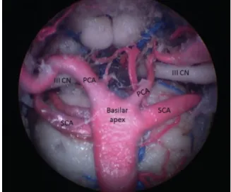
With the method just described we were able to expose the basilar artery in all of its extension, focusing primarily on the BA, the posterior cerebral artery segments p1 and p2, and the superior cerebellar artery (►Fig. 1). The average
length for the posterior cerebral artery exposed was 0.670.2 cm on the right side and 0.590.2 cm on the left side. Both right and left superior cerebellar arteries were exposed with an average length of 0.60.2 cm and 0.70.3 cm, respectively. We identified cranial nerve (CN) III running between the posterior cerebral and superior cerebellar arteries in all specimens, and we were able to avoid manipulation of this nerve when applying the clips. The length of the basilar artery exposed was 2.60.3 cm on average. The distance from the nasal vestibule to the posterior cerebral artery, superior cerebellar artery, and BA was 10 cm with an SD of0.7, 0.6, and 0.8 cm, respectively. We were able to place clips in the posterior cerebral artery, superior cerebellar artery, and BA using the standard clip applicators in different portions of the length exposed in each artery with minimal alteration of surrounding normal tissue in all the specimens (►Figs. 2and3;►Video 1).
Video 1
Endoscopic video showing the technique used to clip the posterior cerebellar artery and basilar artery. Online content including video sequence is available at: https://www.thieme-connect.com/products/
ejournals/html/10.1055/s-0035-1566252
Discussion and Literature Review
BA aneurysms are the most commonly reported in the posterior cerebral arterial circulation, accounting for 50%3,4 of all posterior circulation aneurysms. BA aneurysms are associated with higher risk of bleeding, mortality, and mor-bidity compared with those seen in other posterior circula-tion locacircula-tions.4
Fig. 1 Endoscopic view obtained in a cadaver showing basilar apex. CN, cranial nerve III; PCA, posterior cerebral arteries; SCA, superior cerebellar arteries.
Different variations of the anatomy of BA aneurysms have been described. Nanda et al5designated and reported in his clinical series the complex anatomy of a BA aneurysm in which criteria included an aneurysm>10 cm, calcified or thrombosed, neck4 mm (up to 60% of all BA aneurysms),4 posterior direction, and retro/subsellar aneurysms. Aneur-ysms that did not have these criteria were considered simple aneurysms. In addition to the posterior projection, the ante-rior projection, toward the dorsum sellae, and the upward projection, toward the posterior diencephalon, have also been described.3,6A great proportion of BA aneurysms are
associated with posterior cerebral and superior cerebellar aneurysms. The sum of all of these features makes this pathology one of the most difficult to treat with surgery and endovascular interventions.
Posterior cerebral artery aneurysms have a lower inci-dence of occurrence compared with BA aneurysms. They represent only 1% of all intracranial aneurysms.7Due to the many perforating branches that originate in the posterior cerebral artery, and the different anatomical territories that it supplies, an excellent knowledge of the posterior cerebral artery anatomy is necessary. P1 and P2 segments of the Fig. 2 Endoscopic view obtained in a cadaver showing the technique used to measure the basilar apex, superior cerebellar artery, and posterior cerebral artery. Measurement of the superior cerebellar artery is shown.
Fig. 3 Endoscopic view showing clip placement. (A) Basilar artery. (B) Posterior cerebral artery. (C) Superior cerebellar artery.
posterior cerebral artery are the locations most often re-ported in the literature7–10that we were able to expose with the endonasal endoscopic approach. In patients without an antecedent of a subarachnoid hemorrhage, having an aneu-rysm located in the posterior cerebral artery represents an independent higher risk of rupture, as reported in the study of unruptured intracranial aneurysms in 1998.11These features increase the risk of morbidity and mortality related to this pathology.
Superior cerebellar artery aneurysms are rare. Few clinical series have been reported.12,13They arise mainly at the origin of the superior cerebral artery from the basilar artery or between the origin of the superior cerebral and posterior cerebral arteries.4,13 Compared with BA and posterior cerebral aneurysms, the size of superior cerebellar artery aneurysms are smaller, and they often present with a narrow neck. Additionally, inclusion of the artery origin into the aneurysm has been seen. No other morphological features have been described.
Different surgical approaches and endovascular inter-ventions have been used and reported for the management of these aneurysms. The most common approaches used to reach the BA, superior cerebellar artery, and posterior cerebral artery have been the subtemporal approach and the pterional approach. The view of the aneurysm neck is perpendicular and shorter with the subtemporal approach; also there is no need to retract the great vessels, but there is a need to manipulate the third cranial nerve and temporal lobe.3,5,14 The pterional approach widens the distance between the internal carotid artery and the third cranial nerve that avoids manipulation of the latter, but the oblique direction of the approach affects the visibility of the vessels in question, and it is associated with a longer working distance and the manipulation of the posterior clinoid process. Other approaches has been used less frequently: the transcavernous approach with posterior clinoidectomy, middle fossa approach with medial petrosectomy, orbito-zygomatic craniotomy, and transsylvian approach,15all of them trying to increase the range of visibility, improve the ability of sharp dissection, avoid damage of the perforating vessels, and decrease manipulation of CN III. None of these approaches sufficiently negates the associated risks of the procedure. Additionally all the approaches just described involve significant brain retraction.
After the release of the detachable coil device in 1990, the decision regarding which technique, either endovascular or surgery, would bring better outcomes became more complex. The International Subarachnoid Aneurysm Trial16 in 2002 assessed the outcomes in patients with a ruptured aneurysm after the treatment either with endovascular coiling or surgical clipping. The trial showed better outcomes in terms of survival disability free after 1 year of treatment with endovascular treatment compared with neurosurgical treat-ment. Endovascular techniques in a posterior circulation aneurysm, mainly BA, have showed excellent results for small and narrow neck (<4 mm) aneurysms,5but with wide neck aneurysms, the risks of coil prolapse4and the incomplete occlusion of larger aneurysms increase. Different techniques
to avoid coil prolapse have been reported such as the use of stent-assisted coiling.17For a large BA aneurysm, the occlu-sion rates with endovascular treatment ranged between 7.6% and 67%.5
Posterior cerebral artery aneurysms have shown good results when treated with endovascular coiling.7Berry aneur-ysms in nature were the ones with better outcomes, but giant or fusiform type aneurysms presented the same problem as wide neck BA aneurysms, mainly for incomplete occlusion.7 In these cases, the occlusion of the parent vessel is sometimes inevitable.
Superior cerebellar aneurysms have been treated mainly with endovascular coiling; the cases reported presented incomplete occlusion of the aneurysm in the majority. These results12can be explained when considering the aneurysm size-to-neck ratio and the inclusion of the parent artery origin into the aneurysm. In thefirst case the complete occlusion could lead to a prolapse of the coil to the parent vessel; in the second, the complete occlusion of the aneurysm would be impossible.12 Despite this, the endovascular treatment has shown positive long-term results.
In our study we wanted to investigate the feasibility of the endoscopic endonasal technique as a new window to treat this pathology. There are only nine cases18–26reported in the literature that have used this technique for clipping aneur-ysms, and of these, only four cases are located in the posterior circulation (►Table 1). Two cases presented aneurysms in the basilar trunk18,19and the other two in the right vertebral artery.20,23 There are no cases reporting the use of this technique in the BA, posterior cerebral artery, or superior cerebellar artery. All the cases reported had successful results. Kassam et al23 presented a patient with a right vertebral artery aneurysm previously treated with endovascular coil-ing; in this case the need for a subsequent treatment performed by the author was due to the mass effect caused by the coiling mass responsible for new symptomatology. Fewer complications were seen, and all were successfully treated. As in our study this technique represented a direct route to treat posterior circulation aneurysms, also one to avoid brain retraction and extensive manipulation of the surrounding neurovascular structures. The surgical corridor was found to be adequate for visualization and for standard clip applicators to reach the three vessels studied; however, new instruments designed specifically for this purpose must be developed. We believe the expanded endoscopic approach is a potential alternative to treat posterior cerebral, superior cerebellar, and BA artery aneurysms.
Conclusions
The endoscopic endonasal transclival approach represents a direct anatomical route to the posterior cerebral artery, superior cerebellar artery origin, and basilar artery apex. We believe this approach is a feasible surgical corridor to treat aneurysms arising in these vessels through the use of standard vascular clip applicators without the need for excessive brain retraction and manipulation, thus adding an alternative treatment to the armamentarium of the skull base surgeon.
Note
This article was presented in the poster session at the North American Skull Base Society Conference; Febru-ary 20–22, 2015; Tampa, FL, USA.
Acknowledgments
We thank Matt Pillsbury, manager of the North Carolina Eye Bank Multidisciplinary Surgical Skills Laboratory, University of North Carolina at Chapel Hill.
References
1 Zanation AM, Carrau RL, Snyderman CH, et al. Nasoseptalflap reconstruction of highflow intraoperative cerebral spinalfluid leaks during endoscopic skull base surgery. Am J Rhinol Allergy 2009;23(5):518–521
2 Patel MR, Taylor RJ, Hackman TG, et al. Beyond the nasoseptalflap: outcomes and pearls with secondaryflaps in endoscopic endo-nasal skull base reconstruction. Laryngoscope 2014;124(4): 846–852
3 Hernesniemi J, Korja M. At the apex of cerebrovascular surgery— basilar tip aneurysms. World Neurosurg 2014;82(1–2):37–39 4 Marlin ES, Ikeda DS, Shaw A, Powers CJ, Sauvageau E. Endovascular
treatment of basilar aneurysms. Neurosurg Clin N Am 2014;25(3): 485–495
5 Nanda A, Sonig A, Banerjee AD, Javalkar VK. Microsurgical man-agement of basilar artery apex aneurysms: a single surgeon’s experience from Louisiana State University, Shreveport. World Neurosurg 2014;82(1–2):118–129
6 Peerless SJ, Hernesniemi JA, Gutman FB, Drake CG. Early surgery for ruptured vertebrobasilar aneurysms. J Neurosurg 1994;80(4): 643–649
7 Ciceri EF, Klucznik RP, Grossman RG, Rose JE, Mawad ME. Aneur-ysms of the posterior cerebral artery: classification and endovas-cular treatment. AJNR Am J Neuroradiol 2001;22(1):27–34 8 Ferrante L, Acqui M, Trillò G, Lunardi P, Fortuna A. Aneurysms of
the posterior cerebral artery: do they present specific character-istics? Acta Neurochir (Wien) 1996;138(7):840–852
9 Gerber CJ, Neil-Dwyer G. A review of the management of 15 cases of aneurysms of the posterior cerebral artery. Br J Neurosurg 1992; 6(6):521–527
10 Pia HW, Fontana H. Aneurysms of the posterior cerebral artery. Locations and clinical pictures. Acta Neurochir (Wien) 1977;38; (1–2):13–35
Table 1 Cases reported in the literature of posterior circulation aneurysms treated with endoscopic endonasal approach
Study Age, y/Sex
Initial symptoms/ Findings
Location/size Approach Outcome
Kassam et al23
51/W Progressive clumsiness,
weakness, headache, neck pain, sensory alterations, incoordination
Partially thrombosed, right vertebral artery aneurysm, 11 mm
First: Endovascular therapy successful but mass effect.
Second: Clipping and aneurysmorrhaphy through a transclival transcondylar approach
Excellent healing; im-provement of preoper-ative symptoms; no CSF leakage
Eloy et al19
28/W HH grade III
subarach-noid hemorrhage
Basilar trunk aneurysm,
2.52.21.7 mm
Extended sublabial ap-proach to the sella and sphenoid
Stable; presented vaso-spasm that required antihypertensive treat-ment; it was necessary to place a stent for per-sistent fusiform dilation;
patient’s poststenting
course was unremarkable
Enseñat et al20
74/W Sudden onset of severe
headache, decreased level of consciousness, spatial and time dis-orientation, subarach-noid hemorrhage
Right vertebral artery saccular aneurysm, 1.2 mm Extended endonasal transclival approach Postoperative course uneventful; complete obliteration and paten-cy of vertebral and PICA arteries
CSF leakage 2 wk after surgery completely repaired
Drazin et al18
59/W Sudden onset of severe
headache; subarach-noid hemorrhage HH grade II
Upper basilar artery with an associated feeding vessel to a small cerebellar hemisphere AVM
First: Endovascular treatment. Placement of stent unsuccessful. Second: Clipping trough a suboccipital cranioto-my unsuccessful. Third: Extended endo-nasal transclival approach
Partial occlusion of the aneurysm and
contin-uedfilling of the AVM
vessel.
Returned to the OR, for reclipping that resulted in immediate occlusion and full recovery
Abbreviations: AVM, arteriovenous malformation; CSF, cerebrospinalfluid; HH, Hunt and Hess (scale); OR, operating room; PICA, posteroinferior cerebellar artery.
11 International Study of Unruptured Intracranial Aneurysms Inves-tigators. Unruptured intracranial aneurysms—risk of rupture and risks of surgical intervention. N Engl J Med 1998;339(24): 1725–1733Erratum in: N Engl J Med. 1999; 340:744
12 Haw C, Willinsky R, Agid R, TerBrugge K. The endovascular management of superior cerebellar artery aneurysms. Can J Neurol Sci 2004;31(1):53–57
13 Uda K, Murayama Y, Gobin YP, Duckwiler GR, Viñuela F. Endovas-cular treatment of basilar artery trunk aneurysms with Guglielmi detachable coils: clinical experience with 41 aneurysms in 39 patients. J Neurosurg 2001;95(4):624–632
14 Drake CG. The treatment of aneurysms of the posterior circulation. Clin Neurosurg 1979;26:96–144
15 Hsu FP, Clatterbuck RE, Spetzler RF. Orbitozygomatic approach to basilar apex aneurysms. Neurosurgery 2005;56(1, Suppl):172–177; discussion 172–177
16 Molyneux A, Kerr R, Stratton I, et al; International Subarachnoid Aneurysm Trial (ISAT) Collaborative Group. International Sub-arachnoid Aneurysm Trial (ISAT) of neurosurgical clipping versus endovascular coiling in 2143 patients with ruptured intracranial aneurysms: a randomized trial. J Stroke Cerebrovasc Dis 2002; 11(6):304–314
17 Fargen KM, Mocco J, Neal D, et al. A multicenter study of stent-assisted coiling of cerebral aneurysms with a Y configuration. Neurosurgery 2013;73(3):466–472
18 Drazin D, Zhuang L, Schievink WI, Mamelak AN. Expanded endo-nasal approach for the clipping of a ruptured basilar aneurysm and feeding artery to a cerebellar arteriovenous malformation. J Clin Neurosci 2012;19(1):144–148
19 Eloy JA, Carai A, Patel AB, Genden EM, Bederson JB. Combined endoscope-assisted transclival clipping and endovascular stenting
of a basilar trunk aneurysm: case report. Neurosurgery 2008; 62(3, Suppl 1):142–143; discussion 143–144
20 Enseñat J, Alobid I, de Notaris M, et al. Endoscopic endonasal clipping of a ruptured vertebral-posterior inferior cerebellar artery aneurysm: technical case report. Neurosurgery 2011;69 (1, Suppl Operative):E121–E127; discussion E127–E128 21 Froelich S, Cebula H, Debry C, Boyer P. Anterior communicating artery
aneurysm clipped via an endoscopic endonasal approach: technical note. Neurosurgery 2011;68(2, Suppl Operative):310–316; discus-sion 315–316
22 Germanwala AV, Zanation AM. Endoscopic endonasal approach for clipping of ruptured and unruptured paraclinoid cerebral aneurysms: case report. Neurosurgery 2011;68(1, Suppl Operative):234–239; discussion 240
23 Kassam AB, Mintz AH, Gardner PA, Horowitz MB, Carrau RL, Snyderman CH. The expanded endonasal approach for an endo-scopic transnasal clipping and aneurysmorrhaphy of a large vertebral artery aneurysm: technical case report. Neurosurgery 2006;59(1, Suppl 1):E162–E165; discussion E162–E165 24 Kassam AB, Gardner PA, Mintz A, Snyderman CH, Carrau RL,
Horowitz M. Endoscopic endonasal clipping of an unsecured superior hypophyseal artery aneurysm. Technical note. J Neuro-surg 2007;107(5):1047–1052
25 Kassam AB, Thomas AJ, Zimmer LA, et al. Expanded endonasal approach: a fully endoscopic completely transnasal resection of a skull base arteriovenous malformation. Childs Nerv Syst 2007; 23(5):491–498
26 Kitano M, Taneda M. Extended transsphenoidal approach to anterior communicating artery aneurysm: aneurysm incidentally identified during macroadenoma resection: technical case report. Neurosur-gery 2007;61(5, Suppl 2):E299–E300;discussion E300