Glutamate Neurotransmission in Rodent
Models of Traumatic Brain Injury
Christopher R. Dorsett,1Jennifer L. McGuire,2Erica A. K. DePasquale,2Amanda E. Gardner,2 Candace L. Floyd,3and Robert E. McCullumsmith2
Abstract
Traumatic brain injury (TBI) is a leading cause of death and disability in people younger than 45 and is a significant public health concern. In addition to primary mechanical damage to cells and tissue, TBI involves additional molecular mechanisms of injury, termed secondary injury, that continue to evolve over hours, days, weeks, and beyond. The trajectory of recovery after TBI is highly unpredictable and in many cases results in chronic cognitive and behavioral changes. Acutely after TBI, there is an unregulated release of glutamate that cannot be buffered or cleared effectively, resulting in damaging levels of glutamate in the extracellular space. This initial loss of glutamate homeostasis may initiate additional changes in glutamate regulation. The excitatory amino acid transporters (EAATs) are expressed on both neurons and glia and are the principal mechanism for maintaining extracellular glutamate levels. Diffusion of glutamate outside the synapse due to impaired uptake may lead to increased extrasynaptic glutamate signaling, secondary injury through activation of cell death pathways, and loss of fidelity and specificity of synaptic transmission. Coordination of glutamate release and uptake is critical to regulating synaptic strength, long-term potentiation and depression, and cognitive processes. In this review, we will discuss dysre-gulation of extracellular glutamate and glutamate uptake in the acute stage of TBI and how failure to resolve acute disruptions in glutamate homeostatic mechanisms may play a causal role in chronic cognitive symptoms after TBI.
Keywords:EAAT; extrasynaptic; glutamate; spillover; traumatic brain injury
Introduction
T
raumatic brain injury(TBI) is a leading cause of morbidityand mortality worldwide, resulting in an estimated 10 million hospitalizations or deaths per year.1Injury is most likely during three distinct age periods: early childhood, late adolescence, and late adulthood.2Within the United States, TBI is the primary cause of mortality in individuals younger than 45. The majority of TBIs result from automobile accidents, followed by falls and recreational in-juries.2Additionally, members of the active duty military are at el-evated risk of TBI, with an estimated incidence of TBI among wounded soldiers as high as 22%.3TBI involves both primary me-chanical damage to brain tissues as well as molecular cascades that propagate injury into surrounding tissue, a phenomenon known as secondary injury.4These metabolic effects include unregulated neurotransmitter and ion release, cell swelling, diffuse axonal injury, free radical production and oxidative stress, mitochondrial dys-function, inhibited ATP production, inflammation, and altered gene transcription.4–9These secondary events further exacerbate the ef-fects of the primary injury, increasing blood–brain barrier damage,
edema, ischemia, and hypoxia—ultimately augmenting the cell death process.4,8Due to these secondary processes, TBI continues to evolve over weeks and months, making behavioral outcomes, par-ticularly in mild injuries without focal lesions, difficult to predict.10,11
Increases in extracellular glutamate play an important role in initiating secondary injury cascades. Extracellular glutamate levels are regulated by a family of plasma membrane excitatory amino acid transporters (EAATs), localized to post-synaptic neurons and as-trocytes.12In the initial stages of TBI, extracellular glutamate levels increase and glutamate buffering and clearance is impaired.13Loss of synaptic fidelity due to increased extrasynaptic glutamate sig-naling may mediate persistent cognitive and emotional symptoms after TBI. Intracellular signaling in response to injury changes lo-calization and binding efficiency of glutamate transporters and impairs regulation of extracellular glutamate levels. Altered gluta-mate buffering and reuptake modulate synaptic function, leading to neuroplastic changes in learning and memory.14 Thus, if acute deficits in regulation of extracellular glutamate persist chronically, they may form the molecular basis for the long-term cognitive and emotional deficits many persons afflicted with TBI exhibit.
1
Biological and Biomedical Sciences Doctoral Program, University of North Carolina at Chapel Hill, Chapel Hill, North Carolina. 2
Department of Psychiatry and Behavioral Neuroscience, University of Cincinnati, Cincinnati, Ohio. 3
Department of Physical Medicine and Rehabilitation, University of Alabama at Birmingham, Birmingham, Alabama. DOI: 10.1089/neu.2015.4373
Animal Models of Traumatic Brain Injury
Given the high incidence of TBI, its deleterious effects, and the inability to invasively study the progression of the injury in hu-mans, various animal models have been developed to investigate the pathophysiology of TBI. Because of their smaller size and cost effectiveness, rodents are the primary animals used to model TBI.4 However, versions of these have been adapted for use in non-rodent models, such as pig.15These experimental models mimic various
aspects of human TBI, allowing for comparison of injury type, location, region, and severity.16 Four primary injury models in rodents are commonly used: fluid percussion injury (FPI), con-trolled cortical impact (CCI), weight-drop impact acceleration (WDIA), and blast injury.4
Fluid percussion injury
The FPI model of injury is perhaps the most widely-used and well-characterized model of TBI.17 FPI injury is produced by striking of a pendulum to the back of a fluid reservoir, generating a wave of pressure through an opening in the skull onto the dural surface that results in temporary displacement and mechanical deformation of the brain. FPI injuries are scalable to produce mild, moderate, or severe TBI, and by adjusting the position of the cra-niotomy in relation to the sagittal suture, can produce midline or lateral models of injury, with lateral positioning being the most common.4FPI produces both focal and diffuse injury, including subdural hematoma, intracranial hemorrhage, brain swelling, and axonal shearing characteristic of TBI pathology in humans.18 Ad-ditionally, FPI reliably produces the cognitive and behavioral deficits associated with TBI in humans.19FPI is used due to its reproducibility and the ease with which injury severity can be ad-justed; however, the need for a craniotomy, the cost of the FPI device, and a high mortality rate due to compromises in brainstem function are limitations of the model.20
Controlled cortical impact
The CCI model uses a pneumatic impactor device to drive a rigid rod into the surface of the exposed brain.21 The model induces deformation of the cortex around the injury site and generates widespread degradation to cortical, thalamic, and hippocampal brain regions, resulting in tissue loss, axonal shearing, and contu-sion.21A primary advantage of this model is the ease with which factors such as velocity and depth of injury can be controlled.4 The extent of cortical displacement correlates with both histo-pathological markers of neuronal dysfunction and behavioral deficits; thus, the model can be easily adjusted to fit experimen-tal parameters.22 CCI represents a more focal injury than other commonly-used models, which can have implications in behav-ioral and anatomical characterizations.20However, the low mor-tality rate and reproducible pathology make CCI a useful model for biomechanical studies of TBI.4
Weight-drop impact acceleration
The WDIA model induces trauma via a free falling guided weight striking a metal disk cemented to the rodent’s skull.23A scalable injury is achieved by varying the mass of the weight and the distance that it falls.4The impacting force generates rapid ac-celeration of the brain within the skull, resulting in diffuse brain injury, including petechial hemorrhage and edema in regions from the cortex to the brainstem without fracturing the skull.16WDIA produces characteristic pathological features, including widespread
and bilateral axonal and neuronal damage and extensive diffuse axonal injury, as well as similar behavioral and cognitive defects found in FPI and CCI models.4While cost effective and useful in evoking diffuse axonal injury, this model has drawbacks due to the relative variability in injury severity.20 Despite this, the WDIA model is useful for the study of multiple concussions, an area of increasing importance in the study of sport-related injuries.
Blast injury models
In recent combat operations, explosive blast forces, such as those generated by an improvised explosive device, posed considerable risk of TBI to deployed personnel.3Even individuals who do not experience any external injuries subsequent to an explosion can experience TBI as a result of the forces generated by the blast.3In
order to understand the mechanisms involved in the propagation of the injury from an explosive force, animal models have been de-veloped that seek to recreate the blast injury. In these models, a shock tube and compression forces simulate non-impact blast in-juries.3 Characteristic features of blast injury include cerebral edema, hyperemia, and delayed vasospasm, as well as diffuse ax-onal injury.4Blast injuries also lead to behavioral and cognitive
deficits similar to other models of TBI.4The largest drawback to blast models is the difficulty in standardizing the injury procedure and in replicability of results between research groups.24
The Tripartite Synapse and Glutamate in the Healthy Brain
Glutamate release
Glutamate release, activity as a ligand, and reuptake involves the coordinated action of pre- and post-synaptic neurons, as well as astrocytes.25Receptors, enzymes, and transporters comprise a neu-ron–astroglia coupled system modulating synaptic, perisynaptic, and extrasynaptic glutamate levels.26,27In the pre-synaptic neuron, glu-tamine may be converted to glutamate by glutaminase and packaged by vesicular glutamate transporters (VGLUT1-3) for release into the synapse.28,29Once released, free glutamate in the extracellular space activates post-synaptic receptors, is removed into astrocytes by glutamate transporters, or spills over into the extrasynaptic space. Glutamate in the synapse may occupy and activate ionotropic (N-methyl-D-aspartate (NMDA) receptor, a -amino-3-hydroxy-5-methyl-isoxazole propionate (AMPA), and kainate) or metabo-tropic glutamate receptors on both neurons and astrocytes.25,30,31
Neuronal and glial glutamate transporters
EAATs are the most significant means of extracellular glutamate regulation and are expressed in neurons and glia throughout the brain in a region- and cell-specific manner.36,37EAAT1 and EAAT2 are primarily localized to astroglia, while EAAT3-4 and EAAT5 are primarily localized to neurons and the retina, respectively.36,38 EAATs mediate glutamate transport by an electrogenic exchange of 3 Na+, 1 H+, and 1 glutamate molecule into the cell and 1 K+ion out of the cell, with the net inward movement of positive charges.38–40 The glial transporters are situated in perisynaptic processes facing the synaptic cleft.41In the prefrontal cortex, glial transporters ac-count for the majority of synaptic glutamate reuptake.42,43Cortical homogenates from EAAT2-deficient mice exhibit less than 5% of uptake activity, compared with wild-type mice,44 and additional investigations show that EAAT2 accounts for more than 90% of the glutamate reuptake activity in most brain regions.45
Following EAAT-mediated uptake into astrocytes or neurons, glutamate may enter the tricarboxylic acid cycle via conversion toa -ketoglutarate, be converted to glutamine and transported back into the synapse, or be released into the extracellular space by a variety of mechanisms.46Depending on the cell type, recovered glutamate also may contribute to lactic acid formation. Lactate production is favored in astrocytes, while lactate breakdown is favored in neurons.47Lactate is efficiently shuttled from astrocytes to neurons and may be a pre-ferred energy substrate in neuronal structures enveloped by astrocytic processes.47Metabolism of glutamate in the synaptic terminals is necessary both to provide sufficient energy to sustain transmission and to replenish transmitter pools.48Consequently, inadequate as-trocytic reuptake in response to neuronal activity may both increase signaling though extrasynaptic glutamate receptors as glutamate spills out of the synapse and/or fail to sustain neuronal energy requirements and transmitter pools during periods of high demand.
Regulation of glutamate reuptake
The expression of EAATs is regulated on multiple levels, in-cluding transcription, messenger RNA (mRNA) splicing, protein synthesis, and post-translational modification.38Receptor tyrosine kinase (RTK) signaling appears to represent a primary starting point for second messenger cascades, converging on the mitogen-activated protein kinases (MAPKs) p42 and p44, and ultimately regulating expression of EAAT2. The dual phosphorylation of p42/ p44 MAPKs at threonine-202 and tyrosine-204 increases expres-sion levels of EAAT2 in the presence of neuron-conditioned media, while inhibition of RTKs by the cell-permeable typhostin A23 blocks the induction of EAAT2.49Growth factors can bypass the RTK-p42/44 MAPK pathway and still influence the expression of EAAT2 by directly activating transcription factors such as cAMP-responsive element modulator, cAMP cAMP-responsive element-binding protein (CREB), and activating transcription factor 1 (ATF-1), al-though the induction of EAAT2 via these pathways is weaker than through RTK activation.49
Akt kinase also regulates the expression of EAAT2 by increasing its rate of transcription.50Transfection with a dominant-negative AKT lentiviral vector decreased the effects of epidermal growth factor signaling on EAAT2 expression in astrocyte cultures, while constitutively active AKT increased EAAT2 expression, protein levels, and transport activity in a dose- and time-dependent man-ner.50Growth factor activation of PI-3K can activate not only Akt, but also increases phosphorylation of p42/44 MAPKs, suggesting a converging network regulating EAAT2 expression.51 Thus, in-creased activity of a number of intracellular signaling pathways
regulate EAAT2 expression, with RTK signaling cascades, p42/44 MAP kinases, and AKT protein kinase representing key mediators promoting EAAT2 expression.
Glutamate uptake also may be regulated through either seques-tration of transporters into intracellular storage sites or by ubiquitin-mediated degradation of transporters. Of primary importance in selective EAAT downregulation is the activity of protein kinase C (PKC). PKC has differential effects on the EAAT subtypes; in mixed neuronal and astrocyte cultures, activation of PKC rapidly (within minutes) decreased cell-surface expression of EAAT252 and in-creased surface expression of the neuronal transporter EAAT3.53 These differential effects are thought to represent a switching mechanism from astrocytic to neuronal glutamate uptake. However, as astrocyte transport represents the primary uptake mechanism, the overall effect in a mixed-cell culture would be reduced reuptake and elevated extracellular levels of glutamate.53The deletion of amino acids 475–517 on EAAT2 abolishes the effects of PKC-induced internalization, indicating a carboxyl-terminal phosphorylation site on the transporter.52This decrease in cell-surface expression did not correspond with a reduction in total cellular levels of EAAT2, sug-gesting the immediate effect of phorbol ester activation of PKC involves internalization of the transporter to an intracellular se-questration site.53Internalization can be blocked in astrocyte cultures expressing a dominant-negative variant of clathrin54or by inhibition of the ubiquitin enzyme E1,55demonstrating an ubiquitin-dependent, clathrin-mediated endocytic mechanism of sequestration.
In contrast to short-term activation of PKC, long-term exposure to phorbol ester was accompanied by an overall decrease in total cellular EAAT2 expression.54Lysosomal inhibitors attenuate this decrease in EAAT2 protein, suggesting a cellular mechanism by which PKC modulates EAAT2 levels under physiological or pathological conditions.54 In summary, astrocytic and neuronal kinase signaling mechanisms may positively or negatively regulate EAAT expression and transport activity through transcriptional control or regulation of cell surface expression in a time- and concentration-dependent manner.
Extrasynaptic glutamate signaling
Removal of glutamate from the synaptic cleft involves: 1) the high affinity binding of glutamate by perisynaptic transporters (e.g., EAAT2) and 2) the transport of bound glutamate by the transporter across the plasma membrane.41,56Once bound, glutamate may be ‘‘unbound’’ or released instead of transported across the plasma membrane.41,56The relatively low rate of transport of bound
glu-tamate relative to its binding affinity suggests that the EAATs first act as buffers for released glutamate.41Thus, glutamate molecules may bounce from one transporter-binding site to another until transported, thereby limiting glutamate spillover from the synaptic cleft into extrasynaptic areas.
the spatial arrangement of glutamate synapses, their glutamate transporter buffering zones, and extrasynaptic glutamate receptors will determine the extent and effect of glutamate spillover.66,67
Glutamate in Acute TBI
Neurotoxicity
Elevated extracellular glutamate levels, such as through appli-cation of exogenous glutamate and related analogues into nervous tissue, are extremely toxic to cells.68,69In humans, severe TBI results in elevated cerebrospinal fluid glutamate that may persist in some brain structures for days or perhaps weeks.70,71Following severe TBI in rats, microdialysis determination of extracellular glutamate levels found a 9-fold increase over non-injured control rats.72High levels of glutamate deplete ATP stores due to over-stimulation and the energy expenditure involved in reuptake.68TBI also transiently increases extracellular potassium levels by 6-fold above baseline.7The effects of increased extracellular glutamate and potassium on ionic homeostasis and ATP expenditure are ex-acerbated by changes in astrocytic potassium conductance fol-lowing TBI.73Resting astrocytes are able to effectively remove glutamate from the synapse due to their high potassium conduc-tance and a negatively charged resting membrane potential main-tained in part by the inwardly rectifying potassium channel KIR4.1.74Conditional knockout of the KIR4.1 result in cells with a diminished capacity to transport glutamate.75,76KIR4.1 is not well studied after TBI; however, it was downregulated along with GLT-1 (EAAT2) from 4 h to at least 72 h after CCI in mice.77The central role of potassium channels in the fidelity of glutamate transport highlights the importance of sodium/potassium gradient, as well as ATP production in recovery from TBI.
Increases in extracellular glutamate in TBI also disrupt ionic homeostasis of sodium and calcium. Supra-physiological activa-tion of glutamate receptors increases intracellular levels of sodium ions and leads to swelling of cells from osmotic pressure.78Other pathologic effects of increased extracellular glutamate involve the actions of the neurotransmitter on ionotropic glutamate receptors that are calcium channels.7,79Calcium is a critical signaling mol-ecule governing myriad cellular processes and as such is tightly regulated with intracellular concentrations maintained around 100 nM.79 Prolonged increases in intracellular calcium disrupt mitochondrial production of ATP and activate proteases and ki-nases, including nitric oxide synthase. Increased activity of these enzymes may generate reactive oxygen species, disrupt cytoskel-etal architecture, and increase transcription of genes associated with apoptosis pathways.79 Thus, the pathological results of in-creased extracellular glutamate are mediated in part by increases in intracellular levels of sodium and calcium, leading to cell swelling and the activation of deleterious signaling pathways, respectively. In the absence of calcium, glutamate-mediated increases in in-tracellular sodium can lead to pathological cell swelling and cell death in hippocampal cultures.80However, changes in intracellular sodium and calcium ions have disparate effects on mixed cortical cultures. In an extracellular environment with physiological con-centrations of both sodium and calcium, neurons exhibited imme-diate morphological changes followed later by significant cell degradation and death. Removal of both ions from the extracellular environment was protective to the cells, even with prolonged glu-tamate exposure. However, ion substitution studies (cell cultures with extracellular environments lacking one of the two ions) sug-gested unique roles for each of these ions in the excitotoxic cascade. Mixed-cell cultures in a calcium-free media exhibited morphological
changes, including increased swelling and granulation, but were largely spared from cell death and returned to previous size within an hour.81Cultures in media where choline was substituted for sodium showed markedly less swelling and other morphological changes when exposed to toxic glutamate levels, but exhibited roughly the same amount of cell death as cultures with both ions at physiological levels.81These results indicate that the acute excitotoxic event pre-cipitated by sodium appears to be transient and largely non-toxic to cortical cells, while the glutamate induced changes in calcium ion concentrations play a key role in subsequent cell death by over-whelming the calcium regulatory mechanisms and activating downstream signaling pathways.5Thus, in clinical TBI, it is likely that the calcium-regulated mechanisms responsible for cell death are overwhelmed and contribute to widespread neuronal cell death.
Pathophysiological activation of downstream signaling pathways depends on changes in calcium levels, calcium’s route of entry into the cell, and the location of its release from intracellular storage.5Of particular importance for glutamate mediated neurotoxicity is the ionotropic NMDA receptor. Studies examining calcium’s level and route of entry into the cell found that absolute levels of intracellular free calcium roughly corresponded with neuronal survival, but survival was better predicted by the route of calcium entry and duration of calcium loading.82Neuronal cell cultures were likely to survive excessive intracellular calcium levels when entry occurred through voltage-sensitive channels triggered by cell depolarization; however, similar levels produced by prolonged ligand-gated (glu-tamate) activity resulted in significantly more cell death.82These effects seem to be restricted to glutamate’s activity on NMDA re-ceptors, as increases in calcium stimulated by glutamate from non-NMDA receptors did not have the same impact on cell survival.82 The neurotoxic effects of glutamate-evoked calcium influx are likely due to activity of the NMDA receptor signaling pathways that promote cellular degeneration.82
Transporters in acute TBI
Antisense oligonucleotide knockdown of EAATs show they are critical for maintaining glutamate below toxic levels. EAAT2 knock-down significantly increased hippocampal cell death, compared with controls, after TBI.83Knockout of either GLAST (EAAT1) or GLT-1 in mice yields excitotoxic levels of glutamate similar to those ex-perienced following TBI.84These studies demonstrate the central role of astrocytic EAATs, especially EAAT2, in the maintenance of extracellular glutamate within physiological norms, and illustrate how the pathology of TBI can be exacerbated when EAAT function or expression is compromised. Studies in astrocyte cultures suggest that the half-life of EAAT2 is longer than 24 h85; thus decreases in EAAT2 expression in the hours immediately following TBI86might not be accounted for solely by altering transcription or translation of the transporter. Along with degradation of EAAT2 by caspase-3,87 modulation by PKC could possibly represent a mechanism by which the secondary injury phase of TBI induces dysfunction in glutamate reuptake transporters and exacerbates the deleterious effects of elevated extracellular glutamate.
ipsilateral cortex are reported at 7 days post-injury.88,90,91 More-over, decreases may be in a specific splice variant for EAAT2, suggesting a shift in the cellular or subcellular localization of this transporter in TBI.91
These findings in animal models replicate analyses of human postmortem brain in which protein levels of the astrocytic gluta-mate transporters may be decreased.88,92Compared with moderate EAAT2 staining found in control patients, extensive EAAT2 staining is present in the ipsilateral cortex of TBI cases, with sur-vival times between 1 and 24 h post-injury, indicating an increase in EAAT2 expression in this region.92In survival times less than 1 h or longer than 24 h, weak and sporadic EAAT2 staining patterns were observed. These data suggest dynamic regulation of EAAT2 expression after TBI. This transient increase in EAAT2 expression between 1 and 24 h may represent a compensatory reaction to the elevated extracellular glutamate levels following TBI. At longer survival times, patients exhibit a decrease in EAAT2 expression, possibly as a result of the activity of intracellular signaling path-ways promoting EAAT2 degradation. It is worth noting that short-term TBI survival is correlated with a higher injury severity93; thus,
these findings likely reflect the influence of both of these factors on EAAT2 expression.
While astrocytic EAATs are responsible for the majority of the removal of glutamate from the extracellular space, the neuronal EAATs also are affected by TBI. Increases in expression of EAAT4 were found in hippocampal astrocytes 3 to 7 days following lateral FPI.94 This finding is interesting in that EAAT4 is typically ex-pressed in neurons.57EAAT4 expression in hippocampal astrocytes may represent an endogenous neuroprotective mechanism or may be part of a phenotypic switch in reactive astrocytes.94 Other studies have found similar instances of phenotypic switching of typically ‘‘astrocytic’’ EAATs onto neuronal processes. For ex-ample, hypoxia results in early loss of EAAT2 in the pig hippo-campus, which is followed by the aberrant induction of an EAAT2 splice variant in neuronal cell types.95Additionally, detection of an alternately spliced variant of EAAT1 in neuronal cell cultures following hypoxia is an early and highly sensitive marker for neurons at risk of cell death, as normal expression of glutamate transporters may be negatively modulated by co-expression of these splice variants.96Similarly, engineered expression of EAAT2 on neuronal cell types increases vulnerability to excitotoxicity in hippocampal slice cultures.97It is plausible that glial EAAT ex-pression in neurons may be a compensatory attempt by the brain to offset increases in extracellular glutamate following TBI; however, these efforts may ultimately harm neuronal tissue by making it more vulnerable to excitotoxicity due intracellular glutamate levels beyond the buffering or metabolic capacity of neurons.98
Extracellular glutamate concentrations may modulate the ex-pression of glutamate transporters. Exposure of cultured cortical astrocytes to high levels of extracellular glutamate (20 mM) de-creased EAAT2 expression by 25 and 40% following 24 h or 72 h exposure times, respectively.99The decline in protein expression was not due to astrocyte death, but corresponded to increased glutamine synthetase protein. The decline in transporter expression was not attenuated by non-competitive antagonists for NMDA or AMPA receptors, supporting a receptor-independent mechanism for decreased transporter expression.99In summary, the acute ef-fects of TBI on expression of glutamate transporters are mediated by a number of paracrine and intracellular signaling factors influ-encing the function, location, and phenotype of the transporters. Long-term compromises in the tripartite glutamate system exac-erbate the pathological effects of the primary injury and contribute
to the persistent deficits in cognition characteristic of the secondary injury phase.
Glutamate in Chronic TBI
Glial dysfunction
Eight weeks following moderate lateral FPI, hippocampal cell loss was accompanied by increased gliosis and increased GFAP staining in the thalamus, frontal cortex, and hippocampus, indi-cating a persistent abnormality in astrocytes.100 Additionally, single-cell polymerase chain reaction analysis of glial acidic fi-brillary protein (GFAP)–positive astrocytes found a dramatic shift in gene expression 14 days after ischemic injury in populations of reactive astrocytes.101Reactive astrocytes have increased expres-sion of transcripts for EAAT1, synaptosomal-associated protein 25 (SNAP25), and glutamate receptor subunits, indicating that these cells may be starting to express ‘‘neuronal’’ genes such as SNAP25 and some of the neuronal hyperpolarization activated cyclic nu-cleotide gated potassium channels (HCNs).101
Few studies have evaluated glutamate levels in chronic (>14 days post-injury) animal models of TBI. A series of studies implicated chronic dysregulation of glutamate in chronic TBI by examining late onset behavioral morbidity as indicated by in-creased whisker sensitivity following diffuse experimental TBI. Over 8 weeks following moderate midline FPI, rats in both injured and sham experimental conditions received manual whisker stim-ulation, and behavioral responses were recorded. While sham in-jury animals were ambivalent or soothed by whisker stimulation, animals in the injury condition demonstrated aggravated behavioral responses beginning at 1 week post-injury, which became signifi-cant at 4 weeks and persisted throughout the rest of the 8 weeks.102 A follow up study examining the molecular underpinnings for this increased whisker sensitivity supported hypersensitive glutamate circuitry as a possible causal mechanism.103Microelectrode array studies found increased extracellular glutamate levels in the ventral posterior medial hypothalamus, compared with sham-injured ani-mals, 4 weeks following injury.103Imaging studies after human TBI suggest the balance of glutamate and the inhibitory transmitter c-aminobutyric acid (GABA) may be chronically altered after TBI.71,104,105 Thus, persistent impairments in glutamate circuitry represent a possible mechanism for the chronic cognitive and emotional symptoms experienced by some patients after TBI.
In addition, several studies indirectly examine the roles of gluta-mate transporter expression, glutagluta-mate reuptake, and extracellular glutamate in chronic FPI using novel pharmacological therapeutics. Pharmacological blockade of sodium channels, which inhibits glu-tamate release, attenuated GFAP immunoreactivity 2 weeks fol-lowing moderate-to-severe lateral FPI.106 Another study found treatment with riluzole, a pharmacological agent which inhibits so-dium channels and decreases glutamate release, decreased cortical lesion size 2 weeks following moderate parasagittal FPI.107Further, microtubule-associated protein 2 immunoreactivity 2 weeks after moderate lateral FPI was reduced following treatment with the glu-tamate receptor antagonist kynurenate, compared with untreated animals.108These studies indirectly demonstrate the role of gluta-mate in chronic TBI by showing that inhibiting glutagluta-mate release or modulating glutamate receptors decreases chronic pathophysiology.
Signaling in chronic TBI
kinase (ERK) and CREB activation was decreased in hippocampal slices following a stimulation protocol 12 weeks after parasagittal FPI; in this same study, resting-state levels of phospho-CREB protein were decreased in the hippocampus, suggesting persistent signaling abnormalities.109Interestingly, ERK and CREB are pos-itive modulators of EAAT2 expression.49,51Decreased activity in this signaling pathway is consistent with diminished EAAT ex-pression and activity. Further, ERK and CREB are molecular markers of memory consolidation, and disruptions in these path-ways downstream of glutamate neurotransmission may contribute to the chronic cognitive difficulties observed after TBI. Using elec-trophysiological measures, one group found increased frequency of spontaneous inhibitory post-synaptic currents in the dentate gyrus 1 month after FPI, while another found decreased capacity for LTP in mild, moderate, and severe TBI 8 weeks after injury.110,111These data suggest a mechanism for persistent alterations in the molecular correlates of learning and memory in the chronic FPI model of TBI. There may be a threshold level of LTP that is required to initiate
protein synthesis–dependent consolidation of memory112; thus, the reduced capacity for LTP found 8 weeks following TBI represents a potential mechanism for the cognitive impairment exhibited in both human patients and animal models of TBI.
Glutamate reuptake mechanisms are closely linked to the elec-trophysiological changes reported in chronic TBI discussed above. In the hippocampus, baseline (i.e., ‘‘normal’’) LTP in the CA1 subfield is associated with increased glutamate reuptake; for ex-ample, fear conditioning increased EAAT-dependent reuptake, and blockade of EAAT3 co-localization with the NMDA receptor– signaling complex attenuated synaptic strength.14In a similar study in ischemia, a disorder where glutamate excitotoxicity is believed to play a similar role as in TBI, transient global ischemia decreased protein expression of EAAT2 in area CA1 of the hippocampus corresponding to decreased amplitude of glutamate evoked cur-rents from astrocytes in this area.113 Further, reverse transcrip-tion polymerase chain reactranscrip-tion demonstrated diminished EAAT2 mRNA in post-ischemic cells in hippocampal area CA1.113These
results suggest that disruption in EAAT2 expression and function contributes to astrocytic cell death in the hippocampus, and that glutamate reuptake is important for maintaining synaptic strength and initiating LTP in the hippocampus. Thus, impairment of glu-tamate reuptake could account for observed electrophysiological deficits in this region in chronic TBI.
Therapeutics addressing glutamate
Attempts to offset glutamate excitotoxicity following TBI have largely been disappointing in clinical trials.114Numerous therapeu-tics agents have attempted to address the glutamate imbalance using pre-synaptic glutamate inhibiting agents, competitive and non-competitive NMDA receptor antagonists, cannabinoid agonists, ni-tric oxide synthase inhibitors, AMPA antagonists, serotonin ago-nists, and magnesium sulfates.114,115While initially promising in phase II clinical trials, the synthetic cannabinoid receptor agonist dexanabinol failed to produce statistically relevant improvements in Glasgow Outcome Score (GOS) in phase III trials.116The compet-itive NMDA receptor antagonist selfotel also was brought to phase III clinical trials with similarly disappointing results. Patients in the placebo group had statistically similar GOS scores and mortality rates to those in the drug treatment condition and the study was discontinued.117Extracellular magnesium, which endogenously acts as a non-competitive NMDA antagonist also has been investigated. However, in moderate-to-severe TBI patients, magnesium sulfate administration resulted in no significant improvements in survival, seizure activity, or behavioral outcomes.118Recent animal studies attempting to offset the loss of glutamate homeostasis following TBI involve the use of therapeutic drugs that upregulate glutamate transporter expression. For example, the b-lactam antibody cef-triaxone reversed the loss of EAAT2 expression in the ipsilateral frontal cortex of TBI mice 7 days post-injury and resulted in lower levels of pro-inflammatory mediators.90,119A number of additional compounds, including riluzole, the tricyclic antidepressant amitrip-tyline (which upregulates EAAT expression), and the beta-carboline alkaloid harmine (which stimulates EAAT2 activity), show promise in multiple models of CNS injury and neurodegeneration.120
By focusing on neuronal protection, pharmacological interven-tion may be ignoring the crucial role of astrocytes in the propaga-tion and sustainment of the disease state. We posit that there is also a need for glioprotective agents in therapeutic treatments of chronic TBI. There is still much to be learned about how traumatic injury affects EAAT expression, splice variants, trafficking, localization, activity, and turnover that could identify new therapeutic oppor-tunities in both acute and chronic secondary injury processes. Ac-cumulating evidence suggests that comprehensive examination of the glutamate reuptake mechanism and associated signal trans-duction processes may be a high yield substrate for understanding the pathophysiology of excitatory circuits in chronic TBI.
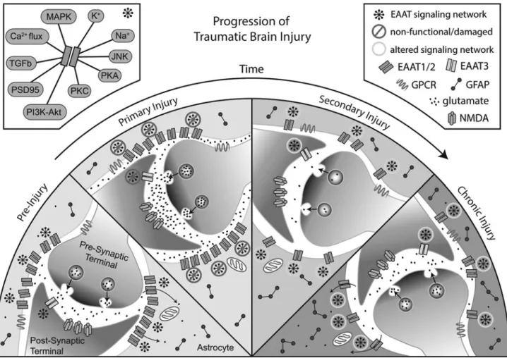
Conclusion
As with the proximal primary injury, alterations in glutamate homeostasis may represent a driving event in the ongoing patho-physiology of TBI. Failure to resolve the secondary injury may be due in part to changes in localization and function of glutamate transporters and their associated signaling molecules, anchoring proteins, and metabolic pathways necessary for normal glutamate transmission (Fig. 1). Given the strong link between proper gluta-mate reuptake and the maintenance of synaptic strength, LTP, and learning and memory, dysfunction in glutamate transport systems could play a causal role in the persistent cognitive symptoms
as-sociated with chronic TBI. Prolonged morphological and molecular changes in astroglia and astroglial glutamate uptake mechanisms may be particularly important as a single astrocyte may intercon-nect with thousands of synapses.121
Diminished glutamate reuptake capacity could have profound effects on cognitive and behavioral outcomes associated with chronic TBI. Inadequate management of glutamate in the synapse and glu-tamate spillover into extrasynaptic domains would affect initiation, amplitude, and sustainability of LTP and LTD, both of which are critically important for synaptic efficiency and cognition. Insufficient astrocytic response to neuronal activity produces electrophysiologi-cal changes consistent with glutamate spillover from the syn-apse.122,123Similar mechanisms may be present in the neocortex in schizophrenia, an illness with profound cognitive impairment.33
Taken together, these mechanisms may explain the diffuse global nature of cognitive and emotional symptoms experienced following TBI; the initial injury may reset glutamate regulatory mechanisms and molecular systems associated with cognitive performance dur-ing the secondary injury phase. If not resolved, these changes con-solidate, resulting in long-term alterations in glutamate homeostasis which may diminish the ability of neurons to effectively initiate or maintain synaptic plasticity (Fig. 1). This model suggests a thresh-old for patient symptomology, below which a patient is asymp-tomatic but nevertheless increasingly susceptible to developing chronic symptoms after each subsequent injury or other CNS stress due to incremental increases in damaged astroglia and associated signaling deficits.
Acknowledgments
This work was partially supported by R01NS075162 (CLF).
Author Disclosure Statement
No competing financial interests exist.
References
1. Langlois, J.A., Rutland-Brown, W., and Wald, M.M. (2006). The epidemiology and impact of traumatic brain injury: a brief overview. J. Head Trauma Rehabil. 21, 375–378.
2. Bruns, J. Jr. and Hauser, W.A. (2003). The epidemiology of trau-matic brain injury: a review. Epilepsia 44 Suppl 10, 2–10. 3. Martin, E.M., Lu, W.C., Helmick, K., French, L., and Warden, D.L.
(2008). Traumatic brain injuries sustained in the Afghanistan and Iraq wars. J. Trauma Nurs. 15, 94–99.
4. Xiong, Y., Mahmood, A., and Chopp, M. (2013). Animal models of traumatic brain injury. Nat. Rev. Neurosci. 14, 128–142.
5. Arundine, M. and Tymianski, M. (2004). Molecular mechanisms of glutamate-dependent neurodegeneration in ischemia and traumatic brain injury. Cell. Mol. Life Sci. 61, 657–668.
6. McIntosh, T.K. (1993). Novel pharmacologic therapies in the treat-ment of experitreat-mental traumatic brain injury: a review. J. Neuro-trauma 10, 215–261.
7. Katayama, Y., Becker, D.P., Tamura, T., and Hovda, D.A. (1990). Massive increases in extracellular potassium and the indiscriminate release of glutamate following concussive brain injury. J. Neurosurg. 73, 889–900.
8. Gaetz, M. (2004). The neurophysiology of brain injury. Clin. Neu-rophysiol. 115, 4–18.
9. Raghupathi, R. (2004). Cell death mechanisms following traumatic brain injury. Brain Pathol. 14, 215–222.
10. Boyle, E., Cancelliere, C., Hartvigsen, J., Carroll, L.J., Holm, L.W., and Cassidy, J.D. (2014). Systematic review of prognosis after mild traumatic brain injury in the military: results of the International Collaboration on Mild Traumatic Brain Injury Prognosis. Arch. Phys. Med. Rehabil. 95, S230–S237.
Borg, J. (2014). Nonsurgical interventions after mild traumatic brain injury: a systematic review. Results of the International Collabora-tion on Mild Traumatic Brain Injury Prognosis. Arch. Phys. Med. Rehabil. 95, S257–S264.
12. Danbolt, N.C., Chaudhry, F.A., Dehnes, Y., Lehre, K.P., Levy, L.M., Ullensvang, K., and Storm-Mathisen, J. (1998). Properties and lo-calization of glutamate transporters. Prog. Brain Res. 116, 23–43. 13. Hinzman, J.M., Thomas, T.C., Quintero, J.E., Gerhardt, G.A., and
Lifshitz, J. (2012). Disruptions in the regulation of extracellular glutamate by neurons and glia in the rat striatum two days after diffuse brain injury. J. Neurotrauma 29, 1197–1208.
14. Levenson, J., Weeber, E., Selcher, J.C., Kategaya, L.S., Sweatt, J.D., and Eskin, A. (2002). Long-term potentiation and contextual fear con-ditioning increase neuronal glutamate uptake. Nat. Neurosci. 5, 155–161. 15. Fievisohn, E.M., Sajja, V.S., Vandevord, P.J., and Hardy, W.N. (2014). Evaluation of impact-induced traumatic brain injury in the Gottingen Minipig using two input modes. Traffic Inj. Prev. 15 Suppl 1, S81–S87.
16. Hallam, T.M., Floyd, C.L., Folkerts, M.M., Lee, L.L., Gong, Q.Z., Lyeth, B.G., Muizelaar, J.P., and Berman, R.F. (2004). Comparison of behavioral deficits and acute neuronal degeneration in rat lateral fluid percussion and weight-drop brain injury models. J. Neuro-trauma 21, 521–539.
17. Thompson, H.J., Lifshitz, J., Marklund, N., Grady, M.S., Graham, D.I., Hovda, D.A., and McIntosh, T.K. (2005). Lateral fluid percus-sion brain injury: a 15-year review and evaluation. J. Neurotrauma 22, 42–75.
18. Graham, S.H., Chen, J., and Clark, R.S. (2000). Bcl-2 family gene products in cerebral ischemia and traumatic brain injury. J. Neuro-trauma 17, 831–841.
19. McIntosh, T.K., Vink, R., Noble, L., Yamakami, I., Fernyak, S., Soares, H., and Faden, A.L. (1989). Traumatic brain injury in the rat: characterization of a lateral fluid-percussion model. Neuroscience 28, 233–244.
20. Cernak, I. (2005). Animal models of head trauma. NeuroRx 2, 410– 422.
21. Hall, E.D., Sullivan, P.G., Gibson, T.R., Pavel, K.M., Thompson, B.M., and Scheff, S.W. (2005). Spatial and temporal characteristics of neurodegeneration after controlled cortical impact in mice: more than a focal brain injury. J. Neurotrauma 22, 252–265.
22. Goodman, J.C., Cherian, L., Bryan, R.M. Jr., and Robertson, C.S. (1994). Lateral cortical impact injury in rats: pathologic effects of varying cortical compression and impact velocity. J. Neurotrauma 11, 587–597.
23. Marmarou, A. (2007). A review of progress in understanding the path-ophysiology and treatment of brain edema. Neurosurg. Focus 22, E1. 24. Gupta, R.K. and Przekwas, A. (2013). Mathematical models of
blast-induced TBI: current status, challenges, and prospects. Front. Neurol. 4, 59.
25. Salt, T.E. and Eaton, S.A. (1996). Functions of ionotropic and me-tabotropic glutamate receptors in sensory transmission in the mam-malian thalamus. Prog. Neurobiol. 48, 55–72.
26. Vernadakis, A. (1996). Glia-neuron intercommunications and syn-aptic plasticity. Prog. Neurobiol. 49, 185–214.
27. Rusakov, D.A. and Kullmann, D.M. (1998). Extrasynaptic glutamate diffusion in the hippocampus: ultrastructural constraints, uptake, and receptor activation. J. Neurosci. 18, 3158–3170.
28. Bellocchio, E.E., Reimer, R.J., Fremeau, R.T. Jr., and Edwards, R.H. (2000). Uptake of glutamate into synaptic vesicles by an inorganic phosphate transporter. Science 289, 957–960.
29. Takamori, S., Rhee, J.S., Rosenmund, C., and Jahn, R. (2000). Identification of a vesicular glutamate transporter that defines a glutamatergic phenotype in neurons. Nature 407, 189–194. 30. Hollmann, M., Boulter, J., Maron, C., and Heinemann, S. (1994).
Molecular biology of glutamate receptors. Potentiation of N-methyl-D-aspartate receptor splice variants by zinc. Renal Physiol. Bioche-mist. 17, 182–183.
31. Hollmann, M. and Heinemann, S. (1994). Cloned glutamate recep-tors. Annu. Rev. Neurosci. 17, 31–108.
32. Nicoll, R.A. and Malenka, R.C. (1999). Expression mechanisms underlying NMDA receptor-dependent long-term potentiation. Ann. N. Y. Acad. Sci. 868, 515–525.
33. McCullumsmith, R.E., Clinton, S.M., and Meador-Woodruff, J.H. (2004). Schizophrenia as a disorder of neuroplasticity. Int. Rev. Neurobiol. 59, 19–45.
34. McGee, A.W. and Bredt, D.S. (2003). Assembly and plasticity of the glutamatergic postsynaptic specialization. Curr. Opin. Neurobiol. 13, 111–118.
35. Contractor, A. and Heinemann, S.F. (2002). Glutamate receptor trafficking in synaptic plasticity. Science’s STKE 2002, re14. 36. Arriza, J.L., Kavanaugh, M.P., Fairman, W.A., Wu, Y.N., Murdoch,
G.H., North, R.A., and Amara, S.G. (1993). Cloning and expression of a human neutral amino acid transporter with structural similarity to the glutamate transporter gene family. J. Biol. Chem. 268, 15329– 15332.
37. Utsunomiya-Tate, N., Endou, H., and Kanai, Y. (1996). Cloning and functional characterization of a system ASC-like Na+-dependent neutral amino acid transporter. J. Biol. Chem. 271, 14883–14890. 38. Danbolt, N.C. (2001). Glutamate uptake. Prog. Neurobiology 65, 1–
105.
39. Zerangue, N. and Kavanaugh, M.P. (1996). Flux coupling in a neu-ronal glutamate transporter. Nature 383, 634–637.
40. Levy, L.M., Warr, O., and Attwell, D. (1998). Stoichiometry of the glial glutamate transporter GLT-1 expressed inducibly in a Chinese hamster ovary cell line selected for low endogenous Na+-dependent glutamate uptake. J. Neurosci. 18, 9620–9628.
41. Tzingounis, A.V. and Wadiche, J.I. (2007). Glutamate transporters: confining runaway excitation by shaping synaptic transmission. Nat. Rev. Neuroscience 8, 935–947.
42. Kanai, Y. (1997). Substrate binding sites of glutamate transporters and structurally related neutral amino acid transporters. Jpn. J. Physiol. 47 Suppl 1, S55–S56.
43. Regan, M.R., Huang, Y.H., Kim, Y.S., Dykes-Hoberg, M.I., Jin, L., Watkins, A.M., Bergles, D.E., and Rothstein, J.D. (2007). Variations in promoter activity reveal a differential expression and physiology of glutamate transporters by glia in the developing and mature CNS. J. Neurosci. 27, 6607–6619.
44. Tanaka, K., Watase, K., Manabe, T., Yamada, K., Watanabe, M., Takahashi, K., Iwama, H., Nishikawa, T., Ichihara, N., Kikuchi, T., Okuyama, S., Kawashima, N., Hori, S., Takimoto, M., and Wada, K. (1997). Epilepsy and exacerbation of brain injury in mice lacking the glutamate transporter GLT-1. Science 276, 1699–1702.
45. Danbolt, N.C., Storm-Mathisen, J., and Kanner, B.I. (1992). An [Na+ +
K+]coupled L-glutamate transporter purified from rat brain is located in glial cell processes. Neuroscience 51, 295–310.
46. Malarkey, E.B. and Parpura, V. (2008). Mechanisms of glutamate release from astrocytes. Neurochem. Int. 52, 142–154.
47. Stobart, J.L. and Anderson, C.M. (2013). Multifunctional role of astrocytes as gatekeepers of neuronal energy supply. Front. Cell. Neurosci. 7, 38.
48. Tani, H., Dulla, C.G., Farzampour, Z., Taylor-Weiner, A., Hugue-nard, J.R., and Reimer, R.J. (2014). A local glutamate-glutamine cycle sustains synaptic excitatory transmitter release. Neuron 81, 888–900.
49. Gegelashvili, G., Dehnes, Y., Danbolt, N.C., and Schousboe, A. (2000). The high-affinity glutamate transporters GLT1, GLAST, and EAAT4 are regulated via different signalling mechanisms. Neu-rochem. Int. 37, 163–170.
50. Li, L.B., Toan, S.V., Zelenaia, O., Watson, D.J., Wolfe, J.H., Rothstein, J.D., and Robinson, M.B. (2006). Regulation of astrocytic glutamate transporter expression by Akt: evidence for a selective transcriptional effect on the GLT-1/EAAT2 subtype. J. Neurochem. 97, 759–771.
51. Abe, K. and Saito, H. (2001). Possible linkage between glutamate transporter and mitogen-activated protein kinase cascade in cultured rat cortical astrocytes. J. Neurochem. 76, 217–223.
52. Kalandadze, A., Wu, Y., and Robinson, M.B. (2002). Protein kinase C activation decreases cell surface expression of the GLT-1 subtype of glutamate transporter. Requirement of a carboxyl-terminal domain and partial dependence on serine 486. J. Biol. Chem. 277, 45741–45750. 53. Gonzalez, M.I. and Robinson, M.B. (2004). Protein kinase
C-dependent remodeling of glutamate transporter function. Mol. In-terventions 4, 48–58.
54. Susarla, B.T. and Robinson, M.B. (2008). Internalization and deg-radation of the glutamate transporter GLT-1 in response to phorbol ester. Neurochem. Int. 52, 709–722.
56. Tong, G. and Jahr, C.E. (1994). Block of glutamate transporters potentiates postsynaptic excitation. Neuron 13, 1195–1203. 57. Massie, A., Cnops, L., Smolders, I., McCullumsmith, R., Kooijman,
R., Kwak, S., Arckens, L., and Michotte, Y. (2008). High-affinity Na+/K+-dependent glutamate transporter EAAT4 is expressed throughout the rat fore- and midbrain. J. Comp. Neurol. 511, 155–172. 58. Chalifoux, J.R. and Carter, A.G. (2011). Glutamate spillover promotes
the generation of NMDA spikes. J. Neurosci. 31, 16435–16446. 59. Hardingham, G.E. and Bading, H. (2002). Coupling of extrasynaptic
NMDA receptors to a CREB shut-off pathway is developmentally regulated. Biochim. Biophys. Acta 1600, 148–153.
60. Hardingham, G.E., Fukunaga, Y., and Bading, H. (2002). Extra-synaptic NMDARs oppose Extra-synaptic NMDARs by triggering CREB shut-off and cell death pathways. Nat. Neurosci. 5, 405–414. 61. Leveille, F., El Gaamouch, F., Gouix, E., Lecocq, M., Lobner, D.,
Nicole, O., and Buisson, A. (2008). Neuronal viability is controlled by a functional relation between synaptic and extrasynaptic NMDA receptors. FASEB J. 22, 4258–4271.
62. Lozovaya, N., Melnik, S., Tsintsadze, T., Grebenyuk, S., Kirichok, Y., and Krishtal, O. (2004). Protective cap over CA1 synapses: ex-trasynaptic glutamate does not reach the postsynaptic density. Brain Res. 1011, 195–205.
63. Tsvetkov, E., Shin, R.M., and Bolshakov, V.Y. (2004). Glutamate uptake determines pathway specificity of long-term potentiation in the neural circuitry of fear conditioning. Neuron 41, 139–151. 64. Rinholm, J.E., Slettalokken, G., Marcaggi, P., Skare, O.,
Storm-Mathisen, J., and Bergersen, L.H. (2007). Subcellular localization of the glutamate transporters GLAST and GLT at the neuromuscular junction in rodents. Neuroscience 145, 579–591.
65. Kullmann, D.M. and Asztely, F. (1998). Extrasynaptic glutamate spillover in the hippocampus: evidence and implications. Trends Neurosci. 21, 8–14.
66. Weng, H.R., Chen, J.H., Pan, Z.Z., and Nie, H. (2007). Glial gluta-mate transporter 1 regulates the spatial and temporal coding of glu-tamatergic synaptic transmission in spinal lamina II neurons. Neuroscience 149, 898–907.
67. Sem’yanov, A.V. (2005). Diffusional extrasynaptic neurotransmission via glutamate and GABA. Neurosci. Behav. Physiol. 35, 253–266. 68. Olney, J.W. (1990). Excitotoxicity: an overview. Can. Dis. Wkly.
Rep. 16 Suppl 1E, 47–57.
69. Liu, Z., Stafstrom, C.E., Sarkisian, M.R., Yang, Y., Hori, A., Tandon, P., and Holmes, G.L. (1997). Seizure-induced glutamate release in mature and immature animals: an in vivo microdialysis study. Neuroreport 8, 2019–2023.
70. Zhang, H., Zhang, X., Zhang, T., and Chen, L. (2001). Excitatory amino acids in cerebrospinal fluid of patients with acute head in-juries. Clin. Chem. 47, 1458–1462.
71. Kierans, A.S., Kirov, II, Gonen, O., Haemer, G., Nisenbaum, E., Babb, J.S., Grossman, R.I., and Lui, Y.W. (2014). Myoinositol and glutamate complex neurometabolite abnormality after mild traumatic brain injury. Neurology 82, 521–528.
72. Faden, A.I., Demediuk, P., Panter, S.S., and Vink, R. (1989). The role of excitatory amino acids and NMDA receptors in traumatic brain injury. Science 244, 798–800.
73. D’Ambrosio, R., Maris, D.O., Grady, M.S., Winn, H.R., and Janigro, D. (1999). Impaired K(+) homeostasis and altered electrophysio-logical properties of post-traumatic hippocampal glia. J. Neurosci. 19, 8152–8162.
74. Olsen, M. (2012). Examining potassium channel function in astro-cytes. Methods Mol. Biol. 814, 265–281.
75. Djukic, B., Casper, K.B., Philpot, B.D., Chin, L.S., and McCarthy, K.D. (2007). Conditional knock-out of Kir4.1 leads to glial membrane depolarization, inhibition of potassium and glutamate uptake, and enhanced short-term synaptic potentiation. J. Neurosci. 27, 11354– 11365.
76. Olsen, M.L., Higashimori, H., Campbell, S.L., Hablitz, J.J., and Sontheimer, H. (2006). Functional expression of Kir4.1 channels in spinal cord astrocytes. Glia 53, 516–528.
77. Gupta, R.K. and Prasad, S. (2013). Early down regulation of the glial Kir4.1 and GLT-1 expression in pericontusional cortex of the old male mice subjected to traumatic brain injury. Biogerontology 14, 531–541.
78. Hablitz, J.J. and Langmoen, I.A. (1982). Excitation of hippocampal pyramidal cells by glutamate in the guinea-pig and rat. J. Physiol. 325, 317–331.
79. Arundine, M. and Tymianski, M. (2003). Molecular mechanisms of calcium-dependent neurodegeneration in excitotoxicity. Cell Cal-cium 34, 325–337.
80. Rothman, S.M. (1985). The neurotoxicity of excitatory amino acids is produced by passive chloride influx. J. Neurosci. 5, 1483–1489. 81. Choi, D.W. (1987). Ionic dependence of glutamate neurotoxicity.
J. Neurosci. 7, 369–379.
82. Tymianski, M., Charlton, M.P., Carlen, P.L., and Tator, C.H. (1993). Source specificity of early calcium neurotoxicity in cultured em-bryonic spinal neurons. J. Neurosci. 13, 2085–2104.
83. Rao, V.L., Dogan, A., Bowen, K.K., Todd, K.G., and Dempsey, R.J. (2001). Antisense knockdown of the glial glutamate transporter GLT-1 exacerbates hippocampal neuronal damage following trau-matic injury to rat brain. Eur. J. Neurosci. 13, 119–128.
84. Rothstein, J.D., Dykes-Hoberg, M., Pardo, C.A., Bristol, L.A., Jin, L., Kuncl, R.W., Kanai, Y., Hediger, M.A., Wang, Y., Schielke, J.P., and Welty, D.F. (1996). Knockout of glutamate transporters reveals a major role for astroglial transport in excitotoxicity and clearance of glutamate. Neuron 16, 675–686.
85. Zelenaia, O.A. and Robinson, M.B. (2000). Degradation of glial glutamate transporter mRNAs is selectively blocked by inhibition of cellular transcription. J. Neurochem. 75, 2252–2258.
86. Rao, V.L., Baskaya, M.K., Dogan, A., Rothstein, J.D., and Dempsey, R.J. (1998). Traumatic brain injury down-regulates glial glutamate transporter (GLT-1 and GLAST) proteins in rat brain. J. Neurochem. 70, 2020–2027.
87. Boston-Howes, W., Gibb, S.L., Williams, E.O., Pasinelli, P., Brown, R.H. Jr., and Trotti, D. (2006). Caspase-3 cleaves and inactivates the glutamate transporter EAAT2. J. Biol. Chem. 281, 14076–14084. 88. van Landeghem, F.K., Stover, J.F., Bechmann, I., Bruck, W.,
Un-terberg, A., Buhrer, C., and von Deimling, A. (2001). Early ex-pression of glutamate transporter proteins in ramified microglia after controlled cortical impact injury in the rat. Glia 35, 167–179. 89. Floyd, C.L., Gorin, F.A., and Lyeth, B.G. (2005). Mechanical strain
injury increases intracellular sodium and reverses Na+/Ca2+ ex-change in cortical astrocytes. Glia 51, 35–46.
90. Goodrich, G.S., Kabakov, A.Y., Hameed, M.Q., Dhamne, S.C., Rosenberg, P.A., and Rotenberg, A. (2013). Ceftriaxone treatment after traumatic brain injury restores expression of the glutamate transporter, GLT-1, reduces regional gliosis, and reduces post-traumatic seizures in the rat. J. Neurotrauma 30, 1434–1441. 91. Yi, J.H., Pow, D.V., and Hazell, A.S. (2005). Early loss of the
glu-tamate transporter splice-variant GLT-1v in rat cerebral cortex fol-lowing lateral fluid-percussion injury. Glia 49, 121–133.
92. Ikematsu, K., Tsuda, R., Kondo, T., and Nakasono, I. (2002). The expression of excitatory amino acid transporter 2 in traumatic brain injury. Forensic Sci. Int. 130, 83–89.
93. Raj, R., Skrifvars, M.B., Bendel, S., Selander, T., Kivisaari, R., Siironen, J., and Reinikainen, M. (2014). Predicting six-month mortality of patients with traumatic brain injury: usefulness of common intensive care severity scores. Crit. Care 18, R60. 94. Yi, J.H., Herrero, R., Chen, G., and Hazell, A.S. (2007). Glutamate
transporter EAAT4 is increased in hippocampal astrocytes following lateral fluid-percussion injury in the rat. Brain Res. 1154, 200–205. 95. Pow, D.V., Naidoo, T., Lingwood, B.E., Healy, G.N., Williams, S.M., Sullivan, R.K., O’Driscoll, S., and Colditz, P.B. (2004). Loss of glial glutamate transporters and induction of neuronal expression of GLT-1B in the hypoxic neonatal pig brain. Brain Res. Dev. Brain Res. 153, 1–11.
96. Sullivan, S.M., Macnab, L.T., Bjorkman, S.T., Colditz, P.B., and Pow, D.V. (2007). GLAST1b, the exon-9 skipping form of the glutamate-aspartate transporter EAAT1 is a sensitive marker of neuronal dys-function in the hypoxic brain. Neuroscience 149, 434–445. 97. Selkirk, J.V., Stiefel, T.H., Stone, I.M., Naeve, G.S., Foster, A.C.,
and Poulsen, D.J. (2005). Over-expression of the human EAAT2 glutamate transporter within neurons of mouse organotypic hippo-campal slice cultures leads to increased vulnerability of CA1 pyra-midal cells. Eur. J. Neurosci. 21, 2291–2296.
98. Lin, C.L., Bristol, L.A., Jin, L., Dykes-Hoberg, M., Crawford, T., Clawson, L., and Rothstein, J.D. (1998). Aberrant RNA processing in a neurodegenerative disease: the cause for absent EAAT2, a glutamate transporter, in amyotrophic lateral sclerosis. Neuron 20, 589–602. 99. Lehmann, C., Bette, S., and Engele, J. (2009). High extracellular
100. Bramlett, H.M., Dietrich, W.D., Green, E.J., and Busto, R. (1997). Chronic histopathological consequences of fluid-percussion brain injury in rats: effects of post-traumatic hypothermia. Acta Neuro-pathol. 93, 190–199.
101. Rusnakova, V., Honsa, P., Dzamba, D., Stahlberg, A., Kubista, M., and Anderova, M. (2013). Heterogeneity of astrocytes: from devel-opment to injury—single cell gene expression. PloS One 8, e69734. 102. McNamara, K.C., Lisembee, A.M., and Lifshitz, J. (2010). The whisker nuisance task identifies a late-onset, persistent sensory sen-sitivity in diffuse brain-injured rats. J. Neurotrauma 27, 695–706. 103. Thomas, T.C., Hinzman, J.M., Gerhardt, G.A., and Lifshitz, J.
(2012). Hypersensitive glutamate signaling correlates with the de-velopment of late-onset behavioral morbidity in diffuse brain-injured circuitry. J. Neurotrauma 29, 187–200.
104. Chamard, E., Lassonde, M., Henry, L., Tremblay, J., Boulanger, Y., De Beaumont, L., and Theoret, H. (2013). Neurometabolic and mi-crostructural alterations following a sports-related concussion in fe-male athletes. Brain Inj. 27, 1038–1046.
105. Tremblay, S., Beaule, V., Proulx, S., de Beaumont, L., Marjanska, M., Doyon, J., Pascual-Leone, A., Lassonde, M., and Theoret, H. (2013). Relationship between transcranial magnetic stimulation measures of intracortical inhibition and spectroscopy measures of GABA and glutamate+glutamine. J. Neurophysiol. 109, 1343–1349. 106. Sun, F.Y. and Faden, A.I. (1995). Pretreatment with antisense oli-godeoxynucleotides directed against the NMDA-R1 receptor en-hances survival and behavioral recovery following traumatic brain injury in rats. Brain Res. 693, 163–168.
107. Zhang, C., Raghupathi, R., Saatman, K.E., Smith, D.H., Stutzmann, J.M., Wahl, F., and McIntosh, T.K. (1998). Riluzole attenuates cor-tical lesion size, but not hippocampal neuronal loss, following trau-matic brain injury in the rat. J. Neurosci. Res. 52, 342–349. 108. Hicks, R.R., Smith, D.H., and McIntosh, T.K. (1995). Temporal
re-sponse and effects of excitatory amino acid antagonism on microtubule-associated protein 2 immunoreactivity following ex-perimental brain injury in rats. Brain Res. 678, 151–160.
109. Atkins, C.M., Falo, M.C., Alonso, O.F., Bramlett, H.M., and Die-trich, W.D. (2009). Deficits in ERK and CREB activation in the hippocampus after traumatic brain injury. Neurosci. Lett. 459, 52–56. 110. Santhakumar, V., Ratzliff, A.D., Jeng, J., Toth, Z., and Soltesz, I. (2001). Long-term hyperexcitability in the hippocampus after ex-perimental head trauma. Ann. Neurol. 50, 708–717.
111. Sanders, M.J., Sick, T.J., Perez-Pinzon, M.A., Dietrich, W.D., and Green, E.J. (2000). Chronic failure in the maintenance of long-term potentiation following fluid percussion injury in the rat. Brain Res. 861, 69–76.
112. Nguyen, P.V. and Kandel, E.R. (1996). A macromolecular synthesis-dependent late phase of long-term potentiation requiring cAMP in the medial perforant pathway of rat hippocampal slices. J. Neurosci. 16, 3189–3198.
113. Yeh, T.H., Hwang, H.M., Chen, J.J., Wu, T., Li, A.H., and Wang, H.L. (2005). Glutamate transporter function of rat hippocampal
as-trocytes is impaired following the global ischemia. Neurobiol. Dis. 18, 476–483.
114. McConeghy, K.W., Hatton, J., Hughes, L., and Cook, A.M. (2012). A review of neuroprotection pharmacology and therapies in patients with acute traumatic brain injury. CNS Drugs 26, 613–636. 115. Maas, A.I. (2001). Neuroprotective agents in traumatic brain injury.
Expert Opin. Investig. Drugs 10, 753–767.
116. Maas, A.I., Murray, G., Henney, H., 3rd, Kassem, N., Legrand, V., Mangelus, M., Muizelaar, J.P., Stocchetti, N., and Knoller, N.; Pharmos TBI investigators. (2006). Efficacy and safety of dex-anabinol in severe traumatic brain injury: results of a phase III ran-domised, placebo-controlled, clinical trial. Lancet. Neurol. 5, 38–45. 117. Morris, G.F., Bullock, R., Marshall, S.B., Marmarou, A., Maas, A., and Marshall, L.F. (1999). Failure of the competitive N-methyl-D-aspartate antagonist Selfotel (CGS 19755) in the treatment of severe head injury: results of two phase III clinical trials. The Selfotel In-vestigators. J. Neurosurg. 91, 737–743.
118. Temkin, N.R., Anderson, G.D., Winn, H.R., Ellenbogen, R.G., Britz, G.W., Schuster, J., Lucas, T., Newell, D.W., Mansfield, P.N., Ma-chamer, J.E., Barber, J., and Dikmen, S.S. (2007). Magnesium sulfate for neuroprotection after traumatic brain injury: a randomised con-trolled trial. Lancet. Neurol. 6, 29–38.
119. Wei, J., Pan, X., Pei, Z., Wang, W., Qiu, W., Shi, Z., and Xiao, G. (2012). The beta-lactam antibiotic, ceftriaxone, provides neuropro-tective potential via anti-excitotoxicity and anti-inflammation re-sponse in a rat model of traumatic brain injury. J. Trauma Acute Care Surg. 73, 654–660.
120. Fontana, A.C. (2015). Current approaches to enhance glutamate transporter function and expression. J. Neurochem. 134, 982–1007. 121. Parpura, V. and Verkhratsky, A. (2012). The astrocyte excitability
brief: from receptors to gliotransmission. Neurochem. Int. 61, 610–621. 122. Tanaka, M., Shih, P.Y., Gomi, H., Yoshida, T., Nakai, J., Ando, R., Furuichi, T., Mikoshiba, K., Semyanov, A., and Itohara, S. (2013). Astrocytic Ca2+signals are required for the functional integrity of tripartite synapses. Mol. Brain. 6, 6.
123. Shen, H.W., Scofield, M.D., Boger, H., Hensley, M., and Kalivas, P.W. (2014). Synaptic glutamate spillover due to impaired glutamate uptake mediates heroin relapse. J. Neurosci. 34, 5649–5657.
Address correspondence to: Robert E. McCullumsmith, MD, PhD University of Cincinnati Department of Psychiatry and Behavioral Neuroscience MSB 5255A 231 Albert Sabin Way Cincinnati, OH 45267-0838