Eugenia Timofeev Monaghan
A thesis submitted to the faculty at University of North Carolina at Chapel Hill in partial fulfillment of the requirements for the degree of Master of Science in Periodontology in
the School of Dentistry.
Chapel Hill 2018
© 2018
preservation and socket seal after tooth extraction. (Under the direction of Thiago Morelli)
This study describes a randomized clinical trial involving twenty-four subjects recruited at the University of North Carolina. Subjects were randomized into two groups: 1) Control: Extraction treated with xenograft bone substitute (Bio-Oss Collagen®) + collagen dressing (HeliPlug®), 2) Test: Extraction treated with xenograft bone substitute (Bio-Oss Collagen®) + 3D-collagen matrix (Mucograft Seal®). Cone Beam Computed Tomography (CBCT) was obtained at baseline and at 6 months to compare linear and volumetric hard-tissue changes. CBCT images were analyzed by a non-contact reverse engineering system (Geomagic Control®).
TABLE OF CONTENTS
List of Tables ... vi
List of Figures ... vii
Literature Review ... 1
1. Wound Healing ... 1
1.1 Undisturbed Extraction Socket Healing ... 1
1.2 Ridge Remodeling ... 3
2. Alveolar Ridge Preservation ... 5
2.1 Materials and Methods for Alveolar Ridge Preservation ... 5
2.2 Outcomes of Alveolar Ridge Preservation ... 6
3. Soft-Tissue Grafts over Augmented Sockets ... 8
4. 3D-Collagen Matrix (Mucograft Seal®) ... 8
References ... 10
Manuscript ... 13
1. Introduction... 13
2. Materials and Methods ... 15
2.1 Randomization ... 16
2.2 Surgical Procedure ... 16
2.3 Radiographic Analysis ... 17
2.4 Statistical Analysis... 18
3. Results ... 19
3.1 Bone Linear Analysis ... 19
4. Discussion ... 22
References ... 27
Appendix 1: Inclusion criteria ... 29
Appendix 2: Exclusion criteria ... 30
LIST OF TABLES
LIST OF FIGURES
Figure 1: CBCT slice section ... 35
Figure 2: Mean linear bone loss ... 36
Figure 3. Mean linear bone loss, premolar teeth only ... 37
Figure 4: Mean linear bone loss, incisor teeth only ... 38
Figure 5: Mean volumetric bone loss ... 39
Figure 6: Mean volumetric bone loss, premolar teeth only ... 40
LITERATURE REVIEW
1. Wound Healing
Extraction of a tooth is generally indicated when a tooth can no longer be maintained or restored for long-term health, function, and/or esthetics. Extracting a tooth from alveolar bone triggers a biological cascade that leads to significant local changes to the alveolar ridge anatomy. The wound healing events and bone remodeling have long held an interest for clinicians, even more so once dental implant therapy was introduced. After a tooth is extracted, the bundle bone that lines that alveolar socket is the first to be resorbed. Further remodeling of both bundle bone and the outer surface of alveolar bone results in reduction of both ridge height and width (Araujo and Lindhe, 2005). The bone loss and ridge atrophy later have a large impact on the ability to replace lost dentition with implant therapy.
1.1 Undisturbed Extraction Socket Healing
Early investigations into the process of healing were of relatively short duration, looking only at the immediate after effects of the procedure. One of the first to
could be seen as early as Day 5 post-extraction, with complete bony fill completed by Day 31. Additionally, epithelium had completely covered the socket opening by Day 7. Claflin theorized that the healing process in extraction wounds in human would be the same as in dogs, only much slower.
Indeed, when analyzing healthy human subjects, Amler noted a similar sequence of events, albeit with the expected extended timeline. In humans, clot formation was also observed on Day 1, but replacement of the clot by granulation tissue and appearance of osteoid at the base of the socket was not until Day 7. Fusion of the epithelium over the open socket was seen as early as day 24 and as late as day 35, and it took until Day 38 for 2/3 of the socket to have trabecular bone fill (Amler 1969).
The animal studies noted above do have the limitation of being relatively short, thereby limiting the amount of information that can be obtained about further alveolar healing and remodeling. In addition, the human studies by Amler and others are from biopsy limited to the superficial tissue, with no inclusion of hard tissue. Additionally, only samples from marginal portions of the socket were taken, thereby events that occurred in the central and apical compartments were not investigated.
More recently, a group set out to evaluate a longer-term healing progression in a dog model that included assessment of multiple zones within the socket (Cardaropoli 2003). Similar to previous studies, a coagulum of blood cells and platelets in a fibrin network could be seen at the end of Day 1. On Day 3, osteoclasts were seen on the marginal surfaces of the socket walls, while neutrophils and macrophages had begun to degrade the blood clot. By Day 7, granulation tissue had completely filled the extraction socket, with early vascularized tissue present at the coronal portion of the socket, and a more mature late tissue with large amounts of fibroblasts presented more apically. A
30, the entire socket was filled with this immature woven bone. On Day 60, a cortical hard tissue bridge over the top of the socket was evident, while inside the socket, bone marrow began to replace the woven bone. By Day 180, more mature lamellar bone and bone marrow had replaced the majority of the woven bone within the socket
(Cardaropoli 2003). The findings from this study indicated that healing progresses
differently at different portions of the extraction socket, with progression from the lateral walls inward and in an apical-to-coronal direction. This finding was also replicated in a study utilizing a monkey model, indicating that regeneration begins at the walls of the extraction socket and at the apex, with further remodeling to form trabecular bone and bone marrow (Scala 2014).
1.2 Ridge Remodeling
Extraction of a tooth leads to remodeling and resorption not only in the extraction socket itself, but also in the alveolar bone adjacent to and surrounding the socket housing (Schropp 2003). While the changes that occur after tooth extraction have long held interest for clinicians, the advent of dental implant therapy has created a new wave of concern in terms of how these changes affect the ability to restore lost dentition. Adequate dimensions of alveolar bone are crucial in obtaining ideal 3D implant positioning for both functional and esthetic prosthetic restorations following implant placement (Schneider 1999).
Consequently, a shift of the center of the edentulous ridge occurred toward the palatal and lingual of both arches.
This finding that the buccal wall resorbs more than the lingual wall was mirrored in 2005 in a dog study by Araujo and Lindhe, who theorized that the resorption of the buccal and lingual walls occurs in two distinct stages (Araujo and Lindhe 2005). In the first stage, the bundle bone that lines the extraction socket was resorbed and replaced with woven bone, creating a reduction in vertical ridge dimension. In the second stage, the buccal aspect of the ridge began to remodel, with additional vertical loss, as well as horizontal resorption of the edentulous ridge. By day 60 in the dog model, the crest of the buccal wall was located apical to the crest of the lingual wall. This difference in healing was likely due to the relative amounts of bundle bone presented on the buccal versus the lingual aspects of the ridge. Since the crest of the buccal plate was composed solely of bundle bone, the vertical reduction was much greater on the buccal than the lingual aspect. In addition, the buccal plate of bone was generally thinner than the lingual plate, which might also have contributed to what appears to be a greater amount of vertical reduction on the buccal aspect of the ridge than on the lingual.
The amount of alveolar ridge resorption that occurs will vary from person-to-person, however it is estimated that approximately 50% of the ridge width will be lost in the first 12 months post-extraction, with 2/3 of this reduction occurring in the first 3 months (Schropp 2003). The amount of resorption can be affected by numerous factors,
walls of <1mm being at significantly higher risk for pronounced resorption of alveolar bone, especially in the vertical aspect (Chappuis 2013).These vertical and horizontal ridge changes that occur during post-extraction healing can greatly limit the ability for functional and esthetic implant placement in the created edentulous space.
2. Alveolar Ridge Preservation
While it is possible to attempt ridge regeneration procedures utilizing grafting materials and barrier membranes, these procedures are generally costly, have varying degrees of success, and can delay implant placement and restoration for six months or more. Therefore, it would be of interest and benefit to both the patient and clinician to minimize this post-extraction resorptive process at the time of extraction, rather than dealing with the repercussions later. Ideal treatment should aim to preserve existing hard and soft tissue contours in preparation for forthcoming implant placement. Alveolar ridge preservation was introduced with the idea that filling an extraction site with a biomaterial would prevent extensive bone remodeling, thus facilitating future implant placement in the site.
2.1 Materials and Methods for Alveolar Ridge Preservation
The materials that have been utilized for ridge preservation (socket preservation, site preservation, socket grafting, etc.) are the same materials that are used for other
(MacBeth 2017).
Barrier membranes can be included either on their own or in conjunction to cover the graft material placed into the socket. The theory for the use of barrier membranes lies in its ability to prevent epithelial cells from penetrating the extraction socket, thereby allowing bone to form first. This concept was first discussed by Melcher in 1976, and was a theory used to describe the healing of periodontal wounds, not necessarily extraction sockets. As discussed previously, extraction socket healing results in alveolar bone formation up to the crest, indicating that the blood clot and granulation tissue are likely enough to prevent epithelial down growth(Pagni 2012), at least in a 4-walled extraction socket defect. Barrier membranes may be more useful in cases of buccal or lingual plate fenestration or dehiscence, where the extraction socket is not completely surrounded by alveolar bone. Types of membranes that have been utilized include e-PTFE, titanium, titanium-reinforced PTFE, collagen, and acellular dermal matrix (Pagni 2012, Darby 2009).Membranes can either be covered partially or completely with soft tissue, with significant coronal advancement of soft tissue usually necessary if primary closure over the extraction socket is the ultimate goal.
Other commonly used materials include resorbable sponges, made either of collagen or polylactic/polglycolic acid. A simple collagen sponge is generally used in combination with a bone substitute material in the socket, which the collagen sponge covers and protects the graft material from the oral environment. Collagen sponges can also act as a carrier for other regenerative materials, such as recombinant human bone
morphogenic protein 2 (rhBMP-2) (Melcher 1976). 2.2 Outcomes of Alveolar Ridge Preservation
implant placement, even if it is not possible to preserve 100% of the previous ridge width (Mardas 2015). In a systematic review by Darby, six studies were evaluated that included a comparison between a non-grafted socket to a grafted socket. In all but one study, alveolar ridge preservation resulted in a significantly greater amount of preserved ridge width and height in favor of the grafted groups. Interestingly, when comparing different methods and materials for ridge preservation, there was no evidence that could support the benefit of one technique over the other. In addition, when bone substitute was utilized in the extraction socket, there were a significant amount of graft particle remnants, even in long-term evaluations. How this may affect implant osseointegration and stability in the long-term is still unknown, as long-term studies evaluating such effect are lacking.
In histologic evaluations, it is evident that at early stages of healing, sockets that were grafted displayed encapsulation of the graft material with connective tissue, as opposed to sites that were left to heal spontaneously, which instead already displayed new woven bone in the majority of the socket at the same timepoint. These findings suggest a foreign-body healing reaction, and resulted in delayed healing of the
extraction wound. In addition, it is likely that the majority of these grafted particles are not resorbed in the long-term, and thus remain encapsulated (Araujo and Lindhe 2009). This observation has contributed to concern that placement of dental implants into grafted ridges may affect osseointegration, and thus interfere with rehabilitative success of the patient. However, several histological studies have examined this effect and determined that bone-to-implant contact at grafted sites is comparable to that of implants placed into native bone (Fiorellini 2007, Santis 2011). In addition, similar primary stability could be achieved at the time of implant placement, with healing proceeding normally, and with similar long-term loading and restorative results
particles, with one study reporting that 79% of grafted sites undergo less than 20% loss of buccal plate, while 71% of non-grafted sites undergo more than 20% loss of buccal plate (Nevins 2006).
3. Soft-Tissue Grafts over Grafted Sockets
The first description of utilizing a soft tissue graft to cover a grafted socket was the socket seal technique described by Landsberg and Bichacho in 1994 as a technique meant to optimize the preservation of the hard and soft tissue components of the alveolar ridge immediately following tooth extraction (Landsberg and Bichacho 1994). The placement of the soft-tissue graft was proposed to minimize the shrinkage and loss of soft tissue volume during wound healing to optimize the esthetic results of future implant restorations, as well as to protect the grafted socket from the effects of the oral cavity exposure (Pagni 2012).
The original socket seal technique utilized a free-gingival graft shaped to the outline of the coronal portion of the extraction socket, however other techniques have utilized connective tissue graft as well (Tal 1999). Tal evaluated the results of utilization of a connective tissue graft placed over an extracted socket augmented with either
Demineralized Freeze-Dried Bone Allograft (DFDBA) or xenograft (Bio-Oss®). At 1 week, 18/42 grafts were vital, 13/42 were partially vital, and 11/42 were non-vital. No matter the vitality of the graft, complete closure of all sockets was completed by week-4. In addition, nourishment for the survival of these grafts likely originated from the
underlying blood clot in the extraction socket rather than from the adjacent tissues at the periphery, as partially vital grafts exhibited better vitality in their center than at the periphery of the graft (Tal 1999).However, this technique requires second surgical sites, thus increasing morbidity and cost for the patient.
4. 3D-Collagen Matrix (Mucograft Seal®)
sockets. The rationale is that the use of this porcine matrix may result in faster wound healing, improving hard and soft tissue regeneration, as well as reducing morbidity to the patient due to the absence of a second surgical site that is required with traditional soft tissue grafting utilizing palatal tissue. The porcine matrix collagen is processed to favor immediate blood clot stabilization, and in a previous study was shown to lead to early vascularization, facilitate soft tissue cell ingrowth, and have excellent integration with surrounding tissues (Ghanaati 2011).
In 2012, a clinical trial was completed to compare the results of the 3D-collagen matrix versus natural spontaneous healing in extraction sockets. This evaluation
indicated faster healing time for the test groups in terms of defect area closure at day 4 (19.33mm2 vs. 21.3mm2) and at day 8 (11.7mm2 vs. 13.6mm2) compared to the control group. In addition, the color match for adjacent tissues was more favorable for the test group (Thoma 2012).
Another clinical trial evaluating different methods of alveolar ridge preservation was completed in 2013 (Jung 2013). Investigators compared a control group of spontaneous healing with three test groups: 1) Bovine bone mineral with 10% collagen (Bio-Oss® Collagen) combined with free-gingival graft, 2) Bovine bone mineral with 10% collagen (Bio-Oss® Collagen) combined with Mucograft Seal®, and 3) ß-tricalcium-phosphate-particles with polylactic coating (ß-TCP). The most successful radiographic outcomes in terms of preservation of alveolar ridge height and width were seen in the groups utilizing Bio-Oss® Collagen, with no significant difference in outcomes between utilization of the free gingival graft versus the Mucograft® in combination with the bone mineral material (Jung 2013). This study demonstrated a potential benefit of this material for achieving better outcomes during socket grafting procedures while eliminating the need for a second surgical site.
REFERENCES
Amler, M. H. (1969). The time sequence of tissue regeneration in human extraction wounds. Oral Surgery, Oral Medicine, Oral Pathology, 27(3), 309-318.
Araújo, M. G., & Lindhe, J. (2005). Dimensional ridge alterations following tooth extraction. An experimental study in the dog. Journal of clinical periodontology, 32(2), 212-218.
Araújo, M., Linder, E., & Lindhe, J. (2009). Effect of a xenograft on early bone formation in extraction sockets: an experimental study in dog. Clinical Oral Implants
Research, 20(1), 1-6.
Araújo, M. G., & Lindhe, J. (2009). Ridge alterations following tooth extraction with and without flap elevation: an experimental study in the dog. Clinical oral implants
research, 20(6), 545-549.
Cardaropoli, G., Araujo, M., & Lindhe, J. (2003). Dynamics of bone tissue formation in tooth extraction sites: an experimental study in dogs. Journal of clinical
periodontology, 30(9), 809-818.
Chappuis, V., Engel, O., Reyes, M., Shahim, K., Nolte, L. P., & Buser, D. (2013). Ridge alterations post-extraction in the esthetic zone: a 3D analysis with CBCT. Journal of dental research, 92(12_suppl), 195S-201S.
Claflin, R. S. (1936). Healing of disturbed and undisturbed extraction wounds. Journal of the American Dental Association, 23(6), 945-959.
Cryer, M. H. (1916). The internal anatomy of the face. Lea & Febiger..
Darby, I., Chen, S. T., & Buser, D. (2009). Ridge preservation techniques for implant therapy. Int J Oral Maxillofac Implants, 24(Suppl), 260-271.
Euler, H. (1923). Healing of Extraction Wounds: An Experimental Study. Dtsch. Monat. Zahnheilkd, 41, 687.
Fickl, S., Zuhr, O., Wachtel, H., Bolz, W., & Huerzeler, M. (2008). Tissue alterations after tooth extraction with and without surgical trauma: a volumetric study in the beagle dog. Journal of Clinical Periodontology, 35(4), 356-363.
Fiorellini, J. P., & Nevins, M. L. (2003). Localized ridge augmentation/preservation. A systematic review. Annals of Periodontology, 8(1), 321-327.
Fiorellini, J. P., Kim, D. M., Nakajima, Y., & Weber, H. P. (2007). Osseointegration of titanium implants following guided bone regeneration using expanded
polytetrafluoroethylene membrane and various bone fillers. International Journal of Periodontics & Restorative Dentistry, 27(3).
Ghanaati, S., Schlee, M., Webber, M. J., Willershausen, I., Barbeck, M., Balic, E., ... & Kirkpatrick, C. J. (2011). Evaluation of the tissue reaction to a new bilayered collagen matrix in vivo and its translation to the clinic. Biomedical materials, 6(1), 015010. Heggeler, T.,, J. M. A. G., Slot, D. E., & Van der Weijden, G. A. (2011). Effect of socket preservation therapies following tooth extraction in non‐molar regions in humans: a
systematic review. Clinical oral implants research, 22(8), 779-788.
Jung, R. E., Philipp, A., Annen, B. M., Signorelli, L., Thoma, D. S., Hämmerle, C. H., ... & Schmidlin, P. (2013). Radiographic evaluation of different techniques for ridge
preservation after tooth extraction: a randomized controlled clinical trial. Journal of clinical periodontology, 40(1), 90-98.
Landsberg, C. J., & Bichacho, N. (1994). A modified surgical/prosthetic approach for optimal single implant supported crown. Part I--The socket seal surgery. Practical periodontics and aesthetic dentistry: PPAD, 6(2), 11-7.
MacBeth, N., Trullenque‐Eriksson, A., Donos, N., & Mardas, N. (2017). Hard and soft
tissue changes following alveolar ridge preservation: a systematic review. Clinical oral implants research, 28(8), 982-1004.
Mardas, N., Trullenque‐Eriksson, A., MacBeth, N., Petrie, A., & Donos, N. (2015). Does
ridge preservation following tooth extraction improve implant treatment outcomes: a systematic review: Group 4: Therapeutic concepts & methods. Clinical oral implants research, 26, 180-201.
Melcher, A. H. (1976). On the repair potential of periodontal tissues. Journal of periodontology, 47(5), 256-260.
Nevins, M., Camelo, M., De Paoli, S., Friedland, B., Schenk, R. K., Parma-Benfenati, S., ... & Wagenberg, B. (2006). A study of the fate of the buccal wall of extraction sockets of teeth with prominent roots. International Journal of Periodontics & Restorative Dentistry, 26(1).
Pagni, G., Pellegrini, G., Giannobile, W. V., & Rasperini, G. (2012). Postextraction alveolar ridge preservation: biological basis and treatments. International journal of dentistry, 2012.
Pietrokovski, J., & Massler, M. (1967). Alveolar ridge resorption following tooth extraction. Journal of Prosthetic Dentistry, 17(1), 21-27.
Rogers, W. M., & Applebaum, E. (1941). Changes in the mandible following closure of the bite with particular reference to edentulous patients. The Journal of the American Dental Association, 28(10), 1573-1586.
Santis, E. D., Botticelli, D., Pantani, F., Pereira, F. P., Beolchini, M., & Lang, N. P. (2011). Bone regeneration at implants placed into extraction sockets of maxillary incisors in dogs. Clinical Oral Implants Research, 22(4), 430-437.
Scala, A., Lang, N. P., Schweikert, M. T., de Oliveira, J. A., Rangel‐Garcia Jr, I., &
Schneider, R. (1999). Prosthetic concerns about atrophic alveolar ridges. Postgrad Dent, 6(2), 3-7.
Schropp, L., Wenzel, A., Kostopoulos, L., & Karring, T. (2003). Bone healing and soft tissue contour changes following single-tooth extraction: a clinical and radiographic 12-month prospective study. International Journal of Periodontics & Restorative
Dentistry, 23(4).
Tal, H. (1999). Autogenous masticatory mucosal grafts in extraction socket seal
procedures: a comparison between sockets grafted with demineralized freeze‐dried bone
and deproteinized bovine bone mineral. Clinical oral implants research, 10(4), 289-296. Thoma, D. S., Sancho‐Puchades, M., Ettlin, D. A., Hämmerle, C. H., & Jung, R. E.
(2012). Impact of a collagen matrix on early healing, aesthetics and patient morbidity in oral mucosal wounds–a randomized study in humans. Journal of clinical
MANUSCRIPT
1. Introduction
Extraction of a tooth is generally indicated when a tooth can no longer be maintained or restored for long-term health, function, and/or esthetics. Removing a tooth from the alveolar bone triggers a biological cascade that leads to significant local changes in the ridge anatomy. After tooth extraction, the bundle bone that lines that alveolar socket is the first to be resorbed. Further remodeling of both bundle bone and the outer surface of alveolar bone results in reduction of both ridge height and width (Araujo and Lindhe, 2005). The bone loss and ridge atrophy later may have a large impact on the ability to replace the lost dentition with implant therapy.
extraction with socket grafting was observed in the buccolingual width dimension (1.89mm) and at the midbuccal height dimension
(2.07mm) (Avila-Ortiz, 2015). Another analysis by Nevins and collaborators indicated that 79% of sites that were grafted underwent less than 20% loss of buccal plate, while 71% of sites that were not grafted demonstrated more than 20% loss of buccal plate (Nevins et al, 2006). Overall, socket grafting is widely recommended at the time of tooth extraction in order to limit the future need for more extensive grafting procedures in case of future implant placement.
Over the past several decades, numerous studies have evaluated the efficacy of a variety of biomaterials and combinations for use in alveolar ridge preservation
procedures, including autografts, allografts, xenografts, and alloplasts (Darby, 2009). On average, the use of xenograft or allograft demonstrated better outcomes, especially in the midbuccal alveolar bone height preservation, compared to alloplastic materials (Avila-Ortiz, 2015). The use of a collagen wound dressing material has been suggested to both protect the graft material from washing out of the extraction socket, as well as to promote blood clot formation and wound stability (Wang, 2007). Collagen materials have an inherent ability to act as a hemostatic agent, stimulate platelet aggregation, and enhance fibrin linkage (Sableman, 1985).
In the United States, xenograft or allograft material in combination with a collagen wound dressing (HeliPlug®) is a common method of site preservation. A different method that has been proposed is to place a free soft-tissue graft over a bone graft in an extraction socket (Landsberg and Bichacho, 1994). It has been suggested that a soft-tissue graft might minimize soft soft-tissue shrinkage, optimize aesthetic results of an
An innovative 3D-collagen matrix has been suggested by its parent company as an alternative to autogenous soft tissue graft over mineralized bone graft in an extraction socket to preserve hard and soft tissue volume for future implant placement. The rationale is that the use of this 3D-collagen matrix may result in faster wound healing, and improved hard and soft tissue regeneration in the management of extraction sockets. The collagen is processed to favor immediate blood clot stabilization, and in a previous study, was shown to lead to early vascularization, facilitate soft tissue cell ingrowth, and have excellent integration within the surrounding tissues (Ghanaati, Schlee et al. 2001). These findings have not yet been proven in a randomized clinical trial, nor has a direct comparison with commonly used collagen dressings been completed.
The aim of the present study was to evaluate the clinical and radiographic alveolar bone changes following socket grafting covered with a 3D-collagen matrix compared to socket grafting covered with a collagen dressing over a six-month period. A novel method utilizing digital 3D superimposition based on CBCT scans obtained at baseline and month-6 was used to assess the extent of buccal bone resorption that occurred, both linear and volumetric.
2. Materials and Methods
The study was reviewed in accordance with federal regulations governing human research and obtained IRB approval by full board review (Study #16-0832).
Twenty-four subjects (10 males, 14 females) were recruited by means of advertisements and flyers from the patient, student, and staff population at the University of North Carolina at Chapel Hill. All subject visits occurred at the Go-Health Clinic at the University of North Carolina, School of Dentistry (Chapel Hill, North
Individuals with dehisced, fenestrated, or fractured labial/buccal alveolar bone plates as determined after baseline CBCT or tooth extraction were excluded from the study. Complete inclusion and exclusion criteria are listed in Appendix 1 and 2.
At the initial examination, all patients completed a medical health history
questionnaire, and full mouth clinical measurements were completed, including probing pocket depth, clinical attachment level, bleeding on probing, and gingival index on all teeth. A standardized cone beam tomography (CBCT) scan was obtained. A second CBCT was obtained at the 6-month (24 week) post-extraction visit.
2.1 Randomization
All qualified subjects were randomly assigned into two groups, with 12 subjects per group: 1) Extraction treated with xenograft bone substitute (Bio-Oss Collagen®) + collagen dressing (HeliPlug®), or 2) Extraction treated with xenograft bone substitute (Bio-Oss Collagen®) + 3D-collagen matrix (Mucograft Seal®). The study coordinator placed 24 identical slips of paper, 12 reading “Test” and 12 reading “Control” into identical sealed envelopes. One sealed envelope was selected for each subject, and the randomization result was recorded.
2.2 Surgical Procedure
In the control group subjects, Bio-Oss Collagen® (Geistlich Pharma, Wolhusen, Switzerland) was placed into the debrided socket in the necessary amount to
successfully fill the extraction socket. The bone substitute material was rehydrated with the subject’s blood or sterile saline solution. Subsequently, a collagen dressing
(HeliPlug®) was used to cover the grafted extraction socket and sutured with resorbable suture (5-0 Vicryl, Ethicon Inc., Somerville, NJ, USA) to stabilize the wound.
In the test group subjects, Bio-Oss Collagen® (Geistlich Pharma, Wolhusen, Switzerland) was placed into the debrided socket in the necessary amount to
successfully fill the extraction socket. The bone substitute material was rehydrated with the subject’s blood or sterile saline solution. A 3D-collagen matrix (Mucograft Seal®) was used to cover the grafted extraction socket and sutured with non-resorbable suture (6-0 Prolene, Ethicon Inc., Somerville, NJ, USA) and resorbable suture (5-0 Vicryl, Ethicon Inc., Somerville, NJ, USA) to stabilize collagen matrix over the extraction socket.
Medication prescribed to all subjects is listed in Appendix 3. Subjects in both groups were instructed to rinse with 0.12% chlorhexidine gluconate for 30 seconds twice daily, and to avoid brushing or touching the surgical site for 2 weeks. Sutures were removed at 2 weeks following the surgical appointment. Patients were recalled at 1, 2, 4, 12, and 24 weeks for monitoring of the healing process. Patients were permitted to wear a temporary appliance to replace the missing tooth if one had been provided to them by their referring dentist. If an appliance was available, it was adjusted to remove any direct pressure on the extraction site.
2.3 Radiographic Analysis
time of 15 seconds. The voxel size was 150 microns. To evaluate radiographic linear and volumetric changes from baseline to 6-months, surface mesh models were generated using each timepoint’s CBCT volume. The baseline and 6-month models were
superimposed using selected areas of the data set where no changes had taken place over the study period, such as the zygomatic process and non-treated teeth. The two data sets were aligned and manually checked for discrepancies. The average error in alignment of the two data sets was kept below 0.1mm for all subjects.
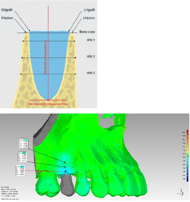
Measurements were obtained and analyzed using a non-contact reverse engineering software (Geomagic Control®). Linear changes were assessed at 1-, 3-, and 5-mm below the alveolar bone crest at the extraction site (Figure 1). Total volume change at the buccal surface area related to the extracted tooth using the same reference points. To set a reference, the most apical point of the extraction socket was defined in the baseline image, and two references lines were drawn. The vertical reference line was drawn in the center of the extraction socket crossing the apical reference point. The horizontal reference line was drawn perpendicular to the vertical line crossing the apical reference point.
2.4 Statistical Analysis
A power analysis was performed prior to the start of the study using a statistical power calculator (SAS Power Procedure, Cary, NC). The sample size of 24 subjects, 12 in each of two groups, allows 90% power (α = 0.05) to detect a difference of 2.5mm in the horizontal ridge width measured at 3mm below the crest, assuming a standard deviation of 1.6mm, as was determined by a previous study (Jung 2013). This power calculation accounts for a 10% subject drop-out rate. Differences were considered statistically significant at p<0.05.
with a two-tailed distribution was performed comparing the two groups for each of the linear measurements (1mm, 3mm, and 5mm) as well as for the volumetric analysis. 3. Results
A total of 24 subjects were admitted and completed the study. Ten males between the ages of 38 and 69 years (mean age 50.6±12.29) and fourteen females between the ages of 31 and 68 years (mean age 51.0±11.42) were enrolled. Patient demographics are listed in Table 1. One control subject’s data sets could not be used for the final analysis due to a dehiscence of the buccal plate that was overlooked at the time of enrollment. Therefore, this subject’s data sets were omitted when calculating the final results. Thus, the test group comprised one central incisor, four lateral incisors, and seven premolars (n=12). The control group comprised three central incisors, three lateral incisors, one canine, and four premolars (n=11). Tooth distribution is listed in Table 2. No serious adverse events were reported. Healing proceeded normally in all subjects, with primary closure over the extraction socket completed in all subjects by week-2 in the test group (Mucograft Seal®) and week-4 in the control group (collagen plug).
3.1 Bone Linear Analysis
Two test subjects and four control subjects were excluded from the linear
measurement analysis due to unacceptable discrepancies during the alignment of the baseline and 6-month CBCT data set. Thus, a total of ten patient data sets were used for the test group linear analysis, and seven patient data sets were used for the control group linear analysis.
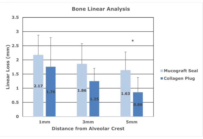
a mean linear loss of bone of 1.75mm (SD=1.03), 1.25mm (SD=0.45), and 0.85mm (SD=0.52) at 1mm, 3mm, and 5mm below the alveolar crest, respectively. The test group (Bio-Oss Collagen® + Mucograft Seal®) had a mean linear bone loss of 2.17mm (SD=0.70), 1.85mm (SD=0.72), and 1.63mm (SD=0.65) at 1mm, 3mm, and 5mm below the alveolar crest, respectively. Statistical analysis of the linear bone remodeling at each mark showed no significant differences at 1mm (p=0.616) or 3mm (p=0.111). There was a statistically significant decrease in amount of bone resorption at 5mm (p=0.029) in favor of the control group.
Next, the groups were further broken down to analyze the differences in linear ridge resorption when compared by tooth type. Premolars in the test and control groups were compared to one another, followed by incisor comparison. For the premolar linear analysis, n=6 for the test group, and n=2 for the control group. For the incisor linear analysis, n=4 for the test group, and n=5 for the control group.
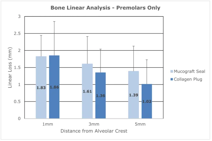
When analyzing a comparison between premolars in the test and control groups (Figure 3), the test group (Bio-Oss Collagen® + Mucograft Seal®) had mean linear loss of bone of 1.83mm (SD=0.62), 1.61mm (SD=0.80), and 1.39mm (SD=0.74) at 1mm, 3mm, and 5mm below the alveolar crest, respectively. The control group (Bio-Oss Collagen® + HeliPlug®) had a mean linear loss of bone of 1.86mm (SD=0.99), 1.36mm (SD=0.69), and 1.02mm (SD=0.71) at 1mm, 3mm, and 5mm below the alveolar crest, respectively. Statistical analysis of the premolar site linear bone remodeling at each mark showed no significant differences at 1mm (p=0.959), 3mm (p=.708), or at 5mm (p=0.552).
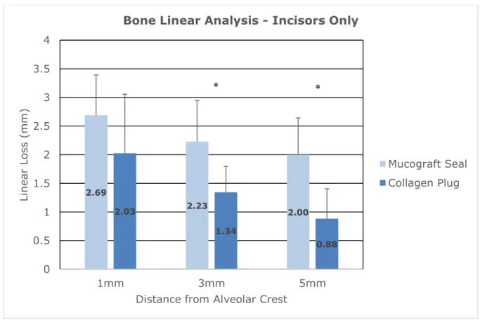
and 0.88mm (SD=0.54) at 1mm, 3mm, and 5mm below the alveolar crest, respectively. Statistical analysis of the incisor site linear bone remodeling at each mark showed no significant differences at 1mm (p=0.236). However, results were significant at 3mm (p=.0.009) and at 5mm (p=0.006) in favor of the control group.
3.2 Bone Volumetric Analysis
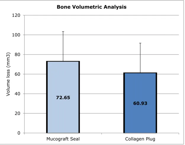
All subject data was used to complete the volumetric analysis comparison from baseline to 6-months (n=23). Results derived from the volumetric analysis of hard tissue changes over six months revealed similar findings in terms of bone volume remodeling from baseline (Figure 4). The control group (Bio-Oss Collagen® + HeliPlug®) demonstrated a mean volumetric hard tissue loss of 60.9mm3 (SD=37.32). The test group (Bio-Oss Collagen® + Mucograft Seal®) demonstrated a mean volumetric hard tissue loss of 72.6mm3 (SD=30.75). The difference between the test and control group was not statistically significant (p=0.668).
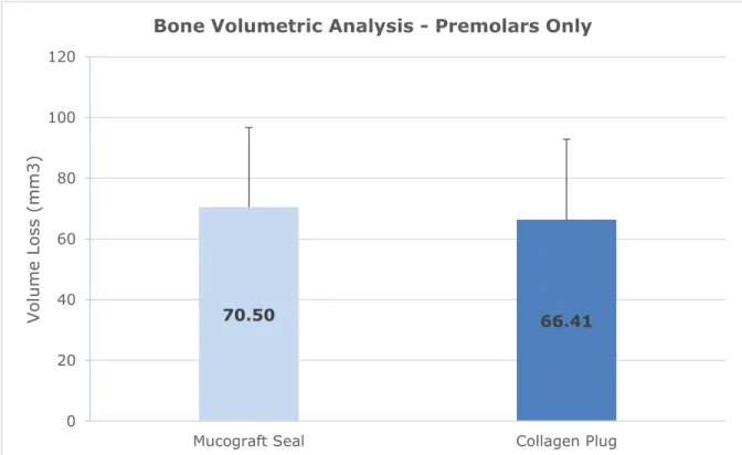
Next, the groups were further broken down to analyze the differences in volumetric ridge resorption when compared by tooth type. Premolars in the test and control groups were compared to one another, followed by incisor comparison. For the premolar
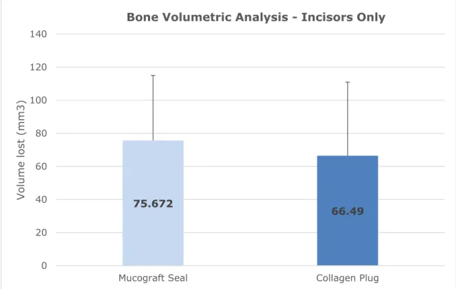
volumetric analysis, n=7 for the test group, and n=4 for the control group. For the incisor volumetric analysis, n=5 for the test group, and n=7 for the control group.
When analyzing a comparison between premolars in the test and control group (Figure 5), the test group (Bio-Oss Collagen® + Mucograft Seal®) had a volumetric mean loss of buccal bone of 70.49mm3 (SD=26.26), while the control group (Bio-Oss
Collagen® + HeliPlug®) had a volumetric mean loss of buccal bone of 66.42 mm3
(SD=26.45). Statistical analysis of the premolar site volumetric buccal bone remodeling indicated no significant difference (p=0.810) between the two groups considering the premolar teeth.
while the control group (Bio-Oss Collagen® + HeliPlug®) had a volumetric mean loss of buccal bone of 66.49mm3 (SD=44.41). Statistical analysis of the incisor site volumetric buccal bone remodeling indicated no significant difference (p=0.719) between the two groups of incisors.
4. Discussion
The present randomized controlled trial demonstrated that the use of either collagen plug or Mucograft Seal® over an extraction socket grafted with Bio-Oss Collagen®
It has been extensively reported that the amount of ridge resorption that occurs after tooth extraction is heavily influenced by the initial thickness of the buccal wall (Nevins 2006, Sanz 2010, Tomasi 2010), with a thicker initial thickness generally
leading to a smaller amount or resorption that occurs. It has also been reported that the buccal plate thickness decreases as one moves anteriorly along the maxillary arch. Huynh-Ba and collaborators analyzed socket wall dimensions in the upper maxilla, and found that the mean width of the buccal plate at premolar sites was 1.1mm, and dropped to a mean width of 0.8mm at anterior sites (canine to canine) (Huynh-Ba 2010). Vera et al. found that the median buccal alveolar bone thickness at the midroot was 1.03mm in premolars, and decreased to 0.70mm for incisors (Vera 2012). Yet another study reported that the mean buccal plate width in maxillary incisors and canines was only 0.6mm (Januario 2011). In a study by Araujo and Lindhe, ridge reduction in premolar sites was 18%, while in anterior sites, ridge reduction was
significantly higher at 34%. Therefore, it is possible that grouping premolar and incisor teeth together may mask differences between the two that could affect the reported mean measurements. In the present study, this theory was tested to compare the data between only premolar teeth in each group, and then to compare the data utilizing only the anterior teeth in each group. Even when grouped by tooth type, volumetric
The present trial also demonstrated that even with alveolar ridge preservation, resorption of buccal bone from baseline still occurs. This finding is in agreement with previous studies that have reported that placement of a biomaterial in the socket immediately after tooth extraction fails to fully prevent resorption of the edentulous alveolar ridge (Darby 2009, Pagni 2012, Araujo and Lindhe 2014). A review by Heggeler and collaborators in 2011 found that natural healing produces a ridge width reduction of 2.6-4.6mm, and even when looking at the material with the best results, there was still a loss of ridge width of 1.2mm post-extraction and socket grafting (Heggeler 2011). However, even though ridge resorption cannot be fully prevented, there is evidence that ridge preservation does reduce the need for further ridge augmentation for implant placement over spontaneous healing (Mardas 2015).
For calculation of linear and volumetric alveolar bone changes over the six-month study period, a non-contact reverse engineering system was utilized (Geomagic Control®). For analysis, the original STL data created using the CBCT data from the initial and 6-month timepoints was imported into the software, and then used to generate a computer-assisted superimposition by selecting areas of the data set in which no changes had taken place over the 6-month healing period, such as zygomatic arches and teeth that had been untouched. A previous precision analysis study
determined that 3-dimensional surface reconstruction using the Geomagic Control® software provides a reliable results breakdown, with a maximum superimposition deviation of 0.06mm ± 0.003, and an average error of 0.002mm (Wang 2014). The American Academy of Oral and Maxillofacial Radiology recommends CBCT imaging as the method of choice for evaluation and assessment of proposed implant sites due the high diagnostic yield and low radiation dose risk. Thus, CBCT volumetric data sets have become standard practice in implant treatment planning (Tyndall 2012).
two test subjects and four control subjects were excluded from linear analysis due to lack of reproducibility and accuracy of baseline and 6-month STL file superimposition. This was likely due to CBCT scatter created by pre-existing metal and ceramic
restorations on subject’s teeth. The scatter may make it difficult to create an accurate superimposition data, and increased the error measurement of the final superimposition. For future studies, it is recommended to exclude potential subjects who have a heavily restored dentition in the arch of interest. As an alternative, once could procure an optical impression and merge it with the 3D data set, thus reducing the effect that the scatter may have on the image analysis.
An interesting finding during the study duration was that complete epithelialization over the extraction socket was completed earlier in the test group (week-2) than in the control group subjects (week-4). This result likely has an effect on early wound healing characteristics, with earlier primary closure resulting in less exposure to the oral cavity. Thus, the test group may have expressed faster angiogenesis, connective tissue and bone maturation over the control group, which might not be evident if only bone quantity is being assessed. While results from this study were not significant for
volumetric differences in quantity of bone that had been lost, there may be a difference in the quality of bone that is present for future implant placement based on earlier healing characteristics and earlier primary closure of the wound. Future histological analysis of the alveolar bone from these subjects is warranted since trephine samples were obtained at the time of implant placement.
socket, it is possible that soft tissue preservation outcomes may be more significant with the use of this product compared to control materials. Further evaluation is merited to assess the effects of Mucograft Seal® on soft tissue volumetric outcomes.
In conclusion, the results of this study indicated that there is no statistically
REFERENCES
Araújo, M. G., da Silva, J. C. C., de Mendonça, A. F., & Lindhe, J. (2015). Ridge alterations following grafting of fresh extraction sockets in man. A randomized clinical trial. Clinical oral implants research, 26(4), 407-412.
Araújo, M. G., & Lindhe, J. (2005). Dimensional ridge alterations following tooth extraction. An experimental study in the dog. Journal of clinical periodontology, 32(2), 212-218.
Artzi, Z., & Nemcovsky, C. E. (1998). The application of deproteinized bovine bone mineral for ridge preservation prior to implantation. Clinical and histological observations in a case report. Journal of periodontology, 69(9), 1062-1067.
Avila‐Ortiz, G., De Buitrago, J. G., & Reddy, M. S. (2015). Periodontal regeneration– Furcation defects: A systematic review from the AAP regeneration workshop. Journal of periodontology, 86, S108-S130.
Darby, I., Chen, S. T., & Buser, D. (2009). Ridge preservation techniques for implant therapy. Int J Oral Maxillofac Implants, 24(Suppl), 260-271.
Ghanaati, S., Schlee, M., Webber, M. J., Willershausen, I., Barbeck, M., Balic, E., ... & Kirkpatrick, C. J. (2011). Evaluation of the tissue reaction to a new bilayered collagen matrix in vivo and its translation to the clinic. Biomedical materials, 6(1), 015010.
Heggeler, T, J. M. A. G., Slot, D. E., & Van der Weijden, G. A. (2011). Effect of socket preservation therapies following tooth extraction in non‐molar regions in humans: a systematic review. Clinical oral implants research, 22(8), 779-788.
Huynh‐Ba, G., Pjetursson, B. E., Sanz, M., Cecchinato, D., Ferrus, J., Lindhe, J., & Lang, N. P. (2010). Analysis of the socket bone wall dimensions in the upper maxilla in relation to immediate implant placement. Clinical oral implants research, 21(1), 37-42.
Januário, A. L., Duarte, W. R., Barriviera, M., Mesti, J. C., Araújo, M. G., & Lindhe, J. (2011). Dimension of the facial bone wall in the anterior maxilla: a cone‐beam computed tomography study. Clinical oral implants research, 22(10), 1168-1171.
Jung, R. E., Philipp, A., Annen, B. M., Signorelli, L., Thoma, D. S., Hämmerle, C. H., ... & Schmidlin, P. (2013). Radiographic evaluation of different techniques for ridge
preservation after tooth extraction: a randomized controlled clinical trial. Journal of clinical periodontology, 40(1), 90-98.
Landsberg, C. J., & Bichacho, N. (1994). A modified surgical/prosthetic approach for optimal single implant supported crown. Part I--The socket seal surgery. Practical periodontics and aesthetic dentistry: PPAD, 6(2), 11-7.
Nevins, M., Camelo, M., De Paoli, S., Friedland, B., Schenk, R. K., Parma-Benfenati, S., ... & Wagenberg, B. (2006). A study of the fate of the buccal wall of extraction sockets of teeth with prominent roots. International Journal of Periodontics & Restorative Dentistry, 26(1).
Nevins, M., & Mellonig, J. T. (1994). The advantages of localized ridge augmentation prior to implant placement: a staged event. International Journal of Periodontics & Restorative Dentistry, 14(2).
Pagni, G., Pellegrini, G., Giannobile, W. V., & Rasperini, G. (2012). Postextraction alveolar ridge preservation: biological basis and treatments. International journal of dentistry, 2012.
Sableman, E. E. (1985). Biology, biotechnology and biocompatibility of collagen. Biocompatibility of tissue analogs, 21-66.
Sanz, M., Cecchinato, D., Ferrus, J., Pjetursson, E. B., Lang, N. P., & Lindhe, J. (2010). A prospective, randomized‐controlled clinical trial to evaluate bone preservation using implants with different geometry placed into extraction sockets in the maxilla. Clinical oral implants research, 21(1), 13-21.
Tomasi, C., Sanz, M., Cecchinato, D., Pjetursson, B., Ferrus, J., Lang, N. P., & Lindhe, J. (2010). Bone dimensional variations at implants placed in fresh extraction sockets: a multilevel multivariate analysis. Clinical Oral Implants Research, 21(1), 30-36.
Tyndall, D. A., Price, J. B., Tetradis, S., Ganz, S. D., Hildebolt, C., & Scarfe, W. C. (2012). Position statement of the American Academy of Oral and Maxillofacial Radiology on selection criteria for the use of radiology in dental implantology with emphasis on cone beam computed tomography. Oral surgery, oral medicine, oral pathology and oral radiology, 113(6), 817-826.
Vera, C., De Kok, I. J., Reinhold, D., Limpiphipatanakorn, P., Yap, A. K., Tyndall, D., & Cooper, L. F. (2012). Evaluation of buccal alveolar bone dimension of maxillary anterior and premolar teeth: a cone beam computed tomography investigation. International Journal of Oral & Maxillofacial Implants, 27(6).
Wang, W., Gao, D. Q., & Yi, J. M. (2014). Precision Analysis of the Surface Reconstruction Model Based on Geomagic Qualify. In Applied Mechanics and Materials (Vol. 618, pp. 443-447). Trans Tech Publications.
APPENDIX 1: INCLUSION CRITERIA
Subjects must be adult males or females age 18 to 80 years (inclusive).
Subjects must be able and willing to follow study procedures and instructions in English.
Subjects must have read, understood and signed an informed consent form in English.
Subjects must have a maxillary premolar, canine, lateral incisor, or central incisor with a restorative or periodontal hopeless prognosis in which an implant is indicated without any sinus lift required.
APPENDIX 2: EXCLUSION CRITERIA
Individuals who have a chronic disease with oral manifestations. Individuals who exhibit gross oral pathology.
The use of either antibiotics or chronic use (more than 7 days) of NSAIDs within 1 month prior to screening examination.
Individuals that require antibiotic prophylaxis prior to dental treatment.
Chronic treatment (i.e. two weeks or more) with any medication known to affect periodontal status (e.g. phenytoin, calcium antagonists, cyclosporine, Coumadin) within 1 month prior to screening examination.
Uncontrolled diabetes mellitus (HbA1c >7) within 3 months prior to screening examination.
Individual with uncontrolled parafunctional habits, such as clenching and bruxing on objects, that could adversely impact implant survival.
Individuals with a history of intravenous bisphosphonates.
Individuals with active infectious diseases such as hepatitis, HIV or tuberculosis. Current cigarette smokers.
Individuals who are known to be pregnant, breastfeeding or planning to become pregnant within 6 months.
Individuals with blood disorders (hemophilia) and /or currently taking anticoagulant medications, such as heparin, warfarin, or clopidogrel.
Individuals receiving any therapy known to affect healing, such as high dose corticosteroids, radiation therapy or chemotherapy.
Individuals allergic to topical or local anesthesia.
Individuals who require maxillary sinus augmentation prior to dental implant therapy.
APPENDIX 3: PRESCRIPTION MEDICATIONS FOLLOWING EXTRACTION
Following Extraction Surgery
Rx Amoxicillin 500mg
Disp 30 tabs
Sig Take 1 tab three times daily
If Penicillin Allergy: Rx Azithromycin 250mg
Disp 6 tabs
Sig Take 2 tabs 1st day then 1 tab qd for 4 days
Rx Ibuprofen 600mg
Disp 20 tabs
Sig Take 1 tab every 6h for two days then prn pain
Rx Hydrocodone/APAP 5/325mg
Disp 20 tabs
Sig Take 1 tab every 4-6h prn pain
Rx 0.12% Chlorhexidine Rinse Disp 1 X 1.8 oz bottle
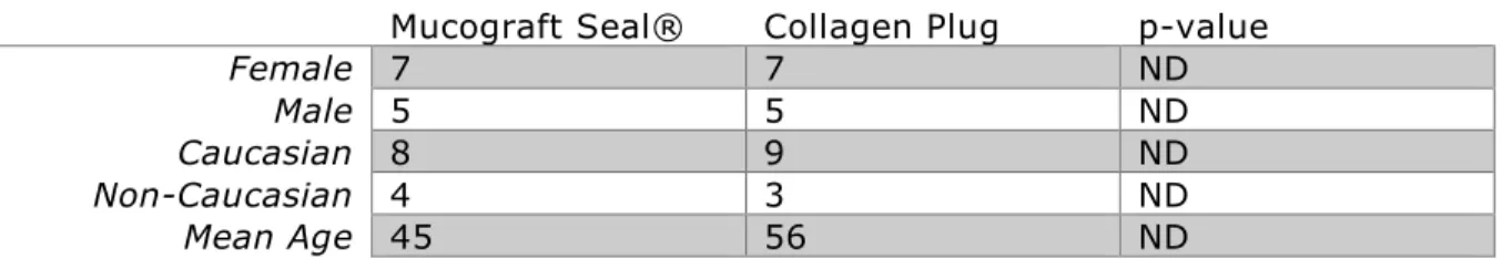
Table 1. Subject demographics
Mucograft Seal® Collagen Plug p-value
Female 7 7 ND
Male 5 5 ND
Caucasian 8 9 ND
Non-Caucasian 4 3 ND
Table 2. Tooth distribution (n=23)
Mucograft Seal® Collagen Plug
Central Incisor 1 3
Lateral Incisor 4 3
Canine 0 1
1st Premolar 4 3
Figure 2: Mean linear bone loss for test (n=10) and control (n=7) groups at six-months post-extraction and ridge preservation. Measures were obtained at 1mm, 3mm, and 5mm below the alveolar crest at the mid-buccal of the extraction site. No statistically significant differences at 1mm and 3mm (p>0.05). Statistically significant difference at 5mm (p<0.05) at 5mm in favor of the control group (*).
2.17
1.86
1.63 1.76
1.25
0.86
0 0.5 1 1.5 2 2.5 3 3.5
1mm 3mm 5mm
Li
nea
r
Loss
(mm
)
Distance from Alveolar Crest
Bone Linear Analysis
Figure 3. Mean linear bone loss for premolar teeth only. Test (n=6) and control (n=2) groups at six-months post-extraction and ridge preservation. Measures were obtained at 1mm, 3mm, and 5mm below the alveolar crest at the mid-buccal of the extraction site. No statistically significant differences between the groups (p>0.05).
1.83
1.61
1.39 1.86
1.36
1.02
0 0.5 1 1.5 2 2.5 3
1mm 3mm 5mm
Li
n
ear
Lo
ss
(
mm
)
Distance from Alveolar Crest
Bone Linear Analysis - Premolars Only
Figure 4: Mean linear bone loss for incisor teeth only. Test (n=4) and control (n=5) groups at six-months post-extraction and ridge preservation. Measures were obtained at 1mm, 3mm, and 5mm below the alveolar crest at the mid-buccal of the extraction site. No statistically significant differences at 1mm (p>0.05). Statistically significant
difference at 3mm and 5mm (p<0.05) in favor to the Control group (*).
2.69 2.23 2.00 2.03 1.34 0.88 0 0.5 1 1.5 2 2.5 3 3.5 4
1mm 3mm 5mm
Li n ear Lo ss ( mm )
Distance from Alveolar Crest
Bone Linear Analysis - Incisors Only
Mucograft Seal Collagen Plug
*
Figure 5: Mean volumetric bone remodeling for test (n=12) and control (n=11) groups at six-months post-extraction and ridge preservation. No statistically significant
difference between the groups (p>0.05).
72.65
60.93
0 20 40 60 80 100 120
Mucograft Seal Collagen Plug
Vol
u
me
lo
ss
(
mm
3)
Figure 6: Mean volumetric bone remodeling for premolar teeth only. Test (n=7) and control (n=4) groups at six-months post-extraction and ridge preservation. No statistically significant difference between the groups (p>0.05).
70.50 66.41
0 20 40 60 80 100 120
Mucograft Seal Collagen Plug
Vol
u
me
Lo
ss
(
mm
3)
Figure 7: Mean volumetric bone remodeling for incisor teeth only. Test (n=5) and control (n=7) groups at six-months post-extraction and ridge preservation. No statistically significant difference between the groups (p>0.05).
75.672
66.49
0 20 40 60 80 100 120 140
Mucograft Seal Collagen Plug
Vol
u
me
lo
st (m
m3
)