DEVELOPMENT OF A NOVEL METHOD FOR GENOME-WIDE IDENTIFICATION OF PROTEINS EXPORTED DURING INFECTION AND FUNCTIONAL STUDIES OF ONE IN
VIVO EXPORTED PROTEIN IN MYCOBACTERIUM TUBERCULOSIS
Ellen Foot Perkowski
A dissertation submitted to the faculty of the University of North Carolina at Chapel Hill in partial fulfillment of the requirements for the degree of Doctor of Philosophy in the Department
of Microbiology and Immunology in the School of Medicine.
Chapel Hill 2015
Approved by:
Miriam Braunstein
Thomas Kawula
Bill Goldman
Peggy Cotter
© 2015
ABSTRACT
Ellen Foot Perkowski: Development of a Novel Method for Genome-wide Identification of Proteins Exported during Infection and Functional Studies of One in vivo Exported Protein in
Mycobacterium tuberculosis
(Under the direction of Miriam Braunstein)
Intracellular pathogens manipulate and outwit the host’s immune defenses and reprogram
the hostile intracellular environment into a hospitable replicative niche. The intracellular
pathogen Mycobacterium tuberculosis is responsible for the disease tuberculosis, which kills approximately 1.5 million people per year. M. tuberculosis produces many proteins that are
exported: transported out of the bacterial cytoplasm to the bacterial cell surface and out into the
host environment. Exported proteins are located at the host-pathogen interface, in an ideal
location to manipulate the host response and allow for intracellular growth, and exported proteins
contribute significantly to virulence. Unfortunately, approaches used to identify proteins
exported by the bacteria are limited to bacteria growing in laboratory media (in vitro). Because in
vitro conditions cannot mimic the complexity of the host environment, there are likely critical exported virulence factors that have been missed because they are only exported in the context of
infection. The main objective of the research described in this dissertation was to develop a
method to identify proteins that are exported by bacterial pathogens during infection of a host (in vivo). We developed a novel method that we refer to as EXIT, EXported In vivo Technology,
proteins exported significantly more in vivo than in vitro, suggesting that temporal or spatial control of their export is important to infection. 21 of these 38 proteins have unknown function,
making them particularly interesting for future functional characterization. We focused on one of
the EXIT identified in vivo exported proteins, OmasA, a protein of unknown function for further
study. We demonstrated that OmasA was required for M. tuberculosis virulence in a mouse model of tuberculosis. We further demonstrated a function for OmasA in stabilizing
multi-protein Mce transporters required for lipid import. Future studies will focus on assigning
function to new EXIT identified exported proteins, in particular proteins whose export is
TABLE OF CONTENTS
LIST OF FIGURES ... ix
LIST OF TABLES ... xi
LIST OF ABBREVIATIONS AND SYMBOLS ... xii
CHAPTER 1: INTRODUCTION ... 1
Bacterial exported proteins are critical to the virulence of intracellular pathogens ... 6
Mechanisms of protein export in M. tuberculosis ... 6
The General Secretion System: SecA1 dependent protein export ... 8
Co-translational Export of Transmembrane Proteins ... 9
Accessory Sec Export Pathway: SecA2 dependent protein export ... 9
Export of Pre-folded Substrates: The Twin-Arginine Translocation Pathway ... 10
Type VII secretion: ESX Specialized Secretion Systems ... 11
Identification of M. tuberculosis exported proteins ... 13
Prediction of Exported Proteins by Bioinformatics (in silico) ... 14
Experimental Identification of Exported Proteins: Mass Spectrometry ... 17
Experimental Identification of Exported Proteins: Genetic Reporters ... 20
Exported proteins play important roles in M. tuberculosis virulence ... 27
Exported proteins important to M. tuberculosis virulence ... 27
Role of Lipid Import and Catabolism in M. tuberculosis virulence ... 28
Summary ... 30
CHAPTER 2: PROBING FOR BACTERIAL PROTEINS AT THE
HOST-PATHOGEN INTERFACE ... 44
Introduction ... 44
Current methods for identifying exported proteins ... 45
Protein export in M. tuberculosis ... 49
Identification of proteins exported during infection ... 50
Results ... 51
Construction of a comprehensive EXIT library for M. tuberculosis ... 53
Optimizing selection of exported ‘BlaTEM-fusions in β-lactam treated mice ... 56
Performing EXIT in M. tuberculosis infected mice ... 59
EXIT successfully identified exported proteins ... 62
EXIT identified new exported proteins ... 67
EXIT fusions identify proteins exported in the lungs ... 70
EXIT exported fusions provide topology information for membrane proteins ... 71
Identification of proteins exported significantly more in vivo than in vitro ... 80
Discussion ... 93
Methods: ... 101
Attributions ... 110
REFERENCES ... 133
CHAPTER 3: AN ORPHANED MCE-ASSOCIATED PROTEIN OF MYCOBACTERIUM TUBERCULOSIS IS A VIRULENCE FACTOR THAT STABILIZES MCE TRANSPORTERS ... 149
Introduction ... 149
Results ... 153
OmasA is important for murine infection ... 153
Deletion of omasA in Mycobacterium smegmatis leads to a mce mutant
morphology phenotype ... 162
OmasA is required for cholesterol utilization ... 164
OmasA is required for cholesterol uptake ... 166
OmasA stabilizes the Mce1 transport complex ... 167
Discussion ... 171
Experimental Procedures ... 177
Attributions ... 183
REFERENCES ... 188
CHAPTER 4: DISCUSSION ... 193
Introduction ... 193
Probing for bacterial proteins at the host-pathogen interface ... 193
Characterization of in vivo exported proteins ... 202
Conclusion ... 211
REFERENCES ... 212
APPENDIX I: CALCULATIONS TO DETERMINE GENOMIC LIBRARY DENSITY AND MOUSE NUMBERS ... 219
Determine size for genomic DNA library ... 219
Determine the number of mice required for comprehensive library testing ... 221
REFERENCES ... 224
APPENDIX II: EXIT RESULTS... 225
REFERENCES ... 259
APPENDIX III: FUNCTIONAL GENOMICS DATABASE FOR MYCOBACTERIUM TUBERCULOSIS ... 261
LIST OF FIGURES
Figure 1.1. Structure of a classic human granuloma ... 4
Figure 1.2. Cellular structure of M.tuberculosis ... 5
Figure 1.3. Exported proteins of M.tuberculosis ... 7
Figure 1.4. Genetic reporters identify a high proportion of proteins with export Signals ... 21
Figure 1.5. MS based methods identify (ID) high abundance cytoplasmic proteins as Exported... 23
Figure 1.6. ‘BlaTEM reporter activity is dependent on export ... 25
Figure 2.1.The ‘BlaTEM reporter ... 48
Figure 2.2 The EXported In vivo Technology (EXIT) strategy ... 52
Figure 2.3 The EXIT library comprehensively represented the Mycobacterium tuberculosis proteome ... 55
Figure 2.4. The ‘BlaTEM reporter is compatible with β-lactam treatment during murine infection ... 58
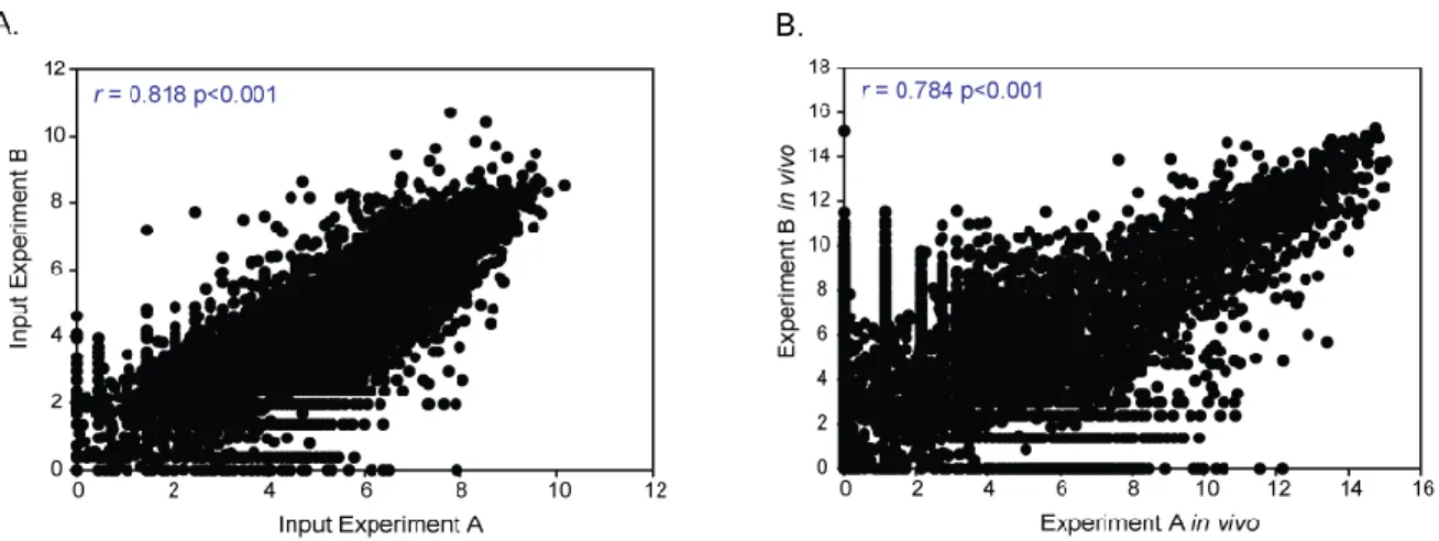
Figure 2.5. EXIT replicate experiments demonstrated high reproducibility and correlation ... 61
Figure 2.6. EXIT identified 593 proteins as exported during murine infection ... 63
Figure 2.7. EXIT successfully and reliably identified exported proteins ... 65
Figure 2.8. EXIT identified new exported proteins ... 68
Figure 2.9. EXIT exported fusions clarify the topology of membrane protein MmpL3 ... 73
Figure 2.10. EXIT exported fusions support composite model of MmpL and MmpS proteins ... 75
Figure 2.11. EXIT exported fusions clarify topology of EMB proteins important to drug resistance in M. tuberculosis ... 77
Figure 2.12. EXIT exported fusions clarify topology of ESX Type VII secretion system membrane proteins ... 79
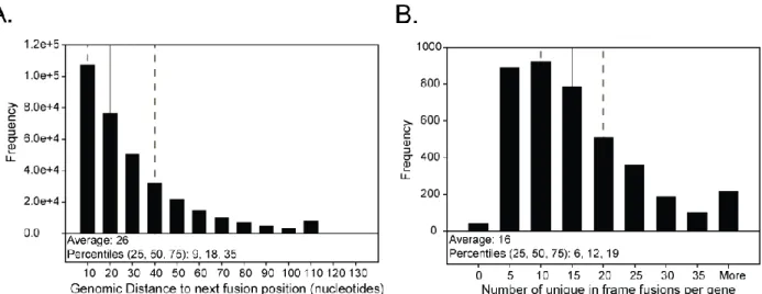
Figure 2.14. Statistical modeling identified 38 proteins exported significantly
more in vivo than in vitro ... 83 Figure 2.15. Proteins of the Mce2 and Mce3 lipid transporters of M. tuberculosis
are reliably exported more in vivo than in vitro ... 91 Figure 3.1. Rv0199 (OmasA) is a transmembrane protein predicted to be a
Mce-associated protein ... 151
Figure 3.2. OmasA is required for early growth and virulence during murine
infection ... 154
Figure 3.3. Mice infected with the omasA mutant have reduced histopathology
compared to WT infected mice ... 156
Figure 3.4. Alignment of all Mas proteins ... 158
Figure 3.5. Phyre 2, an online structural prediction program, predicts with high
confidence that all Mas and Omas proteins form a NTF2-like fold ... 159
Figure 3.6. Mce operons in M. smegmatis and M. leprae ... 161 Figure 3.7. The omasAms mutant shares a morphology phenotype with mce operon
mutants ... 163
Figure 3.8. OmasA is required for M. smegmatis and M. tuberculosis to utilize
cholesterol ... 165
Figure 3.9. OmasA is required for cholesterol uptake ... 168
Figure 3.10. Absence of OmasA results in Mce1 protein instability ... 170
Figure 3.11. OmasA is an integral membrane protein that is important to Mce
LIST OF TABLES
Table 1.1. Exported Protein Identification in M. tuberculosis ... 32
Table 2.1. Plasmids used in this study. ... 111
Table 2.2. Bacterial strains used in this study. ... 112
Table 2.3. Primers used in this study. ... 113
Table 2.4. Proof of principle enrichment studies. ... 114
Table 2.5. Known exported proteins. ... 120
Table 2.6. Most abundant cytoplasmic proteins. ... 124
Table 2.7. EXIT identified proteins with no in silico export signals ... 126
Table 2.8. EXIT exported fusions in unannotated regions. ... 128
Table 2.9. EXIT exported proteins only identified in the lungs. ... 129
Table 2.10. EXIT identified proteins exported significantly more in vivo than in vitro. ... 132
Table 3.1. Bacterial strains used in this study ... 185
Table 3.2. Plasmids used in this study ... 186
Table 3.3. Primers used in this study ... 187
LIST OF ABBREVIATIONS AND SYMBOLS
hydrophobic amino acid
C degrees Celsius
µL microliter
ABC ATP-binding cassette
ADS Albumin Dextrose Saline
Ag antigen
ANL azidonorleucine
ANOVA analysis of variance
ATP adenosine triphosphate
BCG bacillus Calmette-Guerin attenuated vaccine strain
Bla β-lactamase
Bp nucleotide base pairs
BSL-3 Biosafety level 3
C- carboxy terminus of a protein
C14 carbon-14
CD cluster of differentiation
cDNA complementary DNA
CFP culture filtrate protein
cfu colony forming units
Cl chlorine
Cs cesium
D aspartic acid
dATP deoxyadenosine triphosphate
dH2O distilled deionized water
DNA deoxyribonucleic acid
Dpi days post infection
E glutamic acid
Ecc ESX conserved component
ESAT-6 early secreted antigenic target of 6 kDa
Esp ESX specific protein
ESX ESAT-6 secretion system
EXIT EXported In vivo Technology
FDR false discovery rate
g grams
g gravity
GC guanine and cytosine
Glu glutamic acid
GR genetic reporter
H hydrogen
HA Influenza hemagglutinin epitope
HAI histological activity index
HIV Human Immunodeficiency Virus
HRP horseradish peroxidase
Hyg hygromycin
ICM/Dot intracellular multiplication/defective organelle trafficking
IVET in vivo expression technology
Kan kanamycin
kb kilobase
kD kiloDalton
kDa kiloDalton
Lab laboratory
LB Luria Bertani media
Log10 logarithm base 10
Log2 logarithm base 2
M. smegmatis Mycobacterium smegmatis
M. tuberculosis Mycobacterium tuberculosis
Mas Mce associated protein
Mce mammalian cell entry
MEM membrane
Mg magnesium
mg milligram
mL milliliter
MmpL mycobacterial membrane protein large
MS mass spectrometry
Msmeg Mycobacterium smegmatis
N- amino-terminus of a protein
Na sodium
NEB New England Biolabs
NH4 ammonium
NIH National Institutes of Health
OD optical density
omasA orphaned Mce associated protein A
ORF open reading frame
p plasmid
PBS phosphate buffered saline
PCR polymerase chain reaction
PDIM phthiocerol dimycocerosate
PE proline glutamic acid repeat containing protein
PGRS polymorphic guanine-cytosine rich sequence
PhoA alkaline phosphatase
Pi inorganic phosphate
PO4 phosphate
PPE proline proline glutamic acid repeat containing protein
Pro proline
qRT-PCR quantitative reverse-transcriptase PCR
R arginine
R
resistant
Res resolvase cassette
RNA ribonucleic acid
RND Resistance Nodulation Division
Rv M. tuberculosis strain H37Rv attenuated S
sensitive
SDS-PAGE sodium dodecyl sulfate polyacrylamide gel electrophoresis
Sec secretory pathway
SL-1 sulfolipid-1
SOL soluble
SP signal peptide
SPI Salmonella pathogenicity island
SRP signal recognition particle
T3SS Type III secretion system
Tad Tight adherence
Tat Twin-arginine translocation
TM transmembrane domain
Tn transposon
Tn-seq Transposon deep sequencing
TraSH Transposon site hybridization
tRNA transfer RNA
Tw polyoxyethylene sorbitan monoleate
Tween polyoxyethylene sorbitan monoleate
WCL whole cell lysate
WT wild type
X variable amino acid
Y tyrosine
CHAPTER 1: INTRODUCTION
In 1882 Robert Koch described the bacterium Mycobacterium tuberculosis as the causative agent of the disease then known as “consumption” and now better known as
tuberculosis (Sakula, 1983). M. tuberculosis fulfilled “Koch’s postulates”, cultures of M. tuberculosis could be isolated from patients, purified, and used to infect laboratory animals (Koch, 1932). These animals would then develop characteristic tuberculosis disease, and M.
tuberculosis bacilli could be isolated from their lung lesions (Koch, 1932). Prior to Koch’s discovery, Jean-Antoine Villimen had demonstrated that a rabbit could develop tuberculosis after
inoculation of material from a human granuloma; however, many still attributed the disease to
poor environmental conditions or genetics (Daniel, 2006). Koch’s discovery led to a more broad
understanding of the infectious nature of tuberculosis (Daniel, 2006). One of the key symptoms
of tuberculosis, prolonged coughing, is now know to be instrumental in allowing the bacteria to
escape the lungs of an infected individual and be transmitted through the air to surrounding
people. This transmission is so effective that one-third of the world’s population (two billion
people), are thought to be infected with M. tuberculosis (World Health Organization, 2014). The development of the antibiotic streptomycin in the 1940s led to the first treatment for
tuberculosis (Schatz et al., 1944). Even early on, it was clear that tuberculosis was harder to treat than many other bacterial pathogens. Treatment regimens had to be very long or else patients
would quickly relapse, often with drug resistant strains (Sakula, 1983). Despite 70 years since
the first anti-mycobacterial drug was introduced, we are still struggling with effective treatments
presenting active tuberculosis each year, approximately 480,000 patients are infected with
multi-drug resistant strains (World Health Organization, 2014). In the absence of effective antibiotic
treatment tuberculosis is usually fatal (Tiemersma et al., 2011). Today, despite the advances of modern medicine, 1.5 million people continue to die from tuberculosis every year (World Health
Organization, 2014).
Upon inhaling M. tuberculosis, the bacteria are transferred down into the lungs and into the alveolar spaces, where they can be engulfed by resident alveolar macrophages (Russell et al.,
2010). These macrophages are normally the immune system’s first line of defense against
inhaled material, and will usually engulf and destroy invading bacteria. M. tuberculosis,
however, has developed many strategies to avoid destruction, and is able to grow inside
macrophages, in a compartment called the phagosome (Russell et al., 2010). M. tuberculosis reconstructs the phagosome into a hospitable environment, blocking phagosomal maturation and
preventing fusion with lysosomes, which would normally lead to destruction of the phagosomal
contents (Rohde et al., 2007; Armstrong & Hart, 1971). M. tuberculosis will undergo many
rounds of replication inside macrophages. Although these macrophages fail to eliminate the M. tuberculosis bacilli, they will produce chemoattractant molecules resulting in an influx of inflammatory cells including macrophages, dendritic cells, and neutrophils (Russell et al., 2010;
Orme & Basaraba, 2014). Macrophages present antigens to dendritic cells at the site of infection,
and these dendritic cells migrate to lymph nodes where they prime T-cells. Antigen specific T
cells are then recruited to the site of lung infection where they recognize and activate M. tuberculosis infected macrophages, which serves to limit further intracellular bacterial
replication, and they organize the inflammatory cells (Russell et al., 2010; Orme & Basaraba,
focus (Russell et al., 2010; Orme & Basaraba, 2014). This process ultimately leads to formation of a highly specialized structure called a granuloma (Figure 1.1).
Once granulomas have formed, most infected individuals will have no symptoms, a
condition known as latent tuberculosis. Although the immune system contains M. tuberculosis
within granulomas, the bacteria are not fully eradicated and for approximately 10% of infected
individuals this containment will ultimately fail during their lifetime (Flynn & Chan, 2001).
Individuals who fail to contain the M. tuberculosis bacilli will develop active tuberculosis.
Although certain risk factors are known, such as immune dysfunction, the cause of this
reactivation and the factors determining when it will occur are still mysterious (Flynn & Chan,
2001). The underlying ability for M. tuberculosis to cause disease, survive in the macrophage environment and avoid complete destruction by the immune system remains poorly understood.
M. tuberculosis is challenging to treat with antibiotics for several reasons. Drugs must
penetrate granulomas and host cells to reach intracellular M. tuberculosis bacilli. M. tuberculosis bacilli can persist in a non-replicating state, which is resistant to most antibiotics. Additionally,
the M. tuberculosis cellular structure is unique, and poses further complications to drug
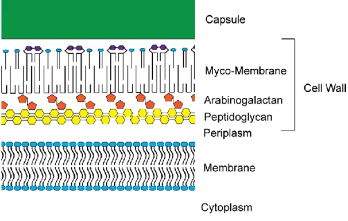
treatment. M. tuberculosis has both a cytoplasmic membrane and a specialized outer membrane referred to as the myco-membrane (Figure 1.2). The outermost layer of M. tuberculosis is a thick
capsule surrounding the myco-membrane. The myco-membrane is composed of specialized long
chain lipids including mycolic acids. The myco-membrane is covalently linked to
arabinogalactan, which is attached to peptidoglycan within the periplasm (Figure 1.2). As well as
Figure 1.2. Cellular structure of M. tuberculosis. M. tuberculosis is an acid-fast organism with an cytoplasmic membrane consisting of a phospholipid bilayer (represented in blue) surrounded by a layer of peptidoglycan (yellow). Unique features of the M. tuberculosis cell wall include arabinogalactan (orange) which covalently crosslinks the outer myco-membrane to the peptidoglycan. The myco-membrane primarily consists of specialized lipids composed of mycolic acids (Marchand et al., 2012). The
Bacterial exported proteins are critical to the virulence of intracellular pathogens
We are beginning to understand the important role that exported proteins play in
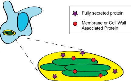
virulence for many bacterial pathogens. This dissertation will refer to exported proteins as any
protein produced in the bacterial cytoplasm and actively transported to the bacterial cytoplasmic
membrane, cell wall, or out into the host environment (Figure 1.3). Exported proteins lie at the
host-pathogen interface, and thus are in an ideal location to interact with the host and contribute
to virulence. Many of the best studied virulence factors are exported proteins. For example,
virulence of the bacterial pathogen Legionella pneumophila requires a type IV secretion system (ICM/Dot), which delivers nearly 300 proteins out of the bacterium and directly into the
cytoplasm of host cells (Isaac & Isberg, 2014). Effectors of the ICM/Dot system mediate
construction of the Legionella-containing vacuole, intracellular replication, and evasion of lysosome fusion (Isaac & Isberg, 2014). Salmonella enterica, another well studied bacterial
pathogen, relies on exported proteins to attach to host cells, invade, program the fate of its
intracellular vacuole, and ultimately replicate to cause infection (Agbor & McCormick, 2011).
Given the importance of exported proteins to intracellular pathogens, it is not surprising that M. tuberculosis also relies on numerous exported proteins to survive and grow in macrophages and cause disease in a host. The goal of this dissertation was to identify proteins that are exported in
the host, and begin the process of assigning function to in vivo exported proteins of M. tuberculosis.
Mechanisms of protein export in M. tuberculosis
Currently there are four types of systems known to export proteins out of the cytoplasm
of M. tuberculosis: the general Sec pathway, the SecA2-dependent pathway, the Tat
transport mechanism as currently understood for each of these export pathways, and a review of
the general importance of each export pathway to M. tuberculosis is described below. Because
each of these systems exports multiple proteins, when mutants defective in any one of these
pathways are studied the cumulative effect of many exported proteins not being properly
localized is observed. Much of what we currently know about the role of M. tuberculosis exported proteins in virulence comes not from studying and identifying the role of individual
proteins, rather from studying genetic deletions disrupting entire export systems.
The General Secretion System: SecA1 dependent protein export
The general secretion system (Sec) is the housekeeping system that carries out the bulk of
export/secretion, as recently reviewed (Ligon et al., 2012). The Sec pathway is essential and is required for the majority of protein export that occurs in all bacteria. Sec exported proteins are
synthesized as precursors containing N-terminal signal peptides, which direct them to be
post-translationally exported by the Sec pathway (Ligon et al., 2012). SecA1 is an ATPase which recognizes signal peptides of proteins for export, delivers them to the SecYEG membrane
channel, and undergoes successive rounds of ATP hydrolysis to transport the protein across the
cytoplasmic membrane (Ligon et al., 2012). After export, a signal peptidase cleaves the signal peptide and releases the mature domain of the protein into the periplasm. Sec exported proteins
can remain in the cell wall or get fully secreted out of the bacterial cell by a poorly defined
second mechanism. As in other bacteria, the Sec system is predicted to export the majority of
proteins in M. tuberculosis, including proteins required for basic cellular physiology, cell maintenance, and virulence (Ligon et al., 2012). Lipoproteins are a subset of Sec exported proteins that have a lipid moiety attached to their N-terminus responsible for localizing
peptide and are cleaved by a specialized lipoprotein signal peptidase (Ligon et al., 2012). The lipoprotein signal peptidase is required for virulence, highlighting the importance of exported
lipoproteins to M. tuberculosis (Sander et al., 2004). SecA1, SecYE, and the general signal peptidase of mycobacteria are all essential for bacterial cell viability (Ligon et al., 2012).
Co-translational Export of Transmembrane Proteins
The SecYEG channel is additionally important to the export of integral membrane
proteins, through a recently reviewed mechanism (Facey & Kuhn, 2010; Ligon et al., 2012). As
polypeptide chains emerge from the ribosome they are scanned for hydrophobic domains that
may constitute transmembrane domains by the signal recognition particle (SRP). Upon detection
of a hydrophobic domain, SRP will bind and transport the growing polypeptide chain and
ribosome complex to the SRP receptor FtsY. FtsY will pass the protein to the SecYEG complex,
where the protein will undergo co-translational export. Transmembrane domains can pass
sideways through a gate in the SecY channel with the help of YidC to stably integrate into the
membrane. A subset of transmembrane proteins can be inserted by YidC alone (Facey & Kuhn,
2010). Transmembrane proteins are required for basic cellular physiology, cell maintenance, and
virulence; therefore, SRP, FtsY, and YidC are all essential to bacterial cell viability (Ligon et al., 2012).
Accessory Sec Export Pathway: SecA2 dependent protein export
An unusual property of mycobacteria is that they have two non-redundant SecA
homologues (SecA1 and SecA2) (Braunstein et al., 2001). The SecA2 protein of mycobacteria is thought to function much like SecA1, binding to SecA2 substrates, transporting them to the
SecYEG complex, and transporting preproteins through SecY with rounds of ATP hydrolysis
peptides, and these signal peptides are required for export (Gibbons et al., 2007). Surprisingly, the signal peptide alone does not confer SecA2 specificity (Feltcher et al., 2013). Undefined
elements within the mature domain determine export through the SecA2 pathway, and the
leading hypothesis is that the SecA2 pathway in mycobacteria may exist to export proteins that
tend to fold prior to export (Feltcher et al., 2013). Furthermore, not all SecA2 substrates have signal peptides, highlighting the importance of additional targeting domains within the mature
domain of SecA2 exported proteins (Braunstein et al., 2001; Braunstein et al., 2003).
SecA2 is not essential for M. tuberculosis growth in vitro, but mutants lacking a functional SecA2 dependent protein export pathway are significantly attenuated (Braunstein et
al., 2003; Kurtz et al., 2006). Mice infected with a ΔsecA2 mutant of M. tuberculosis have lower bacterial burden and survive longer than WT M. tuberculosis infected mice (Braunstein et al., 2003). Additionally, the ΔsecA2 mutant is unable to grow in macrophages (Kurtz et al., 2006).
These results indicate that one or more proteins exported through the SecA2 dependent protein
export pathway are required for virulence. Recent evidence suggests that SecA2 exported
proteins play roles in preventing phagosomal maturation (Sullivan et al., 2012) and promoting nutrient acquisition (Feltcher et al., 2015), although the contribution of individual SecA2 exported proteins to attenuation of the ΔsecA2 mutant has yet to be determined.
Export of Pre-folded Substrates: The Twin-Arginine Translocation Pathway
Mycobacteria use the Twin-Arginine Translocation (Tat) pathway to export pre-folded
proteins in a Sec-independent pathway, which has been recently reviewed (Ligon et al., 2012). Tat exported proteins contain distinct N-terminal signal peptides which direct them for export
through the TatABC complex. The TatBC membrane protein complex binds Tat precursors and
forms homo-oligomers of varying sizes to form a channel large enough to transport the
pre-folded substrate. Transport of pre-proteins through the TatABC complex is energized by the
proton motive force. After export, the signal peptides of Tat exported proteins can be cleaved by
signal peptidases (Ligon et al., 2012).
The Tat export pathway has been studied in many bacterial pathogens and is frequently
required for export of virulence factors and, thus, contributes to pathogenesis (Ligon et al., 2012). The prediction of Tat exported proteins is clouded by the limited agreement between
prediction algorithms; however, as many as 95 proteins may be exported by the Tat pathway in
M. tuberculosis (McDonough et al., 2008). Characterized Tat substrates include proteins
important to both drug resistance and virulence. The Tat pathway is not essential for the in vitro viability of most bacterial, thus it is surprising that the Tat pathway is essential to M. tuberculosis viability in vitro (Saint-Joanis et al., 2006).
Type VII secretion: ESX Specialized Secretion Systems
M. tuberculosis encodes five Type VII secretion systems, named ESX-1 to ESX-5, which
are named for the first known exported substrate of these systems, ESAT-6. ESX secretion was
recently reviewed in (Houben et al., 2014). ESX gene clusters contain genes encoding ESX conserved components (Ecc), ESX specific proteins (Esp), and mycosins (MycP) which form the
export machinery. Some genes encoding ESX substrates are also located within these genomic
regions, including genes encoding the classical ESAT-6 like proteins and Pro-Glu and
Pro-Pro-Glu repeat containing proteins (PE/PPE) (Houben et al., 2014). The export mechanism of Type VII systems is under active investigation. These systems appear to form multi-protein transport
complexes with components in the cytoplasm and cytoplasmic membrane (Houben et al., 2014).
prior to export and are predicted to function as chaperones (Houben et al., 2014). The exported substrates are thought to be targeted, possibly with the assistance of chaperones, to the large ESX
membrane complex consisting of conserved components EccBCDE, all of which are required for
ESX secretion; however, the individual role of each protein within the complex remains unclear
(Houben et al., 2014). EccD is a multi-membrane spanning protein predicted to form a channel for protein transport (Houben et al., 2014). EccC contains three nucleotide binding domains, and is predicted to energize transport of exported substrates through the complex by ATP hydrolysis
(Houben et al., 2014). The subtilisin-like protease, MycP, cleaves at least one ESX substrate and may additionally regulate export in a protease-independent process. Because many ESX
substrates are fully secreted into the culture filtrate, it has been hypothesized that the ESX
secretion system spans both the cytoplasmic membrane and the outer myco-membrane, to direct
proteins out of the bacterium in a single step from the cytoplasm to the extracellular environment
(Houben et al., 2014). However, so far, no ESX secretion components have been identified within the myco-membrane.
The full repertoire of exported substrates of each ESX system is still undefined. ESX
exported proteins do not contain classical signal peptides; rather, a YxxxD/E conserved motif has
been shown to be required for export (Daleke et al., 2012). Surprisingly, the classical substrate
ESAT-6 does not contain this motif. ESX substrates are often exported as heterodimers, and
often it is only one of the proteins in the complex that contains the YxxxD/E motif (Houben et
al., 2014). ESX-1 is the best characterized ESX system in M. tuberculosis. ESX-1 has been shown to export a small number of proteins, including EsxA (ESAT-6), EsxB (CFP-10), and
EspB (Houben et al., 2014). Mycobacterium marinum, a pathogenic mycobacterial species
7% of the M. tuberculosis proteome are Pro-Glu or Pro-Glu Glu repeat containing proteins (PE/PPE proteins), most of which are thought to be surface localized (Goldberg et al., 2014;
Banu et al., 2002; Brennan et al., 2001). In M. marinum, the ESX-5 system was shown to export a large number of PE/PPE proteins, including those encoded outside of the ESX-5 locus
(Abdallah et al., 2009). However, only a subset of PE/PPE proteins in M. tuberculosis are exported through ESX-5, although proteins in M. tuberculosis containing PE domains do have the YxxxD/E motif (Bottai et al., 2012). Future studies are necessary to characterize exported
substrates of the additional ESX systems.
ESX-1 plays a clear role in M. tuberculosis virulence, as shown by attenuation of esx-1
mutants during mouse and macrophage infection (Pym et al., 2002; Lewis et al., 2003). ESX-2 and ESX-4 have not been previously studied. ESX-3 is essential for M. tuberculosis growth in vitro due to a role in iron acquisition(Siegrist et al., 2009). Additionally, ESX-5has recently
been shown to be important during M. tuberculosis infection, as shown by attenuation of esx-5 mutants during mouse and macrophage infection (Bottai et al., 2012).
Identification of M. tuberculosis exported proteins
Comprehensive study of the role of exported proteins in M. tuberculosis virulence first requires a fundamental understanding of which proteins are exported. Currently, in silico
predictions are most frequently used to predict the exported nature of a protein; however, in silico predictions require experimental validation. The approaches currently used to assign a
protein as being exported by M. tuberculosis are reviewed below, and Chapter 2 of this
dissertation describes a novel method we developed to experimentally identify proteins that are
Prediction of Exported Proteins by Bioinformatics (in silico)
The use of computational, or in silico,predictions to identify exported proteins is by far
the easiest and most common approach. Exported proteins comprise approximately 20% of
bacterial proteomes, and include proteins exported by a large diversity of systems. There are a
wide variety of programs designed to predict the cellular location of proteins through identifying
signal peptides and transmembrane domains, common features of exported proteins. One
consistent difficulty with using these prediction programs is that the prediction algorithms have
been trained on datasets from Gram positive and/or Gram negative bacteria; none have been
trained on GC rich acid-fast mycobacteria. Despite the fact that no protein export prediction tools
are optimized for mycobacteria, in silico analyses remain the most common method used to predict the subcellular location of a protein in M. tuberculosis. However, prediction algorithms are far from perfect, and in silico predictions must be followed up with experimentally
validation.
Prediction programs for Sec exported proteins inspect the N-terminus of a protein for a
signal peptide. Signatures of a Sec signal peptide include a positively charged N-terminus, a
hydrophobic core, and a polar region C-terminal domain containing the cleavage site (Feltcher et al., 2013). Programs designed to predict Sec signal peptides include SignalP (Petersen et al.,
2011), Psort (Nakai & Horton, 1999), and PrediSi (Hiller et al., 2004). Recent data suggests that Signal P is the most accurate of the available algorithms when predicting signal peptides in
mycobacteria (Leversen et al., 2009). Lipoproteins, a subset of Sec exported proteins, contain distinct lipoprotein signal peptides with a lipobox motif, often L-A-G/A-↓C where ↓ represents
the signal peptide cleavage site (Juncker et al., 2003). The conserved cysteine residue
Lipoproteins can be predicted using LipoP (Juncker et al., 2003), and a comprehensive in silico analysis of lipoproteins in mycobacteria has recently been published (Sutcliffe & Harrington,
2004).
Several programs are designed to specifically predict signal peptides for the Tat pathway
including Tatfind (Rose et al., 2002), TatPred (Taylor et al., 2006), TatP (Bendtsen et al., 2005), and TigrFAM (Selengut et al., 2007). These Tat prediction algorithms are built on slight
variations of the twin arginine motif, commonly expressed as R-R-X-- ( = hydrophobic) (Ligon et al., 2012), and some incorporate additional requirements for the signal peptide regions
surrounding the Tat motif (Bendtsen et al., 2005; Rose et al., 2002). As previously described (McDonough et al., 2008), variations between Tat prediction algorithms results in disagreement
as to which M. tuberculosis proteins are predicted to contain a Tat signal peptide.
Programs specific to identification of membrane proteins include TMHMM (Krogh et al., 2001; Sonnhammer et al., 1998) TMPRED (Hofmann & Stoffel, 1993), TopPred (Claros & von
Heijne, 1994), and MEMSAT2 (Jones et al., 1994) which predict transmembrane domains through the identification of large regions of hydrophobic residues forming alpha-helices (Krogh
et al., 2001; Sonnhammer et al., 1998; Hofmann & Stoffel, 1993; Jones et al., 1994; Claros & von Heijne, 1994), as well as charge analysis based on positively charged residues on the
cytoplasmic face of the membrane (von Heijne, 1992; Krogh et al., 2001). Studies generally predict that 20-30% of proteins in most genomes contain transmembrane domains (Punta et al., 2007). Transmembrane prediction algorithms which rely only on hydrophobicity analysis lead to
a false identification of hydrophobic regions in globular proteins as transmembrane domains, and
care should be taken when choosing a program to identify membrane proteins (Punta et al.,
positive prediction of globular proteins (Punta et al., 2007). Consistent with previously analyzed bacterial genomes, TMHMM predicts 802 proteins (20% of all proteins) in M. tuberculosis to be
membrane proteins (Perkowski, unpublished data). All transmembrane prediction algorithms
falsely identify N-terminal signal peptides as transmembrane domains due to their inherent
hydrophobicity (Punta et al., 2007). Therefore, predicted transmembrane domains at the far terminus of proteins should be further inspected for cleavage sites to discriminate between
N-terminal transmembrane domains and signal peptides.
Transmembrane prediction programs will also predict the final topology of the integral
membrane proteins and the protein domains that will be localized to the cytoplasm or periplasm.
Unfortunately, these predictions frequently disagree, and currently no method is accurate at
predicting topology from sequence information alone (Punta et al., 2007). Prediction algorithms are built based on high resolution protein structure information; however, very few membrane
proteins have solved crystal structures (Punta et al., 2007). Additionally, transmembrane topology may be determined not only by sequence, but also by interactions with the export
machinery (Punta et al., 2007). For these reasons, it is essential that in silico predicted topologies of transmembrane proteins be experimentally validated.
For the SecA2 and ESX protein export systems of mycobacteria there currently exist no
bioinformatics prediction programs, primarily due to the fact that the precise requirements for
export through these pathways are only beginning to be discovered. Of the known SecA2
exported proteins many contain signal peptides that are identified with the existing signal peptide
bioinformatics prediction programs mentioned above (Gibbons et al., 2007; Feltcher et al., 2015). However, some proteins thought to be exported by the SecA2 pathway in M. tuberculosis
al., 2003; van der Woude et al., 2014; Feltcher et al., 2015). Additionally, there are features of the mature domain of SecA2 exported proteins that are currently poorly defined and required for
export, and the leading hypothesis is that the mature domain of SecA2 exported proteins tends to
fold in the cytoplasm (Feltcher et al., 2013). Export through ESX Type VII secretion systems
requires a YxxxD/E secretion motif within the exported protein or a partner co-secreted protein
(Daleke et al., 2012). However, the YxxxD/E motif is not sufficient for export because undefined elements outside of this motif determine which ESX system a protein is exported through
(Daleke et al., 2012). Overall, while bioinformatics programs play an important role in
predicting exported proteins in M. tuberculosis, they have limitations and should not be the only
method used to determine if a given protein is exported.
Experimental Identification of Exported Proteins: Mass Spectrometry
Mass spectrometry based methods can be used to identify the proteins localized to
exported fractions of M. tuberculosis (cytoplasmic membrane, cell wall or fully secreted culture filtrate) as recently reviewed (de Souza & Wiker, 2011). For these studies, M. tuberculosis is
grown in vitro and then fractionated by various methods to isolate subcellular compartments. The power of mass spectrometry based approaches lies in the sheer quantity of proteins that are
identified and the potential to acquire information about protein abundance (Solis & Cordwell,
2011; de Souza & Wiker, 2011). Quantitative mass spectrometry, in particular, is a powerful
method for identifying substrates of specific export systems, by comparing the abundance of
proteins in exported fractions between wild-type and export system mutants (Feltcher et al., 2015; Champion et al., 2014; Altindis et al., 2015; Fritsch et al., 2013). However, there are limitations to mass spectrometry of subcellular fractions as a means of comprehensive
al., 2015; Schmidt & Volker, 2011). While enrichment of exported proteins is successfully accomplished with subcellular fractionation methods, contamination with cytoplasmic material
can never be fully avoided. This often leads to the identification of highly expressed cytoplasmic
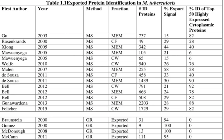
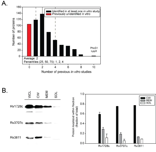
protein contaminants in exported fractions (Table 1.1) (de Souza & Wiker, 2011). Furthermore,
as mass spectrometry has become increasingly sensitive, the potential for false positive
identification of cytoplasmic contaminants as exported proteins has grown. In recent mass
spectrometry proteomics studies, only 30% of proteins identified in exported fractions have a
predicted export signal, highlighting the abundance of non-exported proteins being detected
(Table 1.1). Further, in one of the most comprehensive proteomics studies of M. tuberculosis to
date, of 1,050 M. tuberculosis proteins identified 98% of the proteins were detected in both cytoplasmic and exported fractions (Bell et al., 2012). Thus, caution should be exercised when a protein is identified as being exported solely on the basis of mass spectrometry based
proteomics.
An additional challenge facing exported protein identification by mass spectrometry is
the use of this technology to identify proteins exported by a pathogen during in vivo growth in a host (Schmidt & Volker, 2011; Yang et al., 2015). Mass spectrometry based approaches are biased by abundance, and low abundance proteins in a sample are difficult to identify. This
becomes particularly difficult when attempting to identify relatively rare bacterial proteins from
among host cells proteins in an infection model (Schmidt & Volker, 2011; Yang et al., 2015;
Kruh et al., 2010). Isolation and purification of bacterial cells out of the host tissue, as well as very high bacterial cell numbers is generally required (Xia et al., 2007; Twine et al., 2006; Becker et al., 2006; Schmidt et al., 2010; Liu et al., 2012; Pieper et al., 2009). Further, it is hard
exported. Analysis of crude extracts of infected organs does not allow for discrimination between
exported and non-exported proteins (Schmidt & Volker, 2011; Yang et al., 2015). Isolation of
bacterial cells from infected host cells could be useful for identifying proteins exported to the
bacterial cytoplasmic membrane and cell wall, but current methods will not provide sufficient
material for subcellular fractionation and further analysis (Schmidt & Volker, 2011; Yang et al., 2015). An alternative approach is to search for bacterial proteins secreted away from the bacteria
into the host cytosol. However, due to the sensitivity of mass spectrometry this demands an
efficient method to eliminate intracellular bacteria from the host cytosol which represents
another technical challenge. As a result, the exported proteomes of pathogens during infection
remain to be defined.
Metabolic labeling and biorthogonal chemistry promises to improve many fields,
including the study of bacterial exported proteins (Ngo et al., 2009; Horisawa, 2014; Mahdavi et
al., 2014; Siegrist et al., 2015). Incorporating labels amenable to click-chemistry can allow for downstream chemical reactions which can tag labeled compounds with fluorescent markers, or
allow for isolation of tagged molecules out of complex mixtures (Mahdavi et al., 2014; Dieterich et al., 2007; Siegrist et al., 2015). Recently, bio-orthogonal noncanonical amino acid tagging, BONCAT (Dieterich et al., 2007), has been used in Escherichia coli, Salmonella enterica
serovar Typhimurium and Yersinia pseudotuberculosis to incorporate azidonorleucine (ANL) into proteins (Tanrikulu et al., 2009; Grammel et al., 2010; Mahdavi et al., 2014). In one study,
S. Typhimuriumwas used to infect mammalian cells in vitro, cells were pulsed with ANL to label all bacterial proteins, and then click chemistry was used to selectively tag bacterial ANL
labeled proteins with biotin for isolation (Grammel et al., 2010). A second study used the same
Y. pseudotuberculosis into in vitro cultured cells (Mahdavi et al., 2014). Exported proteins delivered into the host cell were identified through the comparison of wild type Y.
pseudotuberculosis to a T3SS mutant unable to translocate effectors (Mahdavi et al., 2014). Unfortunately, a large number of background cytoplasmic bacterial proteins were also identified
in the host cell, which prevented applying this technology to the identification of new exported
proteins (Mahdavi et al., 2014). Overall, while this technology holds promise for facilitating studies of proteins exported in the context of infection, there remain significant technical hurdles.
In addition, incorporation of unnatural amino acids by bacteria has yet to be performed during in vivo infection.
Experimental Identification of Exported Proteins: Genetic Reporters
An alternative strategy to biochemical identification of exported proteins is the use of
genetic reporters of protein export. A protein export reporter is an enzyme whose activity or
function depends on the reporter being exported/localized out of the cytoplasm. Protein export
reporters are missing their native signal for export and, consequently, they require in-frame
fusion with a protein capable of exporting the reporter to be active. Genetic reporters require a
genetically tractable organism and they need to be compatible with the specific export system
under investigation. Genetic reporter approaches for identifying exported proteins have the
benefit over mass spectrometry of identifying fewer false positives and thus cytoplasmic proteins
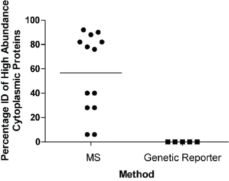
are less of a concern with export reporters. Over 90% of proteins identified with a genetic
reporter contain a predicted export signal (Table 1.1, Figure 1.4), which reinforces their
identification as being exported. This is compared to by mass spectrometry based methods which
only identify approximately 30% of proteins with predicted export signals. Comparing studies of
approaches reveals high abundance cytoplasmic proteins (as identified by PaxDB (Wang et al., 2015; Wang et al., 2012)) as frequently identified as exported by mass spectrometry but not by
genetic reporter approaches (Table 1.1, Figure 1.5).
The classic genetic reporter of bacterial protein export is the enzyme alkaline phosphatase
(PhoA) (Taylor et al., 1987; Kaufman & Taylor, 1994; Cleavinger et al., 1995; Lim et al., 1995; Manoil & Beckwith, 1985; Manoil et al., 1990). Bacterial colonies exporting PhoA can be detected as blue colonies when plate on agar containing a colorimetric PhoA substrate.
Consequently, a truncated ‘PhoA reporter lacking its native signal for export can “report” on the
presence of an export signal in a protein to which it is fused, by producing blue colored colonies.
In the first study to identify M. tuberculosis exported proteins using a ‘PhoA reporter, ‘PhoA was encoded on a plasmid, and random fragments of M. tuberculosis genomic DNA were fused upstream of the reporter (Lim et al., 1995). The resulting plasmids were screened in
Mycobacterium smegmatis, a non-pathogenic model species of M. tuberculosis,and blue colonies were identified when the transformants were plated on the PhoA colorimetric substrate
(Lim et al., 1995). This first study identified three M. tuberculosis proteins as exported (Lim et al., 1995). Further studies utilized the ‘PhoA reporter combined with in vitro transposition into M. tuberculosis cosmids. These transposon mutagenized cosmids were transformed into M.
smegmatis to screen for active PhoA fusions, resulting in the identification of 31 M. tuberculosis exported proteins (Braunstein et al., 2000). Unfortunately, due to endogenous phosphatase activity, the ‘PhoA reporter is not compatible with use in M. tuberculosis, and thus all ‘PhoA
screens have to be carried out in the non-pathogenic M. smegmatis (Lim et al., 1995).
Generation of a β-lactam sensitive ΔblaC mutant of M. tuberculosis (Flores et al., 2005;
Figure 1.5. MS based methods Identify (ID) High Abundance Cytoplasmic Proteins as Exported.
Percentage of high abundance cytoplasmic proteins identified in exported fractions as described in Table 1.1. The Top 50 most abundant cytoplasmic proteins were identified by PaxDB (Wang et al., 2015; Wang
exported proteins directly in pathogenic M. tuberculosis. The E. coli β-lactamase, BlaTEM, cleaves β-lactam antibiotics and is able to produce β-lactam resistance. Like ‘PhoA, removal of
the native signal peptide of ‘BlaTEM prevents export but when fused to a signal peptide, or the
extracytoplasmic portion of an exported protein, the exported ‘Bla reporter confers β-lactam
resistance (Figure 1.6). This was first worked out in Escherichia coli (Broome-Smith & Spratt, 1986), and holds true when the ‘BlaTEM reporter is used in the ΔblaC mutant of M. tuberculosis (McCann et al., 2007). The ‘BlaTEM reporter is compatible with both Sec and Tat export
systems of M. tuberculosis (McCann et al., 2007). An additional β-lactamase reporter (‘BlaC) has been developed for M. tuberculosis that is specific for export by the Tat pathway
(McDonough et al., 2005; McDonough et al., 2008). For the ‘BlaC reporter, only fusion to Tat exported proteins (and not signals for other export systems) works to confer resistance to
β-lactams (McDonough et al., 2008). Generation of a library of plasmids containing the ‘blaC
reporter fused downstream of random fragments of M. tuberculosis genomic DNA led to the identification of 13 proteins exported by the Tat pathway in M. tuberculosis (McDonough et al.,
2008). The ‘blaC reporter has additionally been used to directly test individual proteins for Tat dependent export (Ligon et al., 2012).
In other bacterial pathogens, transposons carrying the ‘PhoA reporter proved a powerful
means to identify exported proteins with roles in virulence (Taylor et al., 1987; Kaufman &
Taylor, 1994; Manoil & Beckwith, 1985; Manoil et al., 1990). With the development of -lactamase reporters that work in mycobacteria, this approach could be directly applied to the
identification of exported virulence factors of M. tuberculosis Using a Himar1-based mariner transposon carrying the ‘BlaTEM reporter in M. tuberculosis and plating on β-lactam containing
identified (McCann et al., 2011). The same in-frame transposon insertion that identifies an exported protein will, in most cases, also disrupt the function of that exported protein. By
subsequent screening of 111 transposon insertion mutants in genes encoding exported proteins
for virulence defects in a macrophage intracellular growth assay a total of six exported proteins
with roles in virulence were identified (McCann et al., 2011). One of these proteins, Rv0199, had no previously predicted function. Chapter 3 will focus on further characterization of the rv0199 transposon mutant culminating in assigning function to Rv0199, and shedding light on the
function of a previously uncharacterized exported protein family.
There are advantages to using a β-lactamase based genetic reporter over other genetic
reporters. First, β-lactamases are selectable markers, dramatically reducing the otherwise
laborious nature of screening for reporter positive colonies (McCann et al., 2011). Second, β-lactamases have the potential of overcoming the requirement for using in vitro grown bacteria
when identifying exported proteins. Many β-lactam antibiotics are available and safe for use in
cell culture and animal models. Proof of principle studies demonstrated, that the ΔblaC M.
tuberculosis mutant is susceptible to β-lactams during intracellular growth in a cultured
macrophage cell line (McCann et al., 2007). Furthermore, McCann et al. demonstrated that the ‘BlaTEM reporter can report on the export of a protein to which it is fused during intracellular
growth. Model strains were constructed that produce the ‘BlaTEM reporter with no export signal
(‘BlaTEM, β-lactamsensitive) or the ‘BlaTEM reporter fused to a signal peptide (sp-‘BlaTEM,
β-lactam resistant) in M. tuberculosis (McCann et al., 2007). Macrophages were infected with
each strain, and treated by addition of the β-lactam carbenicillin to the media. In the absence of
treatment both strains could replicate in macrophages; however, carbenicillin treatment restricted
sp-‘BlaTEM reporter was protected from carbenicillin treatment, and grew normally in the
macrophage. This demonstrated that the ‘BlaTEM reporter can report on export during growth in
macrophages (McCann et al., 2007). These studies with macrophages, suggested the possibility of using the ‘BlaTEM reporter in screening for M. tuberculosis proteins exported during
infection in -lactam treated mice. Chapter 2 will focus on the development of a method called
EXIT (EXported In vivo Technology) which we developed to specifically identify proteins exported by M. tuberculosis during murine infection.
Exported proteins play important roles in M. tuberculosis virulence
Exported proteins are vital to the survival of intracellular pathogens, such as M.
tuberculosis, where they not only maintain cellular physiology, but also provide protection from
intracellular stresses, control the host cell response, and allow for nutrient acquisition in a
purposefully nutrient poor environment (McCann, 2009). Several examples of exported virulence
factors are highlighted below.
Exported proteins important to M. tuberculosis virulence
M. tuberculosis encodes several classical exported virulence factors, for example,
proteins involved in detoxification of radical oxygen and radical nitrogen species (McCann,
2009). Additionally, some of the best studied virulence factors in M. tuberculosis are exported
proteins required for the biosynthesis specialized lipids within the highly complex and
impermeable mycobacterial cell wall and outer membrane (membrane). The
myco-membrane consists of several specialized lipid structures, many including long chain fatty acids
called mycolic acids. Synthesis and transport of many lipids within the myco-membrane of M. tuberculosis is required for bacterial cell viability; however, some lipids are not required for in
synthesis and transport of PDIM to the myco-membrane results in attenuation of M. tuberculosis during infection of macrophages and mice (Forrellad et al., 2013). Interestingly, PDIM appears
to be important for immune modulation, and the attenuation of PDIM mutants appears to result
from reduced control of the host (Forrellad et al., 2013).
M. tuberculosis has not only had to develop strategies to evade destruction by the host immune system, but also to acquire nutrients for survival. The phagosome is nutritionally
restricted as a way to prevent growth of intracellular bacteria (Appelberg, 2006). Perhaps the
most well studied nutritional restriction, the host’s ability to restrict iron is overcome by M. tuberculosis through the production of siderophores, molecules with very high affinity for iron
which are able to chelate it away from host iron stores (Fang et al., 2015). Disruption of siderophore biosynthesis or function attenuates the virulence of M. tuberculosis in cell culture and animal models of infection (Fang et al., 2015).
Role of Lipid Import and Catabolism in M. tuberculosis virulence
During infection, M. tuberculosis is thought to primarily consume lipids as a carbon
source (Munoz-Elias & McKinney, 2006; Eisenreich et al., 2010). This lifestyle is reflected in the numerous enzymes required for catabolism of lipids in M. tuberculosis (Cole et al., 1998). Additionally, the M. tuberculosis genome encodes several specialized multi-protein transporters
predicted to import lipids, four Mce transporters (Mce1, 2, 3, and 4). All of the mce operons are important to virulence in mice (Sassetti & Rubin, 2003; Shimono et al., 2003; Lima et al., 2007;
Uchida et al., 2007; Senaratne et al., 2008; Marjanovic et al., 2010). Study of mce operons in M. tuberculosis has shown that one of them, mce4, imports cholesterol (Pandey & Sassetti, 2008; Klepp et al., 2012; Mohn et al., 2008). M. tuberculosis is fully able to catabolize cholesterol
metabolic pathway, and they are severely attenuated (Miner et al., 2009). The mce4 mutant is attenuated in a mouse model of infection, as demonstrated by reduced bacterial burden, increased
survival time in mice, and decreased histopathology, demonstrating an importance for
cholesterol import during infection (Senaratne et al., 2008; Pandey & Sassetti, 2008). The other
mce operons are not as well studied as mce4, but they are also thought to be lipid transporters. Mce1 is required during infection of macrophages as well as during murine infection (McCann et al., 2011; Rengarajan et al., 2005). Recent evidence suggests that Mce1 imports mycolic acids,
specialized lipids which are incorporated into many complex molecules in the myco-membrane
(Forrellad et al., 2014; Cantrell et al., 2013). Mce1 functions potentially as a mechanism of
recycling for control of the mycolic acid concentration within the mycobacterial outer
membrane. The lipids transported by Mce2 or Mce3 have yet to be identified.
Although the contribution of Mce systems to M. tuberculosis virulence has been
characterized, the role of individual proteins within the Mce transport complex has yet to be
characterized. Mce operons are thought to be analogous to ABC transporters, and the genes
encoded in the operons have been assigned function based on our knowledge of ABC
transporters (Casali & Riley, 2007). However, the sequence similarity to ABC transporters is
limited, and Mce transporters are distinct from ABC transporters in that mce operons contain
genes encoding multiple predicted permease and solute binding protein components. mce operons additionally contain genes encoding proteins with no homology to ABC transporter
components that have been named Mce-associated (Mas) proteins. Therefore, the mechanism of
Mce transport and the function of the individual Mce transporter components in lipid transport
in vivo exported protein of unknown function and assign it a role in the stabilization and function of Mce transporter systems.
Summary
While the technology for identifying exported proteins has improved dramatically, there
are still technical hurdles to accurate genome-wide identification of exported proteins.
Furthermore, current technologies remain limited to identifying exported proteins using in vitro grown bacteria. Transcriptional analysis has identified large shifts in the M. tuberculosis lifestyle
during infection (Talaat et al., 2004; Schnappinger et al., 2006). While transcriptional analysis has informed our perspectives on M. tuberculosis metabolism, and the intracellular lifestyle,
there is mounting evidence that transcriptional analysis alone is not representative of cellular
protein changes (de Souza & Wiker, 2011). Thus, it is important to develop methods to
specifically study protein behavior during infection.
We propose that a subset of exported proteins may be selectively expressed or exported
during infection, that are missed by current analyses focusing on in vitro grown M. tuberculosis.
Such proteins could be incredibly important to pathogenesis, and define interactions between the
host and pathogen. In this dissertation, we sought to comprehensively identify the in vivo exported proteome of M. tuberculosis. Accomplishment of this goal, which has not been
attempted previously in any bacterial pathogen, required the development of a novel method,
which we termed EXIT for EXported In vivo Technology. Chapter 2 will describe the process of
developing and optimizing EXIT, as well as describing key results from analyzing the in vivo exported proteome of M. tuberculosis. We identified 593 proteins as exported during murine infection, including 32 proteins with no in silico predicted export signal which represent
significantly upregulated during infection as compared to in vitro growth. Because these proteins are subject to regulation, they may have important functions during infection and could
understand the host-pathogen interface. Finally, we applied our reporter fusion data to determine
the topology of important M. tuberculosis virulence factors. The proteins identified in this study,
and the vast resource of topology data, represent a valuable resource for the M. tuberculosis research community.
Identification of exported proteins is a proximal goal. With the knowledge that exported
proteins are critical to M. tuberculosis virulence we also seek to better understand the roles of individual exported proteins during infection. Previous studies analyzing transposon mutants
lacking individual exported proteins during intracellular growth identified six attenuated mutants
(McCann et al., 2011). Of these, one mutation was in a membrane protein of unknown function, Rv0199. Rv0199 was also identified by EXIT as exported in vivo. Chapter 3 describes further
characterization of the rv0199 mutant, resulting in a functional assignment of rv0199 as an orphanedmce-associated gene (mas). Further study into the role of Rv0199 in Mce transport
determined that Rv0199, and potentially all Mas family proteins, provides stability to the large
Table 1.1Exported Protein Identification in M. tuberculosis
First Author Year Method Fraction # ID
Proteins
% Export Signal
% ID of Top 50 Highly Expressed Cytoplasmic Proteins
Gu 2003 MS MEM 737 15 82
Rosenkrands 2000 MS CF 49 29 28
Xiong 2005 MS MEM 342 44 40
Mawuenyega 2005 MS MEM 105 21 6
Mawuenyega 2005 MS CW 65 15 6
Wolfe 2010 MS CW 540 26 76
Malen 2007 MS MEM 255 58 28
de Souza 2011 MS CF 458 33 40
de Souza 2011 MS MEM 1439 30 90
Bell 2012 MS CW 791 21 92
Bell 2012 MS MEM 666 24 78
Bell 2012 MS CF 508 29 82
Gunawardena 2013 MS MEM 2203 28 88
Feltcher 2015 MS CW 1729 29 82
Braunstein 2000 GR Exported 31 94 0
Gomez 2000 GR Exported 9 100 0
McDonough 2008 GR Exported 13 100 0
McCann 2011 GR Exported 111 95 0
Table 1.1. Exported Protein Identification in M. tuberculosis.Exported proteins of M. tuberculosis
have been identified by mass-spectrometry (MS) based methods as well as use of genetic reporters (GR) (Gu et al., 2003; Rosenkrands et al., 2000; Xiong et al., 2005; Mawuenyega et al., 2005; Wolfe et al., 2010; Malen et al., 2007; de Souza et al., 2011; Bell et al., 2012; Gunawardena et al., 2013; Braunstein et al., 2000; Gomez et al., 2000; McDonough et al., 2008; McCann et al., 2011). MS based methods have analyzed proteins in exported fractions including the membrane (MEM), cell wall (CW), and extracellular or culture filtrate (CF) fractions. For comparison, all lists were analyzed using the H37Rv RefSeq genome annotation released January 9 2012. The total number of proteins identified (# ID Proteins) in the given fraction is reported and analyzed for percentage containing an export signal (% Export Signal) and the percentage of top 50 most abundant cytoplasmic proteins identified (% ID Top Cytoplasmic), as
described below. Export signals including transmembrane domains and Sec and Tat signal peptides were predicted by SignalP, TatP, TMHMM, (Sutcliffe & Harrington, 2004), and (McDonough et al., 2008). The Top 50 most abundant cytoplasmic proteins were identified by PaxDB, a database calculating the relative abundance of proteins from published proteomics datasets (Wang et al., 2015; Wang et al., 2012). Proteins identified as abundant in PaxDB but containing predicted export signals, or known to be