APPLYING A MOTHER-INFANT DYAD PERSPECTIVE TO EXAMINE THE NUTRITIONAL INTERRELATIONSHIPS OF HIV-INFECTED MALAWIAN
MOTHERS AND THEIR EXCLUSIVELY BREASTFED INFANTS
Elizabeth Marie Widen
A dissertation submitted to the faculty of the University of North Carolina at Chapel Hill in partial fulfillment of the requirements for the degree of Doctor of Philosophy in the
Department of Nutrition, Gillings School of Global Public Health.
Chapel Hill 2012
ii © 2012
iii
Abstract
ELIZABETH MARIE WIDEN: Applying a mother-infant dyad perspective to examine the nutritional interrelationships of HIV-infected Malawian women and their exclusively
breastfed infants
(Under the direction of Linda Adair)
iv
v
Acknowledgments
The MaMi study group has provided me with an excellent environment to study maternal and child nutrition in the context of HIV. Linda in particular has provided superior mentoring and guidance to train me as a nutritional epidemiologist, critical thinker and passionate teacher. Thank you Linda for your time and thoughtful insight during our many meetings, I am forever appreciative of your time and guidance. Linda, Peggy, Valerie, Betsy, Diana, Eric, Krishna and all other group members have been superb colleagues and friends.
I would like to also thank the mothers and infants for their participation, as well as all members, past and present, of the BAN and MaMi study teams, and UNC Project Malawi. My dissertation committee members—Charlie van Der Horst, Anna Maria Siega-Riz, Cathy Zimmer and Michael Hudgens—have also been excellent mentors and have provided much guidance, insight, support and encouragement throughout this process.
vi
vii
Table of Contents
List of Tables ... ix
List of Figures ... x
List of Abbreviations and Symbols... xi
Chapter 1. Introduction ... 1
Overview ... 1
Overall objectives and specific aims... 2
Chapter 2. Literature Review ... 4
The mother infant dyad ... 4
Why anthropometry and iron? ... 4
Methods to evaluate the maternal supply and infant demand relationships have not been developed ... 5
Maternal anthropometry patterns are highly variable during lactation and are not well understood in the context of HIV ... 7
Maternal nutritional status and lactation performance ... 7
Anthropometry patterns of infants during exclusive breastfeeding ... 8
Few studies have examined how maternal weight changes translate into infant weight and length gain with a longitudinal design ... 9
The effects of maternal supplementation on infant growth ... 9
By extending our analyses from anthropometry to iron status, we will explore the mother-infant nutritional dyad in two domains... 11
viii
Chapter 3. Maternal weight loss during exclusive breastfeeding is associated with reduced weight and length gain
in daughters of HIV-infected Malawian women ... 14
Overview ... 14
Introduction ... 15
Methods... 18
Results ... 23
Discussion ... 24
Chapter 4. Maternal transferrin receptors and ferritin are associated with infant values in exclusively breastfed HIV-exposed infants ... 34
Overview ... 34
Introduction ... 35
Subjects and Methods ... 36
Results ... 44
Discussion ... 48
Chapter 5: Synthesis ... 67
Overview of findings ... 67
Maternal weight loss is associated with reduced length and weight gain in daughters of HIV-infected Malawian women ... 68
Maternal hemoglobin and transferrin receptors are associated with infant values during exclusive breastfeeding ... 69
Limitations and Strengths ... 70
Significance and public health impact ... 72
Direction for future research ... 73
ix
List of Tables
Table 3.1 Characteristics of 1,309 BAN mother-infant pairs in the primary analysis of the effects of maternal weight loss on infant growth... 29 Table 3.2 Gender stratified linear regression models showing the effects of maternal weight loss on infant weight and length gain from 0 to 24 wk ... 30 Table 3.3 Predictors of maternal weight loss among BAN mothers ... 32 Table 4.1 Characteristics of mother-infant dyads in both analysis samples ... 54 Table 4.2 Infection, Hb and inflammation adjusted and unadjusted markers of iron status of MaMi subsample mother-infant dyads ... 55 Table 4.3 Linear regression models showing the effects of change in maternal iron status on change in infant iron status outcomes (log transferrin
receptors, hemoglobin, log ferritin) from 2/6 to 24 weeks, adjusted and unadjusted for inflammation ... 56 Table 4.4 Associations between the BAN study interventions and maternal and infant Hb, TfR and ferritin outcomes in MaMi subsample ... 57 Table 4.5 Associations between study interventions and odds of impaired infant iron status at 24 weeks in the MaMi subsample ... 58 Supplemental Table 4.1 Composition of daily ration (140 g) of lipid-based nutrient
supplements given to BAN Study mothers ... 63 Supplemental Table 4.2 Longitudinal random effects model with first order
autoregressive disturbance terms showing the associations between maternal Hb and infant Hb (g/dL) in 1926 BAN mother- infant pairs in the longitudinal Hb sample ... 64 Supplemental Table 4.3 Longitudinal random effects model with first order
autoregressive disturbance terms showing the associations between BAN study intervention arm and maternal Hb
outcomes (g/dL) in 1765 BAN mothers from 6 to 24 weeks ... 65 Supplemental Table 4.4 Longitudinal random effects model with first order
x
List of Figures
xi
List of Abbreviations and Symbols
α – alpha
AGP – alpha-1-acid glycoprotein
AIDS – acquired immune deficiency syndrome ARV – antiretrovirals
BAN – Breastfeeding, Antiretrovirals and Nutrition Study BAN intervention arms –
mLNS-mARV – maternal LNS/maternal ARV mLNS-iARV – maternal LNS/infant ARV mLNS – maternal LNS
mARV – maternal ARV iARV – infant ARV C – control
BMI – body mass index (kg/m2)
CD4 – cluster of differentiation – used to define stage of HIV or AIDS infection CRP – C-reactive protein
Hb – hemoglobin
HIV – human immunodeficiency virus
HIV-EU – human immunodeficiency virus exposed, uninfected LAZ – length-for-age Z-score
LNS – lipid-based nutrient supplement MaMi – Malawi Mothers and Infants Study
xii TfR – soluble transferrin receptor
WAZ – weight-for-age Z-score WFP – World Food Program WHO – World Health Organization wk- week
Chapter 1. Introduction
Overview
The mother is the sole source of nutrition for the infant during exclusive
breastfeeding, yet there is little known about how maternal and infant nutritional statuses are interrelated and interdependent throughout this time. Though maternal health and nutritional status are important determinants of breastmilk quality and quantity, maternal nutritional status during lactation has primarily been examined as a predictor of child nutritional outcomes; rarely have the effects of infant nutritional status on maternal nutritional status been explored. As we are interested in the health and nutritional status of the mother-infant dyad, we framed our analyses to explore the interrelationships between maternal and child nutrition statuses, to understand the balance between infant nutrient demand and maternal nutritional supply during lactation, and to determine how this relates to infant and maternal anthropometry and iron status.
We developed novel methods to explore the intricate interplay and dynamics between the mother and infant during this time period. In resource poor settings with high HIV
prevalence, these relationships may be especially important for long-term maternal and child survival, health, and nutritional status. There currently is a dearth of literature on the
2
HIV. Therefore our research provides urgently needed evidence regarding how maternal and infant nutritional statues are interrelated and interdependent between HIV-infected women and their infants in resource poor settings.
Overall objectives and specific aims.
This study focused on the relationships between maternal and infant nutritional status in HIV-infected lactating women and their exclusively breastfed HIV-exposed, uninfected (HIV-EU) infants. We used data from the Breastfeeding, Antiretrovirals and Nutrition Study (BAN), a randomized controlled trial of 2,369 Malawian HIV infected women and their infants to prevent HIV transmission during exclusive breastfeeding conducted between March 2004 and January 2010 in Lilongwe, Malawi, as well as a sub-study of BAN, the Malawi Mothers and Infants Study (MaMi).1 BAN is unique as it has parallel longitudinal maternal and infant measures of nutritional status, including anthropometry and
micronutrient status, during exclusive breastfeeding. We focused on the HIV exposed,
uninfected infants and their mothers in BAN, who were followed during pregnancy, birth and 1, 2, 4, 6, 8, 12, 18, 21 and 24 weeks postpartum.1 The overall goal of this study was to determine the interrelationships of maternal and infant nutrition status during exclusive breastfeeding. Specific aims for this study are as follows:
Aim 1: Determine the interrelationships between maternal and infant anthropometry
during exclusive breastfeeding in HIV-infected mothers and their infants.
First, we developed models to determine how maternal weight loss between 2 and 24 weeks postpartum influenced infant growth. We hypothesized that infants of mothers losing weight during lactation will have suboptimal growth.
3
status during exclusive breastfeeding in HIV-infected mothers and their infants.
Chapter 2. Literature Review
The mother infant dyad
The mother-infant dyad is a conceptual construct to define the connections between the mother and her infant. Previous research examining the mother-infant dyad has focused particularly on mother-infant interactions and attachment during breastfeeding and infancy.2,3 The dyad has also been studied with regard to evolutionary adaptations to the composition of breast milk.4,5 Numerous authors have indicated that breastmilk production is focused not only on the survival of the infant, but also on the survival of the mother-infant dyad;4 suggesting that “maximum evolutionary gain is obtained when protein and energy levels in breastmilk are just high enough to prevent prohibitive infant mortality rates, but low enough to spare the mother.”5 Although we did not test this hypothesis, it provided a framework for our analyses, as we included the health and nutritional status of the mother as a critical component of breastfeeding as well as infant health and survival.
Why anthropometry and iron?
5
influence infant anthropometry and conversely if and when infant growth influences maternal anthropometry is even more important in the context of HIV.
As iron deficiency is a major public health problem affecting 42% of pregnant women and almost half of preschool aged children globally,6 it is vital to understand if and when maternal and infant iron statuses are related during exclusive breastfeeding. In the context of HIV, iron deficiency is common among HIV-infected women; as such their infants are at heightened risk of iron deficiency. Elucidating this relationship is essential as iron is critical for growth, brain development and immune function.7 Both iron status and
anthropometry are commonly measured in public health settings; therefore, our findings can be translated into public health screening and interventions that can be broadly implemented.
Methods to evaluate the maternal supply and infant demand relationships have not been developed
6
infected mothers, as energy needs are already elevated due to HIV infection; thus examining how maternal supply relates to infant demand is even more imperative in the context of HIV.
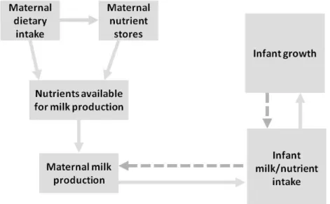
Figure 2.1 Maternal supply and infant demand
The dashed arrows indicate where infant demand may exert influence on maternal nutritional status.Image adapted from “Influence of maternal nutrition on lactation” by Kathy
Rasmussen.8
7
incorporation of feedback loops and for testing of complex causal relationships are urgently needed; but difficult to implement.
Maternal anthropometry patterns are highly variable during lactation and are not well understood in the context of HIV
During the first six months postpartum, most lactating women experience mild and gradual weight loss.12 Among well-nourished women, breastfeeding is usually associated with earlier return to pre-pregnancy weight.13,14 Among undernourished women, though, weight loss, weight maintenance and even moderate weight gain have been reported.15-18 Anthropometry patterns during lactation have been less well described in HIV infected women. Similar to HIV uninfected women, though, the findings are inconsistent with studies reporting both gains and losses during lactation.13
Maternal nutritional status and lactation performance
Studies have shown that breastfeeding can deplete energy and nutrient reserves of women with inadequate dietary intake and poor maternal nutrition can influence breastmilk quantity and nutrient quality.8,18,19 Brown et al (1986) examined the lactational capacity of marginally nourished mothers of Bangladeshi infants by examining relationships between maternal nutritional status and breastmilk quantity and quality.20 In mothers (n=60) with infants who were 90 days old, an association was found between the change in maternal weight from baseline and the quantity of milk, as well as nitrogen and energy content of milk.20 Furthermore, women who gained at least 200 grams from baseline produced more breastmilk with higher amounts of nitrogen and energy regardless of initial maternal
8
nutritional status did not severely impair lactational capacity; though, it limited breastmilk quantity and energy concentration compared to more well nourished mothers.20
How maternal HIV infection influences breastmilk supply and lactation performance is not well documented. Prior studies regarding lactation of HIV infected women have focused on issues related to HIV transmission including breastmilk viral load and
micronutrient content after heat treatment,21,22 rather than the relation of maternal nutritional status to the nutrient content and quantity of breastmilk.
Anthropometry patterns of infants during exclusive breastfeeding
After birth infants typically lose weight in the first few days of life, and then return to birth weight by the seventh to tenth day of life.23 Subsequent growth proceeds at a “rapid but decelerating rate.”23 Birthweight is usually doubled by four to six months of age, and tripled by one year of age.23 Among well-nourished infants, length is increased by 50% in the first year, and is doubled by four years of age.23
9
Few studies have examined how maternal weight changes translate into infant weight and length gain with a longitudinal design
In the East Java Pregnancy Study, increasing maternal post-partum body mass index (BMI) (kg/m2), measured between 4-6 months, was associated with higher infant weight and length gains between birth and 6 months.26 Interestingly, infants with lower birthweight had higher length and weight gains compared to infants with higher birthweight; indicating that the breastmilk quality and quantity was sufficient to promote growth of smaller infants, but the growth of infants with higher nutritional needs was compromised.26 Most of the infants in another study in this population were partially breastfed starting within the first month of life; thus partial breastfeeding (and subsequent exposure to pathogens and other foods) may have biased the effect of post-partum BMI on weight and length gain in this analysis.26 In Bolivian mother-infant pairs, weight and height at 3 months postpartum were positively correlated with infant weight gain between 3 and 6 months.27 These findings suggest associations between maternal and infant anthropometry; however, maternal body composition during early lactation and potential confounding variables such as maternal education, parity and morbidity were not accounted for in the models. Moreover, as exclusive breastfeeding may not have continued through the duration of these prior studies, the mother may no longer be the sole source of nutrition for her infant; thus effect estimates may be biased.
The effects of maternal supplementation on infant growth
10
were supplemented with a protein-calorie supplement (‘atole’, gruel typically made with corn) or caloric supplement (‘fresco’, cool drink).28 Though not significant, maternal
supplementation had a positive effect (0.05 < p < 0.10) on infant weight gain from birth to 6 months.28 Moreover, infants were also supplemented with atole or fresco, thus the effect of maternal supplementation on infant growth cannot be isolated from the effect of infant supplementation.28 Infant supplementation before 3 months had negative effects on infant weight gain (p<0.001), whereas infant supplementation between 3 and 6 months of age was found to positively influence infant growth (p<0.01).28 In the Bacon Chow Study conducted in rural Taiwan starting in 1967 for 6.5 years, the effect of maternal supplementation on infant growth was examined in mothers who gave birth to two infants during course of the study.29 Mothers were randomized to supplementation with a milk-based formula, providing 800 calories and 40 grams of protein per day, or a placebo, providing 40 calories per day, starting at 15 days after the birth of the first infant and continuing through weaning of the second infant.29 There were no significant differences between maternal supplementation versus placebo; whereas the timing of maternal supplementation was important, yet
inconsistent.29 Boys were heavier in the second infant cohort (p=0.02) compared to the first cohort.29 Conversely, girls were heavier in the first infant cohort (p=0.05) compared to the second.29
11
maternal supplementation with lipid base nutrient supplement (LNS) in BAN participants had no sustained or consistent effects on infant growth from birth to 24 weeks.32
By extending our analyses from anthropometry to iron status, we will explore the mother-infant nutritional dyad in two domains
Maternal iron status may influence infant iron status during exclusive breastfeeding; however, many additional factors influence infant iron needs and status. Infant iron stores at birth reflect maternal pregravid and pregnancy iron status, gestational age and birthweight of the infant, and timing of cord clamping.33 Many factors influence post-natal infant iron status including redistribution of body iron, growth rate, infections and inflammation, and possibly breastmilk iron status.33 Postnatally, infant Hb exhibits dynamic changes with a peak at birth of approximately 17 g/dL and a nadir of 11.2 g/dL at 2 months of age.33 Few studies have characterized the iron status of exclusively breastfed HIV-exposed infants.34
Most literature suggests maternal iron status strongly affects infant iron status only during pregnancy and has little effect during lactation;33,35 however, we theorize that in our population of HIV-infected Malawian women, whose iron stores are likely depleted
postpartum, maternal iron status may be an important determinant of infant iron status during exclusive breastfeeding. Further investigation of this association is essential, particularly in women who are already at risk of anemia, such as our population of HIV infected women, as their infants may be at heightened risk of iron deficiency.36,37
Why Malawi?
12
breastfeeding was 2.5 months, while the median duration of predominant breastfeeding was 4.8 months.38 Though the WHO recommends six months of exclusive breastfeeding, 37% of infants ages 4-5 months were given complementary foods.38
13
with 40 HIV positive mothers. 85% of HIV positive mothers reported giving water or porridge to their infant before 4 months of age.42 Moreover, 60% of the 17 mothers with undisclosed HIV status had introduced complementary foods before 6 months of age.42 One mother reported: “Traditionally, we are told that if a child cries it is a sign of hunger.
Chapter 3. Maternal weight loss during exclusive breastfeeding is associated with reduced weight and length gain in daughters of HIV-infected Malawian women
Overview
Maternal weight loss during exclusive breastfeeding may influence growth of exclusively breastfed infants through impaired quality or quantity of breastmilk. This study evaluated how maternal weight loss from 2-24 wk was related to infant weight and length gain in 1,390 lactating HIV-infected mothers and their exclusively breastfed infants. Malawian mother-infant pairs in the Breastfeeding, Antiretrovirals, and Nutrition (BAN) study were randomized to receive a lipid-based nutrient supplement (LNS), meeting nutritional needs of lactation, or no LNS and a maternal, infant or no antiretroviral (ARV) regimen. Linear regression models were used to relate maternal weight loss (weight loss vs. no weight loss) to infant weight and length gain from birth to 24 wk, stratifying by gender, and controlling for infant birthweight/length and maternal BMI at 2 wk (mean: 23.1 kg/m2) and interacting maternal BMI with weight loss. Length (β: -3.24 cm, p=0.01) and weight gain (β: -1.21 kg, p=0.008) were lower in girls whose mothers had lower BMI at 2 wk postpartum coupled with the weight loss, compared to girls of women who did not lose weight. Though associations were only observed in girls, suggesting possible gender differences in suckling and feeding behavior, these findings indicate that maternal weight loss with low energy reserves represents a risk factor for poor infant growth outcomes.
15
Introduction
During exclusive breastfeeding, the mother is the sole source of nutrition for her infant. Breastfeeding can deplete energy and nutrient reserves of women with inadequate dietary intake and very poor maternal nutrition can impair breastmilk quantity and nutrient quality.8,18-20 Increases in maternal weight during lactation have previously been linked to improved quantity and nutrient quantity of breastmilk in marginally nourished Bangladeshi women;20 yet, it is not well understood how these weight changes translate into breastmilk output and infant growth due to the complex nature of this relationship.8
Maternal breastmilk supply is regulated to match infant demand through frequency and intensity of infant suckling.8,44 With increased infant demand, maternal breastmilk
production will increase. In the absence of adequate dietary intake to meet the energy costs of lactation and due to hormonal influences of lactation, maternal fat stores may be mobilized to meet the demand—with implications for maternal weight changes.8 Increased milk intake will subsequently influence infant growth and may lead to further rises in infant demand and additional burden on maternal nutritional status, as a rapidly growing infant will likely
demand more milk.8 Given adequate maternal nutrient stores and dietary intake, most women may be able to meet infant breastmilk demand; however, in cases where mothers are
undernourished and are losing weight, breastmilk energy output may decrease if the mother exceeds her lactational capacity (the ability to produce milk on demand),9 which could adversely affect infant growth.
16
HIV infection and lactation increase metabolic demands,46 lactating HIV infected women may be unable to simultaneously meet their own and their infant’s nutritional needs; thus understanding how maternal weight changes during lactation influence infant growth may be especially important for promoting health and survival of the mother-infant dyad in the context of HIV.
During the first six months postpartum, most lactating women experience mild and gradual weight loss.12 Even among well-nourished women, breastfeeding is usually
associated with earlier return to pre-pregnancy weight, but has also been associated with weight gain and weight maintenance.13,14 Among undernourished women, weight loss, weight maintenance and even moderate weight gain have been reported.15-18 Anthropometry patterns during lactation have been less well described in HIV infected women. Similar to HIV uninfected women, though, the findings are inconsistent with studies reporting both weight gains and losses during lactation.13
Although few studies have reported how the overall pattern of maternal weight changes during breastfeeding influence infant growth, there is evidence of associations between maternal and infant anthropometry in HIV-uninfected and HIV-infected
populations. In Indonesian mother-infant pairs, increasing maternal post-partum body mass index (BMI) (kg/m2), measured between 4-6 months, was associated with higher infant weight and length gains between birth and 6 months.26 In Bolivian mother-infant pairs, maternal weight and height at 3 months postpartum were positively correlated with infant weight gain between 3 and 6 months.27 In thirty-five low income Indian women, a 1 kg loss in maternal fat mass from baseline (measured within 1 month of birth) to 6 months
17
after controlling for baseline maternal weight, infant birthweight and changes in maternal appendicular skeletal mass.47 In HIV-infected and uninfected mothers and infants in Kenya ages 4 to 24 mo, higher maternal weight was associated with higher concurrently measured length-for-age (LAZ), weight-for-age (WAZ) and weight-for-length (WLZ) z-score, while higher maternal height was associated with higher LAZ and WAZ.48 Higher maternal BMI and mid-upper-arm circumference was associated with higher WAZ and WLZ.48 To better understand the relationship between maternal weight changes and infant growth, it may be important to evaluate how the overall pattern of maternal weight change influences infant growth.
Our objective was to examine the how maternal weight loss from 2 to 24 wk relates to infant growth from 0 to 24 wk, corresponding with exclusive breastfeeding in a large sample of mother-infant pairs participating in the Breastfeeding, Antiretrovirals, and Nutrition (BAN) study. The Malawi Mothers and Infants (MaMi) study is an analysis of
anthropometric and nutrition data from the BAN study. Mothers in the BAN study who received lipid based nutrient supplement (LNS) had less weight loss during the exclusive breastfeeding (0-24 wk), than mothers who did not receive LNS regardless of antiretroviral drug assignment.49 In this paper, we examine how infant weight and length gain are
18
the mother has limited stores to mobilize. We expected to observe similar effects on infant weight gain and linear growth.
Methods
The BAN study design has been described in detailed elsewhere.51 Briefly, BAN was a randomized controlled trial of 2,369 mother-infant pairs that was conducted from April 2004 to February 2010 in Lilongwe, Malawi. HIV 1-positive pregnant women (n=3572) were recruited from four antenatal clinics. Initial screening criteria included: age ≥ 18 years (or
≥14 years of age if married), CD4 count ≥ 250 cells/µL(prior to 11/13/2006 CD4 cut off was
≥200 cells/µ L), hemoglobin (Hb) ≥ 7 g/dL, no prior antiretroviral medication use, and no major pregnancy complications.1 Eligible women (n=2791) who delivered at the study site were provided with maternal and infant single dose nevirapine peripartum as well as twice-daily zidovudine and lamivudine for 7 days postpartum. Within 36 hours after delivery mother-infant pairs had to present at the study site and meet secondary eligibility criteria for randomization: infant birthweight ≥ 2 kg, no severe congenital malformations, no other conditions incompatible with survival or that would preclude the use of the study drugs. 2382 mother-infant pairs met these criteria and thirteen women declined further participation. 2369 women completed informed consent and were randomized using a permuted-block method to one of six treatment arms according to a two-arm nutritional and three-arm antiretroviral factorial design. Half of the mothers received daily maternal LNS providing estimated added energy and protein requirements of lactation as well as the recommended daily allowance of micronutrients, excluding vitamin A. Mother-infant pairs were further randomized to
19
diagnosed with HIV-1 within two wk of delivery (n=119) with polymerase chain reaction (PCR) were withdrawn from the study, and mother-infant pairs were referred for care.1 In June 2006, the BAN study initiated cotrimoxazole preventive therapy for infants 6-36 wk of age, based on WHO recommendations.52
To buffer the effects of seasonal food shortages and to prevent sharing of maternal LNS, all participants were given 2 kg of maize per week for family consumption. During a drought from February to August 2005, the World Food Program (WFP) provided food aid to all HIV-infected women in Lilongwe, consisting of a monthly ration of corn/soy flour— similar to the quantity in the BAN maize supplement—and 1 liter of vitamin-A-fortified corn oil. This was provided in lieu of the BAN maize package to an estimated 260 BAN mothers and was evenly distributed across study arms.32
In accordance with the World Health Organization prevention of maternal to child transmission of HIV (PMTCT) guidelines when the study was designed, 53 all BAN mothers were provided intensive counseling to exclusively breastfeed their infants for 24 wk and to rapidly wean by 28 wk.43 Only data up to 24 wk are included in this analysis, corresponding with the period of exclusive breastfeeding.
The Malawi National Health Science Research Committee and the institutional review boards at University of North Carolina at Chapel Hill and the U.S. Centers for Disease Control and Prevention approved the BAN protocol.
Anthropometrics and study procedures
20
0.1 kg unit using Tanita digital electronic scales, which were calibrated regularly. Maternal height was measured with a wall-mounted stadiometer. Infant recumbent length was measured using a wooden length board made to UNICEF specifications.54 Nurses and nutrition assistants were trained in anthropometrics and their measurements were obtained using standard methods.54
Infant HIV status was tested with Amplicor 1.5 DNA PCR at 0, 2, 12 and 28 wk. Postnatal HIV tests were confirmed with a specimen obtained at the following visit. Dried blood spots, collected at all study visits excluding the visit at 21 wk, were tested using Gen-Probe Aptima HIV-1 Qualitative assay to further refine the timing of infection in infants with positive PCR results.
Maternal report of parity, marital status, and years of maternal education was
obtained at the screening visit. Maternal report of infant’s exclusive breastfeeding status was obtained at 4, 8, 12, 18, 21 and 24 wk postpartum. Maternal report of maternal and infant morbidity occurring prior to the visit was obtained at each visit. Physician report of maternal or infant illness was obtained if the mother reported illnesses.
21
Statistical Analyses
Due to normal physiologic changes in the immediate post-partum period and the various factors that could potentially influence the maternal weight measurement at delivery (edema, timing of measurement relative to delivery), we analyze the pattern of maternal weight change from 2 to 24 wk. For simplicity of interpretation and to test the theory proposed by Brown and Dewey,9 where women with insufficient energy reserves will have suboptimal milk energy output if they are losing weight,9 we utilized a dichotomous maternal weight change pattern (weight loss vs. no weight loss) in this analysis. Maternal weight at 2 weeks was subtracted from maternal weight at 24 weeks to determine whether the mother was losing weight (weight change <0) or not losing weight (weight change ≥0) during exclusive breastfeeding. Alternate specifications of maternal weight change, as a continuous and categorical variable (tertiles and quartiles of maternal weight change), were examined as potential primary exposure variables. Compared to the categorical weight loss (weight loss vs. no weight loss), continuous maternal weight change and quantile categories (tertiles and quartiles) were observed to have similar model fit and effects.
22
given the significant gender differences observed in infant weight and length gain, and a significant three-way interaction between maternal weight loss, maternal BMI and gender in the adjusted infant growth models (Length gain interaction p=0.04; Weight gain interaction p=0.08).
Logistic regression was used to evaluate predictors of maternal weight loss, including seasonality, parity, BAN treatment arm, education and maternal CD4 count. Seasonality was measured as month of birth in order to allow us to estimate the pattern of seasonal effects on maternal and infant anthropometry. Birth month allows us to understand the timing of maternal exposure to food insecurity during pregnancy, as well as food availability to the mother following delivery. October was selected as the referent birth month, as mothers were not exposed to periods of food insecurity during the latter part of pregnancy, but were
exposed during breastfeeding. Given no observed differences in the likelihood of maternal weight loss between the months of Sept-Nov (p>0.1), these months were combined together as the reference month for seasonality.
Mother-infant pairs were included in the analysis sample if they had data at 0 and 24 wk for the infant, and 2 and 24 wk for the mother. Multiple births (n=49) were excluded from this analysis, as multiples exhibit different growth patterns than singletons and mothers of multiples have different gestational weight gain and postpartum weight change patterns.56 Infants who were mixed fed or weaned prior to 24 wk (n=248) were also excluded, as the mother was no longer the sole source of nutrition for her infant. Owing to differences in growth and nutrient dynamics of HIV infected infants, HIV infected infants (n=58)
23
weights and lengths at 24 wk were replaced by values interpolated from prior and subsequent measurements. Statistical analyses were conducted with STATA 12.1 (College Station, TX).
Results
Mean weight loss of mothers from 2 to 24 wk was 0.93 kg (SD: 3.4 kg) (Table 3.1). 63.3% of mothers lost weight from 2 to 24 weeks, while 36.7% gained weight or had no weight changes during this period. The proportion of mothers receiving the LNS did not differ between the weight loss and no weight change groups (p=0.85). Of the women that lost weight (n=829), mean weight loss was 2.5 kg. Of the mothers that gained weight (n=469), mean weight gain was 2 kg from 2 to 24 wk.
Maternal weight loss was associated with less length and weight gain in girls,
24
Taller maternal height (p=0.04) and the maternal ARV arm (p<0.001) were associated with increased odds of maternal weight loss, while the LNS intervention, primiparity, low CD4 count, and maternal BMI at 2 wk were not associated with the likelihood of maternal weight loss (Table 3.3). Birth month was significantly associated with maternal weight loss [χ2 (9) =81.33, p<0.001]. Compared to Sept-Nov, births from January to June were
associated with reduced odds of weight loss (all p-values <0.001).
Sensitivity analyses showed that including HIV infected infants who had maternal and infant weight data at birth, 2 wk and 24 wk and were still exclusively breastfed at 24 wk (n=28) did not influence the interpretation of results.
Discussion
In our population, we expected to observe adverse effects of low maternal BMI in both boys and girls; however, lower maternal BMI only adversely affected the growth of girls. Initial maternal BMI was not different by infant gender; but mothers of boys lost marginally more weight than mothers of girls (p=0.07). Given our differential findings by gender, it appears that the milk production of undernourished mothers of girls could have been limited by substrates available for milk biosynthesis or their lactational capacity.8 Perhaps mothers of boys with lower BMIs were better able to mobilize existing nutrient stores or dietary intake into breastmilk production than mothers of girls; yet we are unable to test this as we do not have macronutrient content of breastmilk. Gender preferences in feeding practices are unlikely to explain our findings, as there is little prior evidence of gender preferences in Malawi.57
25
enhance their susceptibility to environmental insults, such as suboptimal breastmilk quality and quantity. Boys are more vulnerable to undernutrition in utero, as they have faster growth rates and their placentas are believed to have less reserve capacity compared to girls.59 As a result of this fetal environment, boys may be programmed to be more adaptive to nutritional insults postpartum, and may have an enhanced ability to demand more milk through suckling and feeding behavior despite suboptimal maternal milk output.
Though previous studies have reported associations between maternal weight change and quantity and energy content of milk,20 sex differences in milk energy output have not been reported in humans. In rhesus macaques, mothers of males, especially if primiparous produced more energy dense, high fat milk, but produced less milk overall, compared to mothers of females.60 Though mothers produced similar milk energy amounts between males and females, it is unclear whether reduced quantity but higher energy content adversely affects growth.60 As higher breastmilk energy and protein consumption has been observed to positively influence infant growth,20 our observed sex differences could possibly be
attributable to sex differences in human milk energy output.
Notably few factors influenced infant weight and length gain in the adjusted models, consistent with previous findings in BAN infants.32 Since BAN infants were exclusively breastfed, a requirement for inclusion in this analysis, this suggests that previously observed predictors of infant growth such as seasonality and parity are not as influential when the infant is exclusively breastfeeding. Exclusive breastfeeding appears to buffer the adverse effects of seasonality on infant weight and length gain. Infant breastmilk intake was observed to decline during the rainy season in The Gambia, which is when mothers usually lose
26
supplementation of lactating women in a subsequent study had no effects on maternal milk volume in The Gambia.61 Given these findings and additional studies reporting seasonal variations breastmilk intake in Kenya and Zaire, the authors concluded that the observed seasonal variations in breastmilk output were attributed to increased infant morbidity and altered breastfeeding patterns due to strenuous maternal farm work.61 Since BAN infants were closely followed and treated for illnesses throughout the study, seasonal morbidity effects on breastmilk intake could have been limited. Moreover, mothers were intensively counseled to exclusively breastfeed. As such, their infants were protected by the
immunological components of milk and were not exposed to pathogens through
contaminated foods and fluids. In adjusted models, most morbidities were not predictors of infant weight or length gain. In the length gain adjusted models, vomiting was associated with diminished length gain only in the boys.
Similar to other studies in HIV infected and non HIV-infected populations, maternal weight change patterns were heterogeneous during exclusive breastfeeding with some mothers losing and others gaining weight.12,16 While taller stature was associated with the maternal weight loss, the likelihood of weight loss was not related to initial BMI.
Primiparity, low CD4 count, marital status and education beyond primary school were not associated with maternal weight loss. Though maternal CD4 count has previously been associated with weight loss in a population of breastfeeding HIV-infected Zambian women,13 inclusion requirements for BAN (CD4>250 cells/µL) may have reduced our ability to
27
may have contributed to the weight loss in this group.6262 Maternal LNS had no association with maternal weight loss from 2 to 24 wk postpartum. Though LNS was found to have a small protective effect on maternal weight loss from 0 to 28 wk in an intent-to-treat analysis of BAN mothers (n=2369) the difference between treatment arms was reported to be of little clinical importance;49 our lack of LNS findings is not surprising given our much smaller analytic sample (n=1309) and our focus on only the period of exclusive breastfeeding from 2 to 24 wk. Mothers who gave birth from January through June were protected from being in the maternal weight loss group. These women were exposed to food insecurity during pregnancy and in the postpartum period, but were not as exposed to food insecurity for the duration of their postpartum weight changes, compared to mothers of infants born between Sept and November. Though many factors influenced the odds of maternal weight loss, few of these factors affected infant weight and length gain; which indicates that maternal
breastmilk output was sufficient to mitigate the effects of these factors, even though the mother was losing weight.
28
infants included in our analysis (n=1309) had higher birthweight (p=0.005), lower prevalence of low birth weight (p=0.009), higher birth length (p=0.002) and were more likely to receive the ARV intervention (p=0.006); however, there were no differences between included and excluded infants in infant gender (p=0.14) or Hb at birth (p=0.96). As worse off infants were lost-to-follow up or excluded, we may have underestimated the effects of maternal weight changes on infant growth.
This study also has many strengths. Applying a dyadic modeling framework is an innovative approach to examine these mother-infant anthropometry relationships. Supporting this analysis approach are the detailed exclusive breastfeeding data, which allowed us to exclude weaned and mixed fed infants from our analyses, as the mother was no longer the sole source of nutrition for her growing infant. Moreover, our analysis focused on the period of the most rapid infant weight and length gain and highest maternal energy demands,
enhancing our ability to observe effects of maternal weight loss. Finally, understanding these relationships is especially important in the context of HIV in order to guide future
interventions and programs to promote maternal and infant health.
29
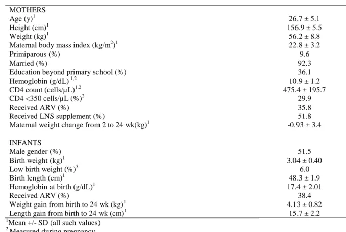
Table 3.1 Characteristics of 1,309 BAN mother-infant pairs in the primary analysis of the effects of maternal weight loss on infant growth
MOTHERS
Age (y)1 26.7 ± 5.1
Height (cm)1 156.9 ± 5.5
Weight (kg)1 56.2 ± 8.8
Maternal body mass index (kg/m2)1 22.8 ± 3.2
Primiparous (%) 9.6
Married (%) 92.3
Education beyond primary school (%) 36.1
Hemoglobin (g/dL) 1,2 10.9 ± 1.2
CD4 count (cells/µ L)1,2 475.4 ± 195.7
CD4 <350 cells/µL (%)2 29.9
Received ARV (%) 35.8
Received LNS supplement (%) 51.8
Maternal weight change from 2 to 24 wk(kg)1 -0.93 ± 3.4
INFANTS
Male gender (%) 51.5
Birth weight (kg)1 3.04 ± 0.40
Low birth weight (%)3 6.0
Birth length (cm)1 48.3 ± 1.9
Hemoglobin at birth (g/dL)1 17.4 ± 2.01
Received ARV (%) 38.4
Weight gain from birth to 24 wk (kg)1 4.13 ± 0.82
Length gain from birth to 24 wk (cm)1 15.7 ± 2.2
1Mean +/- SD (all such values)
2
Measured during pregnancy
3Low birth weight 2-2.5 kg as infants had to weigh
≥2.0 kg to be eligible for BAN
30
Table 3.2 Gender stratified linear regression models showing the effects of maternal weight loss on infant weight and length gain from 0 to 24 wk
Weight gain (kg) Length gain (cm)
Crude Adjusted Crude Adjusted
Coef 95% CI p-value Coef 95% CI p-value Coef 95% CI p-value Coef 95% CI p-value
BOYS n=673 n=653 n=667 n=647
Birth measurement -0.20 [-0.35,-0.04] 0.01 -0.15 [-0.31,0.01] 0.06 -0.42 [-0.51,-0.34] <0.001 -0.43 [-0.51,-0.35] <0.001
Mom lost weight -0.10 [-1.12,0.92] 0.84 -0.09 [-1.14,0.97] 0.87 1.30 [-1.30,3.90] 0.33 1.50 [-1.15,4.14] 0.27
Maternal BMI at 2 wk 0.03 [-0.003,0.07] 0.08 0.02 [-0.02,0.06] 0.27 0.07 [-0.02,0.16] 0.13 0.06 [-0.03,0.15] 0.20
Mom lost *Mom BMI 0.01 [-0.04,0.05] 0.79 0.01 [-0.04,0.05] 0.82 -0.06 [-0.17,0.05] 0.30 -0.07 [-0.18,0.05] 0.26
CD4 <350 0.06 [-0.08,0.20] 0.39 0.12 [-0.24,0.47] 0.52
Fever -0.03 [-0.10,0.04] 0.37 0.16 [-0.004,0.33] 0.06
Vomiting -0.01 [-0.15,0.12] 0.83 -0.38 [-0.72,-0.05] 0.03
Diarrhea -0.05 [-0.17,0.06] 0.38 0.15 [-0.14,0.45] 0.31
Primiparous 0.01 [-0.22,0.24] 0.91 -0.25 [-0.82,0.32] 0.39
Birthmonth (ref: Sept-Nov)
January -0.12 [-0.38,0.14] 0.36 -0.76 [-1.40,-0.11] 0.02
February 0.05 [-0.20,0.30] 0.71 -0.22 [-0.85,0.41] 0.49
March 0.22 [-0.04,0.48] 0.10 0.17 [-0.49,0.82] 0.61
April -0.03 [-0.29,0.23] 0.80 -0.21 [-0.87,0.45] 0.54
May 0.01 [-0.22,0.25] 0.92 0.17 [-0.42,0.77] 0.57
June -0.11 [-0.36,0.14] 0.40 -0.26 [-0.89,0.37] 0.42
July 0.30 [0.06,0.55] 0.02 0.30 [-0.33,0.92] 0.35
August 0.17 [-0.09,0.42] 0.20 0.50 [-0.15,1.15] 0.13
Dec 0.03 [-0.22,0.28] 0.80 -0.20 [-0.82,0.43] 0.54
Married 0.17 [-0.06,0.39] 0.15 0.12 [-0.46,0.69] 0.69
Education 0.0005 [-0.13,0.13] 0.99 0.31 [-0.02,0.64] 0.07
Mom ARV -0.13 [-0.29,0.04] 0.12 -0.59 [-1.00,-0.18] 0.01
Mom LNS 0.07 [-0.06,0.19] 0.29 -0.03 [-0.35,0.29] 0.88
Kid ARV -0.07 [-0.23,0.09] 0.42 -0.25 [-0.65,0.15] 0.22
Intercept 4.16 [3.29,5.02] <0.001 4.16 [3.23,5.09] <0.001 35.04 [30.75,39.34] <0.001 35.48 [31.04,39.91] <0.001
GIRLS n=633 n=609 n=632 n=608
Birth measurement -0.26 [-0.43,-0.10] 0.002 -0.30 [-0.47,-0.13] 0.001 -0.46 [-0.55,-0.37] <0.001 -0.46 [-0.56,-0.36] <0.001
Mom lost weight -0.97 [-1.86,-0.08] 0.03 -1.29 [-2.20,-0.38] 0.01 -2.38 [-4.83,0.08] 0.06 -3.36 [-5.88,-0.85] 0.01
Maternal BMI at 2 wk 0.03 [-0.001,0.06] 0.06 0.02 [-0.01,0.05] 0.12 -0.005 [-0.08,0.07] 0.90 -0.03 [-0.11,0.05] 0.49
Mom lost *Mom BMI 0.04 [0.004,0.08] 0.03 0.06 [0.02,0.09] 0.01 0.1 [-0.01,0.20] 0.07 0.14 [0.03,0.25] 0.01
CD4 <350 0.03 [-0.10,0.16] 0.70 -0.19 [-0.55,0.17] 0.31
Fever 0.01 [-0.06,0.07] 0.84 0.11 [-0.07,0.28] 0.23
Vomiting -0.0005 [-0.13,0.13] 1.00 -0.01 [-0.38,0.37] 0.98
Diarrhea -0.03 [-0.16,0.10] 0.63 -0.003 [-0.36,0.35] 0.99
31
Primiparous -0.13 [-0.34,0.07] 0.20 0.21 [-0.35,0.76] 0.46
Birthmonth (ref: Sept-Nov)
January 0.04 [-0.21,0.28] 0.78 0.04 [-0.66,0.73] 0.92
February 0.32 [0.05,0.59] 0.02 0.66 [-0.09,1.41] 0.08
March -0.02 [-0.28,0.24] 0.87 -0.32 [-1.05,0.41] 0.39
April 0.14 [-0.11,0.39] 0.26 -0.08 [-0.78,0.61] 0.82
May 0.09 [-0.14,0.32] 0.43 0.35 [-0.28,0.98] 0.27
June 0.17 [-0.06,0.39] 0.14 0.64 [0.02,1.26] 0.04
July 0.42 [0.17,0.66] 0.001 0.16 [-0.52,0.84] 0.64
August 0.13 [-0.09,0.35] 0.25 0.88 [0.28,1.49] 0.004
Dec 0.23 [-0.04,0.49] 0.09 0.21 [-0.52,0.94] 0.57
Married -0.05 [-0.29,0.19] 0.70 0.10 [-0.56,0.76] 0.77
Education 0.05 [-0.08,0.18] 0.42 0.16 [-0.20,0.52] 0.38
Mom ARV -0.04 [-0.20,0.11] 0.58 -0.43 [-0.87,0.02] 0.06
Mom LNS 0.08 [-0.05,0.20] 0.22 0.21 [-0.12,0.55] 0.21
Kid ARV -0.11 [-0.26,0.05] 0.18 -0.58 [-1.01,-0.15] 0.01
Intercept 4.06 [3.32,4.80] <0.001 4.22 [3.42,5.01] <0.001 37.64 [33.07,42.21] <0.001 38.03 [33.21,42.86] <0.001
BAN, Breastfeeding Antiretoviral and Nutrition; LNS, lipid-based nutrient supplement; RV, antiretroviral intervention
32
Table 3.3 Predictors of maternal weight loss among BAN mothers
Maternal weight loss from 2-24 wk1
OR 95% CI p
BMI at 2 weeks postpartum 1.02 [-0.02,0.06] 0.32
Maternal height (cm) 1.02 [0.00,0.04] 0.04
CD4 <350 cells/µL 1.14 [-0.13,0.40] 0.33
Primiparous 1.01 [-0.41,0.43] 0.96
Birthmonth (ref: Sept, Oct, Nov)2
January 0.35 [-1.52,-0.56] <0.001
February 0.32 [-1.62,-0.64] <0.001
March 0.19 [-2.16,-1.17] <0.001
April 0.29 [-1.74,-0.77] <0.001
May 0.35 [-1.51,-0.62] <0.001
June 0.42 [-1.34,-0.41] <0.001
July 1.05 [-0.48,0.59] 0.85
August 0.77 [-0.75,0.23] 0.30
December 0.62 [-0.99,0.03] 0.07
Married 1.49 [-0.05,0.84] 0.08
Education 1.22 [-0.06,0.46] 0.13
Mom ARV 1.74 [0.29,0.81] <0.001
Mom LNS 0.98 [-0.26,0.22] 0.87
Intercept 0.03 [-7.11,0.11] 0.06
1
Maternal weight loss from 2-24 wk, compared to weight change ≥0 2Chunk test for season effects: chi2(9) 81.33, p<0.001
33
Infant weight gain from 0-24 wk (kg)
Infant length gain from 0 to 24 wk (cm)
Figure 3.1 Predicted infant weight gain and length from 0 to 24 weeks for varying maternal BMI levels
Predicted values of infant weight and length gain from 0 to 24 weeks from a linear regression model relating maternal weight loss at varying maternal BMI levels at 2 weeks postpartum to infant growth outcomes at 24 weeks. Predicted values at each BMI level represent the linear combination of maternal weight loss coefficient, maternal BMI coefficient and the interaction between maternal weight loss and BMI coefficients. Each model also contained the infant’s birth measurement for weight (p=0.01) in the weight gain model and length (p<0.001) in the length gain model. BMI, body mass index.
3 3.5 4 4.5 5 5.5 6 6.5 7
16 21 26 31
Maternal BMI (kg/m2)
Boys: Maternal weight loss Boys: No maternal weight loss
Girls: Maternal weight loss Girls: No maternal weight loss
35 36 37 38 39 40
16 18 20 22 24 26 28 30 32
Maternal BMI at 2 wk (kg/m2)
Boys: Maternal weight loss Boys: No maternal weight loss
Chapter 4. Maternal transferrin receptors and ferritin are associated with infant values in exclusively breastfed HIV-exposed infants
Overview
Infant iron status at birth is influenced by maternal iron status during pregnancy; whether maternal iron status is associated with infant iron status during exclusive
breastfeeding is controversial. We evaluated how maternal and infant hemoglobin (Hb) and iron status [transferrin receptors (TfR) and ferritin] were related during exclusive
breastfeeding in HIV-infected women and their infants. The BAN (Breastfeeding,
Antiretrovirals and Nutrition) Study was a randomized controlled trial in Lilongwe, Malawi, where HIV-infected women were assigned with a 2 x 3 factorial design to lipid-based nutrient supplements (LNS), or no LNS, and maternal antiretrovirals (ARV), infant ARV or no ARV. We used longitudinal models to relate maternal Hb (n=1926) to concurrently measured infant Hb. In a subsample, we regressed change in infant iron status (Hb, log ferritin, log TfR) between 2 (n=355) or 6 wk (n=167), and 24 wk (n=532) on corresponding change in maternal indicator, adjusting for initial values. A one-unit higher maternal Hb at 6, 12, and18 wk was associated with 0.04 g/dL (p=0.02), 0.07 g/dL (p=0.001), and 0.05 g/dL (p=0.008) respective higher value in infant Hb. In the subsample, an increase in maternal TfR and Hb was associated with a respective increase in infant values (TfR β: 0.18 mg/L,
35
influence of initial infant values, optimizing maternal iron status is important to protect infant iron status.
Introduction
Iron deficiency is the most common nutrient deficiency in low-income countries and is associated with increased perinatal and maternal mortality, and impaired infant growth, neurodevelopment, and immune function. Given the initial iron endowment at birth, to date the predominant opinion has been that infant iron stores are sufficient during exclusive breastfeeding from birth to 6 months.63 However, infants in resource poor settings are prone to early depletion of iron stores, especially if maternal iron status was poor before or during pregnancy,33 and among infants with shorter gestational age,33 low birthweight,64,65 male gender,64 and rapid growth.33 The identification of postnatal factors that influence infant’s risk of early iron store depletion and subsequent deficiency is urgently needed.
Whether maternal iron status is associated with infant iron status during
breastfeeding—independent of infant iron stores at birth—is unclear. Studies evaluating the impact of maternal iron status during lactation on breast milk iron content have been
inconsistent.66-70 Several recent studies have shown that maternal Hb and serum iron levels are related to breastmilk iron levels, especially in anemic women or women with impaired iron status markers;69,70,70-72 however, studies that have investigated whether breastmilk iron levels subsequently influence infant iron status are equivocal.72
36
the mother or infant is recommended to promote child survival and prevent mother-to-child transmission of HIV if replacement feedings are not acceptable, feasible, affordable,
sustainable and safe.45 Together, these factors—impaired maternal iron status, antiretroviral prophylaxis,73and exclusive breastfeeding—may result in early depletion of infant iron stores and possibly a greater dependency on breastmilk iron as lactation proceeds.
Our objective was to examine the relationship between maternal and infant
hemoglobin (Hb) and iron status (ferritin and TfR), and furthermore because this was part of a randomized controlled trial, to determine the effects of the study interventions on maternal and infant Hb in participants of the Breastfeeding, Antiretrovirals and Nutrition Study (BAN) and a subset of BAN participants selected for additional biomarker assays, the Malawi
Mothers and Infants (MaMi) subsample. BAN mother-infant pairs were randomized to a 2-arm nutritional intervention with maternal lipid-based nutrient supplementation (LNS) meeting the nutritional needs of lactation and providing 15 mg iron per day and to a 3-arm antiretroviral (ARV) intervention (maternal, infant or no ARV regimen), and were closely followed during the period of exclusive breastfeeding from 0 to 24 weeks postpartum.
Subjects and Methods
Study population
The data for the current study are from the BAN study, whose design51 and primary intervention findings have been reported elsewhere.1,32,49 BAN was a randomized controlled trial of 2,369 mother-infant pairs conducted in Lilongwe, Malawi from April 2004 to
February 2010. HIV 1-positive pregnant women (n=3572) were recruited from four antenatal clinics and screened for initial eligibility criteria including: age ≥ 14 years of age, CD4 count
37
Malawi Ministry of Health guidelines for HIV treatment), Hb ≥ 7 g/dL, no prior antiretroviral medication use, and no major pregnancy complications.1 From the screening visit to 1 week (wk) postpartum, all eligible mothers received daily iron-folic acid supplementation
containing 200 mg ferrous sulfate and 5 mg folic acid according to the Malawi Ministry of Health care guidelines. At delivery, eligible women (n=2791) received maternal and infant single dose nevirapine peripartum as well as twice-daily zidovudine and lamivudine for 7 days postpartum. Within 36 hours of delivery, mother-infant pairs had to present at the study site and meet secondary eligibility criteria for randomization including: infant birthweight ≥
2 kg, no severe congenital malformations, and no other conditions incompatible with survival or that would preclude the use of the study drugs. Of the 2382 mother-infant pairs who met secondary eligibility criteria, thirteen women declined further participation.
38
ARV arms (mLNS-iARV & iARV) received daily oral nevirapine. On March 26, 2008, the data safety monitoring board halted enrollment in the no drug study arms (C & mLNS) because there was evidence that HIV transmission through breastmilk was higher in these groups.74 Mothers enrolled in these arms were allowed to change to the maternal or infant drug regimen for the remainder of exclusive breastfeeding.74 Further details of the ARV regimens are described elsewhere.49,74 Infants diagnosed by HIV-1 with polymerase chain reaction (PCR) within two wk of delivery (n=119) were disenrolled from the study, and mother-infant pairs were referred for care.1
In accordance with the World Health Organization prevention of maternal to child transmission of HIV (PMTCT) guidelines in development when the BAN study was designed,51,53 where exclusive breastfeeding for six months was recommended until acceptable, feasible, affordable, sustainable and safe (AFASS) conditions for replacement feeding are met, intensive counseling regarding exclusive breastfeeding was provided to BAN mothers. Mothers were instructed to exclusively breastfeed their infants for 24 wk and to wean between 24 and 28 wk.43 All participants were given 2 kg of maize per wk for family consumption to mitigate the effects of seasonal food shortages and to prevent sharing of maternal LNS. The World Food Program (WFP) provided food aid to all HIV-infected women in Lilongwe during a drought between February and August 2005. The aid consisted of a monthly ration of corn/soy flour and 1 liter of vitamin-A-fortified corn oil. An estimated 260 mothers received the ration in lieu of the BAN maize package.32 Based on WHO
39
Informed consent was obtained from all participating mothers. The Malawi National Health Science Research Committee and the institutional review boards at University of North Carolina at Chapel Hill and the U.S. Centers for Disease Control and Prevention approved the study.
Anthropometrics, study procedures and laboratory analyses
Participant visits were conducted at the BAN study clinic at Bwaila Hospital in Lilongwe. Mother-infant pairs were followed at birth and 1, 2, 4, 6, 8, 12, 18, 21, and 24 weeks postpartum, which allows us to closely follow mothers and infants during when breast milk is the sole nutrition source for the infant. At birth and 2, 6, 12, 18, and 24 weeks
40
Plasma was separated from red blood cells, aliquoted to 1 mL plastic storage tubes and stored at -70°C. To identify participants for inflammation and infection biomarker assays (n=537), samples were drawn from the LNS and no LNS groups where all multiple births and infants who became HIV positive at any time point were removed, and mother-infant pairs with anthropometry, and dietary data measurements were prioritized. Ferritin,
transferrin receptors (TfR), and markers of inflammation [C-reactive protein (CRP) and α -1-acid glycoprotein (AGP)] concentrations were measured at 2 or 6 and 24 wk postpartum using Cobas Integra 400 (Roche Diagnostics, Indianapolis, IN) in the subsample mother-infant pairs. Initial biomarker measurements were obtained at 2 weeks in mother-infants with sufficient plasma at that time; otherwise assays were conducted with plasma obtained at 6 wk.
Statistical analysis
This paper focuses on two groups of mother-infant pairs in BAN with Hb and iron measurements: mother-infant pairs with longitudinal Hb data (Longitudinal Hb sample), and a subset of these mother-infant pairs selected for additional biomarker analyses (MaMi
subsample), who have additional measurements of iron status (TfR and ferritin), CRP and
AGP. Mother-infant pairs were excluded from analyses if the infant was a multiple (n=49), HIV-infected (n=58). Infants who were weaned early (n=277) were excluded from the longitudinal anlayiss up to the point of cessation of cessation of exclusive breastfeeding. STATA 12.0 (College Station, TX) was used for all statistical analyses.
Longitudinal Hb sample
41
Characteristics of the included mother-infant pairs in the longitudinal Hb sample (n=1926) were compared to characteristics of randomized mothers excluded from the analysis (n=443) to assess for similarity using t-tests for continuous, normally distributed variables, and nonparametric tests for skewed continuous variables. Compared to excluded randomized mothers, mothers in the longitudinal Hb sample were older (26.2 vs. 25.1 years, p<0.001), had lower prevalence of low CD4 (29% vs 35%, p<0.01), higher pregnancy Hb (10.8 vs. 10.6 g/dL, p<0.001) had less anemia during pregnancy (52% vs. 60%, p=0.002), and fewer
women were primiparous (12 vs. 16%, p=0.01). Infants in the longitudinal Hb sample had heavier birthweight (3.02 vs. 2.96 kg, p=0.001), higher birth length (48.2 vs. 47.9 cm,
p<0.001), lower prevalence of low birth weight (6% vs. 12%, p<0.001), and were more likely to be in the ARV arm (38% vs. 29%, p<0.001).
To understand the influence of maternal anemia on infant iron status postpartum, we used linear regression to evaluate whether maternal anemia during pregnancy was associated with infant Hb at birth, adjusting for infant birthweight. We also evaluated whether maternal anemia during pregnancy (Hb <11 g/dL)75 was associated with infant Hb from 2 to 24 weeks using a longitudinal random effects model with a first order autoregressive disturbance term, adjusting for infant birthweight, birth Hb and rate of weight gain and age. Infant age was modeled with a spline with a knot at 9 weeks of age.
42
containing one knot at 9 wks of age. Interactions of age with study interventions, rate of weight gain and infant Hb at birth were evaluated. The BAN study interventions were tested as potential confounders; however, none of the interventions had significant effects compared to mother-infant pairs in the control group (all p-values >0.20) and were not included in the final model.
In secondary analyses in the longitudinal Hb sample, we separately evaluated for intervention effects on maternal Hb and infant Hb. In the mothers, a longitudinal random effects model with a first order autoregressive disturbance terms was used to evaluate the effect of the maternal study interventions (mLNS-mARV, mARV, mLNS, control) on maternal Hb from 6 to 24 wk postpartum, adjusting for maternal Hb at 2 wk and time in weeks. Potential interactions with the study interventions and time were evaluated. In the infants, similar longitudinal random effects models were used to evaluate the effect of the six study interventions on infant Hb from 2 to 24 wk. Infant age was modeled a spline containing one knot at 9 wks of age. Potential interactions of study interventions, infant Hb at birth and rate of infant weight gain were also evaluated.
MaMi Subsample
43
were less likely to be married (90.5% vs. 93.3%, p=0.01) and infants had marginally higher birthweight (3.04 vs. 3.01 kg, p=0.06) and a lower prevalence of low birthweight (4.6% vs. 8% p=0.01), and a larger proportion received the ARV intervention (41% vs. 35%, p=0.02). Hb and ferritin are sensitive to infection and inflammation—Hb is lowered and ferritin is greatly increased.76 Though TfR is thought to be less sensitive to inflammation and infection,76 we observed associations of CRP and AGP with TfR, and therefore adjusted TfR for infection and inflammation. To remove the effects of inflammation from TfR, Hb and ferritin measurements, we used methods proposed by Thurnham and colleagues.77,78 We used previously used cutpoints to define elevated CRP (>5 mg/L) and AGP (>1 g/L) and stage of inflammation [healthy (normal CRP & AGP), incubation (elevated CRP), early convalescence (CRP & AGP elevated), late convalescence (elevated AGP)].78 We then determined correction factors for each inflammation group by dividing the median value of the healthy inflammation group by the median value of the incubation, early convalescence, and late convalescence groups.78 We then multiplied the measured Hb and iron status values by the group specific correction factor.78
44
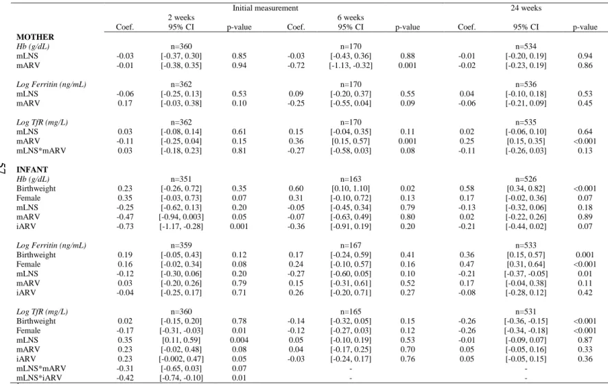
a sensitivity analysis to evaluate how adjustment for infection and inflammation influenced our findings in the MaMi subsample mother-infant pairs. We used linear regression with unadjusted iron markers to evaluate the relationship between change in maternal iron marker on change in infant iron marker outcome, and observed associations similar to the
inflammation adjusted models. In addition to these models, we used logistic regression to evaluate whether depleted maternal iron stores (ferritin <15 ug/L) at 2/6 weeks were
associated with increased odds of depleted infant iron stores (ferritin <12 ug/L) at 2/6 weeks, adjusting for infant birthweight, ARV arm, when initial measurement was obtained and infant gender.
In secondary analyses in the MaMi subsample, we separately evaluated for intervention effects on maternal and infant iron status and Hb, adjusting for infant
birthweight and gender. Interactions between LNS and ARV arms were also evaluated. We also conducted logistic regression models to determine if the study interventions were
associated with increased odds of impaired infant iron status at 24 weeks, adjusting for initial iron status measurement, birthweight, timing of measurement and infant gender. Interactions between LNS and ARV intervention arms were also evaluated.
Results
45
Longitudinal Hb sample
From delivery to 24 wks postpartum, maternal Hb increased (Figure 4.1) and prevalence of maternal anemia (Hb <11 g/dL)75 decreased from about 50% to 31%. Mean infant Hb followed the typical pattern of decline from birth to 24 weeks, as fetal Hb is broken down79 (Figure 4.2). From 12-24 wk, prevalence of low infant Hb (<10.5 g/dL)79 increased from 43% to 50.3%. We observed that pregnancy anemia (Hb<11 g/dL)75 was associated with lower infant Hb at birth (β: 0.23 g/dL, p=0.01), adjusting for infant birthweight, but was not associated with infant Hb from 2 to 24 weeks in a longitudinal model (β: -0.08 g/dL, p=0.2).
The longitudinal model relating maternal and infant Hb over time indicates that higher maternal Hb predicts higher infant Hb over time (Supplemental Table 4.2) with significant associations between 6 and 18 wks (Figure 4.3). Female gender and higher birthweight were associated with higher infant Hb (p<0.001). Higher infant Hb at birth was associated with higher infant Hb over time, while faster weight gain—especially in younger infants—was associated with lower infant Hb over time.
Effects of the study interventions
46
p<0.001); however, at 18 and 24 wks the negative effects were not sustained (all p-values >0.25). Similar effects were observed in mothers who received only ARVs (mARV), where maternal Hb was significantly lower at 6 and 12 wk postpartum (6 wk β: -0.22, p=0.001, 12 wk β: -0.15, p=0.003), but again at 18 and 24 wks the negative effects were not sustained (all p-values >0.1). The longitudinal model of infant Hb shows no effects of the intervention arms on infant Hb (Supplemental Table 4.4).
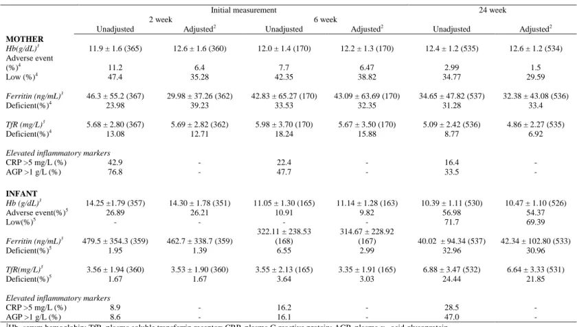
MaMi subsample
In the MaMi subsample, inflammation adjusted maternal Hb remained relatively stable from initial measurement to 24 wk, while maternal TfR declined (Table 4.2). Initially, prevalence of depleted maternal iron stores (adjusted ferritin <15 ug/L) was high, while fewer women had tissue iron depletion, denoted by elevated adjusted TfR (TfR >8.3 mg/L). By 24 weeks, about a third of women had depleted iron stores, but again fewer women had tissue iron depletion. Inflammation adjusted infant Hb declined from initial measurement to 24 wk. Infant ferritin values declined markedly, while TfR increased from initial
measurement to 24 wk. Few infants had abnormal initial adjusted measurements, but by 24 wk many infants began to show signs of poor iron status. From initial measurement to 24 weeks, prevalence of elevated CRP (>5 mg/L) and AGP (>1 g/L) declined in mothers and increased in infants.