R E S E A R C H
Open Access
Pharmacological mechanism underlying
anti-inflammatory properties of two structurally
divergent coumarins through the inhibition of
pro-inflammatory enzymes and cytokines
Salman Khan
1,2, Omer Shehzad
2,3, Mao-Sheng Cheng
4, Rui-Juan Li
4and Yeong Shik Kim
2*Abstract
Background:The aim of the present study is to investigate the effects of two structurally divergent coumarins, calipteryxin (1) and (3’S,4’S)-3’,4’-disenecioyloxy-3’,4’-dihydroseselin (2) fromSeseli recinosum, in lipopolysaccharide (LPS)-stimulated murine macrophages.
Methods:The nitrite production was evaluated using Griess reagent. The protein and mRNA expression levels were investigated through Western blot and quantitative real time-PCR analyses. The NF-κB and AP-1 DNA-binding activities were assessed using an electrophoretic mobility shift assay. The docking studies were performed with Glide XP in Schrödinger suite (version 2013).
Results:The results of the present study revealed that calipteryxin (1) and (3’S,4’S)-3’,4’-disenecioyloxy-3’,4’-dihydroseselin (2) treatment showed potent inhibitory effects on pro-inflammatory enzymes and cytokines associated with molecular signaling pathways. Treatment with calipteryxin and (3’S,4’S)-3’,4’-disenecioyloxy-3’,4’-dihydroseselin also decreased the production of nitric oxide (NO), tumor necrosis factor alpha (TNF-α) and interleukin-1 beta (IL-1β) in a dose-dependent manner. Additionally, both coumarins inhibited the LPS-induced protein and mRNA expression levels of nitric oxide synthase (iNOS) and cyclooxygenase-2 (COX-2) in RAW264.7 cells. To explore the potential mechanisms underlying the inhibitory activity of coumarin derivatives, the protein signaling pathways for NF-κB, mitogen-activated protein kinase (MAPK) and Akt were examined. Calipteryxin and (3’S,4’S)-3’,4’-disenecioyloxy-3’,4’-dihydroseselin markedly reduced the LPS-stimulated phosphorylation of IKKα/β, p-IκBαand IκBαdegradation as well as the nuclear translocation of the p65 subunit of pro-inflammatory transcription factor NF-κB. In addition, calipteryxin and (3’S,4’S)-3’,4’-disenecioyloxy-3’,4’-dihydroseselin) considerably inhibited the LPS-induced expression of ERK, c-Jun N-terminal kinase (JNK), p38 and Akt proteins. Furthermore, both coumarins significantly inhibited c-Jun expression in the nucleus.
Conclusions:Taken together, these results support the therapeutic potential and molecular mechanism of calipteryxin and (3’S,4’S)-3’,4’-disenecioyloxy-3’,4’-dihydroseselin associated with inflammatory diseases.
Keywords:Calipteryxin, (3’S,4’S)-3’,4’-disenecioyloxy-3’,4’-dihydroseselin, NF-κB, MAPK, Akt,Seseli recinosum
* Correspondence:[email protected] 2
College of Pharmacy, Seoul National University, Gwanak-gu, Seoul 151-742, South Korea
Full list of author information is available at the end of the article
Background
Considerable amounts of pro-inflammatory mediators and pro-inflammatory cytokines are released at injury sites during inflammation [1]. These pro-inflammatory mediators respond to numerous stimuli, including bac-terial lipopolysaccharide (LPS), cytokines, and UV ir-radiation, which modulate to their effects by inducing
the activation of NF-κB and AP-1 [2].
NF-κB activates a number of molecules involved in
the inflammatory response, including iNOS, COX-2,
TNF-α, IL-1β, and IL-6 [1]. The production of these
mediators and cytokines through NF-κB stimulation
might reflect the extent of inflammation and has been suggested as a measure to evaluate the effects of anti-inflammatory agents on the anti-inflammatory process.
NF-κB signaling is activated through two diverse pathways:
the canonical (classical) pathway and the non-canonical
pathway [3]. NF-κB signaling through canonical or
non-canonical pathways involves the expression of multiple
genes [4, 5]. The canonical pathway involves the IκBα
kinase (IKK) complex, while the non-canonical pathway
involves NF-κB inducing kinase (NIK), which recruits IKK
to p100 and subsequently activates IKK [4]. In response to
inflammation, the p50 and p65 subunits of NF-κB are
translocated through the canonical pathway, whereas the
p52-containing RelB heterodimer of NF-κB is released and
translocated through the non-canonical pathway [4, 5]. Mitogen-activated protein (MAP) kinases and PI3k/Akt signaling pathways play essential roles in inflammation and tissue remodeling [6, 7]. Consequently, the inhibition of MAP kinases produces anti-inflammatory effects against various inflammatory diseases [6]. However, several studies have demonstrated that the activation of MAP kinases suppresses inflammatory reactions, such as bacterial LPS-induced cytokine production in macrophages.
In the present study, two structurally divergent
cou-marin derivatives, calipteryxin (1) and (3’S,4’S)-3’,4’
-disenecioyloxy-3’,4’-dihydroseselin (2) isolated fromSeseli
resinosum, were studied in terms of LPS-induced
inflam-matory signaling. The genus Seseli L. belongs to the
Apiaceae family, which comprises aromatic herbs used as foods, spices, condiments and ornamentals [8]. This herb has various pharmacological applications, such as anthelmintic, carminative, stomachic and stimulant
prop-erties [8]. It has been reported that Seseli plants show
significant and dose-dependent anti-inflammatory activity and analgesic effects in carrageenan-induced acute inflam-mation in rats [8]. However, only a few pharmacological and biological activity studies concerning the single
com-ponent fromSeselihave been reported. Therefore, the aim
of the present study was to examine LPS-induced inflam-matory signaling in murin macrophages. Moreover, a comparative investigation was performed between two coumarin type compounds.
Methods
Isolation and purification of calipteryxin and (3’S,4’S)-3’,
4’-disenecioyloxy-3’, 4’-dihydroseselin
The separation and purification of calipteryxin and (3’S,4’S)-3’,4’-disenecioyloxy-3’,4’-dihydroseselin, fromSeseli resinosumwere performed using counter-current chroma-tography coupled with an evaporative light scattering
detector (CCC-ELSD). Based on optimum KDvalues of
the target compounds, a two-phase solvent gradient system comprising n-hexane/ethyl acetate/methanol/water at a vol-ume ratio of 3:2:3:2 and 2:1:2:1 was used. The peak frac-tions were collected using CCC-ELSD chromatography. The collected peak fractions were dried under reduced pressure and analyzed through HPLC-ELSD. The results
confirmed that the purity of each compound was≥99 %.
Moreover, the structures of calipteryxin and (3’S,4’S)-3’,4’
-disenecioyloxy-3’,4’-dihydroseselin (Fig. 1) were confirmed
based on ESI-MS/MS,1H-NMR and13C-NMR data, as
de-scribed in previous studies [9].
Cells and culture medium
RAW 264.7 murine macrophages were obtained from the American Type Culture Collection (Manassas, VA). These macrophages were maintained and subcultured according to a previously described procedure [5].
Cell Viability and nitric oxide determination
The cell viability was determined using an MTT assay according to Khan et al. [5]. The RAW 264.7 cells were
treated with various concentrations (2.5 ~ 30μM) of
calip-teryxin and (3’S,4’S)-3’,4’-disenecioyloxy-3’,4’
-dihydrosese-lin or vehicle alone for 2 h prior to LPS (1 μg/ml)
stimulation and subsequently incubated at 37 °C for an additional 18 h. The NO was measured using the Griess reaction as previously described [5].
Fig. 1Chemical structures of calipteryxin and (3’S,4’S)-3’,4’
Western immunoblot analysis
The Western blot procedure was performed according to Khan et al., with some modifications [5].
RNA extraction and quantitative real-time (RT) PCR RT-PCR was performed with total RNA extracted using
easyBlue™ according to the manufacturer’s instructions
(Sigma-Aldrich, St. Louis, MO). The purity and concentra-tions of RNA were determined using an ND-1000 spectro-photometer (Nanodrop Technologies, Wilmington, DE).
All RNA samples were stored at−80 °C until further
ana-lysis. Total RNA (1μg) was converted to cDNA through
RT-PCR (Genius FGEN05TD, Teche, England) using the
iScript™ cDNA Synthesis Kit (BIO-RAD, Hercules, CA)
under the following conditions: 25 °C for 5 min, 42 °C for 30 min and 85 °C for 5 min. Quantitative real-time poly-merase chain reaction (qRT-PCR) analysis was performed using an Applied Biosystems 7300 Real-Time PCR system and software (Applied Biosystem, Carlsbad, CA). qRT-PCR was conducted in 0.2 ml qRT-PCR tubes with forward and reverse primers and the SYBR green working solution
(iTaq™ Universal SYBR Green Supermix, BIO-RAD,
Hercules, CA), using customer PCR master mix under the following conditions: 95 °C for 30 min, followed by 40 cy-cles of 95 °C for 15 s, 55 °C for 20 s and 72 °C for 35 s. The melting point, optimal conditions and specificity of the reactions were determined. The sequences of the PCR primers were previously described [10, 11]. The sense and
antisense primers for iNOS were 5’CCCTTCCGAAGTT
TCTGGCAGC-3’and 5’-GGCTGTCAGAGCCTCGTGG
CTT-3’, respectively. The sense and antisense primers for
COX-2 were 5’
-GGAGAGACTATCAAGATAGTGATC-3’ and 5’-ATGGTCAGTAGACTTTTACA-GCTC-3’,
re-spectively. The following primers were used for; TNF-α,
sense primer, 5’-AGC ACA GAA AGC ATG ATC CG-3’
and antisense primer, 5’-CTG ATG AGA GGG AGG
CCA TT-3’; and for IL-1β, sense primer, 5’-ACCT GCT
GGT GTG TGA CGT T-3’, and antisense primer, 5’-TCG
TTG CTT GGT TCT CCT TG-3’. The sense and
anti-sense primers for rat actin mRNA expression (used as a
control for total RNA content for each sample) were 5’
-TGAAGGTCGGTGTGAACGGATTTGGC-3’ and 5’-CA
TGTAGGCCATGAGGTCCACCAC-3’, respectively.
NF-κB secretory alkaline phosphatase (SEAP) reporter
gene assay in transfected-RAW 264.7 cells
The NF-κB SEAP inhibitory activities of calipteryxin and
(3’S,4’S)-3’,4’-disenecioyloxy-3’,4’-dihydroseselin were
de-termined in LPS-stimulated RAW 264.7 macrophages.
The NF-κB-dependent reporter gene transcription was
an-alyzed using the SEAP assay as previously described, with some modifications [5, 7].
Electrophoretic mobility shift assay (EMSA)
EMSA was performed to investigate the inhibitory
ef-fects on NF-κB and AP-1 DNA binding, as previously
described [5, 7].
Computational methods
Docking studies were performed using Glide XP in Schrödinger suite (version 2013).
Statistical analysis
Unless otherwise stated, the results are expressed as the means ± standard deviations (SD) from three different experiments. One-way analysis of variance (ANOVA)
followed by Dunnett’s t-test was applied to assess the
statistical significance of the differences between the study groups (SPSS version 10.0, Chicago, IL). A value of
p<0.05 was considered statistically significant.
Results
Effects of calipteryxin and (3’S,4’S)-3’,4’
-disenecioyloxy-3’,4’-dihydroseselin on cell viability in LPS- and
SNP-induced macrophages
The cell viability of calipteryxin and (3’S,4’S)-3’,4’
-dis-enecioyloxy-3’,4’-dihydroseselin was evaluated in
LPS-and SNP-stimulated RAW264.7 cells (Fig. 2). No cytotoxic effect was observed in LPS-stimulated macrophages until
treatment with a 30-μM concentration of the derivatives.
Consequently, non-toxic concentrations were used for the following experiments.
Inhibitory effects of calipteryxin and (3’S,4’S)-3’,4’
-dis-enecioyloxy-3’,4’-dihydroseselin on NO production in
LPS- and SNP-induced RAW 264.7 macrophages To determine NO production, we measured the amount of nitrite released into the culture medium. RAW 264.7 cells were treated with various concentrations of
calipter-yxin and (3’S,4’S)-3’,4’-disenecioyloxy-3’,4’-dihydroseselin
(2.5 ~ 30 μM). Incubation with LPS alone markedly
increased NO production from these cells, compared with the NO production generated under the control condi-tions (Fig. 2). However, pre-treatment with calipteryxin
and (3’S,4’S)-3’,4’-disenecioyloxy-3’,4’-dihydroseselin
pre-vented this increased level of NO production in LPS-stimulated RAW 264.7 cells in a concentration-dependent manner (Fig. 2a)., However, SNP-induced NO production was slightly reduced after treatment with calipteryxin and
(3’S,4’S)-3’,4’-disenecioyloxy-3’,4’-dihydroseselin (Fig. 2b).
By comparison, calipteryxin and (3’S,4’S)-3’,4’
-disenecioy-loxy-3’,4’-dihydroseselin inhibition was more remarkable
in LPS-stimulated macrophages than in SNP-induced macrophages. Therefore, the LPS-stimulated RAW264.7 cell model was used for subsequent in vitro experiments.
Effects of calipteryxin and (3’S,4’S)-3’,4’
-disenecioyloxy-3’,4’-dihydroseselin on LPS-induced iNOS and COX-2
As calipteryxin and (3’S,4’S)-3’,4’-disenecioyloxy-3’,4’ -dihydroseselin inhibit NO production, we examined the relationship between the protein and mRNA expression levels of iNOS and COX-2 (Fig. 3). The inhibitory effects
of calipteryxin and (3’S,4’S)-3’,4’-disenecioyloxy-3’,4’
-dihy-droseselin on the protein and mRNA expression levels of iNOS and COX-2 were determined through Western blot-ting and qRT-PCR analyses, respectively. The iNOS and COX-2 protein and mRNA expression levels were mark-edly up-regulated after LPS treatment, and calipteryxin
and (3’S,4’S)-3’,4’-disenecioyloxy-3’,4’-dihydroseselin
sig-nificantly attenuated iNOS and COX-2 mRNA expression in LPS-stimulated macrophages in a concentration-dependent manner (Fig. 3).
Effects of calipteryxin and (3’S,4’S)-3’,4’
-disenecioyloxy-3’,4’-dihydroseselin on LPS-induced NO production using
various inhibitors in macrophages
To investigate the inflammatory signaling pathway in-volved in the inhibitory effects of calipteryxin and
(3’S,4’S)-3’,4’-disenecioyloxy-3’,4’-dihydroseselin on
LPS-induced inflammatory mediators, specific inhibitors of the
NF-κB (TPCK, 20 μM), MAPKs (SB202190, p38 MAPK
inhibitor; SP600125, JNK inhibitor; U0126, ERK inhibitor) and Akt (LY294002) were used (Fig. 4). The pretreatment of RAW 264.7 cells with TPCK, SB202190, SP600125, and LY294002 significantly inhibited LPS-induced nitrite pro-duction in the media, while U0126 showed no effects
at 20μM (Fig. 4). The combination of calipteryxin and
Fig. 2Effects of calipteryxin and (3’S,4’S)-3’,4’-disenecioyloxy-3’,4’-dihydroseselin on cell viability and NO production in LPS- (a) and SNP- (b)
(3’S,4’S)-3’,4’-disenecioyloxy-3’,4’-dihydroseselin with TPCK and specific inhibitors against p38, JNK, ERK and Akt significantly inhibited LPS-induced NO pro-duction (Fig. 4). Overall, these results suggest that p38,
JNK, ERK and Akt, in conjunction with NF-κB
inflamma-tory signaling, might contribute to the inhibiinflamma-tory effects of
calipteryxin and (3’S,4’S)-3’ ,4’-disenecioyloxy-3’,4’
-dihy-droseselin on inflammatory mediators.
Inhibitory effects of calipteryxin and (3’S,4’S)-3’,4’
-dis-enecioyloxy-3’,4’-dihydroseselin on NF-κB signaling
Because the results indicated that calipteryxin and
(3’S,4’S)-3’,4’-disenecioyloxy-3’,4’-dihydroseselin affect
iNOS and COX-2 induction, we focused on two transcrip-tion factors critical in iNOS and COX-2 inductranscrip-tion, i.e.,
NF-κB and AP-1 [5, 12]. Initially, the inhibitory effects of
calipteryxin and (3’S,4’S)-3’ ,4’-disenecioyloxy-3’,4’
-dihy-droseselin on NF-κB in the medium were evaluated. As
shown in Fig. 5, both compounds exhibited remarkable
in-hibitory effects on NF-κB in the culture media.
To analyze whether calipteryxin and (3’S,4’S)-3’,4’
-dis-enecioyloxy-3’,4’-dihydroseselin suppressed the
phosphor-ylation and degradation of IκBα and NF-κB nuclear
translocation, time course experiments were performed in LPS-stimulated macrophages using Western blot analysis
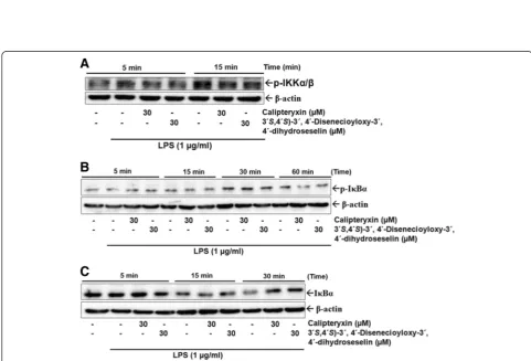
(Fig. 6). After treatment with LPS alone for 1 h, IκBα
activation levels were markedly increased, and calipteryxin
and (3’S,4’S)-3’,4’-disenecioyloxy-3’,4’-dihydroseselin)
sig-nificantly blocked LPS-induced IκBα phosphorylation
after 60 min of LPS stimulation (Fig. 6). However, IκBα
degradation through calipteryxin and (3’S,4’S)-3’,4’
-disene-cioyloxy-3’,4’-dihydroseselin was inhibited after 15 min of
LPS (Fig. 6). Complete inhibition was observed after 30 min of LPS stimulation (Fig. 6).
In an attempt to explore the effects of calipteryxin and
(3’S,4’S)-3’,4’-disenecioyloxy-3’,4’-dihydroseselin on the
inhibition of IκB kinase (IKK) activity in RAW264.7 cells,
Western blot analysis was performed to measure the
phos-phorylation of IKKα/βafter treatment with calipteryxin and
Fig. 3Effects of calipteryxin and (3’S,4’S)-3’,4’-disenecioyloxy-3’,4’-dihydroseselin on the LPS-induced iNOS protein (a), and COX-2 protein (b), iNOS
(3’S,4’S)-3’,4’-disenecioyloxy-3’,4’-dihydroseselin (Fig. 6).
Calipteryxin and (3’S,4’S)-3’,4’-disenecioyloxy-3’,4’
-dihy-droseselin significantly inhibited LPS-induced activation
of IKKα/β(Fig. 6).
Additionally, we examined the DNA-binding affinity of these transcription factors using EMSA (Fig. 7a). Both
Calipteryxin and (3’S,4’S)-3’,4’-disenecioyloxy-3’,4’
-dihy-droseselin attenuated the LPS-induced DNA binding
ac-tivity of both NF-κB (Fig. 7a). The specificity of the bands
was confirmed after adding a 50-fold excess of unlabeled
NF-κB oligonucleotide to the binding reaction (Fig. 7a).
Additionally, the nuclear translocation of p65, a component
of the NF-κB heterodimer, was further evaluated (Fig. 8b).
LPS induced the translocation of p65 from the cytoplasm to the nucleus after treatment for 1 h, and calipteryxin and
(3’S,4’S)-3’,4’-disenecioyloxy-3’,4’-dihydroseselin markedly
prevented the nuclear translocation of p65 (Fig. 7).
Effects of calipteryxin and (3’S,4’S)-3’,4’
-disenecioyloxy-3’,4’-dihydroseselin on LPS-induced MAPKs and Akt
acti-vations in macrophages
MAP kinases are important for the expression of both iNOS and COX-2 [13, 14]. Thus, MAPKs act as specific targets in inflammatory responses. Because MAPK
acti-vation plays an important role in NF-κB stimulation, we
examined whether MAPK’s activity is inhibited through
treatment with calipteryxin and (3’S,4’S)-3’ ,4’
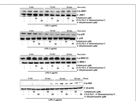
-disene-cioyloxy-3’,4’-dihydroseselin. Herein, we assessed the
phosphorylation levels of MAP kinases, including JNK1,
p38 and ERK 1/2. When RAW 264.7 cells were stimulated
with LPS, in the presence of calipteryxin and (3’S,4’S)-3’,4’
-disenecioyloxy-3’,4’-dihydroseselin, the levels of
phosphor-ylated JNK1, p38 and ERK 1/2 MAPK were observed to significantly start decreasing after 15 min of LPS stimula-tion (Fig. 8). Addistimula-tionally, Akt activastimula-tion was significantly
inhibited after treatment with calipteryxin and (3’S,4’S
)-3’,4’-disenecioyloxy-3’,4’-dihydroseselin after 60 min of
LPS stimulation (Fig. 8). These results suggest that the MAPK and Akt pathways are relevant during the LPS-mediated expression of iNOS and COX-2.
Effects of calipteryxin and (3’S,4’S)-3’,4’
-disenecioyloxy-3’,4’-dihydroseselin on LPS-induced AP-1-DNA binding
and c-Jun expression in macrophages
AP-1 is another transcription factor involved in the regulation of inflammatory processes [13]. To evaluate
the effects of calipteryxin and (3’S,4’S)-3’,4’
-disenecioy-loxy-3’,4’-dihydroseselin, we performed EMSA. The results
clearly demonstrated that calipteryxin and (3’S,4’S)-3’,
4’-disenecioyloxy-3’,4’-dihydroseselin remarkably
inhib-ited AP-1-DNA binding activity (Fig. 9), while LPS-stimulated cells showed significantly high DNA-binding affinity (Fig. 9). In addition, the effects of calipteryxin
and (3’S,4’S)-3’,4’-disenecioyloxy-3’,4’-dihydroseselin on
c-jun were considerably promising (Fig. 9).
Effects of calipteryxin and (3’S,4’S)-3’,4’
-disenecioyloxy-3’,4’-dihydroseselin on pro-inflammatory cytokines in
LPS-stimulated macrophages
Fig. 4Effects of calipteryxin and (3’S,4’S)-3’,4’-disenecioyloxy-3’,4’-dihydroseselin on various inhibitors induced by LPS in RAW 264.7
Fig. 6Effects of calipteryxin and (3’S,4’S)-3’,4’-disenecioyloxy-3’,4’-dihydroseselin on p-IKKα/β(a), p-IκBα(b), and IκBα(c) protein expression levels in time course experiments
Fig. 5Effects of calipteryxin and (3’S,4’S)-3’,4’-disenecioyloxy-3’,4’-dihydroseselin on LPS-induced and NF-κB-dependent alkaline phosphatase (SEAP)
Fig. 7Effects of calipteryxin and (3’S,4’S)-3’,4’-disenecioyloxy-3’,4’-dihydroseselin on NF-κB-DNA binding activity (a) and p65 (b) protein expression
Fig. 8Effects of calipteryxin and (3’S,4’S)-3’,4’-disenecioyloxy-3’,4’-dihydroseselin on the protein expression levels of the MAPKs p-JNK1, p-p38, p-ERK
As calipteryxin and (3’S,4’S)-3’,4’-disenecioyloxy-3’,4’ -dihydroseselin inhibit the activation of the two
pro-inflammatory transcription factors NF-κB and AP-1, we
examined the effects of calipteryxin and (3’S,4’S)-3’,4’
-disenecioyloxy-3’,4’-dihydroseselin on the expression of
pro-inflammatory cytokines using qRT-PCR analysis.
TNF-α and IL-1β are predominantly regulated at the
transcriptional level, whereby the transcription factors
NF-κB and AP-1 play crucial roles [15]. Indeed, the
treatment of LPS-activated cells with calipteryxin and
(3’S,4’S)-3’,4’-disenecioyloxy-3’,4’-dihydroseselin
signifi-cantly reduced the secretion of TNF-α and IL-1β in
RAW 264.7 cells (Fig. 10).
Binding model analysis
To further elucidate the binding of compounds, we per-formed a docking analysis. Docking was simulated using Glide XP (Schrödinger 2013) to examine the interactions
between calipteryxin and (3’S,4’S)-3’,4’-disenecioyloxy-3’
Fig. 9Effects of calipteryxin and (3’S,4’S)-3’,4’-disenecioyloxy-3’,4’-dihydroseselin on Ap-1-DNA binding activity (a) and c-Jun (b) protein expression
Fig. 10Suppression effects of calipteryxin and (3’S,4’S)-3’,4’-disenecioyloxy-3’,4’-dihydroseselin on the mRNA expression of the pro-inflammatory
,4’-dihydroseselin in the NIK active site (Fig. 11).
Calipter-yxin and (3’S,4’S)-3’,4’-disenecioyloxy-3’,4’-dihydroseselin
form two hydrogen bonds with the LYS517 and SER476 residues. The secondary structure of this protein is shown as a solid ribbon (gray). Key residues are displayed in line
style (blue), calipteryxin and (3’S,4’S)-3’,4’
-disenecioyloxy-3’,4’-dihydroseselin are displayed in stick style (carbon
atoms in cyan), and hydrogen bonds are represented as green dotted lines.
Discussion
Macrophages serve as a crucial link between innate and adaptive immunity and play pivotal roles in inflamma-tory signaling [16]. The stimulation of macrophages with bacterial exotoxins, such as LPS, occurs through the spe-cific receptor TLR4 and triggers the recruitment of the cytoplasmic adaptor protein MyD88 and the activation of TIRAP, which subsequently stimulates down-stream
signaling pathways (NF-κB and MAPKs). The
LPS-induced pathways up-regulate the expression of various inflammatory mediators and cytokines involved in the pathogenesis of inflammatory responses [16]. Based on
these hypotheses, the modulation of LPS-induced NF-κB
and MAPK signaling or the regulation of cytokine pro-duction might constitute a therapeutic strategy in many inflammatory diseases.
Natural products have been one of the leading sources for the discovery of new anti-inflammatory agents. Couma-rins are naturally isolated compounds with various remark-able pharmacological and biological properties [8]. In the present study, two different types of coumarins were inves-tigated in terms of LPS-stimulated macrophages through the inhibition of the signaling pathways for the
tran-scription factors NF-κB and AP-1. Initially, the effects
of calipteryxin and (3’S,4’S)-3’,4’-disenecioyloxy-3’,4’
-dihydroseselin on the production of NO and on the regulatory genes for iNOSand COX-2 in LPS-stimulated RAW264.7 macrophages were examined. The LPS-induced down-regulation of the pro-inflammatory
media-tors through calipteryxin and (3’S,4’S)-3’,4’
-disenecioyloxy-3’,4’-dihydroseselin were based on the suppression of the
NF-κB and AP-1 signaling, leading to a therapeutic
ap-proach against inflammatory diseases.
NO is a free radical produced fromL-arginine through
nitric oxide synthases (NOS) that affects immune func-tions by eliciting intracellular signals [2, 5]. The high level of NO causes inflammatory damage to target tissue during infection [17]. Hence, the regulation of NO release via the inhibition of iNOS expression is helpful to alleviate inflammatory damage. In the present study, we also
showed that calipteryxin and (3’S,4’S)-3’,4’
-disenecioyloxy-3’,4’-dihydroseselin significantly suppressed
LPS-induced-iNOS expression at the transcriptional and translational levels in RAW264.7 cells. Additionally, COX-2 is an indu-cible isoform of cyclooxygenase that plays an important
role in inflammation [18]. Calipteryxin and (3’S,4’S)-3’
,4’-disenecioyloxy-3’,4’-dihydroseselin also suppressed
LPS-stimulated COX-2 expression in RAW264.7 cells. We have previously demonstrated that LPS stimula-tion induces pro-inflammatory enzymes and cytokines
through the activation of NF-κB and AP-1 signaling
pathways, which play crucial roles in the control of cellular responses to cytokines and stresses [5, 7].
NF-κB is involved in the regulation of the expression of
pro-inflammatory cytokines and other mediators in-volved in the inflammatory response [19]. Therefore, the inhibition of this signaling might explain the potent
activity of calipteryxin and (3’S,4’S)-3’,4’
-disenecioyloxy-3’,4’-dihydroseselin as suppressors of inflammatory
cyto-kines. Under inactive conditions, NF-κB is located in the
cytoplasm as an inactive NF-κB/IκBα complex and is
controlled through the inhibitory protein IκBα. The
deg-radation of IκBαthrough phosphorylation releases NF-κB
for translocation into the nucleus, thereby initiating the transcription of target genes [2]. Therefore, the
transloca-tion of NF-κB could be evaluated in RAW 264.7 cells
based on NF-κB-DNA binding affinity. In the present
study, LPS induced a marked increase of NF-κB DNA
binding, while treatment with the coumarin derivatives
significantly inhibited NF-κB-DNA binding activity.
To explore a more in-depth mechanism of
calipter-yxin and (3’S,4’S)-3’,4’-disenecioyloxy-3’,4’
-dihydrose-selin, MAPKs and Akt signaling pathways were examined. The results of this study demonstrated that calipteryxin
and (3’S,4’S)-3’,4’-disenecioyloxy-3’,4’-dihydroseselin block
the activation of MAPKs and Akt during early LPS stimulation. This significant inhibition might reflect the potential anti-inflammatory effect of calipteryxin and
(3’S,4’S)-3’,4’-disenecioyloxy-3’,4’-dihydroseselin.
Experi-ments demonstrating the molecular docking of NF-κB
in-ducing kinase (NIK) further supported the molecular analysis data [4]. NIK is a key regulator of inflammation
through the non-canonical NF-κB pathway. In response to
inflammation, the molecular docking molecule (NIK) in
the non-canonical pathway recruits IKKαto p100 for
sub-sequent phosphorylation, ubiquitination and degradation,
resulting in the release and translocation of NF-κB
heterodi-mers [4]. The docking simulation revealed that calipteryxin
and (3’S,4’S)-3’,4’-disenecioyloxy-3’,4’-dihydroseselin form
two hydrogen bonds with LYS517 and SER476 residues. The secondary structure of the protein is shown as a solid ribbon (gray). Key residues are displayed in line style (blue).
Calipteryxin and (3’S,4’S)-3’,4’-disenecioyloxy-3’,4’
-dihydro-seselin are displayed in stick style (carbon atoms in cyan). In addition, the transcription factor AP-1 was inhibited through treatment with both compounds, suggesting that
calipteryxin and (3’S,4’S)-3’ ,4’-disenecioyloxy-3’ ,4’
-dihy-droseselin suppresses the activation of NF-κB and AP-1/
(MAPKs), indicating that the NF-κB pathway is also
in-volved in the anti-inflammatory effects of calipteryxin and (3’S,4’S)-3’,4’-disenecioyloxy-3’,4’-dihydroseselin.
Conclusion
The anti-inflammatory effects of calipteryxin and (3’S,4’S
)-3’ ,4’-disenecioyloxy-3’ ,4’-dihydroseselin were associated
with the inhibition of inflammatory enzymes (iNOS
and COX-2) and cytokines (TNF-α and IL-1β) via
NF-κB, MAPK and Akt pathways. These results suggest
that calipteryxin and (3’S,4’S)-3’,4’-disenecioyloxy-3’,4’
-dihydroseselin represent a potential alternative treatment for inflammation as well, and provide additional clarifica-tion of the underlying molecular mechanisms. More in-depth studies are required for the detailed investigation of the molecular mechanisms and structure activity relation-ships involving these molecules.
Additional file
Additional file 1: Figure S1.Time optimization of coumarins treatment
at various time point by stimulating LPS in RAW 264.7 cells.
Acknowledgments
This work was financially supported through grants (NRF-2013R1A1A2A10005492 and MRC-2009-93146) from the National Research Foundation of Korea.
Author details 1
The Alan Edwards Centre for Research on Pain, McGill University, Montreal, QC H3A 0G1, Canada.2College of Pharmacy, Seoul National University,
Gwanak-gu, Seoul 151-742, South Korea.3Department of Pharmacy, Abdul Wali Khan University, Mardan, Pakistan.4School of Pharmaceutical
Engineering, Shenyang Pharmaceutical University, Shenyang 110016, China.
Received: 8 June 2014 Accepted: 20 June 2015
References
1. Surh YJ, Chun KS, Cha HH, et al. Molecular mechanisms underlying chemopreventive activities of anti-inflammatory phytochemicals: down-regulation of COX-2 and iNOS through suppression of NF-κB activation. Mutat Res. 2001;480–481:243–68.
2. Khan S, Choi RJ, Shehzad O, et al. Molecular mechanism of capillarisin-mediated inhibition of MyD88/TIRAP inflammatory signaling in in vitro and in vivo experimental models. J Ethnopharmacol. 2013;145:626–37.
3. Aggarwal BB, Shishodia S, Sandur SK, Pandey MK, Sethi G. Inflammation and cancer: how hot is the link? Biochem Pharmacol. 2006;72:1605–21. 4. Thair SA, Walley KR, Nakada TA, et al. A single nucleotide polymorphism in
NF-κB inducing kinase is associated with mortality in septic shock. J Immunol. 2011;186:2321–8.
5. Khan S, Shin EM, Choi RJ, et al. Suppression of LPS-induced inflammatory and NF-κB responses by anomalin in RAW 264.7 macrophages. J Cell Biochem. 2011;112:2179–88.
6. Newton K, Dixit VM. Signaling in innate immunity and inflammation. Cold Spring Harb Perspect Biol 2012; 4.
7. Khan S, Shehzad O, Jin HG, et al. Anti-inflammatory mechanism of 15, 16-epoxy-3α-hydroxylabda-8,13(16),14-trien-7-one via inhibition of LPS-induced multicellular signaling pathways. J Nat Prod. 2012;75:67–71. 8. Kupeli E, Tosun A, Yesilada E. Anti-inflammatory and antinociceptive activities
of Seseli L. species (Apiaceae) growing in Turkey. J Ethnopharmacol. 2006;104:310–4.
9. Shehzad O, Khan S, Ha IJ, Park Y, Tosun A, Kim YS. Application of stepwise gradients in counter-current chromatography: a rapid and economical strategy for the one-step separation of eight coumarins from Seseli resinosum. J Chromatogr A. 2013;1310:66–73.
10. Choi RJ, Ngoc TM, Bae K, et al. Anti-inflammatory properties of anthraquinones and their relationship with the regulation of P-glycoprotein function and expression. Eur J Pharm Sci. 2013;48:272–81.
11. Pan MH, Lai CS, Wang YJ, Ho CT. Acacetin suppressed LPS-induced up-expression of iNOS and COX-2 in murine macrophages and TPA-induced tumor promotion in mice. Biochem Pharmacol. 2006;72:1293–303. 12. Hayden MS, Ghosh S. Signaling to NF-κB. Genes Dev. 2004;18:2195–224. 13. Schonthaler HB, Guinea-Viniegra J, Wagner EF. Targeting inflammation by
modulating the Jun/AP-1 pathway. Ann Rheum Dis. 2011;70 Suppl 1:i109–12. 14. Jung WK, Lee DY, Park C, et al. Cilostazol is anti-inflammatory in BV2 microglial
cells by inactivating nuclear factor-κB and inhibiting mitogen-activated protein kinases. Br J Pharmacol. 2010;159:1274–85.
15. Rhoades KL, Golub SH, Economou JS. The regulation of the human tumor necrosis factor alpha promoter region in macrophage, T cell, and B cell lines. J Biol Chem. 1992;267:22102–7.
16. Gomez PF, Pillinger MH, Attur M, et al. Resolution of inflammation: prostaglandin E2 dissociates nuclear trafficking of individual NF-κB subunits (p65, p50) in stimulated rheumatoid synovial fibroblasts. J Immunol. 2005;175:6924–30.
17. Takamata A. Effect of vagotomy on cardiovascular adjustment to hyperthermia in rats. Jpn J Physiol. 1992;42:641–52.
18. Greenhough A, Smartt HJ, Moore AE, et al. The COX-2/PGE2 pathway: key roles in the hallmarks of cancer and adaptation to the tumour microenvironment. Carcinogenesis. 2009;30:377–86.