Comparison of Echocardiographic Measures in a Hispanic/
Latino Population with the 2005 and 2015 American Society of
Echocardiography Reference Limits [The Echocardiographic
Study of Latinos (ECHO-SOL)]:
Qureshi et al: Echocardiography and Cardiac Chamber Quantification
Waqas T. Qureshi, MD1, J. Adam Leigh, MD1, Katrina Swett1, Dharod Ajay, MD1, Matthew A. Allison, MD, MPH2, Jianwen Cai, PhD3, Franklyn Gonzalez II, MS3, Barry E. Hurwitz, PhD4, Sanjiv J. Shah, MD5, Ankit A. Desai, MD6, Daniel M. Spevack, MD7, and Carlos J. Rodriguez, MD1
1Wake Forest School of Medicine, Winston-Salem, NC
2University of California, San Diego School of Medicine, San Diego, CA
3University of North Carolina; Department of Biostatistics, Gillings School of Global Public Health, Chapel Hill, NC
4University of Miami, Miami, FL
5Northwestern University - Feinberg School of Medicine, Chicago, IL
6University of Arizona – Department of Medicine, Tucson, AZ
7Albert Einstein College of Medicine, Bronx, NY
Abstract
Background—Reference limits for echocardiographic quantification of cardiac chambers in Hispanics are not well studied.
Methods and Results—We examined the reference values of left atrium (LA) and ventricle (LV) structure in a large ethnically diverse Hispanic cohort. Two-dimensional transthoracic echocardiography was performed in 1,818 participants of the Echocardiographic Study of Latinos
(ECHO-SOL). Individuals with body mass index ≥30kg/m2, hypertension, diabetes mellitus,
coronary artery disease and atrial fibrillation were excluded leaving 525 participants defined as
healthy reference-cohort. We estimated 95th weighted percentiles of LV end systolic volume, LV
end diastolic volume, relative wall and septal thickness, LV mass and left atrial volume. We then used upper reference limits of the 2005 and 2015 American Society of Echocardiography (ASE)
and 95th percentile of reference cohort to classify the Hispanic Community Health Study/Study of
Latinos (HCHS/SOL) target population into abnormal and normal. Reference limits were also calculated for each of 6 Hispanic origins. Using ASE 2015 defined reference values we
Correspondence to: Waqas T. Qureshi, MD, MS, Department of Internal Medicine, Division of Cardiovascular Medicine, Wake Forest
HHS Public Access
Author manuscript
Circ Cardiovasc Imaging. Author manuscript; available in PMC 2017 January 01.
Published in final edited form as:
Circ Cardiovasc Imaging. 2016 January ; 9(1): . doi:10.1161/CIRCIMAGING.115.003597.
Author Manuscript
Author Manuscript
Author Manuscript
categorized 7%, 21%, 57% and 17% of males and 18%, 29%, 60% and 26% of females as having abnormal LV mass index, relative, septal and posterior wall thickness, respectively. Conversely, 10%, and 11% of males and 4% and 2% of females were classified as having abnormal end-diastolic volume and internal diameter by ASE 2015 cut-offs, respectively. Similar differences were found when we used 2005 ASE cut offs. Several differences were noted in distribution of cardiac structure and volumes among various Hispanic/Latino origins. Cubans had highest values of echocardiographic measures and Central Americans had the lowest.
Conclusions—This is the first large study that provides normal reference values for cardiac structure. It further demonstrates that a considerable segment of Hispanic/Latinos residing in US may be classified as having abnormal measures of cardiac chambers when 2015 and 2005 ASE reference cut-offs are used.
Keywords
reference limits; echocardiography; left ventricle; left atrium; Hispanics; race and ethnicity
Echocardiography has become the dominant cardiac imaging technique for evaluation of cardiac structure and function. An assumption of reliability and validity underlies all medical tests and echocardiography is no exception. The definition of “abnormal” relies on the definition of “normal” and needs to acknowledge normal physiological variation that may arise from factors such as body size, gender, and ethnicity. Reference standards are commonly used in echocardiography to identify abnormal cardiac chamber dimensions, function, and ventricular mass in patients. The American Society of Echocardiography (ASE) has provided recommendations for chamber quantification in 2005 which are recently
updated.1, 2 This guidance is mainly based upon studies using Non-Hispanic Caucasians,
with small numbers of African American and Native American samples and no inclusion of
Hispanics.3, 4 The existing literature suggests a significant difference in left ventricular mass
between Non-Hispanic whites and Hispanics.5
To the best of our knowledge, there is no study that has evaluated echocardiographic reference standards of cardiac structure and geometry of middle aged men and women of Hispanic Origin in United States representative of the six major US Hispanic groups. In this regard, the Echocardiographic Study of Latinos (Echo-SOL) is the largest community-based echocardiographic cohort of Hispanics representative of 6 major Hispanic groups. The aim of this analysis was to examine the reference limits of left ventricle and left atrium chambers quantified by 2-dimensional echocardiography in a cohort of healthy middle aged men and women of Hispanic origin. We also compared these reference limits with the currently accepted 2005 and 2015 chamber quantification ASE guidelines.
Methods
Study Population and Settings
The Hispanic Community Health Study / Study of Latinos (HCHS/SOL) is a population-based longitudinal cohort study designed to examine multiple aspects of chronic disease affecting the Hispanic/Latino population of the United States. Details of sample design and
cohort selection have been published previously. 6, 7 Briefly, diverse Hispanic/Latinos (N =
Author Manuscript
Author Manuscript
Author Manuscript
16,415) ages 18-74 and residing in four U.S. metropolitan areas (Bronx, NY; Chicago, IL; Miami, FL; and San Diego, CA) were recruited between 2008 and 2011. Ineligibility criteria for the HCHS/SOL included being on active military service, not currently living at home, planning to move from the area in the next six months, unable to complete the study in English or Spanish, or unable to attend the clinic examination.
The ECHO-SOL was designed to provide echocardiographic parameters characterizing cardiac structure and function in a representative baseline subsample of the HCHS/SOL population 45 years of age or older. The ECHO-SOL used a stratified sampling design to assure that ECHO-SOL represents not only the overall HCHS/SOL population but also the Hispanic subgroup distribution found in HCHS/SOL. A detailed description of the design,
rational and methods has been described elsewhere.8
Potential participants for Echo-SOL were identified through their participation in HCHS-SOL. Eligibility criteria for inclusion in Echo-SOL were as follows: age 45 years or older, self-reported Hispanic background of Mexican, Puerto Rican, Cuban, Dominican, Central American or South American, and 36 months or fewer from date of baseline visit. Using this, a list of eligible participants was generated by the University of North Carolina at Chapel Hill; the HCHS-SOL Data Coordinating Center. Methods utilized to recruit eligible participants included: direct mailing and phone calls, as well as partnering with other HCHS ancillary studies. The Institutional Review Board (IRB) at Wake Forest School of Medicine (WFSM) and IRBs at each study site provided approval and oversight of all study materials and activities.
Echocardiographic Measurements
To maintain consistency across sites only one ultrasound imaging platform was used: Philips Ultrasound IE-33 or Sonos 5500/7500 interfaced with a standard 2.5- to 3.5-MHz phased-array probe, according to the recommendations of the ASE. At each field imaging center, standard echocardiography ultrasound examination was performed which included; M-mode, two-dimensional (2D), spectral, color flow and tissue Doppler study. With the subjects in partial left decubitus and breathing normally, images are obtained, together with a simultaneous ECG signal, along the parasternal long and short axes and from the apical 4-
and 2-chamber long-axis views.1
2D-echocardiography was used to image the LV in the parasternal long axis view. Left ventricular chamber size and wall thickness were assessed by using 2D measurements. 2D method for determining LVM is widely used, has shown excellent reliability, and has been
well validated in autopsy studies.9, 10
We elected to use chamber dimensions featured in the ASE 2005 and 2015 reference documents for comparison to data from our study population. These included left ventricular mass indexed to body surface area (LVM/BSA, LVMI), left atrial volume index (LAVI), left ventricular end diastolic volume (LVEDV), left ventricular end-systolic volume (LVESV), septal thickness (IVISd), left ventricular posterior wall thickness (LVPWd), left ventricular internal diameter during diastole (LVIDd), and relative wall thickness (RWT). The RWT was measured by the formula 2 * LVPWd/LVIDd. 2D imaging of the LV was performed
Author Manuscript
Author Manuscript
Author Manuscript
from the parasternal long axis, parasternal short axis (basal, mid and apical), apical four-chamber, apical two-four-chamber, and apical long-axis views. The goal of these recordings was to obtain in each view the best possible 2D images of the LV endocardium without
foreshortening of the LV cavity or echo ‘drop out.’
Data management and quality control
For echo data archiving and analysis, we customized a Philips Xcelera® software template with our desired ECHO-SOL measured and derived echocardiographic variables. All ECHO-SOL echocardiograms were read by a registered diagnostic cardiac sonographer certified technical reader and over-read by a Board Certified cardiologist with core cardiovascular training statement level 3 advanced training in echocardiography and National Board of Echocardiography Certification in Comprehensive Adult
Echocardiography (CJR). Over-reads of echocardiograms were performed to confirm the accuracy of key quantitative measurements and to identify clinically important findings. Each study was approved prior to the study data being finalized for transfer to the Coordinating Center. Inter- and intra-reader reproducibility regarding standard 2D echocardiography parameters were assessed. Each study was read by the sonographer and then over-read by a cardiologist. Discrepancies were resolved at the same time to achieve mutual consensus. For the purpose of quality control, 56 studies were randomly selected for assessing inter- and intra-reader variability and inter-class correlation (ICC) data were
shown previously.11 For the inter-reader reproducibility, ICC values ranged from 0.80 to
0.99 with left atrial volume and left ventricular end-diastolic volumes having the highest ICC values (both = 0.97-0.99) in inter-reader assessment. The intra-reader reproducibility values were slightly better than inter-reader values for all measures.
Statistical Analysis
We applied survey methods using sampling weights to provide weighted frequencies of descriptive variables and population estimates. Clinical and sociodemographic
characteristics of the study population are presented as mean (standard error, SEs) for continuous variables and as proportions for categorical variables. Differences in these characteristics by sex were evaluated with the t-test and the Rao-Scott chi-square test for continuous and categorical variables respectively.
Of the 1,818 participants, a healthy subgroup of 525 (29%) participants were selected as the reference sample based on the following criteria: systolic blood pressure <140 mm Hg, diastolic blood pressure < 80 mmHg, no history of drug-treated hypertension, no diagnosis of diabetes, fasting glucose <126 mg/dL, body mass index <30 kg/m2, creatinine <1.3
mg/dL, estimated glomerular filtration rate >60mL/min/1.73m2 and no self-reported history
of CHD. For each echocardiographic variable we calculated summary statistics including
25th, 90th, 95th, and 99th percentile values within the reference sample. The 95th percentile
value was used as the reference upper limit for the specific echocardiographic variables. We classified these reference values according to sex and Hispanic/Latino background group specific percentiles. Using the SAS SURVEYREG procedure, we compared normative values of each of the Hispanic/Latino subgroup with Mexican subgroup and p-values were provided. We also compared echocardiographic measures of Caribbean – Hispanics (Puerto
Author Manuscript
Author Manuscript
Author Manuscript
Ricans, Dominicans and Cubans) to the rest of the Hispanic subgroups and p-values were provided. To assess the proportion of individuals from HCHS/SOL target population (full cohort of ECHO-SOL study) that would have categorized as abnormal based on ASE 2005 and 2015 document vs. our derived reference limits, we applied the 2005 and 2015 sex-specific ASE chamber quantification cutoffs and our derived sex-sex-specific normal reference cutoffs to HCHS/SOL target population to compare estimates of abnormal cardiac structure and function for the specific echocardiographic variables. All statistical analyses were performed with SAS software v. 9.3 (SAS Institute, Cary, NC) and weighted to adjust for sampling probability and non-response.
Results
A total of 525 out of 1,818 participants were included in the healthy target population. Clinical characteristics of HCHS/SOL target population and healthy ECHO-SOL target population are given in Table 1. In the overall target population, the mean age was 56 years and the target population consisted of mostly females (65%). Hispanics with Mexican background were highest in number (25%) and Hispanics with South American background were the fewest (8%). Overall target population was more likely to be older, female, dyslipidemic, have higher systolic and diastolic blood pressure, fasting blood glucose, total cholesterol, body mass index and lower HDL.
In the healthy target population, values of the 95th percentile cutoff were smaller in women
than in men for LVMI, LVESV, LVEDV, RWT, IVISd, LVPWd and LVIDd. (Tables 2)These measures were significantly different between males and females (p <0.001) except RWT. All values were smaller in women except LAVI, which was larger in women than
males (p<0.001). The 95th percentile cutoffs by Hispanic/Latino background are given in
Table 3. Compared to Mexican individuals, Cuban individuals had the highest values of LVMI, IVISd, LVPWd, RWT, LVIDd, LVESV and LVEDV, (p<0.001) while Central
American individuals had the lowest 95th percentile cutoffs of these echocardiographic
measures (Table 3). When Puerto Ricans, Dominicans and Cubans were collectively classified as Caribbean – Hispanics and the echocardiographic measures were compared with the rest of the Hispanic/Latino subgroups; we found that except LVMI (p = 0.13) and LVIDd (p = 0.60), IVISd, LVPWd, RWT, LVESV and LVEDV were significantly higher while LAVI was significantly lower (p for all <0.001).
In both sexes, the 95th percentile of cardiac measurements for LVMI, IVISd, LVPWd and
RWT were higher than upper limit of the normal ranges of ASE 2005 and 2015 (Table 4).
The 95th percentiles derived for both sexes in Hispanic/Latinos were lower than the upper
reference limits given by ASE 2005 and ASE 2015 documents for LVEDV and LVIDd. The
95th percentile of LVESV was also lower for both sexes than ASE 2015 document but only
females LVESV was lower than ASE 2005 specified upper limit.
The 95th percentiles of LAVI in both sexes were higher than those specified in ASE 2005
but similar to those specified in ASE 2015 document. The 95th percentile of LVESV for
males was similar to that of ASE 2005 upper limit but was lower than upper limit specified by ASE 2015 document.
Author Manuscript
Author Manuscript
Author Manuscript
When the upper 2 standard deviation cut offs of ASE 2005 and 2015 guidelines, as well as
95th percentile cut off derived from healthy ECHO-SOL cohort, were used to categorize the
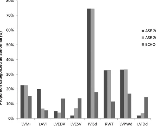
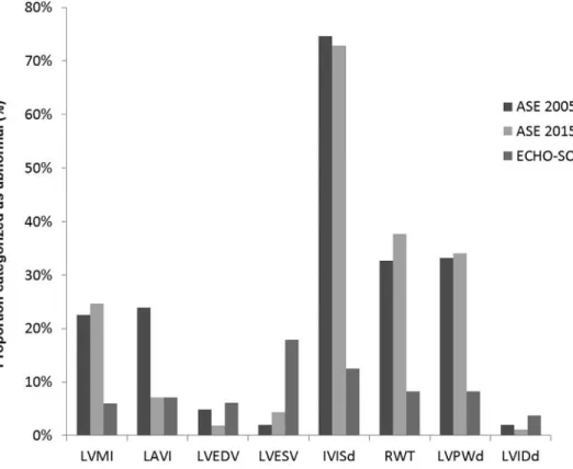
HCHS/SOL target population into abnormal and normal, we identified significant differences for males and females (Figures 1 and 2). Using ASE 2015 defined reference values we categorized ECHO/SOL cohort as having abnormal LVMI, RWT, IVISd and LVPWd in 7%, 21%, 57% and 17% of males and in 18%, 29%, 60% and 26% of females, respectively. Conversely, 10%, and 11% in males and 4% and 2% in females were classified as having abnormal LVEDV and LVIDd by ASE 2015 cut offs, respectively. Similar differences were found when we compared 2005 ASE cut offs. Overall females were more likely to be classified as having abnormal measures of cardiac structure than males such as (LVMI, RWT, IVISd and LVPWd) and males were more likely to be categorized as having abnormal measures of LV volume (LVEDV, LVESV and LVIDd).
Discussion
To the best of our knowledge, there have been no other large scale studies examining normal echocardiographic chamber dimensions in healthy Hispanic/Latinos in the US. In this study of healthy participants of ECHO-SOL, we examined the distribution of echocardiographic measures of left ventricle and left atrium in a healthy cohort and then used the derived values to identify abnormal values in the full cohort. We also used the suggested cut off values from the 2005 and 2015 ASE guidelines to differentiate abnormal values for the Hispanic/Latino cohort. Gross differences were observed in reference limits for Hispanics/ Latino compared with ASE chamber quantification guidelines. Both 2005 and 2015 ASE-suggested cut-offs underestimate the measures of LVMI, IVISd, and RWT. In contrast, these thresholds overestimated the measures of LVEDV, LVIDd and LVPWd in both males and females. These observations depict relatively “thicker and smaller” healthy hearts in individuals with Hispanic/Latino origin compared to ASE guidelines defined reference values. Furthermore, appreciable differences in cardiac chamber measures were noted between participants with different Hispanic/Latino origins.
The current recommended echocardiographic reference ranges, jointly published by the American Society of Echocardiography and the European Association of Echocardiography, were an important advance in quantitative echocardiography. Many of these studies, utilized in the guideline meta-analyses were performed over three to four decades ago with early generation echo technology and acoustic windows that may have improved significantly over time. Hence, the measures from these older studies may not accurately represent the
normative values of the present day. 12
This study adds to the growing discrepancy regarding ethnic-based reference limits. These differences have been highlighted by the EchoNormal study, a meta-analysis of left heart
reference ranges inclusive of a diverse world population.13 In the weighted analysis of the
cohort of Hispanic/Latino individuals, the current study demonstrates that males and females might be inappropriately classified into abnormal categories based on using reference limits provided by ASE chamber quantification guidelines. The study also confirms the prior
literature showing racial differences in cardiac chamber dimensions.14 This observation, in
part, has been attributed to the higher burden of cardiovascular risk factors especially, in the
Author Manuscript
Author Manuscript
Author Manuscript
case of non-Hispanic blacks who are known to have a higher prevalence of hypertension, a higher left ventricular mass and higher degree of abnormal left ventricular geometry than
non-Hispanic whites.15 These differences may partially account for the increased
cardiovascular mortality among non-Hispanic blacks.16 Although the participants in the
current study are healthy, they demonstrated higher left ventricular mass than that of the ASE cut offs.
A prior study of Hispanic/Latinos reported that individuals with Puerto Rican background have the highest degree of cardiovascular risk factors and hypercholesterolemia was most
common in individuals with Central American background.17 The current study observed
that individuals with Cuban background with higher values of all left ventricular and atrial measurements with Central American background exhibited the lowest values. Thus, risk factors alone might not be the only plausible explanation for these findings.
Ethnic variations in cardiac structural measures by echocardiography have significant impact on clinical decision-making. American College of Cardiology, American Heart Association and European Society of Cardiology guidelines for management of valvular heart disease rely heavily on chamber quantification and suggest management based on
various cut offs.18, 19 This study shows potential underestimation of left ventricular end
diastolic volume and internal diameter implicating lower cut off values for Hispanics/ Latinos when considering valvular heart disease management, especially the timing of surgery. The Joint National Committee on prevention, detection, evaluation and treatment of high blood pressure has suggested evaluation of left ventricular hypertrophy in all
hypertensive patients to identify end – organ damage and to be more aggressive in
management of these patients.20 Using the cut offs for septal wall thickness or left
ventricular mass will lead to overestimation of individuals with ventricular hypertrophy which may lead to unnecessary therapy to prevent further end – organ damage. There are differences in cardiac structure with highest degree of left ventricular mass, thickness and volumes noted in Cubans.
There are several limitations of this study. A direct comparison to the ASE reference ranges is not plausible for all cardiac dimensions examined. Those reference ranges were obtained through a combination of means with standard deviations (septal wall thickness, LV mass, LV dimensions / volumes, LA dimensions / volumes), trying to correlate chamber dimension with risk of an adverse event (LV mass, LV dimensions, LA volumes), and expert opinion (septal wall thickness, LA volumes). Previous studies in immigrants from various origin countries have shown that the incidence for certain diseases and behaviors will begin to resemble those of the population of their new home country in a process called acculturation. There has been an observed increase in the prevalence of diabetes mellitus
with acculturation in Hispanics/Latinos not of Mexican origin.21 Increased acculturation and
second-generation participants in the HCHS/SOL population have been found to have a
higher prevalence of cardiovascular disease and cardiovascular disease risk factors.17 This
initial analysis does not differentiate between first or subsequent generations of immigrant nor control for the degree of acculturation. It is noteworthy that the derived reference values are for individuals >45 years of age and cannot be generalized to adults <45 years of age. Adults >45 are known to have differences in cardiac chambers and thus, use of these
Author Manuscript
Author Manuscript
Author Manuscript
normative values are not suitable for use in younger adults. Defining echocardiographic parameters, nonetheless, based on country of origin and degree of acculturation appears too
specific to be useful to implement with regards to standardized guidelines. We used 95th
percentile as the cut off for abnormal values, while historically, it was not used due to the absence of a large patient population sample size. In addition, we did not use outcomes to define the abnormal values here and it is possible that lower levels of LV structure and function in Hispanics/Latinos might be associated with outcomes. We also performed weighted analyses where the echocardiographic reference limits in participants of ECHO-SOL were generalized to the whole HCHS/ ECHO-SOL target population, providing us with robust percentile estimates. When assessing participants from Central and South America only a small percentage of participants were derived from these geographical areas (8% and 11%, respectively), thus only a relatively small number of participants are used to generate the reference values for these sub-populations. This smaller number could bias the normative values and thus it is possible that in a larger sample, the normative values of these groups differ from this study.
In conclusion, we examined the distributions of various echocardiographic chamber measures in a large cohort of Hispanics/Latinos individuals. These measures seemed to be disparate from the measures normally used in echocardiographic laboratories based on ASE chamber quantification guidelines. Further research is needed to identify the risk related cut offs in Hispanics/Latinos as this segment of US population continues to grow.
Acknowledgments
The authors acknowledge the investigators, the staff, and the participants of HCHS-SOL and ECHO-SOL for their dedication and commitment to the success of this study. Investigators website - http://www.cscc.unc.edu/hchs/
Sources of Funding: The Hispanic Community Health Study/Study of Latinos was carried out as a collaborative study supported by contracts from the National Heart, Lung, and Blood Institute (NHLBI) to the University of North Carolina HC65233), University of Miami HC65234), Albert Einstein College of Medicine (N01-HC65235), Northwestern University (N01-HC65236), and San Diego State University (N01-HC65237). The following Institutes/Centers/Offices contribute to the HCHS/SOL through a transfer of funds to the NHLBI: National Institute on Minority Health and Health Disparities, National Institute on Deafness and Other Communication Disorders, National Institute of Dental and Craniofacial Research, National Institute of Diabetes and Digestive and Kidney Diseases, National Institute of Neurological Disorders and Stroke, NIH Institution-Office of Dietary Supplements.
ECHO-SOL was supported by a grant from the National Heart, Lung, and Blood Institute (R01 HL104199, Epidemiologic Determinants of Cardiac Structure and Function among Hispanics: Carlos J. Rodriguez, MD, MPH Principal Investigator).
References
1. Lang RM, Bierig M, Devereux RB, Flachskampf FA, Foster E, Pellikka PA, Picard MH, Roman MJ, Seward J, Shanewise JS, Solomon SD, Spencer KT, Sutton MS, Stewart WJ. Recommendations for chamber quantification: A report from the american society of echocardiography's guidelines and standards committee and the chamber quantification writing group, developed in conjunction with the european association of echocardiography, a branch of the european society of cardiology. Journal of the American Society of Echocardiography : official publication of the American Society of Echocardiography. 2005; 18:1440–1463. [PubMed: 16376782]
2. Lang RM, Badano LP, Mor-Avi V, Afilalo J, Armstrong A, Ernande L, Flachskampf FA, Foster E, Goldstein SA, Kuznetsova T, Lancellotti P, Muraru D, Picard MH, Rietzschel ER, Rudski L, Spencer KT, Tsang W, Voigt JU. Recommendations for cardiac chamber quantification by
Author Manuscript
Author Manuscript
Author Manuscript
echocardiography in adults: An update from the american society of echocardiography and the european association of cardiovascular imaging. Journal of the American Society of
Echocardiography : official publication of the American Society of Echocardiography. 2015; 28:1– 39 e14. [PubMed: 25559473]
3. Ilercil A, O'Grady MJ, Roman MJ, Paranicas M, Lee ET, Welty TK, Fabsitz RR, Howard BV, Devereux RB. Reference values for echocardiographic measurements in urban and rural populations of differing ethnicity: The strong heart study. Journal of the American Society of
Echocardiography : official publication of the American Society of Echocardiography. 2001; 14:601–611. [PubMed: 11391289]
4. Devereux RB, Roman MJ, de Simone G, O'Grady MJ, Paranicas M, Yeh JL, Fabsitz RR, Howard BV. Relations of left ventricular mass to demographic and hemodynamic variables in american indians: The strong heart study. Circulation. 1997; 96:1416–1423. [PubMed: 9315526]
5. Rodriguez CJ, Diez-Roux AV, Moran A, Jin Z, Kronmal RA, Lima J, Homma S, Bluemke DA, Barr RG. Left ventricular mass and ventricular remodeling among hispanic subgroups compared with non-hispanic blacks and whites: Mesa (multi-ethnic study of atherosclerosis). Journal of the American College of Cardiology. 2010; 55:234–242. [PubMed: 20117402]
6. Lavange LM, Kalsbeek WD, Sorlie PD, Aviles-Santa LM, Kaplan RC, Barnhart J, Liu K, Giachello A, Lee DJ, Ryan J, Criqui MH, Elder JP. Sample design and cohort selection in the hispanic community health study/study of latinos. Annals of epidemiology. 2010; 20:642–649. [PubMed: 20609344]
7. Sorlie PD, Aviles-Santa LM, Wassertheil-Smoller S, Kaplan RC, Daviglus ML, Giachello AL, Schneiderman N, Raij L, Talavera G, Allison M, Lavange L, Chambless LE, Heiss G. Design and implementation of the hispanic community health study/study of latinos. Annals of epidemiology. 2010; 20:629–641. [PubMed: 20609343]
8. Rodriguez CJDA, Allison MA, Shah SJ, Hurwitz B, Bangdiwala S, Gonzalez F, Kitzman D, Gillam L, Spevack D, Dadhania R, Langdon S, Kaplan R. Rationale and design of the echocardiographic study of hispanics / latinos (echo-sol). Ethn Dis. 2015; 25:180–6. [PubMed: 26118146]
9. Byrd BF 3rd, Finkbeiner W, Bouchard A, Silverman NH, Schiller NB. Accuracy and reproducibility of clinically acquired two-dimensional echocardiographic mass measurements. American heart journal. 1989; 118:133–137. [PubMed: 2741780]
10. Devereux RB, Alonso DR, Lutas EM, Gottlieb GJ, Campo E, Sachs I, Reichek N.
Echocardiographic assessment of left ventricular hypertrophy: Comparison to necropsy findings. The American journal of cardiology. 1986; 57:450–458. [PubMed: 2936235]
11. Rodriguez CJ, Dharod A, Allison MA, Shah SJ, Hurwitz B, Bangdiwala SI, Gonzalez F, Kitzman D, Gillam L, Spevack D, Dadhania R, Langdon S, Kaplan R. Rationale and design of the echocardiographic study of hispanics/latinos (echo-sol). Ethnicity & disease. 2015; 25:180–186. [PubMed: 26118146]
12. Vasan RS, Larson MG, Levy D, Evans JC, Benjamin EJ. Distribution and categorization of echocardiographic measurements in relation to reference limits: The framingham heart study: Formulation of a height- and sex-specific classification and its prospective validation. Circulation. 1997; 96:1863–1873. [PubMed: 9323074]
13. Poppe KK, Doughty RN, Whalley GA. Redefining normal reference ranges for echocardiography: A major new individual person data meta-analysis. European heart journal cardiovascular Imaging. 2013; 14:347–348. [PubMed: 22872514]
14. Mayet J, Shahi M, Foale RA, Poulter NR, Sever PS, Thom SAM. Racial-differences in cardiac structure and function in essential-hypertension. Brit Med J. 1994; 308:1011–1014. [PubMed: 8068083]
15. Rodriguez CJ, Diez-Roux AV, Moran A, Jin ZZ, Kronmal RA, Lima J, Homma S, Bluemke DA, Barr RG. Left ventricular mass and ventricular remodeling among hispanic subgroups compared with non-hispanic blacks and whites mesa (multi-ethnic study of atherosclerosis). Journal of the American College of Cardiology. 2010; 55:234–242. [PubMed: 20117402]
16. Rodriguez CJ, Sciacca RR, Diez-Roux AV, Boden-Albala B, Sacco RL, Homma S, DiTullio MR. Relation between socioeconomic status, race-ethnicity, and left ventricular mass - the northern manhattan study. Hypertension. 2004; 43:775–779. [PubMed: 14981073]
Author Manuscript
Author Manuscript
Author Manuscript
17. Daviglus ML, Talavera GA, Aviles-Santa ML, Allison M, Cai J, Criqui MH, Gellman M, Giachello AL, Gouskova N, Kaplan RC, LaVange L, Penedo F, Perreira K, Pirzada A,
Schneiderman N, Wassertheil-Smoller S, Sorlie PD, Stamler J. Prevalence of major cardiovascular risk factors and cardiovascular diseases among hispanic/latino individuals of diverse backgrounds in the united states. Jama. 2012; 308:1775–1784. [PubMed: 23117778]
18. Bonow RO, Carabello BA, Chatterjee K, de Leon AC Jr, Faxon DP, Freed MD, Gaasch WH, Lytle BW, Nishimura RA, O'Gara PT, O'Rourke RA, Otto CM, Shah PM, Shanewise JS. 2008 focused update incorporated into the acc/aha 2006 guidelines for the management of patients with valvular heart disease: A report of the american college of cardiology/american heart association task force on practice guidelines (writing committee to revise the 1998 guidelines for the management of patients with valvular heart disease). Endorsed by the society of cardiovascular anesthesiologists, society for cardiovascular angiography and interventions, and society of thoracic surgeons. Journal of the American College of Cardiology. 2008; 52:e1–142. [PubMed: 18848134]
19. Bonow RO, Carabello BA, Kanu C, de Leon AC Jr, Faxon DP, Freed MD, Gaasch WH, Lytle BW, Nishimura RA, O'Gara PT, O'Rourke RA, Otto CM, Shah PM, Shanewise JS, Smith SC Jr, Jacobs AK, Adams CD, Anderson JL, Antman EM, Fuster V, Halperin JL, Hiratzka LF, Hunt SA, Nishimura R, Page RL, Riegel B. Acc/aha 2006 guidelines for the management of patients with valvular heart disease: A report of the american college of cardiology/american heart association task force on practice guidelines (writing committee to revise the 1998 guidelines for the management of patients with valvular heart disease): Developed in collaboration with the society of cardiovascular anesthesiologists: Endorsed by the society for cardiovascular angiography and interventions and the society of thoracic surgeons. Circulation. 2006; 114:e84–231. [PubMed: 16880336]
20. Chobanian AV, Bakris GL, Black HR, Cushman WC, Green LA, Izzo JL Jr, Jones DW, Materson BJ, Oparil S, Wright JT Jr, Roccella EJ. The seventh report of the joint national committee on prevention, detection, evaluation, and treatment of high blood pressure: The jnc 7 report. Jama. 2003; 289:2560–2572. [PubMed: 12748199]
21. Kandula NR, Diez-Roux AV, Chan C, Daviglus ML, Jackson SA, Ni H, Schreiner PJ. Association of acculturation levels and prevalence of diabetes in the multi-ethnic study of atherosclerosis (mesa). Diabetes care. 2008; 31:1621–1628. [PubMed: 18458142]
Author Manuscript
Author Manuscript
Author Manuscript
Clinical Perspective
Hispanics are the largest ethnic minority in United States. Even though Hispanic/Latinos are considered to be a single ethnic group, they are composed of individuals from diverse geographic backgrounds. These individuals differ not only in their ancestry but also in terms of cardiac structure from white non – Hispanics. However, the current guidelines for measurement of cardiac structure recommended by the American Society of Echocardiography (ASE) are derived mainly from white non – Hispanic populations. There is limited literature regarding normative echocardiographic measures of cardiac chambers. Our study demonstrates normal cardiac chamber measures obtained from a healthy subgroup of Echo-SOL cohort and then compares these derived normative values with 2005 and 2015 ASE recommended chamber quantification cut offs in a cohort of 1,818 Hispanic/Latinos. We not only identified significant differences between normative Hispanic/Latino cut offs and guidelines' based cut offs but also among cut offs of six Hispanic subgroups. We examined these differences between Mexicans, Central Americans, South Americans, Dominicans, Puerto Ricans and Cubans. The study emphasizes the need for ethnic specific cut offs for normal echocardiographic measures of chambers and highlights differences in echocardiographic measures within Hispanic subgroups. This is important clinically as some individuals may get classified as abnormal despite having normal echocardiographic measures and vice versa.
Author Manuscript
Author Manuscript
Author Manuscript
Figure 1. Proportion of participants of HCHS/SOL target population categorized as abnormal by upper cut offs of three different criteria in males
LVMI – ratio of left ventricular mass to body surface area; LAVI – left atrial volume; LVEDV – left ventricular end diastolic volume; LVESV – left ventricular end systolic volume; IVISd – interventricular septal diameter; RWT – relative wall thickness; LVPWd – left ventricular posterior wall diameter; LVIDd – left ventricular internal diameter in diastole
ASE 2005, 2005 American society of echocardiography 2005 chamber quantification guidelines; ASE 2015, 2015 American society of echocardiography 2005 chamber quantification guidelines; ECHO-SOL, chamber quantification cut offs based on healthy subcohort of ECHO-SOL
Author Manuscript
Author Manuscript
Author Manuscript
Figure 2. Proportion of participants of HCHS/SOL target population categorized as abnormal by upper cut offs of three different criteria in females
LVMI – ratio of left ventricular mass to body surface area; LAVI – left atrial volume; LVEDV – left ventricular end diastolic volume; LVESV – left ventricular end systolic volume; IVISd – intrerentricular septal diameter; RWT – relative wall thickness; LVPWd – left ventricular posterior wall diameter; LVIDd – left ventricular internal diameter in diastole
ASE 2005, 2005 American society of echocardiography 2005 chamber quantification guidelines; ASE 2015, 2015 American society of echocardiography 2005 chamber quantification guidelines; ECHO-SOL, chamber quantification cut offs based on healthy subcohort of ECHO-SOL
Author Manuscript
Author Manuscript
Author Manuscript
Author Manuscript
Author Manuscript
Author Manuscript
Author Manuscript
Table 1
Clinical characteristics of ECHO-SOL target population
Clinical characteristics Overall Target Population (unweighted N = 1818)
Healthy Target Population (unweighted N = 525)
Age (years)* 56 (0.5) 53 (0.5)
Males‡ 631 (35%) 177 (34%)
Body mass index (kg/m2)* 30.1 (0.1) 25.9 (0.1)
Body surface area (m2)* 1.8 (0.1) 1.8 (0.1)
Systolic blood pressure (mmHg)* 136.2 (1.0) 129.6 (1.0)
Diastolic blood pressure (mmHg)* 77.9 (0.7) 76.0 (0.7)
Smoking‡
Current 304 (17%) 101 (19%)
Former 443 (24%) 112 (21%)
Never 1069 (59%) 311 (59%)
Fasting blood glucose (mg/dL)* 102.2 (0.4) 93.4 (0.4)
Dyslipidemia‡ 722 (40%) 180 (34%)
High density lipoprotein (mg/dL)* 50.4 (0.7) 52.9 (0.7)
Total cholesterol (mg/dL)* 208.6 (2.3) 207.1 (2.3)
Hispanic groups
Mexican‡ 458 (25%) 162 (31%)
Puerto Rican‡ 348 (19%) 86 (16%)
Cuban‡ 356 (20%) 94 (18%)
South American‡ 150 (8%) 59 (11%)
Central American‡ 176 (10%) 44 (8%)
Dominican‡ 326 (18%) 78 (15%)
*
Means with standard error of continuous variables expressed,
‡
Author Manuscript
Author Manuscript
Author Manuscript
Author Manuscript
Table 2
Reference limits of left ventricular and atrial echocardiographic measures in males and females for healthy ECHO-SOL cohort (N = 525)
Males Percentiles Echocardiographic measures Mean 25 th 90 th 95 th 99 th LVMI (grams/m 2) 86 73 109 127 132 LAVI (ml/m 2) 24 19 31 34 43 IVISd (mm) 9.3 10.5 12.5 13.0 14.5 LVPWd(mm) 8.9 7.7 10.8 11.7 12.5 RWT 0.38 0.32 0.49 0.51 0.55 LVIDd (mm) 48 45 53 56 58 LVEDV (ml) 97 85 117 130 145 LVESV (ml) 40 33 52 58 64 Females Mean 25 th 90 th 95 th 99 th LVMI (grams/m 2) 70 60 88 94 104 LAVI (ml/m 2) 23 18 31 35 39 IVISd (mm) 9.3 8.1 11.3 12.4 13.3 LVPWd(mm) 7.9 7.0 9.7 10.2 10.8 RWT 0.37 0.32 0.48 0.49 0.54 LVIDd (mm) 43 40 48 49 52 LVEDV (ml) 70 62 85 90 101 LVESV (ml) 27 23 36 37 44
Author Manuscript
Author Manuscript
Author Manuscript
Author Manuscript
Table 3
Reference limits of left ventricular and atrial echocardiographic measures by Hispanic/Latino background for healthy ECHO-SOL cohort (N = 525)
Author Manuscript
Author Manuscript
Author Manuscript
Author Manuscript
Percentiles Measure Hispanic/Latino Background Mean 25 th 90 th 95 th 99 th p-value RWT Mexican 0.36 0.30 0.46 0.48 0.54 Reference Central American 0.37 0.32 0.46 0.48 0.50 <0.001 South American 0.37 0.31 0.45 0.46 0.49 0.49 Cuban 0.40 0.35 0.50 0.52 0.54 <0.001 Dominican 0.36 0.32 0.44 0.49 0.53 0.84 Puerto Rican 0.38 0.31 0.49 0.51 0.57 <0.001 LVIDd (mm) Mexican 46 42 54 56 58 Reference Central American 44 41 48 50 54 <0.001 South American 44 41 48 49 50 <0.001 Cuban 46 43 51 53 54 0.007 Dominican 45 41 50 51 53 <0.001 Puerto Rican 45 42 50 51 54 0.003 LVEDV (ml) Mexican 83 69 106 111 129 Reference Central American 70 54 91 98 108 <0.001 South American 77 59 101 108 126 <0.001 Cuban 87 72 116 124 147 <0.001 Dominican 79 67 98 102 111 <0.001 Puerto Rican 84 70 110 113 141 0.43 LVESV (ml) Mexican 33 25 46 50 68 Reference Central American 29 21 40 42 53 <0.001 South American 30 22 42 45 60 <0.001 Cuban 36 28 52 54 60 <0.001 Dominican 31 25 45 46 51 0.001 Puerto Rican 33 26 45 47 54 0.73Author Manuscript
Author Manuscript
Author Manuscript
Author Manuscript
Table 4
Comparison of 95
th percentile cut off identified in ECHO-SOL healthy cohort and American Society of Echocardiography upper reference
limits
Females
Males
Measure
ASE 2005
ASE 2015
ECHO-SOL
†
ASE 2005
ASE 2015
ECHO-SOL
LVMI (g/m
2)
88
88
94
102
102
127
LAVI (ml/m
2)
28
34
35
28
34
34
IVISd (mm)
9
9
12.4
10
10
13.0
LVPWd (mm)
9
9
10.2
10
10
11.7
RWT
0.42
0.42
0.49
0.42
0.42
0.51
LVIDd (mm)
53
52
49
59
58
56
LVEDV (ml)
104
106
90
155
150
130
LVESV(ml)
49
42
37
58
61
58