A SYSTEMS BIOLOGY-BASED APPROACH TO INVESTIGATE FORMALDEHYDE’S EFFECTS ON MICRORNA EXPRESSION PROFILES
Julia Elizabeth Rager
A dissertation submitted to the faculty of the University of North Carolina at Chapel Hill in partial fulfillment of the requirements for the degree of Doctor of Philosophy in the Department of Environmental Sciences and Engineering of the Gillings School of Global Public Health
Chapel Hill 2013
Approved by:
Dr. Rebecca C. Fry
Dr. David Diaz-Sanchez
Dr. Ilona Jaspers
Dr. Kenneth G. Sexton
ii
ABSTRACT
JULIA RAGER: A Systems Biology-Based Approach to Investigate Formaldehyde’s Effects on MicroRNA Expression Profiles
(Under the Direction of Dr. Rebecca C. Fry)
Formaldehyde is a common indoor and outdoor air pollutant that adversely impacts
global public health. Many toxicological studies have shown that formaldehyde causes
nasopharyngeal cancer, possibly through tissue damage, increased cell proliferation, and/or DNA
damage. However, there is lack of knowledge regarding formaldehyde’s effects at the systems
biology level and whether epigenetic mechanisms may contribute to cellular responses.
Furthermore, whether formaldehyde is capable of altering genomic and epigenomic processes
throughout sites distal to the respiratory tract is unknown. This topic is of high interest, as the
link between formaldehyde inhalation exposure and leukemia development is currently under
heated debate. Epidemiological studies have shown evidence supporting a link between
formaldehyde exposure and leukemia development, while toxicological investigations have yet
to provide evidence supporting formaldehyde’s ability to influence sites distant to the respiratory
tract. Before this dispute is resolved, further evaluation of the biological mechanisms linking
formaldehyde to disease is clearly necessary. In particular, formaldehyde-induced changes to
epigenetic contributors to transcriptional programs are extremely understudied, where
microRNA (miRNA) expression profiles have yet to be investigated in relation to formaldehyde.
We set out to test the novel hypothesis that miRNAs have altered expression profiles
within the respiratory and hematopoietic systems upon exposure to formaldehyde. Our studies
were the first to show that formaldehyde significantly disrupts miRNA expression patterns in
vitro, within cultured human lung cells, and in vivo, within the nasal epithelium of nonhuman
primates. Using a rodent model, the impact of formaldehyde exposure on miRNA-related
processes in direct contact and distant tissues, including the nasal mucosa, circulating white
iii
miRNA expression profiles within the nose and blood, but not the bone marrow. Evaluating the
epigenetic effects of formaldehyde exposure at the systems biology level, putative
miRNA-mediated responses were mapped onto interacting networks. Signaling related to inflammation,
cell death, and cancer was identified as enriched. Taken together, our research increases the
knowledge of under-studied mechanisms linking formaldehyde exposure to disease, acting as an
iv
ACKNOWLEDGEMENTS
I would first like to thank my advisor, Dr. Rebecca C. Fry, for welcoming me into her
research group at UNC. She has made a huge impact on my life, and it is difficult to think where
I would be without her mentorship these past four years. Her enthusiasm, leadership, and
patience are truly commendable, to say the least. From her, I’ve learned ways to succeed at work
while juggling family and friends. It has been a pleasure seeing Dr. Fry’s lab grow to the success
that it is today, and being able to play a role in that growth is extremely gratifying. I will forever
be grateful to her for making my graduate experience at UNC so incredibly meaningful.
I would also like to thank our Research Specialist and Lab Manager, Lisa Smeester, who
dedicated much of her time to helping me plan experiments and learn valuable laboratory
techniques.
I would also like to thank my committee members, Dr. David Diaz-Sanchez, Dr. Ilona
Jaspers, Dr. Kenneth G. Sexton, and Dr. James A. Swenberg, for their valuable input and advice
in preparing my research project and dissertation.
Lastly, I would like to thank my parents, Brent and Diane Rager, who have been
v
TABLE OF CONTENTS
LIST OF TABLES………...x
LIST OF FIGURES………...…xii
INTRODUCTION ... 1
Formaldehyde Exposure Sources ... 1
Mechanisms Underlying Formaldehyde-Induced Cancer of the Upper Respiratory Tract ... 2
Incongruent Findings Between Epidemiological and Toxicological Studies ... 4
MicroRNAs ... 5
MicroRNAs and Leukemia ... 6
Project Approach ... 6
Dissertation Organization ... 7
CHAPTER 1: FORMALDEHYDE EXPOSURE ALTERS MICRORNA EXPRESSION PROFILES IN HUMAN LUNG CELLS ... 9
1.1 OVERVIEW ... 9
1.2 STUDY OBJECTIVES ... 10
1.3 MATERIALS AND METHODS ... 10
1.3.1 Cell Culture ... 10
1.3.2 Formaldehyde Treatment ... 10
vi
1.3.4 Microarray Processing ... 11
1.3.5 Microarray Analysis... 12
1.3.6 Enriched Biological Functions and Network Analysis ... 12
1.3.7 RT-PCR Verification of miRNA Expression ... 13
1.3.8 Interleukin-8 Measurement ... 13
1.4 RESULTS ... 14
1.4.1 Formaldehyde Exposure Modulates miRNAs in Human Lung Cells ... 14
1.4.2 miRNA Expression Changes are Validated through RT-PCR ... 15
1.4.3 miRNA Targets are Integrated into Biological Networks ... 16
1.4.4 Conservation of Predicted and Observed mRNA Targets ... 18
1.4.5 Inflammatory Cytokine IL-8 is Released in Response to Formaldehyde .... 19
1.5 DISCUSSION ... 19
CHAPTER 2: FORMALDEHYDE AND EPIGENETIC ALTERATIONS: MICRORNA CHANGES IN THE NASAL EPITHELIUM OF NONHUMAN PRIMATES ... 24
2.1 OVERVIEW ... 24
2.2 STUDY OBJECTIVES ... 25
2.3 MATERIALS AND METHODS ... 25
2.3.1 Animals ... 25
2.3.2 Formaldehyde Exposures ... 25
2.3.3 Sample Collection ... 26
2.3.4 Sample Processing ... 26
vii
2.3.6 RT-PCR Confirmation of miRNA Expression Changes ... 27
2.3.7 Predicting Targets of miR-125b and miR-142-3p ... 28
2.3.8 Pathway Enrichment Analysis of Predicted Targets ... 28
2.3.9 Testing miRNA Targets using RT-PCR ... 28
2.4 RESULTS ... 29
2.4.1 Formaldehyde Disrupts miRNA Expression Profiles in Nasal Tissue ... 29
2.4.2 RT-PCR Confirmed Formaldehyde-Induced miRNA Expression Changes ... 30
2.4.3 Transcriptional Targets of miR-125b and miR-142-3p were Predicted ... 31
2.4.4 Apoptosis Signaling is Associated with miR-125b Predicted Targets ... 31
2.4.5 Apoptosis-Related miR-125b Targets are Decreased in Expression ... 32
2.4.6 ILK Signaling is Associated with miR-142-3p Predicted Targets... 33
2.4.7 ILK-Related miR-142-3p Targets are Altered in Expression ... 33
2.5 DISCUSSION ... 34
CHAPTER 3: FORMALDEHYDE-INDUCED CHANGES IN MICRORNA SIGNALING IN THE RAT ... 40
3.1 OVERVIEW ... 40
3.2 STUDY OBJECTIVES ... 41
3.3 MATERIALS AND METHODS ... 41
3.3.1 Animals ... 41
3.3.2 Formaldehyde Exposures ... 42
3.3.3 Sample Collection ... 42
viii
3.3.5 miRNA Microarray Analysis ... 43
3.3.6 Transcript (mRNA) microarray analysis ... 44
3.3.7 Computationally Predicting miRNA Transcriptional Targets ... 45
3.3.8 Systems-Level Analysis of Predicted miRNA-mRNA Interactions ... 45
3.3.9 Confirming miRNA Microarray Results using RT-PCR ... 46
3.3.10 Confirming mRNA Microarray Results using RT-PCR ... 46
3.4 RESULTS ... 47
3.4.1 Study Design ... 47
3.4.2 Formaldehyde Alters miRNA Expression in the Nose and WBC ... 47
3.4.3 Formaldehyde-Responsive miRNAs are Tissue-Specific ... 48
3.4.4 Formaldehyde-Responsive miRNA Expression is Largely Sustained in the Nose ... 48
3.4.5 Formaldehyde Causes Tissue-Specific Gene Expression Changes ... 50
3.4.6 Formaldehyde-Responsive miRNAs May Mediate a Fraction of Transcriptional Effects ... 51
3.4.7 Systems-Level Analysis Reveals miRNA-Mediated Signaling of Critical Pathways ... 52
3.4.8 Microarray Results were Confirmed using RT-PCR ... 55
3.5 DISCUSSION ... 56
DISCUSSION AND CONCLUSIONS ... 61
MiRNAs are Key Responders to Formaldehyde in Direct Target Tissues ... 61
MiRNAs Respond in a Tissue Type-Specific Manner ... 63
Formaldehyde-Responsive miRNAs Regulate Genes Involved in Critical Pathways... 64
ix
Public Health Relevance ... 72
Relationship of Current Research to Future Hypotheses ... 73
Dissertation Conclusion ... 75
SUPPLEMENTARY TABLES ... 77
x
LIST OF TABLES
Table 1: Biological functions significantly associated with all predicted target sets of miR-33, miR-330, miR-181a, and miR-10b, the miRNAs with the greatest
expression alterations upon exposure to formaldehyde in human lung cells ………….18
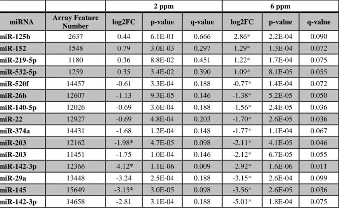
Table 2: Formaldehyde inhalation exposure in the nasal epithelium of nonhuman primates significantly disrupts the expression levels of 13 unique miRNAs,
represented by 15 array probesets ………….……….30
Table 3: Functions and diseases that are enriched within the miRNA-mediated signaling networks associated with formaldehyde exposure in the (A) nose and (B) WBC
of the rat 28-day group ………..….52
Supplementary Table 1: miRNAs significantly differentially expressed upon exposure
to 1 ppm formaldehyde in human lung epithelial cells. ... 77
Supplementary Table 2: Predicted transcriptional targets for miR-33, miR-330,
miR-181a, and miR-10b. ... 79
Supplementary Table 3: 40 networks associated with the predicted targets of miR-33,
miR-330, miR-181a, and miR-10b. ... 92
Supplementary Table 4: Canonical pathways involving at least three molecules present in top networks significantly associated with predicted targets
of miR-33, miR-330, miR-181a, and miR-10b. ... 96
Supplementary Table 5: Biological functions significantly (p-value < 0.005) associated with predicted transcriptional targets of miR-33, miR-330,
miR-181a, and miR-10b. ... 99
Supplementary Table 6: Biological functions significantly (p-value < 0.005) associated with formaldehyde-responsive genes, as identified through
pathway analysis of the Li et. al. 2007 genomic database. ... 103
Supplementary Table 7: Transcriptional targets predicted to be regulated by miR-125b. ... 105
Supplementary Table 8: Transcriptional targets predicted to be regulated by miR-142-3p. ... 109
Supplementary Table 9: Pathways significantly associated with the predicted targets
of miR-125b. ... 110
Supplementary Table 10: Pathways significantly associated with the predicted targets
of miR-142-3p. ... 111
xi
Supplementary Table 12: 42 genes differentially expressed by formaldehyde within
the nose of rats in the 28-day group. ... 116
Supplementary Table 13: 130 genes differentially expressed by formaldehyde within
the WBC of rats in the 28-day group. ... 117
Supplementary Table 14: Individual networks constructed using formaldehyde-associated mRNAs predicted to be regulated by formaldehyde-
xii
LIST OF FIGURES
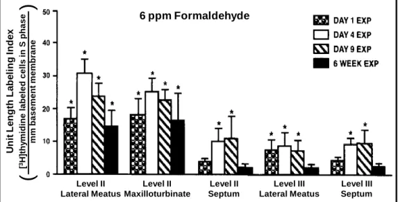
Figure 1: As a part of formaldehyde’s carcinogenic mode of action, formaldehyde
causes significant increases in cell proliferation in direct target cells ... 3
Figure 2: MicroRNAs are important regulators of gene expression and protein production ... 6
Figure 3: Project overview. ... 7
Figure 4: Formaldehyde modulates the expression of 89 miRNAs in human lung cells. ... 14
Figure 5: Microarray results align with RT-PCR results ... 15
Figure 6: Significant molecular networks of miRNA-mediated signaling likely affected by formaldehyde exposure in human lung cells ... 17
Figure 7: Interleukin-8 levels are significantly elevated in formaldehyde-treated lung cells compared to untreated cells ... 19
Figure 8: RT-PCR confirms the altered expression of selected miRNAs upon exposure to formaldehyde within the nonhuman primate nasal epithelium ... 31
Figure 9: Predicted mRNA targets of formaldehyde-altered miR-125b are involved in apoptosis signaling, suggesting that the regulation of the apoptotic machinery may be modified through formaldehyde’s influence on miRNAs ... 32
Figure 10: RT-PCR shows the decreased expression of apoptosis signaling-related genes predicted to be targeted by miR-125b, the miRNA with the greatest increased expression resulting from 6 ppm formaldehyde exposure ... 33
Figure 11: RT-PCR shows the altered expression of ILK signaling-related genes predicted to be targeted by miR-142-3p, the miRNA with the greatest decreased expression resulting from 6 ppm formaldehyde exposure. ... 34
Figure 12: Study design ... 47
Figure 13: Distribution of formaldehyde-responsive miRNAs across three exposure conditions and three tissues in the rat ... 48
Figure 14: Formaldehyde-responsive miRNAs throughout the nose and WBC... 49
Figure 15: Gene expression changes induced by formaldehyde exposure in the rat nose and WBC of the 28-day group ... 51
xiii
Figure 17: Microarray results align with RT-PCR results in the rat nose. ... 55
Figure 18: 28 miRNAs that were significantly altered at the expression level within direct target tissues by formaldehyde exposure in at least two of the
three models tested ... 62
Figure 19: Common biological functions and disease signatures are enriched for by miRNA-mediated signaling responses to formaldehyde exposure across
multiple species/tissues. ... 65
Figure 20: Novel mode of action (MOA) that may link formaldehyde inhalation
exposure to hematopoietic changes ... 67
1
INTRODUCTION
Understanding the biological impacts upon exposure to formaldehyde via inhalation
exposure is crucial, as formaldehyde in a ubiquitous air pollutant present throughout the
environment. The combined studies in this work take several approaches to investigate novel
mediators of response to formaldehyde exposure. The ultimate goal for this investigation is to
increase the current knowledge of mechanisms and biological pathways underlying
formaldehyde-induced effects, acting as an important foundation for future research in public
health and toxicology.
Formaldehyde Exposure Sources
Formaldehyde is a common air toxic that is present in both indoor and outdoor
atmospheres. In outdoor environments, formaldehyde is present due to direct emissions from
anthropogenic and biogenic sources, and is also formed as a secondary chemical product through
hydrocarbon atmospheric chemistry (WHO 2001). Anthropogenic sources of formaldehyde
include automobile exhaust, power plants, manufacturing facilities, and incinerators (NTP 2011;
WHO 2001). Ambient air is estimated to contain formaldehyde at levels between 0.0008 and
0.02 ppm (WHO 2001). High formaldehyde exposures can occur within indoor environments,
where formaldehyde is released from household products (e.g. cleaning agents, carpet, furniture)
and cigarette smoke (NTP 2011). The highest formaldehyde levels are found in certain
occupational environments. For example, high chronic exposures of 2-5 ppm formaldehyde have
been measured in garment and textile industries, during the varnishing of furniture and wooden
floors, and in some manufacturing jobs related to board mills and foundries (Duhayon et al.
2008). Acute exposures to high levels of formaldehyde (≥ 3 ppm) have been reported for
pathologists, embalmers, and paper workers (Duhayon et al. 2008). A range of lower
formaldehyde levels have also been measured in industries related to the production of resins,
2
It is also important to note that formaldehyde is produced endogenously in all cells, as it
is an intermediate of serine, glycine, methionine, and choline metabolism (IARC 2006). It is also
an essential intermediate formed during the biosynthesis of purines, thymidine, and certain
amino acids (IARC 2006). Because of the constant presence of both endogenous and
environmental formaldehyde exposure, understanding the exposure response and biological basis
of formaldehyde-induced health effects is of utmost importance.
Mechanisms Underlying Formaldehyde-Induced Cancer of the Upper Respiratory Tract
The exact mechanisms linking formaldehyde exposure to cancer remain unknown.
Formaldehyde is currently classified as a known human carcinogen (IARC 2006), although the
entire mechanism by which formaldehyde induces cancer is not fully understood (Liteplo et al.
2003; NTP 2011). The genotoxic effects of formaldehyde exposure on direct target cells within
the respiratory tract have been studied extensively. Specifically, formaldehyde has been shown to
cause a variety of types of genetic damage in vitro and in vivo. These types of damage include
DNA-protein crosslinks, which have been detected in vitro (Merk et al. 1998), in the nasal
mucosa of rats (Casanova et al. 1989; Casanova et al. 1994), and in the nasal turbinates,
nasopharynx, larynx, trachea, carina, and proximal bronchi of nonhuman primates (Casanova et
al. 1991) exposed to formaldehyde. DNA double strand breaks have also been shown to result
from formaldehyde exposure in vitro (Noda et al. 2011). Formaldehyde also causes a multitude
of DNA adducts to form in vitro, including N2-hydroxymethyl-deoxyguanosine, N6
-hydroxymethyl-deoxyadenosine, and N4-hydroxymethyl-deoxycytosine (Swenberg et al. 2011).
N2-hydroxymethyl-deoxyguanosine adducts have also been found to be induced exogenously in
the nasal mucosa of rats (Lu et al. 2011) and nonhuman primates (Moeller et al. 2011) exposed
to formaldehyde.
Formaldehyde-induced genetic damage can lead to cell death. For example, in vitro
studies show that the amount of formaldehyde-induced DNA-protein crosslinks inversely relates
to cell survival (Merk et al. 1998; Ross et al. 1980). Furthermore, a pharmacodynamic model has
been created to describe the relationship between formaldehyde exposure, DNA-protein
crosslink formation, and DNA replication arrest using in vivo measurements (Heck et al. 1999).
During DNA replication arrest, lesions can be repaired. However, if the damage is not repaired
3
(Jackson et al. 2009). Alternatively, carcinogenesis can occur from the activation of an oncogene
or the inactivation of a tumor suppressor gene (Jackson et al. 2009). Altogether, the various types
of formaldehyde-induced DNA damage may underlie the observed increases in cytotoxicity in
vitro (Lovschall et al. 2002; Merk et al. 1998; Quievryn et al. 2000) and tissue damage in vivo
(Chang et al. 1983; Monticello et al. 1991; Monticello et al. 1996).
Cytotoxic damage to target cells is known to cause regenerative cell proliferation to
replace and repair damaged tissue (Boobis 2010; Cohen et al. 2008). Increases in the rate of
cellular proliferation can then increase the probability of de novo mutations in critical oncogenes
(Boobis 2010). In the case of formaldehyde, it is postulated that regenerative cell proliferation
resulting from formaldehyde-induced cytotoxicity increases the number of DNA replications,
and thereby, increases the likelihood of DNA damage causing DNA replication errors, resulting
in mutations and eventually carcinogenesis (Liteplo et al. 2003; McGregor et al. 2006). This
proposed mode of action is supported by previous investigations showing that formaldehyde
causes sustained cell proliferation in direct target tissue in vivo (Figure 1). For example, many
studies show that formaldehyde exposure increases cell proliferation rates in the upper
respiratory epithelium of rodents and nonhuman primates (Chang et al. 1983; Monticello et al.
1989; Monticello et al. 1991; Roemer et al. 1993). Furthermore, sites of increased cellular
proliferation rates have been shown to correlate with regions of nasal tumor incidence
(Monticello et al. 1991; Monticello et al. 1996).
U nit Le ngt h La be ling Inde x [ 3H] thy m idine la be le d c e lls in S pha s e m m b asem en t m em b ran e
(
)
Level II Maxilloturbinate Level II Septum Level III Lateral Meatus Level III Septum Level II Lateral Meatus6 ppm Formaldehyde
4
While there is a link between cell proliferation and carcinogenesis, other mechanisms
influencing formaldehyde-induced cancer are understudied, including signaling pathways that
may play a role in altered cell proliferation and carcinogenesis. Our study, therefore, contributes
to the current understanding of mechanisms underlying cancer caused by formaldehyde by
evaluating epigenetically-regulated processes at the systems-level. Our project is the first to use a
systems biology approach to assess the epigenetic effects of formaldehyde on direct target cells
within the respiratory tract.
Incongruent Findings Between Epidemiological and Toxicological Studies
There is disparity between epidemiological and toxicological findings regarding possible
links between formaldehyde and leukemia. Formaldehyde is a known human carcinogen (IARC
2006), but its association with hematological cancers is currently undergoing debate. In June
2011, the U.S. National Toxicology Program added formaldehyde as a human
lymphohematopoietic carcinogen, while stating that the mechanisms by which formaldehyde
causes leukemia are unknown (NTP 2011). This classification is largely based on
epidemiological evidence supporting formaldehyde-induced myeloid leukemia, as identified in
cohorts of embalmers (Hauptmann et al. 2009), garment workers (Pinkerton et al. 2004), and
workers in various formaldehyde-related industries (Beane Freeman et al. 2009; Zhang et al.
2009). However, some of these epidemiological findings are controversial (Bachand et al. 2010).
These types of occupational exposures are common, where more than two million U.S. workers
are exposed to formaldehyde (USDL 2011). Because formaldehyde exposure and leukemia
development are both prevalent, understanding the biological basis linking formaldehyde to
disease is extremely important.
Despite epidemiological evidence supporting formaldehyde as a leukemogen, the
biological plausibility underlying formaldehyde-induced leukemia is still debated. To elaborate,
leukemia is a cancer of the blood or bone marrow, where recognized environmental leukemogens
typically cause hematopoietic toxicity/genotoxicity and subsequent leukemia development
(McHale et al. 2012; Mukherjee et al. 2012). However, formaldehyde is reactive and undergoes
metabolism rapidly (IARC 2006), and formaldehyde blood concentrations do not change after
inhalation exposure (Casanova et al. 1988; Heck et al. 1985). As a result, some scientists believe
5
(Cole et al. 2004; Golden et al. 2006; Heck et al. 2004; Pyatt et al. 2008). Nevertheless, a few
mechanisms underlying formaldehyde-induced leukemia have been proposed (Goldstein 2011;
Zhang et al. 2009). One key event common amongst these proposed mechanisms requires
damages or alterations within hematopoietic stem or progenitor cells. Still, researchers have yet
to elucidate how formaldehyde, either directly or indirectly, may alter hematopoietic cells. With
the goal of filling a component of this research void, our study investigates changes in miRNA
signaling throughout the body, representing an area of research that has yet to be evaluated in
relation to formaldehyde.
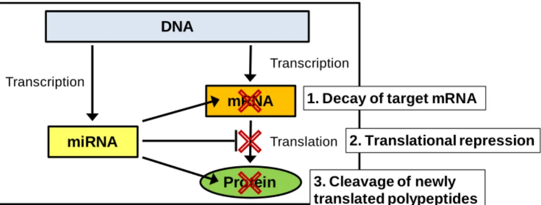
MicroRNAs
MicroRNAs are important epigenetic regulators of gene expression that may play key
roles in formaldehyde-induced health effects. These recently discovered molecules, clearly a part
of the epigenetic machinery (Iorio et al. 2010), play a large role in the regulation of mRNA
abundance and protein production. By base pairing to target mRNAs, miRNAs can cause mRNA
degradation and/or translational repression (Filipowicz et al. 2008; Friedman et al. 2009). In
some cases, miRNAs can even cleave newly translated proteins (Friedman et al. 2009) (Figure
2). To quantify, mammalian miRNAs are estimated to regulate more than 60% of all
protein-coding genes (Friedman et al. 2009). Because miRNAs play such pivotal roles in gene
regulation, miRNAs have received increasing attention throughout medical and toxicological
research fields. Several cancer-related studies have shown that miRNA expression profiles are
drastically altered in tumors in comparison to healthy tissue. For example, miRNA expression
profiles have been shown to be significantly disrupted in nasopharyngeal carcinoma (Chen et al.
2009) and leukemia (Bousquet et al. 2008; Cammarata et al. 2010; Garzon et al. 2008; Wang et
al. 2011). Whether formaldehyde exposure is capable of affecting miRNA expression profiles,
6
MicroRNAs and Leukemia
Leukemia development is known to be influenced by miRNAs. Hematological function is
heavily influenced by miRNAs, since these molecules are important regulators of hematopoietic
stem/progenitor cell differentiation (Marcucci et al. 2011; Yendamuri et al. 2009), cell cycle
(Han et al. 2010), and apoptosis (Garzon et al. 2009). Distinct miRNA expression profiles also
exist in leukemia patients, where miRNAs have been shown to classify various risk groups
(Calin et al. 2006; Marcucci et al. 2009; Marcucci et al. 2011). Further linking miRNAs to
leukemia, several miRNAs have been implicated as leukemia-related tumor suppressors (e.g.
miR-29b) (Garzon et al. 2009) and oncogenes (e.g. miR-125b, miR-155, miR-29a) (Costinean et
al. 2006; Han et al. 2010; O'Connell et al. 2008). Because of the current interest regarding
formaldehyde’s link to leukemia, we find the lack of research on formaldehyde’s epigenetic
effects surprising. We therefore address this scientific gap by investigating formaldehyde-altered
miRNAs throughout multiple target tissues, revealing novel responses that have yet to be
investigated.
Project Approach
This research focuses on epigenetic responses to formaldehyde inhalation exposure
across direct contact and distant tissues. The primary hypothesis to be tested is that miRNAs
have altered expression profiles within the respiratory and hematopoietic systems upon
exposure to formaldehyde.
DNA
miRNA
mRNA
Protein
Transcription
Translation Transcription
1. Decay of target mRNA
2. Translational repression
3. Cleavage of newly translated polypeptides
7
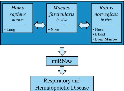
The project employs an integrated approach to perform cross-tissue and cross-species
comparative analyses of epigenetic responses to formaldehyde exposure (Figure 3). We assess
miRNA expression profiles across three separate species in multiple tissues and cells,
representing possible key events linking formaldehyde inhalation exposure to respiratory and
hematopoietic disease. It is notable that our research is the first to: (i) investigate miRNA
expression profiles in formaldehyde-exposed cells, (ii) to compare formaldehyde-induced
miRNA responses across three species, and (iii) to compare miRNA expression profiles between
direct contact and distant targets of formaldehyde inhalation exposure using the rodent model.
Using this strategy, we reveal novel mechanisms underlying formaldehyde exposure-induced
effects on biological signaling.
Figure 3: Project overview. The effect of formaldehyde inhalation exposure on miRNA expression profiles is assessed across three species. Using this experimental design, this research tests the novel hypothesis that miRNAs have altered expression profiles within the respiratory and hematopoietic systems upon exposure to formaldehyde.
Dissertation Organization
This dissertation is organized into three chapters. The first chapter describes an in vitro
study which was the first to show that formaldehyde exposure significantly alters miRNA
expression profiles. The study detailed in the second chapter expanded the initial in vitro
findings using an in vivo model. Here, nonhuman primates exposed to formaldehyde were found Homo
sapiens
in vitro
---•Lung
Macaca fascicularis
in vivo
---•Nose
Rattus norvegicus
in vivo
---•Nose
•Blood
•Bone Marrow
miRNAs
8
to also show significantly altered miRNA expression profiles within the nose, a direct target of
formaldehyde inhalation exposure. The last study, described in chapter three, compares miRNA
expression profiles altered by formaldehyde exposure across the nose, circulating white blood
cells, and bone marrow cells in a rodent model. It is of great interest that miRNA expression
patterns were identified as disrupted by formaldehyde exposure in the nose and white blood
cells, but not the bone marrow. This study also included a time-series analysis, where
formaldehyde was found to disrupt miRNA expression profiles more drastically after shorter
periods of exposure. In all three studies, miRNA responses were further evaluated at the
mechanistic level by mapping results to the biological networks. Significant pathways involved
in cellular regulation were identified as likely disrupted via formaldehyde’s influence on miRNA
expression profiles. The studies contained within these three chapters provide novel insights into
9
CHAPTER 1
FORMALDEHYDE EXPOSURE ALTERS MICRORNA EXPRESSION PROFILES IN HUMAN LUNG CELLS
1.1 Overview
Exposure to formaldehyde, a known air toxic, is associated with cancer and respiratory
disease. Despite its adverse health effects, the mechanisms underlying formaldehyde-induced
disease remain largely unknown. Research investigations have uncovered microRNAs (miRNAs)
as key post-transcriptional regulators of gene expression that may influence cellular disease state.
While studies have compared different miRNA expression patterns between diseased and healthy
tissue, this is the first study to examine perturbations in global miRNA levels resulting from
formaldehyde exposure.
We set out to investigate whether cellular miRNA expression profiles are modified by
formaldehyde exposure in human lung cells. We hypothesized that formaldehyde exposure
disrupts miRNA expression levels within lung cells, representing a novel epigenetic mechanism
through which formaldehyde may induce disease.
Human lung epithelial cells were grown at air-liquid interface and exposed to gaseous
formaldehyde at 1 ppm for 4 hours. Small RNAs and protein were collected and analyzed for
miRNA expression using microarray analysis or IL-8 protein levels by ELISA, respectively.
Gaseous formaldehyde exposure altered the miRNA expression profiles in human lung
cells. Specifically, 89 miRNAs were significantly down-regulated in formaldehyde exposed
samples versus controls. Functional and molecular network analysis of the predicted miRNA
transcript targets revealed that formaldehyde exposure potentially alters signaling pathways
associated with cancer and inflammatory response. IL-8 release was increased in cells exposed to
formaldehyde, and results were confirmed by real-time PCR.
Formaldehyde alters miRNA patterns which regulate gene expression, potentially leading
10
1.2 Study Objectives
For this study, we set out to test the novel hypothesis that formaldehyde exposure can
disrupt miRNA levels within lung cells. We tested this hypothesis by exposing human lung
epithelial cells to formaldehyde using a direct air-liquid interface that physically mimics the
human respiratory tract. Using microarray analysis, we assessed genome-wide miRNA
expression profiles and identified miRNAs altered at the expressed level by formaldehyde
exposure. To expand our findings to the systems-level, we predicted transcriptional targets of the
formaldehyde-altered miRNAs and mapped them onto molecular interaction networks. Here,
critical biological pathways related to putative miRNA-mediated responses to formaldehyde
were identified. Taken together, this research suggests a novel epigenetic mechanism by which
formaldehyde may induce disease.
1.3 Materials and Methods
1.3.1 Cell Culture
Human A549 type II lung epithelial cells derived from a human lung adenocarcinoma
were cultured according to standard protocol (ATCC). Cells were grown in growth media
containing F-12K plus 10% FBS plus 1% penicillin and streptomycin. Cells were plated onto 24
mm diameter collagen-coated membranes with 0.4 μM pores (Trans-CLR; Costar, Cambridge,
MA). Upon confluence, cells were cultured in phenol red-free F-12K nutrient mixture without
FBS. Immediately prior to exposure, media above each membrane was aspirated in order to
create direct air-liquid interface culture conditions. The media beneath each membrane remained
to supply nourishment for cells throughout the exposure.
1.3.2 Formaldehyde Treatment
Gaseous formaldehyde was generated by heating 143 mg paraformaldehyde (Aldrich
Chemical Company, Inc., Milwaukee Wi, lot no. 05910EI) in an air-flushed “U-tube” until the
powder was completely vaporized within a dark un-irradiated 120 m3 environmental chamber.
The walls of the chamber are made of chemically non-reactive film, as detailed previously
11
filtered ambient air during cloudy conditions. This resulted in a formaldehyde concentration of 1
ppm (1.2 mg/m3) which was then drawn through a cellular exposure chamber
(Billups-Rothenberg, Modular Incubator Chamber, Del Mar, CA) at 1.0 L/min. The exposure chamber
was positioned within an incubator where CO2 was added to the formaldehyde exposure source
stream at 0.05 L/min and a small water dish provided proper humidification. Prepared lung cells
were exposed to 1 ppm formaldehyde for 4 hours, while mock-treated control cells were exposed
to humidified air under similar conditions. Experiments were carried out with six technical
replicates for each exposure condition, generating a total of 12 samples. After nine hours, cells
were scraped and stored at -80°C in TRIzol® Reagent (Invitrogen Life Technologies), and
basolateral supernatants were aspirated and stored at -80°C.
1.3.3 Cytotoxicity Analysis
To measure formaldehyde exposure’s cytotoxicity, the enzyme lactate dehydrogenase
(LDH) was measured within the supernatant of each sample. Measurements were acquired using
a coupled enzymatic assay, according to the supplier’s instructions (Takara Bio Inc., Japan). LDH fold increase was calculated as μLDH, FE / μLDH C, where μ represents the mean LDH activity, FE represents formaldehyde exposed samples, and C represents controls.
1.3.4 Microarray Processing
RNA molecules of at least 18 nucleotides in length were isolated using Qiagen’s
miRNeasy® Kit according to the manufacturer’s protocol (Qiagen, Valencia CA). RNA was
quantified with the NanoDrop™ 1000 Spectrophotometer (Thermo Scientific, Waltham MA) and
its integrity was verified with an Agilent Technologies 2100 Bioanalyzer (Santa Clara, CA).
RNA was labeled and hybridized to the human miRNA microarray (version 1) manufactured by
Agilent Technologies (Santa Clara, CA). This microarray measures the expression levels of 534
human miRNAs. Three of the six total samples from each exposure condition, three
formaldehyde-exposed and three mock-treated samples, were hybridized using 400 ng of input
RNA per sample. RNA labeling and hybridization were performed according to the
manufacturer’s protocol, and microarray results were extracted using Agilent Feature Extraction
Software. Data were submitted to NCBI's Gene Expression Omnibus (GEO) database
12
1.3.5 Microarray Analysis
The resulting expression levels for each of the miRNAs measured by the microarrays
were calculated and filtered for miRNAs expressed above a background level (background was
set at 30, approximating the median signal per array). This resulted in a reduction of probesets
from 12033 to 4900 records. Differential miRNA expression was defined as a significant
difference in miRNA expression levels between treated samples and untreated samples, where the following three statistical requirements were set: (1) fold change of ≥ 1.5 or ≤ - 1.5 (treated versus untreated); (2) p-value < 0.05; and (3) false discovery rate (FDR) < 0.05. P-values and
FDRs were generated using the Comparative Marker Selection tool in GenePattern
(www.broadinstitute.org/cancer/software/genepattern/) (Reich et al. 2006). Here, 2000
permutation tests were carried out using the signal-to-noise (SNR) ratio analysis and smoothed
p-values were determined for each miRNA. SNR is defined by the equation SNR = (μA – μB) / (σA + σB), where μ represents average sample intensity and σ represents standard deviation (Golub et al. 1999). SNRs have been shown to provide one of the most accurate classification
prediction methods (Cho et al. 2002). False discovery rates (FDRs) were calculated as the
expected fraction of false positives among probesets reported as significant using the Benjamini
and Hochberg procedure (Benjamini et al. 1995). Targets for the most differentially expressed
miRNAs were identified using miRDB (www.mirdb.org) (Wang 2008) where targets with a
score of >70 were investigated.
1.3.6 Enriched Biological Functions and Network Analysis
Enriched biological functions and molecular network analyses were performed using the
Ingenuity database (Ingenuity® Systems, www.ingenuity.com, Redwood City, CA). The
Ingenuity database provides a collection of gene to phenotype associations, molecular
interactions, regulatory events, and chemical knowledge accumulated to develop a global
molecular network. The lists of putative targets for each miRNA were overlaid onto this global
molecular network, where protein networks significantly associated with the targets were
algorithmically constructed based on connectivity. Associated enriched canonical pathways
within these networks were also identified. Functional analysis was carried out to identify
biological functions and disease signatures most significantly associated with the input targets.
13
exact test. This test generated a p-value signifying the probability that each function or disease
was associated with the miRNA targets by chance alone. Only enriched functions with p-values
< 0.005 were assessed.
1.3.7 RT-PCR Verification of miRNA Expression
Expression levels of the five most significantly modified miRNAs were also tested using
real-time reverse-transcriptase PCR (RT-PCR). The TaqMan® MicroRNA Primer Assays for
hsa-miR-33 (ID 002135), hsa-miR-450 (ID 2303), hsa-miR-330 (ID 000544), hsa-miR-181a (ID
000516), and hsa-miR-10b (ID 002218) were used in conjunction with the TaqMan® Small RNA
Assays PCR kit (Applied Biosystems). The Bio-Rad MyCycler Thermal Cyler was used for the
reverse transcription step, and the Roche Lightcycler 480 was used for the real-time step. The
same three control and three formaldehyde exposed samples from the microarray were used for
RT-PCR, which was performed in technical duplicate. Statistical significance was evaluated
using a t-test.
1.3.8 Interleukin-8 Measurement
The protein abundance of the cytokine interleukin-8 (IL-8) was measured using the
basolateral supernatant from all 12 samples. A BD OptEIATM human IL-8 enzyme-linked
immunosorbent assay (ELISA) was performed and analyzed according to the manufacturers’
protocol (BD Biosciences, San Jose, California). Experiments were carried out with 12 technical
replicates for each exposure condition. Scanned absorbance reading outliers were identified
through the Grubbs’ test (www.graphpad.com) where outliers were identified as those with less
than a 5% probability of occurring as an outlier by chance alone, as based off a normal
distribution (Grubbs 1969). IL-8 fold increase was calculated as μIL-8 FE / μIL-8 C, where μ
represents the mean, FE represents formaldehyde exposed samples, and C represents controls.
Statistical significance of the treated versus untreated IL-8 levels was calculated using a t-test
14
1.4 Results
1.4.1 Formaldehyde Exposure Modulates miRNAs in Human Lung Cells
In this study, we set out to identify whether formaldehyde exposure alters the expression
levels of miRNAs in lung cells. Human lung epithelial cells (A549) were exposed to gaseous
formaldehyde drawn directly from an un-irradiated (dark) environmental chamber into an
exposure chamber or were mock-treated. This exposure resulted in a 6.68 fold increase in LDH
release. Comparisons to cell viability demonstrate that this fold change in LDH is associated
with minimal cell killing. After exposure, small RNAs were collected and their relative
abundance measured using microarrays. A total of 343 unique miRNAs were detectable above
background in these cells. The 343 miRNAs were further assessed for formaldehyde-induced
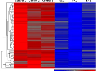
changes in expression level. A total of 89 miRNAs showed a significant decrease in expression
in the formaldehyde exposed lung samples compared to control samples (Figure 4, see
Supplementary Table 1). There were no miRNAs identified with significantly increased
expression levels in response to formaldehyde. The five most significantly differentially
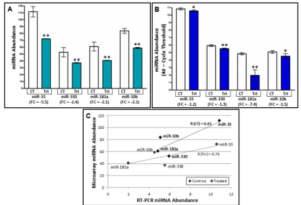
expressed miRNAs, as determined through microarray analysis, were 33 (FC -5.5),
miR-450 (FC -3.6), miR-330 (FC -2.4), miR-181a (FC -2.1), and miR-10b (FC -2.1). Here, fold
change (FC) represents the ratio of miRNA abundance in exposed relative to the control samples.
Figure 4: Formaldehyde modulates the expression of 89 miRNAs in human lung cells. A heat map displays the relative expression levels of the 89 miRNAs, where data are mean standardized and hierarchical clustering is performed. Blue indicates relative low expression while red indicates relative high expression. Formaldehyde-treated samples are abbreviated as FA.
15
1.4.2 miRNA Expression Changes are Validated through RT-PCR
RT-PCR was used to confirm the array-based findings, where the decreased miRNA
expression induced by formaldehyde exposure was verified. Specifically, miR-330 showed a
formaldehyde-induced FC of -1.3, miR-181a showed a FC of -7.4, miR-33 showed a FC of -1.2,
and miR-10b showed a FC of -1.5 (Figure 5). miR-450 showed minimal expression changes
with a FC of -1.04 (data not shown). As it was not validated with RT-PCR, further analysis on
miR-450 was not performed. To assess the similarity of the RT-PCR and array-based expression
level quantification, the average relative miRNA abundances were compared against the raw
microarray expression levels. This analysis shows high correlations (0.81 for control samples,
0.76 for treated samples) between the average miRNA abundance measured with both RT-PCR
and microarray (Figure 5). These analyses support that the direction of miRNA differential
expression induced by formaldehyde was consistent between the RT-PCR and microarray
analyses. It is important to note that there is a difference in the magnitude of expression change
with the microarray results generally greater than those obtained with RT-PCR.
16
1.4.3 miRNA Targets are Integrated into Biological Networks
In order to identify potential biological pathways affected by formaldehyde exposure, the
89 miRNAs that showed significant changes in expression levels were ranked according to their
fold changes in expression, p-values of significance, and RT-PCR results (see Supplementary
Table 1). Here, the four miRNAs with the most significant formaldehyde-induced changes in
expression were further investigated: miR-33, miR-330, miR-181a, and miR-10b. For each of
these four miRNAs, we identified their putative mRNA targets. Using a stringent cutoff of a
match score between each miRNA and its mRNA targets followed by analysis for unique
mRNAs per target list, we identified a total of 67 targets of miR-33, 217 targets of miR-330, 334
targets of miR-181a, and 25 targets of miR-10b (see Supplementary Table 2). Among this list
of 643 mRNAs, there are 42 that are common to at least two of the modulated miRNAs.
Once the predicted transcriptional targets were identified for the most significant
miRNAs, they were overlaid onto molecular pathway maps enabled through the Ingenuity®
Systems Knowledge Base. Networks containing miRNA targets were algorithmically constructed
based on connectivity and known relationships among proteins. The predicted targets of miR-33,
miR-330, miR-181a, and miR-10b resulted in the generation of a total of 40 networks (see
Supplementary Table 3). For each of the miRNA targets, the most significant (p-values range
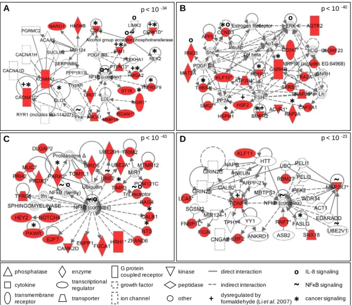
from 10-23 to 10-43) network has been highlighted for further evaluation (Figure 6). The proteins
identified within these networks were queried for their enrichment for various canonical
pathways. A comparison of the canonical pathways highlighted the conservation of a
cancer-associated pathway common to all four miRNA-generated networks (see Supplementary Table
17
Figure 6: Significant molecular networks of miRNA-mediated signaling likely affected by formaldehyde exposure in human lung cells. Protein networks display interactions using the transcriptional targets of (A) miR-33, (B) miR-330, (C) miR-181a, and (D) miR-10b. Networks are displayed with symbols representing predicted miRNA targets (red symbols) or proteins associated with the predicted targets (white symbols).
Using a biological process enrichment analysis, the 40 networks encoded by the
transcriptional targets for each miRNA were queried for biological processes that were most
significantly modulated by formaldehyde exposure. A total of 71 unique biological processes
were found (see Supplementary Table 5). Across the mRNA targets, common enrichment was
found for 13 different cellular biological processes. These processes included inflammatory
response (p-value = 0.0029) and endocrine system development/function (p-value = 0.0018)
which were enriched within the targets of all four miRNAs (Table 1).
p < 10 -40
p < 10 -43 p < 10 -23
p < 10 -34 A C B D phosphatase cytokine enzyme transcriptional regulator G protein coupled receptor other transmembrane receptor transporter direct interaction indirect interaction ion channel growth factor kinase peptidase cancer signaling dysregulated by
formaldehyde (Li et al. 2007)
+
*
+ + + + + + + +*
*
*
*
*
*
*
*
*
*
*
*
*
*
*
*
*
*
*
*
*
*
~
o IL-8 signaling NFĸB signaling
~
~
~
~
~
~
~
~
~
o o o o o o o18
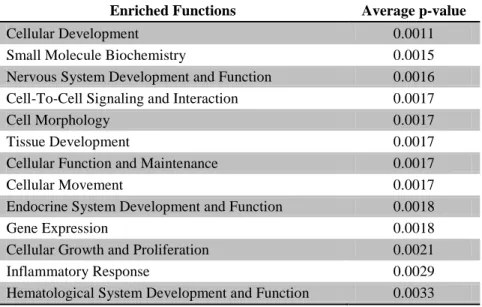
Table 1: Biological functions significantly associated with all predicted target sets of miR-33, miR-330, miR-181a, and miR-10b, the miRNAs with the greatest expression alterations upon exposure to formaldehyde in human lung cells.
Enriched Functions Average p-value
Cellular Development 0.0011
Small Molecule Biochemistry 0.0015
Nervous System Development and Function 0.0016
Cell-To-Cell Signaling and Interaction 0.0017
Cell Morphology 0.0017
Tissue Development 0.0017
Cellular Function and Maintenance 0.0017
Cellular Movement 0.0017
Endocrine System Development and Function 0.0018
Gene Expression 0.0018
Cellular Growth and Proliferation 0.0021
Inflammatory Response 0.0029
Hematological System Development and Function 0.0033
1.4.4 Conservation of Predicted and Observed mRNA Targets
In our analysis, we used a stringent computational metric to match miRNAs to their
predicted mRNA targets to better understand the biological implications of formaldehyde
exposure. As these mRNA targets were computationally predicted, we also compared our results
with those of an existing genomic database established from a study that analyzed human
tracheal fibroblast cells exposed to formaldehyde (Li et al. 2007). In this comparison, we found
overlap between the predicted mRNA targets of the formaldehyde-modulated miRNAs and the
tested formaldehyde-responsive genes previously identified (Li et al. 2007). Specifically,
brain-derived neurotrophic factor (BDNF), bone morphogenetic protein receptor, type II
(serine/threonine kinase) (BMPR2), calcium channel voltage-dependent L type, alpha 1C subunit
(CACNA1C), casein kinase 1 delta (CSNK1D), high mobility group AT-hook 2 (HMGA2), heat
shock transcription factor 2 (HSF2), heat shock 105kDa/110kDa protein 1 (HSPH1), and Pim-1
oncogene (PIM1), are found within the four most significant networks associated with the
identified miRNA targets (Figure 6).
We expanded our comparison by performing network analysis on the
formaldehyde-associated genes identified by Li et al. (2007). Here, networks were constructed and related
19
Networks related to cancer (p-value = 1.9 x 10⁻¹⁹), inflammation (p-value = 1.1 x 10⁻⁸), and
endocrine system disorders (p-value = 3.15 x 10⁻⁴) were generated (see Supplementary Table
6).
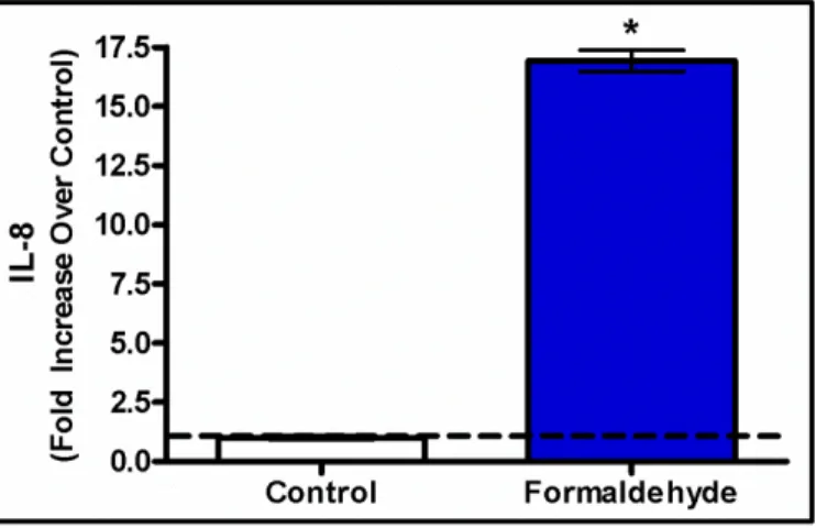
1.4.5 Inflammatory Cytokine IL-8 is Released in Response to Formaldehyde
Based on our findings from the canonical pathway and biological process enrichment
analyses that showed the IL-8 pathway as potentially dysregulated by miRNAs associated with
formaldehyde exposure, we set out to confirm whether IL-8 protein levels may be influenced by
such exposure. After cells were exposed to formaldehyde, IL-8 protein release was assessed. The
investigation of the inflammatory response protein IL-8 showed that human lung cells activate an
inflammatory response after exposure to formaldehyde. Specifically, an average 16.9 fold
increase (p-value < 0.05) in cytokine release was observed in formaldehyde exposed cells
relative to control samples (Figure 7).
Figure 7: Interleukin-8 levels are significantly elevated in formaldehyde-treated lung cells compared to untreated cells. Results are displayed as fold increase over control +/- S.E.M. (*) indicates statistical significance compared to control samples (p-value < 0.05).
1.5 Discussion
In this study, we exposed human A549 lung epithelial cells to formaldehyde using an in
vitro exposure system that physically replicates in vivo human lung gas exposures (Bakand et al.
2005). It is important to note that A549 cells are carcinoma cells that exhibit differences in
20
detoxifying pathways and are more resistant to apoptosis in comparison to normal cells (Kweon
et al. 2006). While we recognize that A549 cells may not completely mimic normal lung cell
response, there are several advantages to using these cells for air toxicant studies. For example,
when exposed to gases at an air-liquid interface, A549 cells secrete enough surfactant to mimic
airway surface tension (Blank et al. 2006). As a result, A549 cells are routinely used to study the
effects of environmental air exposures (Doyle et al. 2004; Doyle et al. 2007; Jaspers et al. 1997;
Sexton et al. 2004), including formaldehyde (Quievryn et al. 2000; Speit et al. 2008; Speit et al.
2010). A549 cells have also shown the same sensitivity and removal efficiency towards
formaldehyde-induced DNA protein crosslinks as primary human nasal epithelial cells (Speit et
al. 2008).
Our microarray analysis revealed that formaldehyde exposure resulted in the
regulation of 89 miRNAs. It was interesting that all of the modulated miRNAs were
down-regulated by formaldehyde exposure. This general trend of miRNA down-regulation has been
observed in rat lung cells exposed to cigarette smoke (Izzotti et al. 2009), as well as in multiple
tumor cell types, including lung cancer, breast cancer, and leukemia (Lu et al. 2005).
We focused a detailed analysis on the four most significantly down-regulated miRNAs,
as determined through microarray analysis and RT-PCR: 33, 330, 181a, and
miR-10b. These miRNAs have been studied, to some extent, and knowledge about their regulation
and association to disease is growing. For example, miR-33 shows decreased expression levels in
tissues from patients with lung carcinomas (Yanaihara et al. 2006). Also, miR-330 expression
has been measured at significantly lower levels in human prostate cancer cells when compared
against nontumorigenic prostate cells (Lee et al. 2009). Furthermore, miR-330 has been
suggested to act as a tumor suppressor by regulating apoptosis of cancer cells (Lee et al. 2009).
In addition, miR-10b shows altered expression levels within breast cancer tissue, and is one of
the most consistently dysregulated miRNAs able to predict tumor classification (Iorio et al. 2005;
Ma et al. 2007). These findings suggest that miR-33, miR-330, and miR-10b may influence
cellular disease state, specifically related to cancer.
Formaldehyde exposure also altered the expression level of miR-181a, which has known
associations with leukemogenesis (Marcucci et al. 2009). The specific link between
formaldehyde exposure and leukemia is currently debated, as numerous epidemiological studies
21
2004; L Zhang et al. 2010), as well as against it (Bachand et al. 2010; Marsh et al. 2004).
However, it is important to note that our study evaluates miRNA expression in lung cells, which
likely differ from leukemia target cells’ responses to formaldehyde exposure, or exposure to
formaldehyde’s metabolic products. Nevertheless, it is worth highlighting the observation of the
dysregulation of miR-181a upon exposure to formaldehyde.
To expand our analysis, we used a systems biology approach to understand the potential
biological implications of the miRNA expression changes induced by acute formaldehyde
exposure. For this analysis, we used a stringent computational matching approach to identify
predicted mRNA targets for miR-33, miR-330, miR-181a, and miR-10b. The identified mRNA
targets were used to construct associated molecular networks and were analyzed for their known
involvement in signaling pathways and biological functions. The identified networks showed
enrichment for various canonical pathways including nuclear factor kappa-B (NFκB) and
interleukin-8 (IL-8) signaling. Although very few predicted targets overlapped between the four miRNAs, proteins involved with cancer mechanisms including that of the NFκB pathway were found within the miRNA target networks. Importantly, NFκB has clear links to inflammation and cancer development (Karin et al. 2005). Also related to inflammation, IL-8-related signaling
molecules were present in the miRNA target networks. Previous studies have shown IL-8 release
in lungs cells representing inflammatory response after exposure to other air pollutants (Jaspers
et al. 1997; Sexton et al. 2004). In addition, investigations have shown increased IL-8 levels in
lungs of patients with diseases such as acute lung injury (McClintock et al. 2008), adult
respiratory distress syndrome (Jorens et al. 1992), and asthma (Bloemen et al. 2007).
Inflammation is a recognized formaldehyde-induced response, as formaldehyde is known to
irritate the respiratory system (Tuthill 1984) and increase asthmatic response (Rumchev et al.
2002; Wieslander et al. 1997). Our findings suggest that the canonical pathways associated with
formaldehyde-induced miRNA alterations may affect the regulation of biological pathways
associated with various disease states, including cancer and inflammation.
As a method to further verify our results, we compared the protein levels of cytokine
interkeukin-8 (IL-8) in formaldehyde-exposed cells versus mock-treated controls. We found that,
indeed, IL-8 showed significantly increased protein expression levels in the
formaldehyde-exposed cells. These results support our findings that IL-8 signaling is altered in lung cells
22
lung cells after pre-sensitization to tumor necrosis factor alpha (TNFα) (Persoz et al. 2010). TNFα is a proinflammatory mediator shown to have increased levels upon exposure to formaldehyde (Bianchi et al. 2004). Our network analyses suggest that cytokine signaling may
be altered through changes in miRNA expression levels. Supporting this is a recent study that
shows modifications to miRNAs may influence the expression of cytokines, including IL-6 and
IL-8 (Jones et al. 2009). Future research will test whether the observed miRNA expression
changes are directly associated with IL-8 signaling.
In an effort to gain further understanding of formaldehyde’s effects on gene expression,
we compared our results with those of an existing genomics database (e.g. mRNA) from a study
that evaluated human lung cells exposed to formaldehyde (Li et al. 2007). Using the predicted
targets in our most significant miRNA networks, we found the following genes overlap with the
existing database: BDNF, BMPR2, CACNA1C, CSNK1D, HMGA2, HSF2, HSPH1, and PIM1.
These genes have been shown to play a role in various diseases. For example, BDNF, or
brain-derived neurotrophic factor, modulates neurogenesis after injury to the central nervous system
(Ming et al. 2005). CSNK1D, or casein kinase 1 delta, has been identified as up-regulated in
breast cancer tissue (Abba et al. 2007). HMGA2, or high mobility group AT-hook 2, is oncogenic
in many cells, including lung carcinoma cells, and is regulated by the tumor-suppressive miRNA
let-7 (Lee et al. 2007). Lastly, PIM1, or Pim-1 oncogene, is found at increased levels within
prostate cancer tissue (Dhanasekaran et al. 2001). Network analysis of all
formaldehyde-responsive genes identified through the Li et. al. (2007) study revealed significant associations
with cancer, inflammation, and endocrine system regulation, which also overlap with our
findings. These genes are therefore linked with formaldehyde-induced changes in miRNA
abundance as well as mRNA alterations, and they are related to a diverse range of cellular
responses including tumorigenesis.
In conclusion, our study provides evidence of a potential mechanism that may underlie
the cellular effects induced by formaldehyde, namely the modification of miRNA expression.
We identify a set of 89 miRNAs that are dysregulated in human lung cells exposed to
formaldehyde. Mapping the most significantly changed miRNAs to their predicted
transcriptional targets and their network interactomes within the cell reveals the association of
formaldehyde exposure to inflammatory response pathways. We also validate our findings by:
formaldehyde-23
induced mRNA expression changes; and (3) examining protein expression changes of a key
inflammatory response mediator, IL-8. Future research will investigate whether the expression
levels of these miRNAs may serve as potential biomarkers of formaldehyde exposure in humans.
Such biomarkers can be utilized to better monitor human exposure to environmental toxicants
and relate them to health effects. Based on our findings, we believe that miRNAs likely play an
important role in regulating formaldehyde-induced gene expression and may represent a possible
24
CHAPTER 2
FORMALDEHYDE AND EPIGENETIC ALTERATIONS: MICRORNA CHANGES IN THE NASAL EPITHELIUM OF NONHUMAN PRIMATES
2.1 Overview
Formaldehyde is an air pollutant present in both indoor and outdoor atmospheres.
Because of its ubiquitous nature, it is imperative to understand the mechanisms underlying
formaldehyde-induced toxicity and carcinogenicity. MicroRNAs (miRNAs) can influence
disease caused by environmental exposures, yet miRNAs are understudied in relation to
formaldehyde. Our previous investigation demonstrated that formaldehyde exposure in human
lung cells caused disruptions in miRNA expression profiles.
Here, we expand our preliminary in vitro findings to an in vivo model. We set out to test
the hypothesis that formaldehyde inhalation exposure significantly alters miRNA expression
profiles within the nasal epithelium of nonhuman primates.
Cynomolgus macaques were exposed by inhalation to approximately 0, 2, or 6 ppm
formaldehyde for 6 hours/day for two consecutive days. Small RNAs were extracted from nasal
samples and assessed for genome-wide miRNA expression levels. Transcriptional targets of
formaldehyde-altered miRNAs were computationally predicted, analyzed at the systems level,
and assessed using RT-PCR.
Expression analysis revealed that 3 and 13 miRNAs were dysregulated in response to 2
and 6 ppm formaldehyde, respectively. Transcriptional targets of the miRNA with the greatest
increase (miR-125b) and decrease (miR-142-3p) in expression were predicted and analyzed at
the systems level. Enrichment was identified for miR-125b targeting genes involved in apoptosis
signaling. The apoptosis-related targets were functionally tested using RT-PCR, where all targets
showed decreased expression in formaldehyde-exposed samples.
Our study reveals that formaldehyde exposure disrupts miRNA expression profiles within
25
2.2 Study Objectives
For this study, we set out to test the novel hypothesis that formaldehyde inhalation
exposure significantly alters miRNA expression profiles within the nasal epithelium of
nonhuman primates. We tested this hypothesis by exposing nonhuman primates (cynomolgus
macaques) to ~ 0, 2, or 6 ppm formaldehyde for 6 hours/day across two days. After exposure,
nasal epithelial tissue was assessed for formaldehyde-induced changes in miRNA expression
profiles across the genome. Transcriptional targets of the miRNAs with the highest increase and
highest decrease in expression were predicted in silico and mapped onto molecular interaction
networks. Important canonical pathways enriched within the constructed networks were
identified and further assessed at the gene expression level. Altogether, this study reveals a novel
epigenetic mechanism through which formaldehyde may influence critical signaling pathways
known to influence disease.
2.3 Materials and Methods
2.3.1 Animals
Cynomolgus macaques were treated humanely and with regard for alleviation of
suffering. Animals were exposed, sedated, and euthanized using protocols approved by the
Lovelace Research Institute’s animal care and use committee (FY10-104A). For this study, eight
male cynomolgus macaques (Macaca fascicularis) were selected from the Lovelace Respiratory
Research Institute colony. Animals were approximately six years of age and weighed between
4.48 and 8.56 kilograms. Animals were conditioned to whole body exposure chambers for 30,
60, 180, and 360 minutes prior to the first day of exposure, as previously described (Moeller et
al. 2011).
2.3.2 Formaldehyde Exposures
Animals were exposed to formaldehyde over the course of two days for six hours each
day using whole body exposure chambers. Target exposure concentrations were 0, 2, and 6 ppm
26
Formaldehyde was isotope-labeled for the purposes of a previous investigation (Moeller et al.
2011). Chamber concentrations were monitored by collecting samples with a Waters XpoSure
Aldehyde Sampler cartridge every five minutes throughout each exposure period. Samples from
the cartridges were analyzed using high-performance liquid chromatography with an attached
detector monitoring ultraviolet absorbance at 360 nm (Lu et al. 2011; Moeller et al. 2011). Two
control animals were placed in whole body exposure chambers containing clean air. Three
monkeys were exposed to a target concentration of 2 ppm formaldehyde, where the measured
concentration averaged 1.9 ppm across the exposure periods. Three monkeys were exposed to a
target concentration of 6 ppm formaldehyde, where the measured concentration averaged 6.1
ppm across the exposure periods. For more detailed methods, see Moeller et al. (Moeller et al.
2011).
2.3.3 Sample Collection
Approximately 15 minutes after the second exposure period, animals were serially
sedated with Ketamine (10 mg/kg, intramuscular) and euthanized with Euthasol (>1 ml/4.5 kg,
intravenous). Animals underwent necropsy one at a time with each necropsy requiring
approximately 45 minutes. All samples were collected within 3 hours of the exposure. Sample
collection started immediately after the last exposure in order to parallel sacrifice and sample
collection times used in our previous studies (Lu et al. 2011; Moeller et al. 2011). During
necropsy, nasal epithelial tissue from the maxilloturbinate regions were collected, placed in
RNAlater® (Qiagen, Valencia, CA), and stored at -80°C. Samples were shipped by overnight
courier on dry ice to the University of North Carolina at Chapel Hill.
2.3.4 Sample Processing
Small RNAs were isolated from nasal tissue samples. Samples were first disrupted and
homogenized using a TissueRuptor (Qiagen) in the presence of TRIzol (Invitrogen Life
Technologies, Carlsbad, CA), and RNA was isolated using the miRNeasy® kit (Qiagen).
Extracted RNA was quantified with a Nanodrop 1000 spectrophotometer (Thermo Scientific,
Waltham, MA) and its integrity verified with a 2100 Bioanalyzer (Agilent Technologies, Santa
Clara, CA). RNA was then labeled and hybridized to the Agilent Human miRNA Microarray
27
11080 probesets. Microarray results were extracted using Agilent Feature Extraction software.
Microarray data have been submitted to National Center for Biotechnology Information (NCBI)
Gene Expression Omnibus repository (Edgar et al. 2002) and are available under accession
number GSE34978 (NCBI 2010).
2.3.5 Microarray Analysis
Microarray data were normalized by quantile normalization. To eliminate background
noise, miRNA probes with signal intensities less than the median signal (signal = 40) across all
replicates were removed. Differential expression was defined as a significant difference in
miRNA levels between exposed versus unexposed samples, where three statistical requirements were set: (i) fold change of ≥ 1.5 or ≤ - 1.5 (average exposed versus average unexposed); (ii) p-value < 0.05 (ANOVA); and (iii) a false discovery rate corrected q-p-value < 0.1. Analysis of
variance (ANOVA) p-values were calculated using Partek® Genomics SuiteTM software (St.
Louis, MO). To control the rate of false positives, q-values were calculated as the minimum
“positive false discovery rate” that can occur when identifying significant hypotheses (Storey
2003).
2.3.6 RT-PCR Confirmation of miRNA Expression Changes
To confirm formaldehyde-induced miRNA expression changes, we performed real-time
reverse transcriptase polymerase chain reaction (RT-PCR) using two miRNAs identified as the
most increased in expression (miR-125b and miR-152) and two miRNAs identified as the most
decreased in expression (miR-145 and miR-142-3p) following 6 ppm formaldehyde exposure.
TaqMan® MicroRNA Primer Assays for hsa-miR-125b (ID 000449), hsa-miR-152 (ID 000475),
hsa-miR-145 (ID 002278) and hsa-miR-142-3p (ID 000464) were used in conjunction with the
TaqMan® Small RNA Assays PCR kit (Applied Biosystems, Carlsbad, CA). The same control
and formaldehyde-exposed samples from the microarray analysis were used for RT-PCR, and
was performed in technical triplicate. The resulting RT-PCR cycle times were normalized