APOBEC3G and APOBEC3F Act in Concert To Extinguish HIV-1
Replication
John F. Krisko,*Nurjahan Begum, Caroline E. Baker, John L. Foster, J. Victor Garcia
Division of Infectious Diseases, Center for AIDS Research, University of North Carolina School of Medicine, Chapel Hill, North Carolina, USA
ABSTRACT
The multifunctional HIV-1 accessory protein Vif counters the antiviral activities of APOBEC3G (A3G) and APOBEC3F (A3F),
and some Vifs counter stable alleles of APOBEC3H (A3H). Studies in humanized mice have shown that HIV-1 lacking Vif
ex-pression is not viable. Here, we look at the relative contributions of the three APOBEC3s to viral extinction. Inoculation of bone
marrow/liver/thymus (BLT) mice with CCR5-tropic HIV-1
JRCSF(JRCSF) expressing a
vif
gene inactive for A3G but not A3F
deg-radation activity (JRCSFvifH42/43D) displayed either no or delayed replication. JRCSF expressing a
vif
gene mutated to
inacti-vate A3F degradation but not A3G degradation (JRCSFvifW79S) always replicated to high viral loads with variable delays. JRCSF
with
vif
mutated to lack both A3G and A3F degradation activities (JRCSFvifH42/43DW79S) failed to replicate, mimicking JRCSF
without Vif expression (JRCSF
⌬
vif). JRCSF and JRCSFvifH42/43D, but not JRCSFvifW79S or JRCSFvifH42/43DW79S, degraded
APOBEC3D. With one exception, JRCSFs expressing mutant Vifs that replicated acquired enforced
vif
mutations. These
muta-tions partially restored A3G or A3F degradation activity and fully replaced JRCSFvifH42/43D or JRCSFvifW79S by 10 weeks.
Surprisingly, induced mutations temporally lagged behind high levels of virus in blood. In the exceptional case, JRCSFvifH42/
43D replicated after a prolonged delay with no mutations in
vif
but instead a V27I mutation in the RNase H coding sequence.
JRCSFvifH42/43D infections exhibited massive GG/AG mutations in
pol
viral DNA, but in viral RNA, there were no fixed
muta-tions in the Gag or reverse transcriptase coding sequence. A3H did not contribute to viral extinction but, in combination with
A3F, could delay JRCSF replication. A3H was also found to hypermutate viral DNA.
IMPORTANCE
Vif degradation of A3G and A3F enhances viral fitness, as virus with even a partially restored capacity for degradation outgrows
JRCSFvifH42/43D and JRCSFvifW79S. Unexpectedly, fixation of mutations that replaced H42/43D or W79S in viral RNA lagged
behind the appearance of high viral loads. In one exceptional JRCSFvifH42/43D infection,
vif
was unchanged but replication
proceeded after a long delay. These results suggest that Vif binds and inhibits the non-cytosine deaminase activities of intact
A3G and intact A3F, allowing JRCSFvifH42/43D and JRCSFvifW79S to replicate with reduced fitness. Subsequently, enhanced
Vif function is acquired by enforced mutations. In infected cells, JRCSF
⌬
vif and JRCSFvifH42/43DW79S are exposed to active
A3F and A3G and fail to replicate. JRCSFvifH42/43D Vif degrades A3F and, in some cases, overcomes A3G mutagenic activity to
replicate. Vif may have evolved to inhibit A3F and A3G by stoichiometric binding and subsequently acquired the ability to target
these proteins to proteasomes.
H
IV-1 Vif is an accessory protein that prevents inhibition of
viral replication by the innate immune system proteins
APOBEC3G (A3G) and APOBEC3F (A3F) (
1–3
). These HIV-1
restriction factors gain access to HIV-1 virions and deaminate
cytosines in negative-strand DNA during reverse transcription of
viral genomic RNA (
4–6
). This activity yields G-to-A mutations in
HIV-1 genes, leading to loss of viral viability (
4
,
6
). Vif prevents
A3G and A3F antiviral activity by linkage to proteosomal
degra-dation (
7–9
). Vif is also found inside virions, where it can
poten-tially inhibit encapsidated A3G and A3F (
10
,
11
). In addition to
directly inhibiting the G-to-A mutagenic activity of A3G and A3F,
virion-encapsidated Vif could also prevent A3G and A3F
inhibi-tion of reverse transcripinhibi-tion, transcripinhibi-tion elongainhibi-tion, and
provi-ral integration and inactivation of the transactivation response
element (
12–16
).
The devastating effect of human APOBEC3s on HIV-1
JRCSF(JRCSF) replication
in vivo
in the absence of Vif has been
previ-ously demonstrated in NSG-hu humanized mice (
17
,
18
). We also
reported that JRCSF lacking Vif function cannot systemically
rep-licate in bone marrow/liver/thymus (BLT) humanized mice and is
quickly extinguished (
17
). In cases where we were able to recover
archival sequences from viral DNA, high levels of G-to-A
tions incompatible with viral replication were present. The
muta-tions were approximately 85% GG to AG and 15% GA to AA (
17
).
A3G is the only APOBEC3 that preferentially mutates GG over GA
sites, pointing to A3G as the dominant restricting factor for HIV-1 (
4
,
19
). An alternate hypothesis is that any of the other APOBEC3s, all
of which have dominantly GA-to-AA mutagenic activity, may
contribute to the totality of the restriction but do so mainly by
Received2 January 2016 Accepted18 February 2016
Accepted manuscript posted online24 February 2016
CitationKrisko JF, Begum N, Baker CE, Foster JL, Garcia JV. 2016. APOBEC3G and APOBEC3F act in concert to extinguish HIV-1 replication. J Virol 90:4681– 4695.
doi:10.1128/JVI.03275-15.
Editor:G. Silvestri
Address correspondence to J. Victor Garcia, [email protected], or John L. Foster, [email protected].
*Present address: John F. Krisko, Argos Therapeutics, Inc., Durham, North Carolina, USA.
mechanisms not involving G-to-A mutation. Further, the
identi-ties of APOBEC3 genes other than A3G genes that could
signifi-cantly contribute to HIV-1 extinction in humanized mice remain
an outstanding question. The leading candidates are A3F,
APOBEC3H (A3H), and APOBEC3D (A3D), in descending order
of likely importance (
2
,
20–22
). However, evidence for significant
nondeaminase activity
in vivo
has not been forthcoming (
22
).
To advance understanding of the role of APOBEC3 genes in
restricting HIV-1
vif
⫺(i.e.,
vif
defective), we infected BLT mice
with JRCSF containing mutated
vif
genes that were defective for
A3G and/or A3F degradation. We found that A3G acting alone
was able to completely terminate HIV-1 infection in some cases,
while in others, replication was greatly delayed but eventually
pro-duced high levels of virions in blood. A3F acting alone had
rela-tively weak effects on viral replication, but Vif mutations partially
restoring A3F degradation activity were nonetheless strongly
se-lected. Strikingly, virus with the doubly mutated
vif
that was
un-able to degrade A3G or A3F mimicked
vif
⫺virus by failing to
replicate in BLT mice. Also, a possible role for A3H in explaining
our observations was addressed. A3H was not a necessary
compo-nent for termination of HIV-1 infection but in combination with
A3F may contribute to delays in viral replication. A case of HIV-1
hypermutation was found in which numerous GA-to-AA
muta-tions could be attributed to A3H.
Therefore, we found that A3G strongly restricts HIV-1, if not
countered by
vif
, but consistent restriction requires an additional
factor which several lines of evidence suggest to be A3F. The major
antiviral impact of A3F may be through a cytosine
deaminase-independent mechanism.
MATERIALS AND METHODS
Cell culture.TZM HeLa cells obtained from the NIH AIDS Research and Reference Reagent Program (23) and human embryonic kidney 293T cells were cultured at 37°C and 10% CO2in Dulbecco’s modified Eagle
me-dium (DMEM) (Sigma) supplemented with 10% fetal bovine serum (SAFC Biosciences), 50 IU penicillin, 50g/ml streptomycin, and 2 mM
L-glutamine (Cellgro). CEM-SS cells, a human T cell line, were cultured at 37°C and 5% CO2in RPMI 1640 (Sigma) supplemented with 10% fetal
bovine serum (SAFC Biosciences), 50 IU penicillin, 50g/ml streptomy-cin, 2 mML-glutamine (Cellgro), and 1 mM sodium pyruvate (Cellgro). Transfections.HEK 293T cells were transfected using Lipofectamine 2000 (Invitrogen) according to the manufacturer’s protocol. Cells (7⫻ 105per well in a 6-well plate or 5⫻106in a 10-cm dish) were cultured in
DMEM in the absence of antibiotics 24 h prior to transfection. Plasmid DNA was resuspended in Opti-MEM (Gibco) at a ratio of 1g of DNA to 2.5l of Lipofectamine 2000. Following a 20-min incubation at room temperature, the DNA-Lipofectamine 2000 mixture was added dropwise to the 293T cells.
Preparation of whole-cell lysates and Western blotting.Mammalian expression vectors for A3G (pcDNA3.1A3GV5) and A3F (pcDNA3.1A3FV5) were obtained from B. M. Peterlin (24). These plasmids were transfected in 293T cells with JRCSF proviral clones. The transfected 293T cells were washed with cold phosphate-buffered saline (PBS) while attached to the culture dish and then lysed in 800l of cold lysis buffer (50 mM Tris, pH 8.0, 10% glycerol [Fisher], 100 mM NaCl [Fisher], 25 mM NaF [Sigma], 2 mM Na3VO2[Sigma], 20 mM-glycerophosphate [Sigma], 25 mM
ben-zamidine [Sigma], 2 mM EDTA, pH 8.0, and 0.5% IGEPAL CA630 [Sigma]) supplemented with one mini-protease inhibitor tablet (Roche) per 10 ml of lysis buffer. The cell lysates were cleared by centrifugation at 15,000⫻gfor 10 min at 4°C, and the protein concentrations of the postnuclear supernatants were determined using IgG protein standards in the Bio-Rad protein assay reagent (Bio-Rad). Total protein (200g) from whole-cell lysates in sodium dodecyl sulfate (SDS) loading buffer was
denatured for 5 min at 95°C and resolved on a 12% polyacrylamide gel for 900 V · h. The proteins were then transferred to a Hybond-C Extra nitro-cellulose membrane (Amersham Biosciences) using a TransBlot semidry transfer cell (Bio-Rad) at 20 V for 42 min. The membrane was then blocked for 1 h in 10% milk in Tris-Tween-buffered saline (TTBS) (10 mM Tris, pH 8.0, 150 mM NaCl, 0.05% Tween 20 [Fisher Scientific]), incubated with primary antibody in 10% milk in TTBS for 12 to 16 h at 4°C, washed three times in TTBS, incubated with horseradish peroxidase (HRP)-conjugated secondary antibody in 10% milk in TTBS for 2 h, and finally washed three times in TTBS. The membrane was incubated in the luminol substrate buffer (100 mM Tris, pH 8.8, 2.5 mM luminol [Fluka], 400Mp-coumaric acid [Sigma], and 5.4 mM H2O2[Sigma]) for 5 min
and then exposed to film for an appropriate amount of time to visualize expression of Vif, A3G, or A3F and glyceraldehyde-3-phosphate dehydro-genase (GAPDH). Wild-type JRCSF Vif effectively depleted A3G and A3F. A3D (pCS2Apobec3DEHA) degradation activity was assayed as described above, with the exception that the SDS loading buffer contained 6 M urea (25).
Antibodies.Rabbit anti-Vif (number 2221), obtained from the NIH AIDS Research and Reference Reagent Program (26), was used at 1:1,000. Mouse anti-V5 (Invitrogen number R960-25) was used at 1:7,500. Mouse anti-hemagglutinin (HA) (Abcam number 1818) was used at 1:1,000. Rabbit anti-GAPDH (clone 14C10; Cell Signaling number 2118L) was used at 1:3,000. Goat anti-rabbit HRP (Cell Signaling number 7074S) was used at 1:10,000. Goat anti-mouse HRP (Invitrogen number 62-6520) was used at 1:10,000.
Generation ofvif-defective HIV-1.All experiments were performed using JRCSF obtained from the NIH AIDS Research and Reference Re-agent Program (27). JRCSF⌬vif was previously described (17). The fol-lowing JRCSF proviral clones with mutations invifwere created by site-directed mutagenesis: JRCSFvifH42/43D, JRCSFvifW79S, JRCSFvifH42/ 43DW79S, JRCSFvifW79F, JRCSFvifW79Y, JRCSFvifH42/43N, and JRCSFvifH42N/H43G. The H42/43D mutation was based on the report by Mehle et al. that mutating H42/H43 to N42/N43 reduced A3G degra-dation (28). We mutated the histidines to aspartates, as they were poten-tially more disruptive of function (29). For a mutation that blocked A3F degradation, we mutated tryptophan 79 to serine (JRCSFvifW79S) based on reports that mutation of this residue to alanine resulted in a specific block in the capacity of the mutant protein to degrade A3F (30,31). Mutation of W79 to serine followed the suggestion of Yu et al. that the hydrophilic serine may be more detrimental to function than alanine (9). Production of HIV-1 stocks and titration.HIV-1 stocks were gener-ated by transfecting proviral DNA into 293T cells using Lipofectamine 2000 (Invitrogen) as indicated above. The virus-containing culture su-pernatant was collected and cleared of cellular debris, first by centrif-ugation at 3,000 rpm for 20 min and then by filtering through a 0.45-m filter (Millipore). Infectious titers of JRCSF and JRCSFvif variants were determined by using TZM-bl cells (23). Infected cells turn blue upon staining with X-Gal (5-bromo-4-chloro-3-indolyl--D
-galac-topyranoside) (Sigma). Blue cells were counted by microscopy, and the counts were used to calculate the titer, which was defined as tissue culture infectious units per milliliter (TCIU).
Viral cultures.CCR5-expressing CEM-SS cells were used to propa-gate both wild-type andvif-defective JRCSF (17). Cells (1⫻106) were
infected in a 24-well plate with 5⫻104TCIU of virus in complete RPMI
containing 4g/ml Polybrene at 37°C and 5% CO2for 4 h. The cells were
then washed extensively with PBS and cultured at 37°C and 5% CO2in
complete RPMI containing 0.5g/ml puromycin to maintain selection of CCR5-expressing cells. The cell cultures were passaged every 3 days, and a sample of the culture supernatant was collected for quantitation of the amount of viral capsid protein by p24 enzyme-linked immunosorbent assay (ELISA).
Alameda, CA) were transplanted into thymus/liver-implanted NOD/ SCID interleukin 2␥⫺/⫺(IL-2␥⫺/⫺) mice (Jackson Laboratories, Bar Har-bor, ME). Human CD45⫹cells in the peripheral blood of these mice were monitored periodically by flow cytometry (FACSCanto; BD Biosciences). The mice were maintained at the Division of Laboratory Animal Medi-cine, University of North Carolina at Chapel Hill (UNC-CH), in accor-dance with protocols approved by the UNC-CH Institutional Animal Care and Use Committee. The mice were inoculated intravenously with either wild-type JRCSF orvif-defective JRCSF by tail vein injection.
Flow cytometry.Peripheral blood obtained from the humanized mice (50l) was first blocked with murine IgG (Sigma) and then stained with mouse anti-human CD45 (clone 2D1; 557873; Becton-Dickinson) to ex-clude murine cells from the analysis. Human lymphocytes were identified by their human CD45⫹expression and further characterized by staining with mouse anti-human CD3 (clone HIT3a; 555573; Becton-Dickinson). Human CD3⫹cells were further analyzed for CD4⫹and CD8⫹ expres-sion with mouse anti-human CD4 (clone RPA-T4; 555749; Becton-Dick-inson) or CD8 antibodies (clone SK1; 347314; Becton-DickBecton-Dick-inson). Data were collected using a FACSCanto instrument and analyzed using FACS Diva software (Becton-Dickinson).
Peripheral blood analysis.Peripheral blood was collected from hu-manized mice from the retro-orbital vein using EDTA-containing capil-lary tubes (Drummond Scientific). Whole blood was centrifuged, and the plasma was removed for analysis of viral RNA. The volume of plasma removed was replaced with solution B (1 liter PBS [Sigma], 5 g bovine serum albumin [BSA] [Sigma], 50 U/ml penicillin [Sigma], 50 mg/ml streptomycin [Sigma], 1% citrate phosphate dextrose [Sigma]). The cel-lular portion of the blood was used for flow cytometric analysis as previ-ously described or for genomic-DNA isolation. Peripheral blood to be used for DNA isolation was resuspended in 1 ml of a hypotonic red blood cell lysis buffer (500 ml distilled H2O [dH2O], 4.15 g NH4Cl [Sigma], 0.5
g KHCO3[Sigma], and 0.019 g EDTA [Sigma]). Following a 10-min
in-cubation at room temperature, the lysed blood was centrifuged at 10,000 rpm for 1 min, and the lysis buffer was removed by aspiration. The result-ing cell pellet was stored at⫺80°C until DNA extraction.
Plasma viral load analysis.Plasma viremia was determined by quan-titating the copy number of cell-free viral RNA. Plasma (20l) from the humanized mice was used to isolate viral RNA using the QIAamp Viral RNA minikit (Qiagen) according to the manufacturer’s protocol. Viral RNA was then quantified using a one-step reverse transcriptase proto-col. RNA isolated from the plasma (5l) was used as the template with the TaqMan RNA-to-Ct 1-step kit (Applied Biosystems). Primers for the real-time PCR were directed against a conserved region ofgagand were pre-viously published (44).
Tissue harvest.The spleen, lymph nodes, and thymus were harvested and disrupted by grinding the tissue through a 70-m-pore-size cell strainer (Falcon) with the plunger of a 3-ml syringe, and the cells were washed through the strainer with cold solution B. Red blood cells from the spleen were lysed with hypotonic lysis buffer, and cells from all the tissues were pelleted by centrifugation at 1,500 rpm for 5 min at 4°C.
Mononuclear cells were isolated from the bone marrow by crushing the long bones from the hind legs using a mortar and pestle, collecting the cells by washing with cold solution B, and filtering the cells through a 70-m cell strainer. Red blood cells were lysed with hypotonic lysis buffer, and the cells were pelleted by centrifugation at 1,500 rpm for 5 min at 4°C. Mononuclear cells were isolated from the liver by grinding the tissue through a 70-m cell strainer and pelleting the cells by centrifugation at 1,500 rpm for 5 min at 4°C. The resulting cell pellet was then resuspended in 40% Percoll, underlayered with 70% Percoll, and centrifuged at 2,400 rpm for 20 min at room temperature. Mononuclear cells were collected from the interface and washed with cold solution B. Cells were isolated from the lungs by first mincing the tissue with scissors and digesting the tissue in 2.5 ml RPMI containing 6 mg collagenase D (Roche) and 50g DNase I (Roche) for 30 min at 37°C. The digested tissue was then ground through a 70-m cell strainer with a 3-ml syringe plunger, washed with
cold solution B, and centrifuged at 1,500 rpm for 5 min at 4°C. As with the liver, lung mononuclear cells were isolated by resuspending the cell pellet in 40% Percoll, underlayering with 70% Percoll, and centrifugation at 2,400 rpm for 20 min at room temperature, and the cells were collected from the interface and washed with cold solution B.
Live cells from all tissues were counted by trypan blue exclusion and pelleted for genomic DNA isolation by centrifugation at 10,000 rpm for 1 min or pelleted by centrifugation at 1,500 rpm for 5 min, resuspended in freezing medium (90% heat-inactivated fetal bovine serum [FBS], 10% dimethyl sulfoxide [Fisher]), frozen live in a cryo-freezing container at
⫺80°C, and then stored in liquid nitrogen.
Genomic-DNA isolation and nested PCR of viral DNA.Genomic DNA was isolated from frozen peripheral blood cell pellets by resuspend-ing a pellet in 0.01 M Tris, pH 7.4, containresuspend-ing recombinant PCR grade proteinase K (Roche) and incubating it at 56°C for 1 h. The proteinase K was then heat inactivated at 95°C for 20 min, and the mixture was frozen at⫺20°C. After thawing, the samples were centrifuged for 1 min at 14,000 rpm, and the DNA-containing supernatant was transferred to a new, un-autoclaved Eppendorf tube.
Genomic DNA from mononuclear cells (5⫻105to 5⫻106) from
animal tissues was prepared using QIAamp DNA blood minicolumns (Qiagen) according to the manufacturer’s protocol. All PCRs were per-formed using the Expand High Fidelity PCR System (Roche). A 1.7-kb region of the JRCSF genome from nucleotides 617 to 2358, encompassing gag, was amplified. The primers for the outer and inner reactions are listed inTable 1. The reaction conditions were as follows: a one-time denaturation at 95°C for 2 min, followed by 35 cycles of 95°C for 15 s, 55°C for 30 s, and 72°C for 1 min 45 s. Amplification concluded with an elongation step at 72°C for 7 min. PCR was performed on a 1.2-kb region of the viral genome from nucleotides 2493 to 4023 including the reverse transcriptase region ofpol. The primers for the outer and inner reactions are listed inTable 1. The reaction conditions were as follows: a one-time denaturation at 95°C for 5 min, followed by 30 cycles of 95°C for 1 min, 55°C for 1 min, and 68°C for 1 min. Amplification concluded with an elongation step at 72°C for 7 min. A 3.4-kb region of the viral genome from nucleotides 1877 to 5227 includ-ing the RNase H region ofpolwas also performed. The primers for the outer and inner reactions are listed inTable 1. The touchdown PCR con-ditions were as follows: a one-time denaturation at 95°C for 2 min; 10 cycles of 95°C for 15 s and 30 s at 65°C and then subtracting 1°C each subsequent cycle; and 68°C for 3.5 min, followed by 25 cycles of 95°C for 15 s, 55°C for 30 s, and 68°C for 3.5 min. Amplification concluded with an elongation step at 68°C for 10 min. A 1.4-kb region from nucleotides 4941 through 6399 of the HIV-1JRCSFgenome that includedvifwas also
ampli-fied with the primers listed inTable 1. The reaction conditions were as follows: a one-time denaturation at 95°C for 5 min, followed by 30 cycles of 95°C for 30 s, 55°C for 30 s, and 72°C for 1.5 min. Amplification con-cluded with an elongation step at 72°C for 7 min.
Amplification of cell-free viral RNA.Viral RNA was isolated from the plasma of JRCSF-exposed humanized mice using a QIAamp viral minikit according to the manufacturer’s recommended protocol (Qiagen number 52904). Purified viral RNA was then reverse transcribed into cDNA with the reversevifouter primer (Table 1) using SuperScript III reverse trans-criptase (Invitrogen number 18080-044) according to the manufacturer’s recommended protocol. The viral RNA was then digested using RNase H (Invitrogen number 18021-014), and the cDNA was used as a template in nested PCRs forvif,gag, andpol. The PCR products were sequenced with inner primers used for amplification. Individual clones from the ampli-fied viral RNA were sequenced following TA cloning (Invitrogen).
APOBEC3H coding sequence amplification.Cellular RNA from fro-zen pellets of peripheral blood mononuclear cells was extracted, and PCR amplification and sequencing of the A3H mRNA coding sequence was performed by the method of Ooms et al. (45).
nucleotide sequences were aligned against the corresponding proviral DNA sequence used to generate the viruses using the Highlighter program (http://www.hiv.lanl.gov/content/sequence/HIGHLIGHT/highlighter .html), which can identify G-to-A mutations indicative of APOBEC mu-tagenesis.
Highlighter output uses a graphical representation of DNA sequences for comparison. To assess the effects of G-to-A transition mutations on the viral protein sequence, amplified DNA sequences were translated us-ing the online ExPASy translation tool (http://au.expasy.org/tools/dna .html).
Statistics.Data are presented as means⫾standard errors of the mean (SEM).
Nucleotide sequence accession numbers.Sequences were deposited in GenBank with the following accession numbers: KU900139 to KU900143, KU926704, KU900138, and KU887644.
RESULTS
We previously reported that JRCSF with an inactivating deletion
in
vif
(JRCSF
⌬
vif) failed to replicate in BLT humanized mice (
17
).
Here, we investigated if the anti-HIV-1 activity of A3G is sufficient
to extinguish HIV-1 replication or if other APOBEC3s have
im-portant roles. A JRCSF proviral clone was constructed with a
vif
gene mutated at histidines 42 and 43, which are known to be
critical for A3G degradation. The histidines were replaced with
aspartates (JRCSFvifH42/43D) (
28
). We also mutated
trypto-phan 79, previously demonstrated to be critical for A3F
degra-dation, to serine (JRCSFvifW79S) (
30
,
31
). The doubly
defec-tive JRCSFvifH42/43DW79S was also constructed (see
Materials and Methods). The three viruses were produced, and
all three had the full replicative capacity of JRCSF
in vitro
(
Fig.
1A
). As shown in
Fig. 1B
, the mutated Vifs were found to be
stably expressed and to be defective for A3G degradation but
not A3F (H42/43D) or defective for A3F degradation but not
A3G (W79S). VifH42/43DW79S failed to degrade both A3G
and A3F. We also tested the abilities of the mutated Vifs to
degrade A3D. Vif and VifH42/43D effectively degraded A3D,
but VifW79S did not. VifH42/43DW79S showed an
intermedi-ate phenotype (
Fig. 1C
).
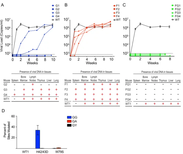
Replication of JRCSFvifH42/43D, JRCSFvifW79S, and
JRCSFvifH42/43DW79S in BLT humanized mice.
In
Fig. 2A
,
top, the time course of viral replication following intravenous
in-jection of 90,000 TCIU of JRCSF or JRCSFvifH42/43D is shown.
Seven wild-type-JRCSF-inoculated mice are plotted in aggregate
(means
⫾
SEM). In two JRCSFvifH42/43D mice (G1 and G2),
viral RNA was not detected in peripheral blood for at least 8 weeks.
The effective degradation of A3F and A3D by VifH42/43D makes
it unlikely that these two APOBEC3s were involved in the
ob-served viral extinction. In the other two mice (G3 and G4), virus
replicated, but the appearance of viral RNA in blood was greatly
delayed.
Upon sacrifice, viral DNA (reverse transcriptase coding
se-quence, nucleotides 2562 to 3881; GenBank accession number,
M38429
) was found in multiple organs of a representative JRCSF
(WT1) mouse, G3, and G4 (
Fig. 2A
, bottom). G1 which gave no
viral replication, was reinfected with JRCSF, and normal
replica-tion was observed (not shown). G2 also had no viral replicareplica-tion,
and viral DNA could be amplified from only one tissue. Therefore,
the replication of JRCSF with a
vif
gene defective for A3G
degra-dation but not A3F degradegra-dation is severely restricted but not
con-sistently extinguished. These variable results are in contrast to
JRCSF with a fully defective
vif
(JRCSF
⌬
vif), which fails to
repli-cate in BLT mice at all (
17
).
Figure 2B
, top, shows the results of the same experiment
per-formed with JRCSFvifW79S. In all cases, there was robust
replica-tion, but one mouse (F1) had delayed replicareplica-tion, with 10
4copies
of viral RNA at 2 weeks. By 4 weeks, all JRCSFvifW79S mice had
10
5or more copies of viral RNA, and by 6 weeks, all the mice had
viral loads of 10
6copies or more. Viral DNA from F1 to F4 was
present in nearly all tissues tested (
Fig. 2B
, bottom). JRCSF
defec-tive for both A3G and A3F degradation (JRCSFvifH42/43DW79S)
was also inoculated into BLT humanized mice (
Fig. 2C
). No viral
RNA was detected in plasma for 6 to 8 weeks postinjection (
Fig.
2C
, top), and no evidence of viral DNA in tissues was found (
Fig.
2C
, bottom). This result mimicked our previously reported results
with JRCSF
⌬
vif and strongly implies that the combined activities
of A3G and A3F rapidly extinguish viral replication (
17
).
Prominence of GG-to-AG mutations in archival viral DNA.
The JRCSF reverse transcriptase coding sequence was amplified
from multiple organs of G3, G4, and F1 to F4 (
Fig. 2A
, bottom,
and B, bottom). The percentage of GG sites mutated to AG and the
percentage of GA sites mutated to AA sites were determined. High
levels of GG-to-AG mutations were noted for G3 and G4, with
TABLE 1Nested-PCR primer sequences
Region amplified Primer Primer sequence
HIV-1 JRCSF 617–2358 Fwd gag Outer 5=-CTCAATAAAGCTTGCCTTGAGTGC-3=
Rev gag Outer 5=-CTTCCAATTATGTTGACAGGTGTAGG-3=
Fwd gag Inner 5=-GTGTGGAAAATCTCTAGCAGTGGC-3=
Rev gag Inner 5=-CTGTATCATCTGCTCCTGTATCTAATAGAGC-3=
HIV-1 JRCSF 1877–5227 Fwd pol Outer 5=-GATGACAGCATGTCAGGGAG-3=
Rev pol Outer 5=-GGTCAGGGTCTACTTGTGTGC-3=
Fwd pol Inner 5=-TGGCTGAAGCAATGAGCCAAG-3=
Rev pol Inner 5=-GTGGGATTTGTACTTCTGAAC-3=
HIV-1 JRCSF 2493–4023 Fwd RT Outer 5=-GCTCTATTAGATACAGGAGC-3=
Rev RT Outer 5=-CCTAATGCATATTGTGAGTCTG-3=
Fwd RT Inner 5=-GTAGGACCTACACCTGTCAAC-3=
Rev RT Inner 5=-CCTGCAAAGCTAGGTGAATTGC-3=
HIV-1 JRCSF 4941–6399 Fwd vif Outer 5=-CAGGGACAGCAGAGATCC-3=
Rev vif Outer 5=-GTGGGTACACAGGCATGTGTGG-3=
Fwd vif Inner 5=-ATTTGGAAAAGGACCAGCAAAGC-3=
34% of GG sites but less than 1% of GA sites mutated (
Fig. 2D
,
H
42/43D). The near absence of GA-to-AA mutations suggested
that A3G is highly specific for GG-to-AG mutations
in vivo
(
4
,
17
).
The four JRCSFvifW79S-inoculated mice had less than 1%
possi-ble GA-to-AA mutations (
Fig. 2D
,
W
79S). This finding raised the
possibility that cytosine deaminase activity is minimally
responsi-ble for the antiviral effect of A3F apparent in the extinction of
JRCSFvifH42/43DW79S replication. However, we could not
as-sess GA-to-AA mutations in JRCSFvifH42/43DW79S mice, as no
viral RNA or DNA was amplified from FG1 to FG4. The
effective-ness of the combined actions of A3G and A3F may result from a
two-pronged restriction of JRCSF by a cytosine deaminase activity
and a non-cytosine deaminase activity.
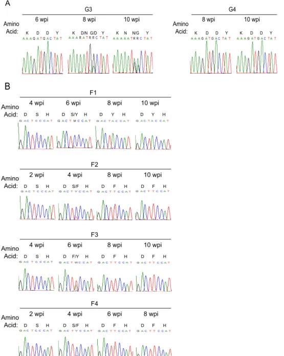
Enforced mutation of defective
vif
.
The mechanism of viral
escape from the antiviral activities of A3G (G3 and G4) or A3F (F1
to F4) was investigated by sequencing
vif
in viral RNA from blood
drawn at 10 weeks.
vif
genes from the terminal time point of the
course of G3 and G4 infection are aligned in
Fig. 3A
. Vif from G3
had the aspartate codon (GAT) for amino acid position 42
re-placed with the asparagine codon (AAT) (
Fig. 3A
). The original
aspartate codon (GAC) for amino acid position 43 was altered to
RRC. No other mutations were noted in G3 Vif. To resolve the
ambiguity at position 43, we cloned the PCR products from G3. In
10 clones, we found seven asparagine (AAC) and three glycine
(GGC) codons. Hence, the H42/43D mutation was replaced with
N42/N43 or N42/G43. Remarkably, there were no mutations in
vif
from G4.
In
Fig. 3B
,
vif
sequence from the viral RNA of the F1 mouse had
the serine codon at position 79 mutated to tyrosine (TCC to TAC)
and F2 to F4 had phenylalanine at position 79 (TCC to TTC). Two
other Vif mutations were found: R19K (AGA to AAA) in F3 and
E45V (GAA to GWA) in F4. R19K, like S79F, first appeared at
week 6 and was dominant at week 8 (not shown). E45V did not
appear in F4 viral RNA until after F79 had replaced S79 (not
shown). Unlike the conserved amino acid residues at positions 42
(H), 43 (H), and 79 (W), amino acid sites 19 and 45 are
polymor-phic (
46
).
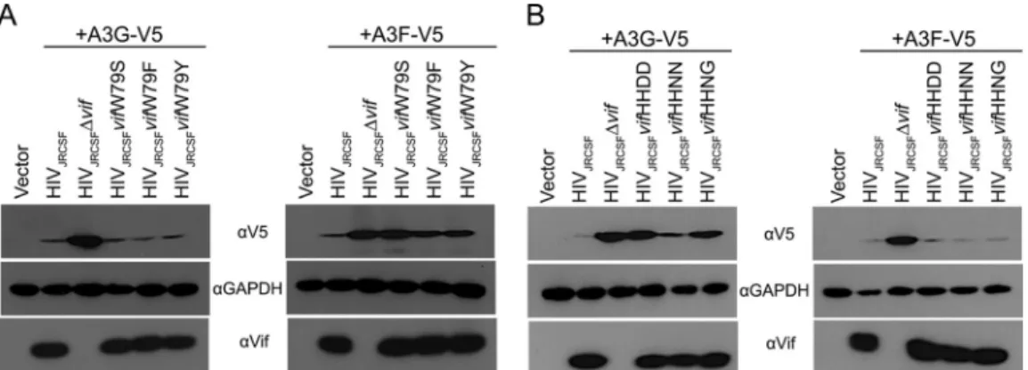
If W79F, W79Y, H42N/H43N, and H42N/H43G were
posi-tively selected, then these mutations should restore at least partial
A3F or A3G degradation activity to Vif. To test this hypothesis, we
constructed proviral clones with Vif mutated to Y79, F79, N42/
N43, and N42/G43 and performed A3G and A3F degradation
as-says in transfected 293T cells as shown in
Fig. 1B
. As demonstrated
in
Fig. 4A
, VifW79Y and VifW79F had A3F degradation activities
intermediate between those of wild-type Vif and the fully defective
VifW79S. Both VifW79Y and VifW79F effectively degraded A3G.
Contrary to a previous report on HIV-1
NL4 –3Vif (
28
), we found
that JRCSF Vif with the N42/N43 mutation (from G3) showed
106 105 104 103 102
5 10 15 20
HIV
JRCSF
HIV
JRCSFvifH42/43D
HIV
JRCSFvifW79S
HIVJRCSFvifH42/43DW79S
101
Days
p24 (pg/ml)
HIV
JRCSF
HIV
JRCSF
∆
vif
HIV
JRCSF
vif
H42/43D
HIV
JRCSF
vif
W79S
HIV
JRCSF
vif
H42/43DW79S
αV5
αGAPDH
αVif
+A3G-V5
HIV
JRCSF
HIV
JRCSF
∆
vif
+A3F-V5
B
HIV
JRCSF
vif
H42/43D
HIV
JRCSF
vif
W79S
HIV
JRCSF
vif
H42/43DW79S
A
HIV
JRCSF
HIV
JRCSF
∆
vif
HIV
JRCSF
vif
H42/43D
HIV
JRCSF
vif
W79S
HIV
JRCSF
vif
H42/43DW79S
+A3D-HA
C
αHA
αGAPDH
αVif
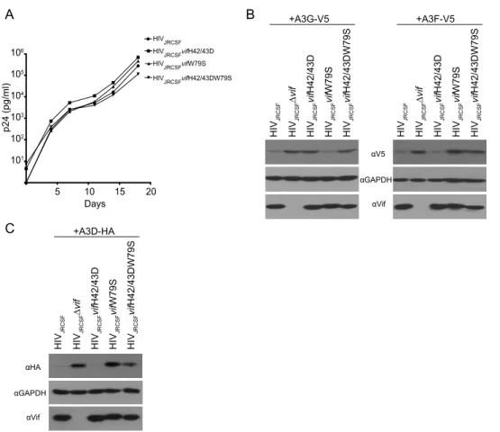
FIG 1Mutations in Vif that disrupt degradation of APOBEC3G or APOBEC3F do not affect virus replicationin vitro. (A) Viruses were produced by transfection
of 293T cell with proviral clones. CCR5-expressing CEM-SS cells were infected with wild-type JRCSF or JRCSF with point mutations in Vif that disrupt degradation of either APOBEC3G, APOBEC3F, or both. The culture supernatant was monitored longitudinally by ELISA for HIV p24gagprotein as a measure of
nearly wild-type A3G degradation activity, while VifN42/G43 had
an intermediate level of activity (
Fig. 4B
). The results of these
in
vitro
assays support the conclusion that JRCSFs with mutated Vifs
overcame initial delays in replication by acquiring partial or
near-wild-type APOBEC3 degradation activities. However, mouse G4
was an exception to these conclusions. Viral loads for G4 were low
for 6 weeks but subsequently increased and nearly matched
wild-type viral loads by 10 weeks (
Fig. 2A
, top). Strikingly, there was no
evidence of mutations in
vif
from G4 (
Fig. 3A
).
Enforced mutation and viral load are not temporally
corre-lated.
To more rigorously test the hypothesis that the ability of
JRCSFvifH42/42D to reach wild-type viral loads in G3 at 10 weeks
was dependent on the N42/N43 and N42/G43 mutations, we
se-quenced
vif
genes from plasma virions at earlier time points. If the
mutations to N42/N43 and N42/G43 accounted for high levels of
replication, then the changes in
vif
sequences should correspond
temporally with increased viral loads. Instead, we found that
de-spite a viral load of over 10
5copies of viral RNA per ml of blood at
4 and 6 weeks, plasma viral RNA still had the D42/D43 double
mutation (
Fig. 2A
and
5A
). By 8 weeks with a viral load at
wild-type levels, the original H42/43D mutation had begun to
disap-pear. G at the first position in codon 42 was almost completely
replaced by A, giving an asparagine codon. G was still the
domi-nant nucleotide at codon position 1 for amino acid 43, but in the
second codon position, G had largely replaced A. These changes
indicated that the D43 codon had mostly mutated to GGC, or
glycine. However, VifN42/G43 was not particularly effective in
degrading A3G
in vitro
(
Fig. 4B
). Finally, at 10 weeks, N42/N43
(7/10) was in the majority at the original D42/D43 site and N42/
G43 (3/10) was in the minority (
Fig. 5A
). Therefore, closer
anal-ysis indicated that the appearance of
in vivo
-generated mutations
at H42/43D lags behind the ability of the virus to replicate. Hence,
the hypothesis that active replication is strictly driven by
restora-tion of A3G degradarestora-tion activity is not supported. Our
observa-tions raise the possibility that Vif is sometimes able to maintain
viral viability despite loss of the A3G degradation function.
Mu-107
103 104 105 106
0 2 4 6 8 10
WT G1 G2 G3 G4 V
iral Load (Copies/ml)
Weeks WT F1 F2 F3 F4 107 103 104 105 106
0 2 4 6 8 10 Weeks 107 103 104 105 106
0 2 4 6 8
Weeks WT FG1 FG2 FG3 FG4 Mouse FG1 FG2 FG3 FG4 Spleen
-Bone Marrow -Lymph Nodes -Thymus -Liver -Lung-WT1
-
+
+
+
+
+
Presence of viral DNA in tissuesMouse F1 F2 F3 F4 Spleen
-Bone Marrow LymphNodes Thymus Liver Lung
WT1
-
+
+
+
+
+
Presence of viral DNA in tissues+
+
+
+
+
+
+
+
+
+
+
+
+
+
+
+
+
+
+
+
+
+
+
Mouse G2 G3 G4 Spleen Bone Marrow LymphNodes Thymus Liver Lung
WT1
-
+
+
+
+
+
Presence of viral DNA in tissues+
+
+
+
+
+
+
+
+
+
+
+
--
-
-
-A
B
C
60
40
20
Percent of
Sites Mutated
WT1 H42/43D W79S
GG GA GY
D
FIG 2Absolute restriction of HIVin vivorequires both APOBEC3G and APOBEC3F. (A) (Top) The plasma of BLT humanized mice infected with JRCSFvifH42/
tations restoring A3G degradation activity may facilitate the virus
reaching wild-type levels of replication.
Similarly, for F1 to F4, the dominant or exclusive codon at 4
weeks for amino acid position 79 is TCC, for serine (
Fig. 5B
). At
this time point, the four viral loads are higher than 10
5copies of
viral RNA per ml of blood (
Fig. 2B
). Not until 6 to 8 weeks had
codon 79 mutated to TAC (tyrosine) or TTC (phenylalanine)
when viral loads were at wild-type levels. Therefore, it is evident
that the mutations restoring partial A3F degradation activities to
Vif do not precede but in fact lag behind active viral replication.
Relevance of APOBEC3H.
One potentially confounding
vari-able for the extinction of viral infection in G1, G2, and FG1 to FG4
mice was the presence or absence of an active A3H allele (
Fig. 2A
and
C
). A3H is highly polymorphic, with stable/active alleles and
unstable/inactive alleles (
20
,
45
,
47–49
). Primary CD4
⫹T cells
homozygous and heterozygous for active/stable alleles of A3H are
strikingly resistant to infection by HIV-1 expressing a Vif that is
specifically defective for A3H degradation (
20
,
45
). It is important
to note that A3H is not countered by JRCSF Vif, since the Vif has
amino acids I39 and N48 (
Table 2
). Vifs with F39 and H48 are
FIG 3In vivoenforced mutation ofvifin JRCSFvifH42/43D and JRCSFvifW79S. (A) HIVvifsequences amplified from plasma virions at terminal time points
highly active for A3H degradation, but mutation of one of these
residues makes Vif inactive for A3H degradation while
maintain-ing A3G and A3F degradation activity (
47
,
50
). Specifically, B-1
and B-3 Vifs (I39 and H48) are inactive for A3H degradation, as is
NL4-3 Vif (F39 and N48), strongly suggesting that JRCSF Vif (I39
and N48) is also nonfunctional for A3H degradation (
20
,
47
,
50
).
JRCSF Vif is fully active for A3G, A3F, and A3D degradation
de-spite the presence of I39 and N48 (
Fig. 1B
and
C
). Therefore, if
A3H can restrict HIV-1 replication
in vivo
, then the presence of an
active A3H allele in combination with an active A3G could
ac-count for the absence of JRCSF replication in G1, G2, and FG1 to
FG4 (
Fig. 2A
and
C
) (
2
,
20
,
45
). To determine if A3H was
neces-sary for JRCSF extinction, we sequenced A3H mRNA from
hu-man tissue in the cohorts of mice that were used in the
experi-ments shown in
Fig. 2
. There were seven cohorts, each with a
common human tissue used for reconstitution of the mice.
Table
3
groups the mice into cohorts and documents the A3H
haplo-types (
20
). We found haplotypes I, III, IV, and V. Of these
haplo-types, only haplotype V encodes an active A3H.
The JRCSFvifH42/43D-inoculated mouse G1 was
homozy-gous for defective A3H alleles, but nonetheless, infection was
aborted (
Fig. 2A
). Given that JRCSFvifH42/43D Vif effectively
degrades A3D and A3F, the extinction of viral replication in G1
appeared to be the result of A3G activity. G3 and G4 were
heterozygous for the active A3H haplotype V. G3 and G4 had
delayed but ultimately full blown systemic infections despite
the presence of an active A3H gene. Therefore, extinction of
JRCSFvifH42/43D can occur in the absence of active A3H (G1),
and JRCSFvifH42/43D replication can occur in the presence of
an active A3H gene (G3 and G4). These results rule out a
con-sistently important role for A3H in viral extinction, though
possible effects of A3H on viral replication could be present but
not discernible because of the greater impact of A3G.
The JRCSFvifH42/43DW79S-inoculated mice also support the
conclusion that A3H is minimally involved in viral extinction.
FG1 to FG3 mice have no A3H activity (
Table 3
). Therefore, A3H
activity is not required for consistent viral extinction, since FG1 to
FG3 mice exhibit no viral replication. JRCSFvifW79S-infected
mice (F1 to F4) were all heterozygous for the active V allele of
A3H, and only F1 had delayed HIV-1 replication (
Fig. 2B
).
There-fore, it is not possible to draw a firm conclusion regarding the role
of A3F or the active alleles of A3H other than that the combination
of A3F and A3H activities is much weaker than the combination of
A3F and A3G activities.
Negative effects of A3F and A3H on JRCSF replication.
Figure
2
provides evidence that A3F can enhance the restriction of HIV-1
by A3G to consistently block viral replication. A role for A3H is
less clear. In this regard, results with primary CD4
⫹T cells suggest
that diminished HIV-1 replication should be observable with
JRCSF replication in cohorts that express active A3H alleles (
20
,
45
). However, multiple cohorts of mice have been infected with
JRCSF by three different groups, and no evidence for individual
cohorts that are highly resistant to infection has been noted (
17
,
18
,
36
). While this evidence suggests only weak anti-HIV-1 effects
for A3H, the population frequencies of A3H active alleles can be
low and are highly dependent on geography (
51
,
52
). It is possible
that none of the cohorts from these three reports expressed an
active allele of A3H. Therefore, we compared JRCSF replication in
five cohorts of mice with one of the cohorts heterozygous for
active A3H. In
Fig. 6
, WT1 is a BLT mouse with A3H haplotypes I
and V, and no difference in the viral load time course was observed
relative to four other cohorts. The dramatic restriction of HIV-1
by active A3H alleles seen in primary CD4
⫹T cells by Refsland et
al. (
20
) and Ooms et al. (
45
) may reflect the
ex vivo
activation of
primary T cells, which are inactive in BLT mice (
39
,
53
).
To further investigate possible inhibitory effects of A3F and
A3H on HIV-1 replication, we inoculated BLT mice with a
10-fold-lower initial dose of JRCSF (9,000 TCIU) to possibly enhance
small effects, in contrast to the 90,000 TCIU employed to
deter-mine the ability of APOBEC3s to extinguish replication. At the
lower dose, JRCSFvifH42/43D failed to replicate in three of four
mice, and replication was greatly delayed in the fourth mouse (
Fig.
7
). Of note, two mice (G5 and G8) in which replication was not
detected had two inactive A3H alleles (
Table 3
). G6 and G7 were
heterozygous for the active A3H allele V (
Table 3
). No viral
repli-cation was observed for G6, but delayed replirepli-cation was observed
with G7 (
Fig. 7
). Again, the combination of A3G and A3H did not
uniformly restrict JRCSF.
FIG 4Mutations that arosein vivopartially restored Vif function. (A) Proviral DNAs with the designated Vif mutations at codon 79 and an expression vector
Two of the three JRCSFvifW79S mice infected with 9,000
TCIU (F6 and F7) were clearly delayed relative to JRCSF, with
replication in mouse F7 the most retarded. Interestingly, mice F5
and F6 were defective for A3H while F7 was heterozygous for A3H
allele V (
Table 3
). These results confirm the conclusion that A3G is
the major restrictor of HIV-1 infection, with A3F capable of
de-laying but not fully restricting JRCSF. Our results also suggest that
an effect of A3H is difficult to detect in the presence of active A3G
but may be seen when A3H and A3F are acting in concert (F7).
A3G and A3H hypermutation in viral DNA.
Mouse G2 was
inoculated with JRCSFvif42/43D and had no detectable HIV-1
RNA in blood, and viral DNA was amplified only from lymph
nodes (
Fig. 2A
, bottom). G2 was from cohort 5 and was
heterozy-gous for the active haplotype V allele of A3H (
Table 3
). The reverse
transcriptase coding sequence (1,320 bp) was determined and
ex-hibited massive G-to-A mutation (
Fig. 8A
). The sequence of the
PCR product lacked ambiguities and may have been amplified
from a discrete template. Based on results from G3 and G4, we
expected extensive GG-to-AG mutation but only minor
GA-to-AA mutation (
Fig. 2D
). On the other hand, if A3H was
signif-K D D YAmino Acid:
6 wpi 8 wpi
K D/N G/D Y
10 wpi
K N N/G Y G3
K D D Y Amino
Acid:
8 wpi 10 wpi
G4
K D D Y
Amino Acid:
4 wpi 6 wpi
S/Y
10 wpi F1
8 wpi
D S H D H D Y H D Y H
2 wpi 4 wpi
S/F
10 wpi F2
8 wpi
D S H D H D F H D F H
Amino Acid:
4 wpi 6 wpi
F/Y
10 wpi F3
8 wpi
D S H D H D F H D F H
Amino Acid:
2 wpi 4 wpi
S/F
8 wpi F4
6 wpi
D S H D H D F H D F H
Amino Acid:
A
B
FIG 5Evolution of the APOBEC3 disrupting mutations in thevifgenein vivo. (A) Sequences of codons 41 to 44 ofvifamplified from the plasma RNA in two
icantly mutating JRCSFvifH42/43D, then a definite GA-to-AA
signature should be observable (
2
). In
Fig. 8B
,
a
relatively high
level of GA-to-AA mutation was observed, accounting for 19% of
the 78 total observed G-to-A mutations (61 at GG, 15 at GA, and
2 at GT). A3H is likely responsible for these GA-to-AA mutations,
because JRCSFvifH42/43D effectively degrades A3F and A3D
(
Fig. 1B
and
C
). It is possible that G2 is an example of HIV-1
hypermutation that was known prior to the discovery of the
anti-viral role of A3G (
1
,
54
).
It should be noted that the massive mutation of DNA
pre-sented in
Fig. 8B
represents a nearly total mutation of the DNA by
A3G. There are 32 guanosine doublets in the RT coding sequence
(excluding GGG and GGGG). Twenty-four are in the context of
GGA, and all 24 were mutated to AGA. Two are in the context
of GGT, and both were mutated to AGT. Six were in the context of
GGC, and none of them has a G-to-A change. The triplet GGG
occurs 14 times (excluding GGGG). Eight were in the context of
GGGA, and five of these were mutated to AAGA, two were
mu-TABLE 2Amino acids 38 through 49 of Vifs with or without A3H
degradation activitya
Vifb Amino acids 38–49c A3H degradation
JRCSF No
LAI Yes
NL4-3 No
Vif B-1 No
Vif B-3 No
Hypo-Vif No
a
All Vifs degrade A3G and A3F.
bLAI, HIV-1
LAI; NL4-3, HIV-1NL4-3. Vif B-1 and B-3 are subtype B Vifs. c
The critical residues for activity are phenylalanine 39 and histidine 48. Both a phenylalanine at position 39 and a histidine at position 48 are required for A3H degradation activity (LAI). Isoleucine at position 39 with histidine at position 48 is inactive (B-1 and B-3), and phenylalanine at position 39 and asparagine at position 48 is inactive (NL4-3). JRCSF has isoleucine at position 39 and asparagine at position 48. B-1 and B-3 are from Binka et al. (47), and Hypo-Vif is from Refsland et al. (20). Conserved residues are italic and underlined; mutated residues are boldface and underlined.
TABLE 3Cohorts and A3H haplotypesa
Cohort Mice
Amino acidbencoded at
position:
Haplotype Activity 15 18 105 121 178
1 G3, G4, F1 N R G K E I No
N R R D E V Yes
2 F2, F3, F4 ⌬ L R D E IV No
N R R D E V Yes
3 F5, F6, G8, FG2, FG3
N R G K E I No
⌬ R R D E III No
4 G1 N R G K E I No
N R G K E I No
5 G2 N R R D E V Yes
⌬ L R D E IV No
6 G5, FG1 ⌬ R R D E III No
⌬ L R D E IV No
7 F7, G6, G7, FG4
N R R D E V Yes
N R G K E I No
aRNA encoding APOBEC3H from BLT mouse peripheral blood mononuclear cells
(PBMCs) was amplified and sequenced. The amino acids encoded at positions 15, 18, 105, 121, and 178 were determined. The seven haplotypes for A3H are as follows: I, NRGKE; II, NRRDD; III,⌬RRDE; IV,⌬LRDE; V, NRRDE; VI,⌬LGKE; and VII, NRRKD. Active forms of A3H have asparagine at position 15 and arginine at position 105. Deletion of the codon for N15 or glycine at position 105 renders the protein unstable and hence inactive. Only four (I, III, IV, and V) of the seven possible haplotypes were present in the seven cohorts. Haplotype V represented the only active A3G allele. Functional A3H was present only in the heterozygous condition. Cohort 7 had two codons for T43, ACG and ACA.
b⌬, 3-base deletion of the N15 codon.
107
103
104
105
106
0 2 4 6 8 10
V
iral Load (Copies/ml)
Weeks
WT1
WT4 WT3 WT2
WT5
WT7 WT6
FIG 6Robust replication of JRCSF in a mouse cohort heterozygous for A3H
haplotype V. Individual plots of the wild-type JRCSF viral load versus time are presented. The seven mice are fromFig. 2(WT), where JRCSF replication is presented in aggregate. WT1 has A3H haplotypes I and V, but viral replication is not diminished. WT2 to WT7 are derived from four separate cohorts.
108
103
104
105
106
0 2 4 6
V
iral Load (Copies/ml)
G5
G8 G7 G6
F5
F7 F6
107
WT8 WT9
Weeks
FIG 7JRCSF, JRCSFvifW79S, and JRCSFvifH42/43D viral load time courses
tated to AAAA (likely representing combined actions of A3G and
A3H), and one was mutated to GAGA. Three sites were GGGT,
and all three were mutated to AAGT. Interestingly, three sites
were GGGC, and all three were mutated to AGGC. There were
five quadruplets mutated to AAAGA, GAAGA, GAAGA,
GAAGT, and AAGGC. These results suggest that
in vivo
a 5
=
G
mutation to a CC doublet in minus-strand DNA (GCC) blocks
deamination (
4
). Taking this inhibitory effect into account,
there were 61 G-to-A mutations out of 65 feasible GG targets
(94%). A3H activity was much less exhaustive than A3G
activ-ity. There were 102 GA sites in the reverse transcriptase coding
sequence, and 15 were mutated (15%).
The sanctity of viral RNA sequence.
The massive potential of
A3G to mutate the minus strand of HIV-1 emphasizes the
impor-tance of Vif to the virus. We accordingly asked if any of this
mas-sive mutational potential resulted in fixed sequence changes in
replicating viruses other than in
vif
. We determined the sequences
of
gag
and the coding sequences for reverse transcriptase and
RNase H from the terminal bleed of virion RNA of G3, G4, and F1
to F4. There were no fixed mutations or even clear cases of
poly-morphism in the viral genomic RNA sequence of
gag
or the reverse
transcriptase coding sequence (not shown). In the case of RNase H
sequences, there were no mutations evident for G3 or F1 to F4, but
G4 had a nonsynonymous point mutation changing valine 27 to
isoleucine (
Fig. 9
). G3 and F1 to F4 had enforced mutations in
vif
(
Fig. 3
), but G4 did not, raising the possibility that RNase H may
FIG 8HIV DNA amplified from an extinguished infection is hypermutated. (A) Highlighter sequence analysis of the reverse transcriptase region of HIVpol. HIV
DNA amplified from the lymph nodes of mouse G2 was G-to-A hypermutated (green lines). HIV-1JRCSFnucleotide numbers are indicated at the bottom. (B)
be a site of extra Vif mutations with impacts on the APOBEC3 axis
(
15
,
55
).
DISCUSSION
A3G is clearly the dominant APOBEC3 in countering HIV-1
in-fection (
2
,
17
,
22
). However, unlike
vif
⫺JRCSF (JRCSF
⌬
vif),
JRCSFvifH42/43D infection is not always extinguished in BLT
humanized mice. This result demonstrates that A3G activity is not
sufficient to consistently extinguish HIV-1 infection. An
impor-tant role in extinguishing HIV-1 is likely played by A3F, since
JRCSFvifH42/43DW79S was not able to replicate.
In vivo
involvement of A3F and A3D.
Considerable
in vitro
evidence exists for an anti-HIV-1 role for A3F (
2
,
22
). The
in vivo
role of this APOBEC3 is less clear. Sato et al. (
22
) infected NSG-hu
mice with chimeric NL4-3 virus, converted to R5 tropism by
re-placing
env
V3 of NL4-3 with JRCSF V3 to give NLCSFV3 (
56
).
NLCSFV3-4A has Vif amino acids 14 to 17 (DRMR) all mutated to
alanine and is defective in A3F degradation (
57
). The virus
repli-cated slowly, and viral loads at 6 weeks were reduced by 80%
relative to those of the wild type, NLCSFV3. While these findings
strongly suggest a significant impact on HIV-1 replication by A3F,
the interpretation of the result is not straightforward because the
tetra-alanine mutation may impact other Vif activities in addition
to A3F degradation (
58
).
A complication regarding A3F’s antiviral effect is the ability of
NLCSFV3-4A to diversify its genomic RNA. G-to-A mutations
were found by Sato et al. in 91 single-genome amplicons of
env
at
the level of 1.6% of total G sites (
22
). Wild-type NLCSFV3 and
A3G degradation-defective NLCSFV3-5A (amino acids 40 to 44,
YRHHY to AAAAA) had only 0.21% and 0.26% of G sites mutated
to A. The inability of NLCSFV3-5A to diversify viral sequence was
attributed to the high propensity of A3G to introduce lethal
mu-tations, thereby preventing mutated virus from replicating. If
greater viral diversity is beneficial to the virus, then A3F may have
to be considered a proviral factor, not an antiviral factor,
particu-larly given its weak effect on viral replication relative to A3G. We
consider this scenario unlikely, since HIV-1 could readily mutate
reverse transcriptase to have a higher error rate if a greater level of
viral diversity was beneficial (
59
).
Our mutational approach to A3F degradation was different
from that of Sato et al. (
22
) in that we mutated only a single
residue. This meant that unlike NLCSFV3-4A, JRCSFvifW79S
could be selected against by enforced mutation. Indeed,
JRCSFvifW79S reverted to the aromatic residue phenylalanine or
tyrosine, which partially restored A3F degradation function. The
partial reversion to wild-type activity straightforwardly defines
A3F as an antiviral factor, as virus with A3F degradation activity
has greater fitness and outgrows JRCSFvifW79S. We also found
support for A3F as an important antiviral factor in the
obser-vation that JRCSFvifH42/43DW79S was not able to infect BLT
mice. Extinction of JRCSFvifH42/43D infection (G1, G2, G5,
G6, and G8, but not G3, G4, and G7) may be stochastic. Under
our experimental infection conditions, viral survival is
possi-ble, but not certain. A negative impact on viral replication by
A3F would limit the opportunity the virus has to mutate H42/
43D. In this scenario, extinction of the virus occurs soon after
inoculation or not at all.
Wild-type JRCSF Vif and Vif mutated to H42/43D degraded
not only A3F, but A3D as well. This rules out a role for A3D in
extinction of JRCSFvifH42/43D in mice G1, G2, G5, G6, and G8.
In the case of JRCSFvifH42/43DW79S, A3D would not have been
degraded, and there is the possibility that it contributed, along with
A3F, to viral extinction in FG1 to FG4. Arguing against such a role are
the findings that in humanized mice, A3D is expressed at lower levels
than A3F, and
in vitro
studies demonstrating weaker inhibition of
HIV-1 by A3D than A3F (
2
,
20
,
22
,
45
,
60
). For these reasons, any
antiviral impact of A3D would be swamped by that of A3F.
In vivo
involvement of A3H.
We also considered the
possibil-ity that A3H could contribute to viral extinction in the cases where
inoculated JRCSFvifH42/43D failed to replicate. It is important to
note that, unlike A3G and A3F, A3H would not be degraded by
JRCSF Vif because of amino acids I39 and N48 (
20
,
47
,
50
). This
situation could result in a combined effect of A3G and A3H,
lead-ing to viral destruction in JRCSFvifH42/43D and JRCSFvifH42/
FIG 9Mutations in HIV RNase H are present only in virus from mouse G4. HIV-1 RNase H sequence amplified from the plasma of viremic humanized mice
43DW79S inoculations. A3H haplotypes were determined in
seven mouse cohorts, and of the four haplotypes present (I, III, IV,
and V) (
Table 3
), only A3H V produces a stable protein. Mice
G5 and G8 were inoculated with 9,000 TCIU of JRCSFvifH42/43D
and displayed no viral replication (
Fig. 7
). In neither case was an
active A3H allele present. The mice inoculated with 90,000 TCIU
of JRCSFvifH42/43D that did not exhibit viral replication, G1 and
G2, had haplotypes I/I and IV/V, respectively. Therefore, viral
replication in G1 was robustly suppressed without a contribution
of A3H (or A3F) activity, and there was not a consistent
relation-ship between the presence of A3H activity and suppression of viral
replication (
Fig. 2A
). Similarly, A3H is not the necessary factor
that causes JRCSFvifH42/43DW79S to consistently fail to
repli-cate. FG1 to FG3 were all homozygous for inactive A3H alleles, yet
no viral replication was observed (
Table 3
and
Fig. 2C
). Additional
evidence for the suggestion that A3H has a minimal impact on
JRCSF restriction is the fact that wild-type JRCSF replication is not
reduced in BLT mice heterozygous for A3H haplotype V (
Fig. 6
).
Hence, A3G is clearly the dominant restrictor of HIV-1, with A3F
likely playing a critical role. We did find evidence of a small
inhib-itory effect of A3H on JRCSFvifW79S. A humanized mouse (F7)
with a low-dose inoculation that was heterozygous for an active
allele of A3H displayed a greater delay in viral replication (
Fig. 7
)
than two JRCSFvifW79S mice that were homozygous for inactive
A3H alleles (
Fig. 7
, F5 and F6).
A role for A3H in hypermutation.
Evidence for GA-to-AA
mutations indicative of A3F and/or A3H cytosine deaminase
ac-tivity was minimal in WT1, G3, G4, and F1 to F4 viral DNA (
Fig.
2D
), but one JRCSFvifH42/43D infection (G2) had 15% of the GA
sites mutated to AA in the reverse transcriptase coding sequence
from lymph node DNA. JRCSFvifH42/43D has A3F degradation
activity, which strongly implicates A3H as the mutagenic factor.
GG-to-AG mutations were also numerous, to the remarkable
ex-tent of 94% of poex-tential sites. This extreme level of G-to-A
muta-tions suggests an abnormal level of cytosine deaminase activity,
which may be related to the hypermutation of viral DNA found in
HIV-1-positive individuals (
1
,
5
).
In vivo
evidence for Vif activity not involving degradation of
A3F or A3G.
A3G mutation of GG to AG is effective in inactivating
virus prior to integration, and A3F’s GA-to-AA mutations are
fewer and relatively ineffective (
61
). Thus, it seems unlikely that
GA-to-AA mutation plays a decisive role in the equivalent
suscep-tibilities of JRCSFvif42/43DW79S and JRCSF
⌬
vif to extinction.
The notion that extinction occurs early after inoculation or not at
all is supported by the fact that plasma viral RNA was not detected
in G1, G2, and FG1 to FG4 despite an inoculum of 90,000 TCIU.
When virus was able to overcome mutations in
vif
(G3, G4, and F1
to F4), viral loads ultimately reached 10
6copies of viral RNA. It is
also important to note that appearance of
vif
mutations
in vivo
that partially restored A3G or A3F degradation lagged behind high
levels of virus or, in the case of G4, failed to appear at all.
Non-cytosine deaminase Vif activities not inhibited by the H42/43D or
W79S mutations may account for these observations. There are
multiple anti-APOBEC3 activities other than APOBEC3
degrada-tion, including A3G and A3F inhibition of reverse transcripdegrada-tion,
transcription elongation, and proviral integration and
inactiva-tion of the transactivainactiva-tion response element (
12–16
). The greater
potency of A3F than A3G in preventing viral integration is an
attractive possible mechanism for the antiviral activity of A3F
(
15
). Early forms of Vif may have exhibited the ability to directly
inhibit encapsidated APOBEC3s prior to acquiring the function of
targeting APOBEC3s to proteasomes.
ACKNOWLEDGMENTS
This work was supported in part by funds from the National Institute of Allergy and Infectious Diseases (grants AI033331, AI096937, and AI96138 [J.V.G.]) and by the UNC Center for AIDS Research (grant P30 AI50410). We thank Nathaniel Schramm for insightful comments and sugges-tions, as well as current and past members of the Garcia laboratory and veterinary technicians at the UNC Division of Laboratory Animal Medi-cine for their assistance with different aspects of this research.
FUNDING INFORMATION
This work, including the efforts of J. Victor Garcia, was funded by HHS | NIH | National Institute of Allergy and Infectious Diseases (NIAID) (AI33331). This work, including the efforts of J. Victor Garcia, was funded by HHS | NIH | National Institute of Allergy and Infectious Diseases (NIAID) (AI096937). This work, including the efforts of J. Victor Garcia, was funded by HHS | NIH | National Institute of Allergy and Infectious Diseases (NIAID) (AI96138). This work was funded by HHS | NIH | National Institute of Allergy and Infectious Diseases (NIAID) (P30 AI50410).
REFERENCES
1.Albin JS, Harris RS. 2010. Interactions of host APOBEC3 restriction
factors with HIV-1 in vivo: implications for therapeutics. Expert Rev Mol
Med12:e4.http://dx.doi.org/10.1017/S1462399409001343.
2.Hultquist JF, Lengyel JA, Refsland EW, LaRue RS, Lackey L, Brown
WL, Harris RS. 2011. Human and rhesus APOBEC3D, APOBEC3F,
APOBEC3G, and APOBEC3H demonstrate a conserved capacity to re-strict Vif-deficient HIV-1. J Virol85:11220 –11234.http://dx.doi.org/10 .1128/JVI.05238-11.
3.Sheehy AM, Erthal J.2012. APOBEC3 versus retroviruses, immunity
versus invasion: clash of the titans. Mol Biol Int2012:974924.http://dx .doi.org/10.1155/2012/974924.
4.Harris RS, Bishop KN, Sheehy AM, Craig HM, Petersen-Mahrt SK,
Watt IN, Neuberger MS, Malim MH.2003. DNA deamination mediates
innate immunity to retroviral infection. Cell113:803– 809.http://dx.doi .org/10.1016/S0092-8674(03)00423-9.
5. Liddament MT, Brown WL, Schumacher AJ, Harris RS. 2004.
APOBEC3F properties and hypermutation preferences indicate activity against HIV-1 in vivo. Curr Biol14:1385–1391.http://dx.doi.org/10.1016 /j.cub.2004.06.050.
6.Wiegand HL, Doehle BP, Bogerd HP, Cullen BR.2004. A second human
antiretroviral factor, APOBEC3F, is suppressed by the HIV-1 and HIV-2 Vif proteins. EMBO J23:2451–2458.http://dx.doi.org/10.1038/sj.emboj .7600246.
7.Kobayashi M, Takaori-Kondo A, Miyauchi Y, Iwai K, Uchiyama T.
2005. Ubiquitination of APOBEC3G by an HIV-1 Vif-Cullin5-Elongin B-Elongin C complex is essential for Vif function. J Biol Chem280:18573– 18578.http://dx.doi.org/10.1074/jbc.C500082200.
8.Xiao Z, Ehrlich E, Yu Y, Luo K, Wang T, Tian C, Yu XF.2006. Assembly
of HIV-1 Vif-Cul5 E3 ubiquitin ligase through a novel zinc-binding do-main-stabilized hydrophobic interface in Vif. Virology349:290 –299.http: //dx.doi.org/10.1016/j.virol.2006.02.002.
9.Yu Y, Xiao Z, Ehrlich ES, Yu X, Yu XF.2004. Selective assembly of
HIV-1 Vif-Cul5-ElonginB-ElonginC E3 ubiquitin ligase complex through a novel SOCS box and upstream cysteines. Genes Dev18:2867–2872.http: //dx.doi.org/10.1101/gad.1250204.
10. Britan-Rosich E, Nowarski R, Kotler M.2011. Multifaceted
counter-APOBEC3G mechanisms employed by HIV-1 Vif. J Mol Biol410:1065– 1076.http://dx.doi.org/10.1016/j.jmb.2011.03.058.
11. Feng Y, Baig TT, Love RP, Chelico L.2014. Suppression of
APOBEC3-mediated restriction of HIV-1 by Vif. Front Microbiol5:450.http://dx.doi .org/10.3389/fmicb.2014.00450.
12. Bishop KN, Holmes RK, Malim MH.2006. Antiviral potency of APOBEC
proteins does not correlate with cytidine deamination. J Virol80:8450 – 8458. http://dx.doi.org/10.1128/JVI.00839-06.
13. Gillick K, Pollpeter D, Phalora P, Kim EY, Wolinsky SM, Malim MH.