ORAL MICROBIOME CHANGES ASSOCIATED WITH FIXED DENTAL PROSTHODONTIC RESTORATION
Sarah Kay Youny Lee
A thesis submitted to the faculty at the University of North Carolina at Chapel Hill in partial fulfillment of the requirements for the degree of Master in Science in the School of Dentistry
(Prosthodontics).
Chapel Hill 2018
© 2018
ABSTRACT
Sarah Kay Youny Lee: Oral microbiome changes associated with fixed dental prosthodontic restoration
(Under the direction of Kimon Divaris)
The oral microbiome is a relatively unexplored component of oral health and disease. Common oral diseases, including dental caries and periodontitis, are now best understood as dysbiotic shifts of the oral microbial ecology. Prosthodontics is a cornerstone of clinical dentistry, serving to promote oral health through dental rehabilitation. However, it remains unknown if and to what degree prosthodontic treatment confers changes in the oral microbiome.
In this observational clinical study, we studied the effects of fixed dental prosthodontic restorative treatment on the oral microbiome composition. Prosthodontic patients’ salivary samples were collected during their treatment course. Microbiome analyses relied upon whole genome sequencing (WGS) shotgun. Reads were aligned, mapped and analyzed to obtain
ACKNOWLEDGEMENTS
To the team members of the UNC School of Dentistry’s Graduate Prosthodontics staff, thank you for your support throughout my training.
To my co-residents and fellow, thank you for your friendship, patience, and facilitation of this study and for being an integral part of my residency experiences both professionally and personally.
To the faculty of Graduate Prosthodontics, thank you for sharing your wisdom and insight and for inspiring me to be a lifelong learner.
To Dr. Dan Bailey, thank you for your positivity and honesty in feedback and appreciation for learning.
To Dr. Kent Healey, thank you for being a wonderful role model and mentor, always so generous to share your time in teaching with respect, appreciation, and humility.
To my program director and mentor, Dr. Ryan Cook, thank you for believing in me and for your enthusiasm, guidance, and effort in helping me to strive to be the best that I can be and for your commitment to our program and residents.
To my previous program director and mentor, Dr. Lyndon Cooper, thank you for providing me the means of starting in this program and for pushing the parameters of my thinking and encouraging me to explore beyond.
To Hunyong Cho, this study’s biostatistician support, who provided support in processing, interpreting, and understanding the results and aiding in my learning process.
To my project mentor, Dr. Kimon Divaris, thank you for sharing your time in teaching me and for inspiring me with your passion and enthusiasm for science and investigation, as well as appreciation for running.
To my committee members: Dr. Ibrahim Duqum, thank you for being the impetus to this field of dentistry and for being a constant source of support. Dr. Ingeborg De Kok, thank you for your congeniality and friendship, for cheering me throughout. Dr. Di Wu, thank you for your wealth of knowledge and encouragement to learn.
To my family: Mom and dad, thank you for everything, I cannot overstate how much your unconditional love and support, as well as patience, have meant to me during the ups and downs of this residency and life. Konny, thank you for your encouragement and being the best big brother. The Grigoris clan, thank you for your good cheer and spirits from afar and always checking in on me.
To Foti, my best friend and partner, thank you for your love, encouragement, support, and for always bringing out the best in me. I love you.
To the Lord, Jesus Christ, for being the constant and steady force in my life and guiding me when all else seems unstable and obscure. Your Love is truly endless, everlasting, eternal, and awesome.
TABLE OF CONTENTS
LIST OF TABLES………...x
LIST OF FIGURES………xi
LIST OF ABBREVIATIONS………...………xii
INTRODUCTION………...…………....1
1. Oral microbial communities and their organization………1
1.1. Defining the oral microbiome………...1
1.2. Biofilms and the oral microbiome………2
1.3. Saliva and the oral microbiome………3
1.4. Next generation sequencing (NGS)………..4
1.4.1. Whole genome sequencing (WGS)………5
shotgun - metagenomics 2. Oral health and disease………6
2.1. Previous models of oral disease and bacteria………...…6
2.1.1. Caries………...…..6
2.1.2. Periodontal disease……….…7
2.2. Current understanding of the oral microbiome……….8
in health and disease 2.2.1. Caries……….9
2.2.2. Periodontal disease………..10
2.3. Oral health and general health………11
3.1. Defining dental prosthetic reconstruction………...11
3.1.1. Fixed dental prosthesis (FDP)………..12
3.2. Indications for dental reconstruction………..12
3.3. Prosthodontic treatment impact on general health………..13
3.4. Biological considerations for tooth-borne FDP restorations………..13
4. Statement of purpose……….14
MATERIALS AND METHODS………..15
1. Study design………..15
2. Patient selection……….15
2.1 Inclusion criteria………..16
2.2. Exclusion criteria………16
2.3 Continuance criterion………..16
3. Data collection and analysis……….17
3.1 Electronic patient record (EPR) data………...17
3.2. Clinical procedures……….17
4. Saliva sample collection………17
5. Sample preparation, nucleic acid extraction, WGS shotgun……….18
6. Bioinformatics pipeline and statistical analyses………18
RESULTS………..19
1. General data………...19
2. Salivary metagenome analysis: Species-level………...21
2.1 Number of genera/species………22
2.3. Alpha diversity and beta diversity………..25
2.4. Longitudinal analysis………..27
3. Salivary metagenomics analysis: Variables………...30
3.1. Diabetes analysis……….30
3.2. Periodontal disease history analysis………34
DISCUSSION………39
CONCLUSION………..45
LIST OF TABLES
LIST OF FIGURES
Figure 1. The average abundance of gene (in RPK) and pathways ………..20
per sample at baseline Figure 2. Gene abundance distribution of each sample (in RPK)……….20
Figure 3. Pathway abundance distribution of each sample………21
Figure 4. Species in each group……….22
Figure 5. NMDS plot of identified species………24
Figure 6. tSNE scatter plot……….24
Figure 7. PCoA and Scree plots……….24
Figure 8. Shannon diversity index at the species-level as mean………25
and range in each group Figure 9. Linear model of Shannon diversity index at the species-level………...26
and age Figure 10. Simpson-Gini diversity index at the species-level as mean……….26
and range in each group Figure 11. Linear model of Simpson-Gini diversity index at the species-level……….26
and age Figure 12. Richness index at the species-level (50%/80%)………...27
Figure 13. Dominance index at the species-level according to caries status……….27
Figure 14. Sample dissimilarity as measured by beta diversity at the species-level……….27
Figure 15. Number of species between visits and subjects………29
Figure 16. Shannon entropy index at the species-level between visits and subjects……….29
Figure 17. Simpson-Gini diversity index at the species-level between visits ………..30
and subjects Figure 18. Dominance index at the species level between visits and subjects………..30
Figure 20. Species in each group (diabetes)………..31
Figure 21. Shannon diversity index at the species-level as mean and………...32
range in each group (diabetes) Figure 22. Simpson-Gini diversity index at the species-level as mean and ………..32
range in each group (diabetes) Figure 23. Richness index at species-level (50%/80%) (diabetes)………32
Figure 24. Dominance index at the species-level as mean and range………33
in each group (diabetes) Figure 25. Sample dissimilarity as measured by beta diversity at the ………..33
species-level (diabetes) Figure 26. NMDS plot of identified species (diabetes)……….33
Figure 27. tSNE scatter plot (diabetes)………..34
Figure 28. PCoA plot for each subject (diabetes)………..34
Figure 29. Species in each group (periodontal disease)……….35
Figure 30. Shannon diversity index at the species-level as mean and………...35
range in each group Figure 31. Simpson-Gini diversity index at the species-level as mean and ………...36
range in each group (periodontal disease) Figure 32. Richness index at the species-level (50%/80%) (periodontal disease)………36
Figure 33. Dominance index at the species-level as mean and……….36
range in each group (periodontal disease) Figure 34. Sample dissimilarity as measured by beta diversity at the………...37
species-level (periodontal disease) Figure 35. NMDS plot of identified species (periodontal disease)………37
Figure 36. tSNE scatter plot (periodontal disease)………37
LIST OF ABBREVIATIONS
DNA Deoxyribonucleic acid
EPR Electronic patient record FDP Fixed dental prosthesis
HOMINGS Human Oral Microbe Identification using Next Generation Sequencing)
MetPHlAn2 Metagenomic Phylogenic Analysis mRNA Messenger ribonucleic acid
MS Mutans streptococci
NA Nucleic acid
NGS Next generation sequencing
NMDS Non-metric Multidimensional Scaling
PC Principal Component
PCoA Principal Component Analysis RPK Reads per kilobase
STROBE Reporting of Observation Studies in Epidemiology rRNA Ribosomal ribonucleic acid
tSNE t-Distributed Stochastic Neighbor Embedding
INTRODUCTION 1. Oral microbial communities and their organization
The oral cavity is a dynamic environment built by communities of microbial cells
consisting of bacteria, fungi, archaea, viruses, and protozoa (Wade 2013; Zaura et al. 2014). It is one of several interrelated systems of the body, including the gut, skin, and rectum, that make up the overall human microbiome and metagenome(Group et al. 2009; Human Microbiome Project 2012b). The cells of these microbiomes account for 90% of cells of the human body (Savage 1977; Wilson 2008). To explore this crucial capacity, relatively recent and extensive
investigations have been done to better understand the oral microbiome organization of the oral cavity and its many parts in health and disease.
1.1. Defining the oral microbiome
The oral cavity is an ecosystem containing various habitats that consist of communities of commensal, symbiotic, and pathogenic microorganisms (Lederberg and McCray 2001). These communities are referred to as microbiomes (Ursell et al. 2012).
Seven major phyla of bacteria dominate the oral microbiome: Firmicutes, Proteobacteria Actinobacteria, Bacteroidetes, candidate division TM7, Spirochaetes, and Fusobacteria
significant variations are also observed with age and the stage of dentition, suggesting that the composition of the oral microbiome has a temporal component (Xu et al. 2015).
The oral microbiome consists of a core set and variable set of microorganisms that are in a flux of symbiosis and dysbiosis based on innate and acquired host factors such as genetics and oral hygiene habits. The core microbiome is a patterned diversity displayed in clinically healthy oral environments and is consistently shared between different individuals. On the other hand, the variable microbiome consists of microbial communities unique to the individual (Bik et al. 2010; Zarco et al. 2012; Zaura et al. 2014).
1.2. Biofilms and the oral microbiome
The oral bacteria are predominantly organized in biofilms within the oral cavity (Marsh 2006; Socransky and Haffajee 2005). The physical integrity of the biofilm is based upon an exopolymer matrix structure that encapsulates a specific compositional and spatial design of microorganisms (Jenkinson and Lamont 2005; Sbordone and Bortolaia 2003). The microbial composition develops by way of inter-species interactions as well as interactions with the host environment and its subsequent reaction to the inhabiting microorganisms that leads to a process of succession (Bick et al. ; Sbordone and Bortolaia 2003; Socransky and Haffajee 2005).
Quorum sensing, a bacterial mode of communication in which quorum sensing molecules are produced and detected by cells, has been suggested as one of the primary mechanisms by which a biofilm regulates the balance of healthy and pathogenic concentrations of microbial species (Wade 2010).
2014). These biotas are essentially biofilms that organize both spatially and temporally(Sbordone and Bortolaia 2003).
1.3. Saliva and the oral microbiome
Saliva is a biological fluid made of water, proteins, and inorganic and organic substances contained within the oral cavity. While the oral microbiome has specific ecological organization, the salivary microbiome has been shown to be representative of the overall oral microbiome due to its ability in collecting the shedding surfaces of these oral niches(Fabian et al. 2008;
Yamashita and Takeshita 2017). It can serve as a global marker of the oral microbiome because of its encompassing nature in which saliva directly contacts and carries components derived from other oral structures such as gingival crevicular fluid, cell debris, plaque, nasal and bronchial secretions, lining cells, blood, exogenous substances, and bacteria(Curtis et al. 2011; Fabian et al. 2008; Fox 1989; Kaczor-Urbanowicz et al. 2017; Kaufman and Lamster 2000; 2002; Lee 2009; Liu and Duan 2012; Mandel and Wotman 1976; Saliva: Its role in health and disease. Working group 10 of the commission on oral health, research and epidemiology (core) 1992). It contains mRNAs, proteins, and DNAs from local tissue as well as tissues from distant sites.
Saliva has been shown to exhibit long-term temporal stability (Belstrom et al. 2016b; Rasiah et al. 2005; Stahringer et al. 2012; Yamanaka et al. 2012; Zhou et al. 2013) and affected by the host and host’s systemic condition (Lira-Junior et al. 2018; Zaura et al. 2017), but its composition has been found to be altered based on external factors such as use of systemic antibiotics, as well as chemo- and radiotherapies (Lazarevic et al. 2013; Xu et al. 2013). However, the salivary bacterial profile variance has been found to be comparable, at around 13.5%, to variations found in distributions of genera in neutral genetic markers between individuals in human populations(Nasidze et al. 2009). This implies that saliva may be
applicable in a global sense in which geographic location may not necessarily impart a distinct composition.
As such, saliva can be used as a tool for assessment and diagnosis, as well as potential predictive indicator of oral microbial health status (Belstrøm 2016; Belstrom et al. 2016a; Kaczor-Urbanowicz et al. 2017; Kaufman and Lamster 2000; 2002; Lee 2009; Liu and Duan 2012).
1.4. Next generation sequencing (NGS)
sequencing and the Human Oral Microbe Identification Microarray (HOMIM) have been used by several studies and were shown to be valid and efficient means of studying the oral
microbiome(Ahn et al. 2011).The core data of the oral microbiome has been accumulated to form the Human Oral Microbiome Database, a collection of microbial reference genomes consisting of known species, unnamed isolates, and unnamed and uncultured phylotypes
recognized by the 16S rRNA sequence information (Ahn et al. 2011; Dewhirst et al. 2010). This database organizes the defined human oral microbial taxa in a standardized manner that is accessible for research and dissemination(Ahn et al. 2011). It is also part of a greater
conglomeration known as the Human Microbiome Project in which bacterial genome sequences are being collected in major ecological sites of the body – the nasal cavity, oral cavity,
gastrointestinal tract, and urogenital tract (Ahn et al. 2011; Dewhirst et al. 2010; Morgan et al. 2013).
1.4.1. Whole genome sequencing (WGS) shotgun - metagenomics
WGS, a type of NGS approach, sequences large quantities of DNA, ordering the nucleotides such that variations can be identified in any part of the genome (Bick et al. ;
Genomic research 2018; Human Microbiome Project 2012a; Ng and Kirkness 2010; Weinstock 2012). It taxonomically profiles a microbial community to the species, and more detailed, the strain level (Weinstock 2012). WGS also enables functional profiling of metagenomic and metatranscriptomic sequence data, including aggregated whole-community level pathway reconstruction (Abubucker et al. 2012; Fodor et al. 2012; Lozupone et al. 2006; Nyvad et al. 2013; Schloss et al. 2009; Segata et al. 2013; Weinstock 2012).
sequence (reads) are compared to an existing reference genome sequence such that a consensus sequence can be formed (Ng and Kirkness 2010). With de novo assembly, the total microbial DNA is directly sequenced through overlapping of comparable sequence reads that are overlapped to form long sequences (contigs) (Ng and Kirkness 2010; Nyvad et al. 2013;
Weinstock 2012). The process of generation, assembly, organization and analysis of WGS data is complex, computationally, time and resource intensive, but can be highly informative (Bick et al. ; Weinstock 2012).
By utilizing WGS, a more comprehensive compositional view of microbial communities can be generated.
2. Oral health and disease
The oral microbiome is a highly individualized, dynamic composition of commensal microbial species with development and interactions influenced from host genetic and environmental factors (Costalonga and Herzberg 2014; Gomez et al. 2017; Wade 2010).
2.1. Previous models of oral disease and bacteria
The mechanisms of oral diseases such as caries and periodontitis have historically been understood as infectious microbial processes, in which disease-specific bacteria induce a negative impact on susceptible oral structures. This pathogenicity-driven model related disease occurrence to certain microorganisms that had the ability to overcome and/or surpass host defenses.
2.1.1. Caries
et al. 1994). Theories that attempted to explain dental decay emerged and were organized into specific versus non-specific plaque hypotheses (Kleinberg 2002; Loesche 1986).
Caries-pathogen acquisition was hypothesized to occur within a “window of infectivity”, in which colonization of MS occurs at the time of primary teeth eruption(Caufield et al. 1993). Furthermore, previous research then linked the source of vertical transmission from mother, or other closest caregiver, to child, describing infants with MS also had mothers and caregivers carrying this pathologic bacteria(Berkowitz 2006; Berkowitz et al. 1975). Horizontal
transmission of MS, in which microorganisms are transferred between members of a group, had also been found to be plausible as children sharing a daycare setting and non-mother members in the immediate family have been found to share genotypes of MS(Mattos-Graner et al. 2001; van Loveren et al. 2000).
2.1.2. Periodontal disease
Periodontal diseases, ranging from gingivitis to aggressive periodontitis, were investigated based on species-association to a particular type of disease (Moore et al. 1983; Moore et al. 1982; Paul 1970; Slots 1979; Socransky Sigmund and Haffajee Anne 1994;
Socransky and Haffajee 1992; Tanner et al. 1979). Microbes distinctive to a diseased state were identified as keystone pathogens and included Porphyromonas gingivalis, Tannerella forsythia
(Socransky Sigmund and Haffajee Anne 1994; Socransky and Haffajee 2005). The complexes were organized by a color-code system that indicated the microbial clustering, community
ordination, and disease states as complexes and their pathogenic nature changed based on disease progression (Socransky and Haffajee 2005). The occurrence of periodontal disease was based on: 1) virulence of a bacterial pathogen, 2) the local environment, and 3) host susceptibility
(Haffajee and Socransky 1994).
2.2. Current understanding of the oral microbiomes in health and disease
It is now understood that the processes of health and disease, as well as their dynamic balance, are community-driven, rather than pathogen-specific(Adler et al. 2013; Cephas et al. 2011; Ling et al. 2010; Sbordone and Bortolaia 2003; Xu et al. 2015). Microbial interactions, over a period of time and with influence from both physiologic host development and
environmental factors such as dietary habits and hygiene, cause changes in the abundance and ergo, metabolism and expression of microorganisms within the oral environment (Bernard et al. 2012; Costello et al. 2012; Sbordone and Bortolaia 2003). The resulting host-bacteria responses are then indicative of whether health is preserved or disease is elicited (Buskermolen et al. 2018; Cosseau et al. 2008; Costello et al. 2012; Krisanaprakornkit et al. 2000; Peyyala et al. 2012).
Microbial biofilm shifts contribute to oral diseases (Dewhirst et al. 2010; Scannapieco 2013; Zarco et al. 2012). Biofilms can be viewed as a subset of a microbiome and demonstrate specific microorganism organization, communication, and functions(Faveri et al. 2015;
conditions but rather clinical manifestations of dysbiosis occurring in the supragingival and subgingival oral microbiome, respectively (Jorth et al. 2014). In a state of symbiosis, the core and variable microbiomes interplay with their host and the host’s factors to promote and maintain health. When disrupted, dysbiosis occurs and thereby, the disease process proceeds. 2.2.1. Caries
Investigations to understand the differences presented in a microbiome of a caries-free versus caries-active person demonstrate the complexity of bacterial diversity and interactions within a temporal scheme. It is understood that in infancy, heritable bacteria within the oral microbiome have been found to not be associated with caries pathogenicity, but with time, have reduced abundance (Gomez et al. 2017). The biofilms associated with a homeostatic state of health become disrupted, causing an ecological change in which microbial species tolerating the unbalanced state, become more dominant, and by which, a compositional change is incurred ((Marsh 2006; Nyvad et al. 2013; Takahashi and Nyvad 2011). Whereas, environmentally
Herzberg 2014; Eriksson et al. 2017; Simon-Soro et al. 2018). Taxa likely associated with caries are impacted by environmental influences and show variation based on sucrose consumption and age (Costalonga and Herzberg 2014; Gomez et al. 2017). Bacterial functions can also be
modified on the basis of the ecological changes that affect the environment, enabling different niches to form as the microorganisms adapt (Duran-Pinedo and Frias-Lopez 2015). Health-associated taxa change over time and interestingly show less abundance with increased sucrose intake (Gomez et al. 2017).
2.2.2. Periodontal disease
The distinctive presence and greater concentration of known disease-associated bacteria in periodontitis has been established. Further, a pattern of previously uncultured, but disease-associated microbes has been identified. But, the process by which this particular community shift occurs and establishes itself remains unclear.
It has been suggested that periodontitis operates in an initiator-promoter like causal mechanism where the microorganism composition sets up the disease process and the host’s response facilitates disease expression (Teles et al. 2013). Periodontitis-associated biofilms not only have their own make-up of microbes, but also a distinctive metabolism and function that promotes virulence (Teles et al. 2013). In conjunction to this, the host immune-inflammatory response is triggered, which sets forth the destruction of tissue.
capacity to impair to immune or modulate immune response to periodontal disease (Lin et al. 2008; Lin and Li 2009; Loenen et al. 2001; Mogensen and Paludan 2001; Slots 2009; Slots et al. 2006).
2.3. Oral health and general health
The oral cavity is one of the key gateways of generalized health (Dewhirst et al. 2010; Oral health in america : A report of the surgeon general 2000). The oral cavity’s condition determines and reflects the state of an individual’s physiological, psychological, and social status of health (Antonoff 1975; Oral health in america : A report of the surgeon general 2000).
Systemic diseases like diabetes, heart disease, and respiratory diseases have been found to be associated with poor oral health status and can both affect and be affected by oral health. Individuals displaying immune-incompetency are particularly susceptible to development of serious, systemically-involved complications from oral sources of infection (Oral health in america : A report of the surgeon general 2000).
3. Prosthodontics and oral health
3.1. Defining dental prosthetic reconstruction
For the restoration of dentitions, utilized prosthodontic restorations can reconstruct a single tooth or multiple teeth, and can amount to the entire mouth. The treatment goal is to promote structural integrity that is supportive of function, via mastication and phonetics, as well as socio-psychological interactions related to one’s appearance and the capability of completing functional tasks in a socially acceptable, unimpeded manner (Franks 1976; Rosenstiel et al. 2016).
3.1.1. Fixed dental prosthesis (FDP)
The fixed dental prosthesis (FDP) is a restoration that is secured to a natural tooth or dental implant abutment(The glossary of prosthodontic terms 2005). A tooth-borne FDP, commonly referred to as a crown, encompasses the majority of a tooth in order to recreate the appearance and functions of the tooth it is restoring. The FDP can also replace missing dentition in its bridge form, by which two teeth, covered by a single FDP, abut and are connected to a single-tooth prosthesis (The glossary of prosthodontic terms 2005).
3.2. Indications for dental reconstruction
The need for treatment through dental prostheses may arise from compromised oral health. Edentulism, the state of missing dentition, continues to be a significant health issue (Cooper 2009; Felton 2009). It mostly occurs as a consequence of the most common oral
diseases (caries and periodontal disease), which can lead to tooth morbidity and tooth loss if left untreated. Dental trauma and cancers of the head and neck area are additional, yet less common indications for dental reconstruction.
health(Franks 1976; Goldman 1949; McLean 1978; Yanase and Le 2014). Occlusion is restored through such that normal and healthy function can occur. With improved function, it was observed that blood circulation is improved(Goldman 1949).
3.3. Prosthodontic treatment impact on general health
Studies have evaluated the outcomes of prosthodontic treatment on quality of life and general health. In general, patients who had undergone prosthetic treatment of any kind,
including removable and fixed prostheses, reported a self-perceived improvement in their general health (Reissmann et al. 2011; Wickert et al. 2014).
3.4. Biological considerations for tooth-borne FDP restorations
Treatment with FDP involves diagnosis, planning, treatment, and maintenance. These steps in the process are aimed towards establishment of reconstruction of the dentition within a stable state of oral health, free of disease.
The process of restoration requires the creation of a sound and intact tooth foundation. This necessitates the removal and management of disease (Rosenstiel et al. 2016; Zitzmann et al. 2010). Though it has been reported that FDP restorations have an average lifespan of 10 years, complications that compromise the restoration or cause failure are anticipated (Tan et al. 2004).
ecology. In return, these interactions can also affect the status of the prostheses and their long-term success.
While technical execution and fabrication of a quality prosthesis are important in the more immediate success and survival of a restoration and its tooth component, a favorable long term prognosis relies on the management of factors surrounding and outside of the parameters of the restoration (Antonoff 1975; Valderhaug 1980; Yanase and Le 2014).
4. Statement of Purpose
Prosthodontic dental treatment is the cornerstone of dental practice as it relates to the care and rehabilitation of a compromised dentition, usually due to the results of common oral
diseases. However, it remains unknown if and to what degree prosthodontic treatment confers any (positive) changes in the oral microbiome. This study seeks to address this knowledge gap via the following aims:
Aim 1. Evaluate the salivary oral microbiome composition and diversity using next-generation sequencing (NGS) methods, arising from various clinical states of oral health (e.g., caries, periodontal disease, diabetes mellitus) as assessed in a cross-sectional manner.
MATERIALS AND METHODS 1. Study design
This study was approved by the Institutional Review Board at UNC (UNC IRB #16-0040) and followed the Strengthening the Reporting of Observational Studies in Epidemiology (STROBE) guidelines on reporting observational human research studies. Participants were recruited from the UNC Graduate Prosthodontics clinic in Chapel Hill, NC between November 2016 and February 2018 and all provided written informed consent.
The study was designed as an observational, prospective cohort study investigating changes in the oral microbiome associated with prosthodontic dental rehabilitation.
Unstimulated, whole saliva samples were obtained before, during, and after the completion of
prosthodontic treatment. The participants are asked to provide three salivary samples at pre-defined time points within their treatment scope (pre-treatment, during, and post-treatment) using the OMNIgene ORAL OM-501 (DNA Genotek, OraSure Technologies, Canada) saliva
collection kit. 2. Patient selection
2.1 Inclusion criteria
Eligible participants must have met the following inclusion criteria: adult males or
females who are years or older, able and willing to follow study procedures and instructions, able to read, understand and sign the consent form, in good general health, agree to undergo
prosthodontic treatment consisting of, at minimum, definitive placement of six (6) units of any type of fixed dental prostheses (full coverage crowns or retainer crowns, pontics, inlays, onlays, implants).
2.2. Exclusion Criteria
Individuals are not be eligible for study participation if any of the following exclusion criteria apply: had a chronic disease with oral manifestations, exhibited gross oral pathology, received treatment with antibiotics for any medical or dental condition within 6 months prior to the screening examination, exhibited active periodontal disease, received chronic treatment (i.e., two weeks or more) with any medication known to affect periodontal status (e.g., phenytoin, calcium antagonists, cyclosporin, coumadin, non-steroidal anti-inflammatory drugs, aspirin) within 6 months of the screening examination, received ongoing medications initiated less than 6 months prior to enrollment (i.e., medications for chronic medical conditions must be initiated at least 6 months prior to enrollment).
2.2. Continuance criterion
3. Data collection and analysis
3.1. Electronic patient record (EPR) data
Dental and medical records are accessed via the school of dentistry EPR system to confirm that participants fit within the study's inclusion criteria and to also derive demographic, medical history and dental treatment information for eligible participants. Each participant’s initial/presenting oral condition (e.g., clinical charting), based on the graduate provider’s clinical examination and the planned restorative reconstruction, is recorded.
3.2. Clinical procedures
The first salivary sample (pre-treatment salivary sample) is obtained at any visit prior to the initiation of fixed dental prosthetic treatment from the patient’s presenting restorative condition. The second sample is obtained at any appointment in which active oral disease have been managed but before placement of any definitive fixed dental prosthetic restorations (e.g., time of final impression). The third salivary sample is planned to be obtained one to two months after completion of the restorative reconstruction, during a post-operative follow-up examination treatment.
4. Saliva sample collection
5. Sample preparation, nucleic acid extraction, WGS shotgun
After total nucleic acid extraction from the saliva samples according to the manufacturers instructions, 1 ng of intact genomic DNA was processed using the Nextera XT DNA Sample Preparation Kit (Illumina, San Diego, CA). Next, tagmented DNA was amplified via a limited-cycle PCR programme adding index 1(i7) and index 2(i5) (Illumina) in unique combination for each sample, as well as primer sequences required for cluster formation. Each library was purified using Agencourt® AMPure® XP Reagent (Beckman Coulter, Brea, CA) and quantified with Quant-iT™ PicoGreen® dsDNA Reagent (Molecular Probes, Thermo Fisher Scientific division, Waltham, MA). The resulting pool was heat denatured before loading on the HiSeq reagent cartridge and on the HiSeq 2500 instrument (Illumina). 150 base-pair paired-ended sequencing was carried out using a HiSeq SBS V4 (250 cycles) sequencing kit according to manufacturer´s instructions in the presence of 10% PhiX control (Divaris et al. In Press). 6. Bioinformatics pipeline and statistical analyses
Pair-ended sequencing reads were de-multiplexed and converted to FASTQ. Quality was assessed with FastQC. Reads were then aligned, human reads were removed, and all reads were converted to a single single-read dataset. Microbial community profiling was done using
19 RESULTS 1. General data
At the 1 year and 3 months since initiation of this investigation, 21 salivary samples were obtained from 17 subjects, with 4 subjects having had samples from 2 visits. Baseline
demographic data and clinical characteristics of the subjects are summarized in Table 1.
Table 1. Demographic and clinical data
A: Implant placement during treatment, B: Implant placement prior to treatment, AB: Implant placement prior to treatment and during treatment, NA: No implant placement
At baseline, samples were grouped based on caries status (absent versus present) for comparisons and distribution analyses.
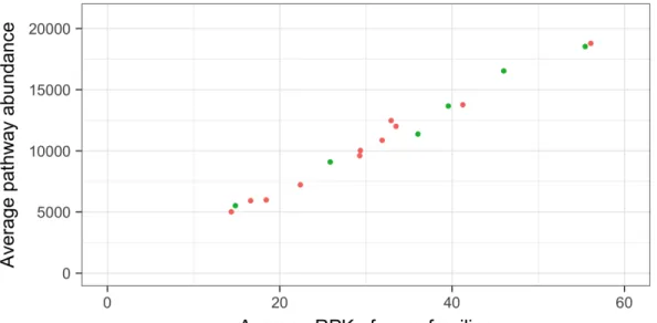
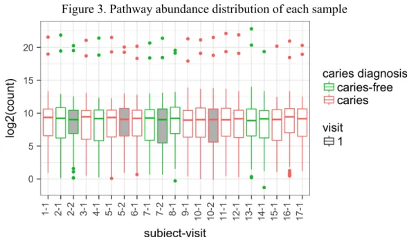
The average abundance of genes and pathways per sample, as normalized for sequencing depth and gene length, was assessed in RPK (Reads Per Kilobase) and presented in Figure 1. The gene abundance distribution of samples and path abundance distribution of samples were further transformed by taking the logarithm to the base of 2 (figure 2, figure 3).
March 2018
1
Summary and screening
1.1
Sample distribution
There are 17 subjects including 4 subjects with 2 visits (21 observations in total). The distribution of the phenotype is given in Table 1.1.1 and some of the joint distributions are illustrated in Figure 1.1.1.
Table 1.1.1: Summary of the phenotypes
Variable No Yes NA Total
Caries 6 11 - 17
Perio 8 9 - 17
Gingivitis 4 13 - 17
Diabetes 5 12 - 17
second visit 13 4 - 17
Gender Female: 4, Male: 13 17 Smoker 0: 3, 1: 12, 2: 1, NA: 1 17 Implant A: 8, B: 1, AB: 5, NA: 3 17 Age Min 40, Med 67, Max 78 17
Figure 1.1.1: Graphical representation of joint distribution
Note that doing exploratory multiple testing with a small sample size following state-ments may not be statistically generalizable.
20
Figure 1. The average abundance of gene (in RPK) and pathways per sample at baseline
Figure 2. Gene abundance distribution of each sample (in RPK)
Zero counts excluded, Gray boxes represent 2nd visit
March 2018
• (Upper right plot) Men are more likely to have dental caries.(Fisher test, p = 0.099, OR= 8.5 )
• (Bottom left plot) No association between caries and gingivitis, and between caries and perio.
If perio = 1, then most likely gingivitis = 1. (Fisher test,p= 0.03, OR=1 )
• (Bottom right plot) Smokers with diabetes are likely to have dental caries. (But logistic model gives a large p-value even with an interaction term because of the single observation at smoker=2)
1.2
The average abundance of gene (in RPK) and pathways per
sample
Figure 1.2.1: The average abundance of gene (in RPK) and pathways per sample at baseline (the first visit)
No subject has too small or too large mean RPK (genes) or mean path abundance, and the abundance do not seem to be associated with the dental caries diagnosis.
The log2 abundance of individual gene families and pathways are presented in Fig-ure 1.2.2. Zero counts are excluded and the gray boxes represent the second visits.
3
March 2018
Figure 3. Pathway abundance distribution of each sample
Zero counts excluded, Gray boxes represent 2nd visit
No subject demonstrated an excessively small or large mean of RPK (genes) or mean path abundance. The abundance also did not demonstrate association with dental caries diagnosis.
2. Salivary metagenome analysis: Species-level
Metagenomic Phylogenic Analysis (MetaPhlAn2), a Maker Gene Based Taxonomic Assignment strategy was applied for identification of the microbial taxa from the salivary metagenome. The number of species, alpha, and beta diversities were studied(Whittaker 1972). For testing group mean differences, two-sample t-tests, or more generally Wald tests, were conducted based on linear models with the continuous covariates. Because this is an exploratory study, p-values were not routinely corrected for the multiple testing.
To compare the similarity of composition of bacteria between samples, the dimension of the compositional data was reduced and possible clustering was assessed. For dimension reduction, the species were screened by detection rate of 0.002 and prevalence threshold of 0.5, and
March 2018
Figure 1.2.2: The abundance of each gene (in RPK, top) and each pathway (bottom) per sample
22
Distributed Stochastic Neighbor Embedding (t-SNE) (van der Maaten et al. 2008), and Principal Component Analysis (PCoA) (Pearson 1901) were applied. For NMDS, Bray-Curtis dissimilarity index was used. For tSNE, Euclidean distance metric and perplexity of 5 were used. For PCoA, the proportion of variability added by additional dimensions was examined through the scree plot.
Top abundant species were compared for each feature group by their rankings within the group. For the subjects with two longitudinal saliva samples, change in alpha diversity, and change in Principal Component (PC) values were studied (Whittaker 1972).
2.1. Number of genera/species
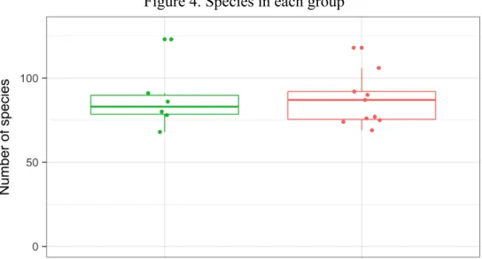
From all samples, 81 genera and 214 species, based on 360,907 gene families and 298 pathways, were identified. In comparing samples, no significant differences were noted in the number of species based on caries status (figure 4).
Figure 4. Species in each group
p-value (t-test) = 0.93
March 2018
2
Ecosystem - WGS Metaphlan2 at baseline
The analyses are
based on baseline data
unless otherwise mentioned.
The microbiome community with dental caries tend to be more diverse.
But this
tendancy is somewhat confounded with the age (See Section 2.4) so
may not be true
after adjusting for the age
.
2.1
Number of genera/species
In total, there are 214 species and 81 genera in metaphlan2 data. (360,907 gene families,
298 pathways)
For each subject, the number of species is given in the figure below. There is not a
big di
↵
erence in number of species between caries groups.
p-value (t-test) = 0.93
2.2
Beta diversity of bacteria (species level)
2.2. Abundance of bacterial species
The species-level abundance ranking of the samples was tabulated (table 2). Differential abundances were observed between caries and caries-free groups: Actinomyces sp HPA0247 and
Actinomyces graevenitzii demonstrated greater mean abundance in carious samples, while
Neisseria subflava had greater mean abundance in caries-free samples.
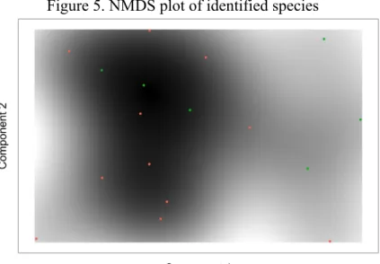
The NMDS plot revealed no significant distinction between samples based on the presence of caries (figure 5). The tSNE plot revealed lack of clustering, likely attributable to a small sample size (figure 6). The PCoA plot showed clustering of subjects with caries at the upper center portion of the plot, with the Scree plot demonstrating that 2-3 principal components contribute to total variance (figure 7).
24
Figure 5. NMDS plot of identified species
Figure 6. tSNE scatter plot
Figure 7. PCoA and Scree plots
March 2018
3
Visualization
Visualization (PCoA, tSNE, PCA plot) is done for core microbiome (67 species out of
213 species)
3.1
Visualization - NMDS
The NMDS plot below is based on the metaphlan2 (species level). Not much distinction
between dental caries diagnosis.
3.2
Visualization - tSNE
The tSNE plot below is based on the metaphlan2 (species level). Hard to see clusters
with a small sample.
3.3
Visualization - PCA
Hard to see clusters with a small sample, but caries group seems to cluster around upper
center. (The scree plot on the right tells us that 2 or 3 principal components will su
ffi
-cient.)
8
25 2.3. Alpha diversity and beta diversity
At baseline, alpha and beta diversities, at the species level, were assessed.
Alpha diversity, demonstrating species richness, through the number of taxa in the salivary microbiome, included Shannon diversity index (figure 8, figure 9), Simpson-Gini diversity index (figure 10), richness, and dominance. Richness was measured as the number of observed species within each sample given only top 50% or top 80% of the species (figure 12). Dominance represented the proportion of the abundance of the most abundant species (figure 13).
Beta diversity represented the differences of taxonomic abundance from different samples (figure 14). Beta diversity was measured using average Bray dissimilarity within each feature group.
The salivary microbiome, as presented by the samples, showed a greater tendency for more diversity in caries-associated samples though there was not a significant difference in number of species between the samples. Specifically, older patients demonstrated less diversity, and with caries status, the diversity observed initially with caries is less clear (figure 9, figure 11).
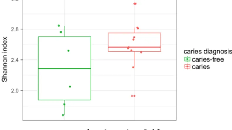
Figure 8. Shannon diversity index at the species-level as mean and range in each group
p-value (t-test) = 0.13
March 2018
2.3
Alpha diversity of bacteria (species level)
A. Shannon entropy
p-value (t-test) = 0.13
B. Simpson diversity
p-value (t-test) = 0.15
C. Richness - species (50%/80%)
p-value (t-test) = 0.93, 0.34
D. Dominance
26
Figure 9. Linear model of Shannon diversity index at the species-level and age
p-value = 0.28 (caries), 0.07 (age)
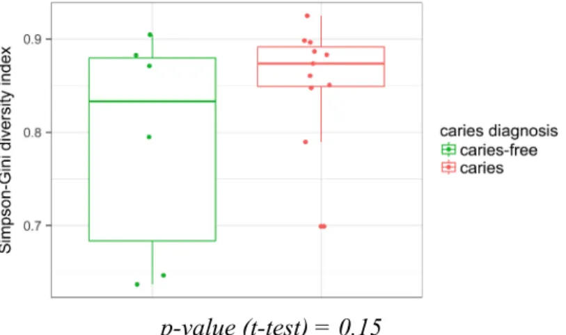
Figure 10. Simpson-Gini diversity index at the species-level as mean and range in each group
p-value (t-test) = 0.15
Figure 11. Linear model of Simpson-Gini diversity index at the species-level and age
p-value = 0.28 (caries), 0.18 (age) March 2018
2.4
diversity by age
Contrary to the result in the Early Childhood Dental Caries study (ZOE), older subjects have less diversity (more dominance).
A. Shannon entropy vs age
When age is considered, we see that among the older, microbiome systems with dental caries have high entropy, and healthy ones low entropy, but not as much as clear as we have seen earlier in this section.
linear model (outcome ⇠caries + age): p-value = 0.28 (caries), 0.07 (age)
B. Simpson diversity vs age Similar for Simpson diversity.
linear model (outcome ⇠caries + age): p-value = 0.28 (caries), 0.18 (age)
7
March 2018
2.3
Alpha diversity of bacteria (species level)
A. Shannon entropy
p-value (t-test) = 0.13
B. Simpson diversity
p-value (t-test) = 0.15
C. Richness - species (50%/80%)
p-value (t-test) = 0.93, 0.34
D. Dominance
p-value (t-test) = 0.28
March 2018
2.4
diversity by age
Contrary to the result in the Early Childhood Dental Caries study (ZOE), older subjects have less diversity (more dominance).
A. Shannon entropy vs age
When age is considered, we see that among the older, microbiome systems with dental caries have high entropy, and healthy ones low entropy, but not as much as clear as we have seen earlier in this section.
linear model (outcome ⇠caries + age): p-value = 0.28 (caries), 0.07 (age)
B. Simpson diversity vs age Similar for Simpson diversity.
linear model (outcome ⇠caries + age): p-value = 0.28 (caries), 0.18 (age)
27
Figure 12. Richness index at the species-level (50%/80%)
p-value (t-test) = 0.93, 0.34
Figure 13. Dominance index at the species-level according to caries status
p-value (t-test) = 0.28
Figure 14. Sample dissimilarity as measured by beta diversity at species-level
p-value (t-test) = 0.61
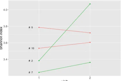
2.4. Longitudinal analysis
Longitudinal changes in the salivary microbiome for 4 subjects with 2 samples were evaluated for comparison (Table 3).
March 2018
2.3
Alpha diversity of bacteria (species level)
A. Shannon entropy
p-value (t-test) = 0.13
B. Simpson diversity
p-value (t-test) = 0.15
C. Richness - species (50%/80%)
p-value (t-test) = 0.93, 0.34
D. Dominance
p-value (t-test) = 0.28
6
March 2018
2.3
Alpha diversity of bacteria (species level)
A. Shannon entropy
p-value (t-test) = 0.13
B. Simpson diversity
p-value (t-test) = 0.15
C. Richness - species (50%/80%)
p-value (t-test) = 0.93, 0.34
D. Dominance
p-value (t-test) = 0.28
6
March 2018
2
Ecosystem - WGS Metaphlan2 at baseline
The analyses are
based on baseline data
unless otherwise mentioned.
The microbiome community with dental caries tend to be more diverse.
But this
tendancy is somewhat confounded with the age (See Section 2.4) so
may not be true
after adjusting for the age.
2.1
Number of genera/species
In total, there are 214 species and 81 genera in metaphlan2 data. (360,907 gene families,
298 pathways)
For each subject, the number of species is given in the figure below. There is not a
big di
↵
erence in number of species between caries groups.
p-value (t-test) = 0.93
2.2
Beta diversity of bacteria (species level)
Between visits 1 and 2, the subjects had the following course:
Subject 002 had undergone 9 months of treatment consisting of 18 natural tooth preparations for FDP, 3 implants placements, bone graft, crown lengthening, and 6 tooth
extractions. The subject also had two, 1-week courses of antibiotic prophylaxis in conjunction to treatment during that time period (one course is 500mg Amoxicillin, 3 times a day, for 7 days).
Subject 005 had undergone 9 months of treatment consisting of 7 natural tooth preparations for FDP, bone graft, and 1 tooth extraction.
Subject 007 had undergone 8 months of treatment consisting of temporary placement of a maxillary removable complete denture, 5 natural tooth preparations for FDP, 4 implant
placements, 4 definitive tooth-borne FDP placement. The subject also had a single, 5-day course of antibiotic prophylaxis in conjunction to treatment during that time period (500 mg
Amoxicillin, 3 times a day, for 5 days).
Subject 010 had undergone 2 months of treatment consisting of 16 natural tooth preparations.
29
Table 3. Demographic and clinical data of subjects with 2 samples (visits)
A: Implant placement during treatment, B: Implant placement prior to treatment, AB: Implant placement prior to treatment and after treatment
Figure 15. Number of species between visits and subjects
Figure 16. Shannon entropy index at the species-level between visits and subjects
March 2018
6
Longitudinal analysis
Using the information from those who visited twice, we plot the trajectories of some of
the diversity measures, and we fit longitudinal models.
The phenotypes of the repeated subjects are tabulated in Table 6.0.1.
Table 6.0.1:
Phenotypes of the subject with 2 visits
id
caries perio gingivitis diabetes smoker age gender implants
002
0
0
1
0
1
75 m
AB
005
1
1
1
1
1
70 m
AB
007(IRB) 0
0
0
0
1
65 f
A
010
1
1
1
0
0
72 m
AB
6.1
Spaghetti plots
The signal is very weak with only four subjects. Overall, the diversity increased after
treatment.
March 2018
6
Longitudinal analysis
Using the information from those who visited twice, we plot the trajectories of some of
the diversity measures, and we fit longitudinal models.
The phenotypes of the repeated subjects are tabulated in Table 6.0.1.
Table 6.0.1:
Phenotypes of the subject with 2 visits
id
caries
perio
gingivitis
diabetes
smoker
age
gender
implants
002
0
0
1
0
1
75
m
AB
005
1
1
1
1
1
70
m
AB
007(IRB)
0
0
0
0
1
65
f
A
010
1
1
1
0
0
72
m
AB
6.1
Spaghetti plots
30
Figure 17. Simpson-Gini diversity index at the species-level between visits and subjects
Figure 18. Dominance index at the species level between visits and subjects
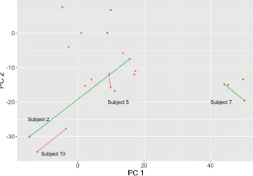
Figure 19. Change in PC between visits and subjects at the species-level
March 2018
March 2018
25
March 20186.2 Change in Principal components
Figure 6.2.1 shows the change in the principal components from the first to the second visit.
Figure 6.2.1:Change in PC
All 4 second visits have regressed to the center of the space. The subject number 2 has the biggest change in both PCs.
6.3 Testing the association of each species with dental caries Longitudinal models are non-identifiable or non-estimable due to small number of repli-cates.
Chen & Li method might have been used for longitudinal compositional data (logistic-beta model), to see if there is any association with a specific bacteria species with the dental caries status, but for most of the core bacteria species, they do not have zero counts. So the test could not be done.
Also simpler model such as linear mixed e↵ect models can be considered. However, to predict the random e↵ects, there should be more sample or replicates, than is now. This test could not be done either.
31 3. Salivary metagenome analysis: Variables
Diversity indices were compared across clinical characteristics and other medical information or comorbidities such as dental caries diagnosis, gingivitis, periodontal disease history, diabetes, gender, and smoking status. Findings related to diabetes and periodontal disease history were notable.
3.1. Diabetes analysis
We found that the salivary microbiome of patients with diabetes was more diverse compared to those without diabetes. Though the number of species did not show any important difference (figure 20), alpha and beta diversity indices were different between the groups (figures 21-24). The NMDS, tSNE, and PCoA plots revealed a distinct clustering of samples associated with diabetes (figures 26-28).
Figure 20. Species in each group (diabetes)
p-value (t-test) = 0.93
March 2018
2
Ecosystem - WGS Metaphlan2 at baseline
The analyses are
based on baseline data
unless otherwise mentioned.
The microbiome community with diabetes tend to be more diverse,
even after
control-ling for the age (See Section 2.4), but the beta diversity is the opposite: i.e. the people in
the diabetes group are more homogeneous than do the people in the non-diabetes group.
2.1
Number of genera/species
In total, there are 214 species and 81 genera in metaphlan2 data. (360,907 gene families,
298 pathways)
For each subject, the number of species is given in the figure below. There is not a
big di
↵
erence in number of species between diabetes groups.
p-value (t-test) = 0.93
2.2
Beta diversity of bacteria (species level)
32
Figure 21. Shannon diversity index at the species-level as mean and range in each group (diabetes)
p-value (t-test) = 0.015
Figure 22. Simpson-Gini diversity index at the species-level as mean and range in each group (diabetes)
p-value (t-test) = 0.013
Figure 23. Richness index at species-level (50%/80%) (diabetes)
p-value (t-test) = 0.48, 0.04
March 2018
2.3
Alpha diversity of bacteria (species level)
A. Shannon entropy
p-value (t-test) = 0.015
B. Simpson diversity
p-value (t-test) = 0.013
C. Richness - species (50%/80%)
p-value (t-test) = 0.48, 0.04
D. Dominance
p-value (t-test) = 0.013
March 2018
2.3
Alpha diversity of bacteria (species level)
A. Shannon entropy
p-value (t-test) = 0.015
B. Simpson diversity
p-value (t-test) = 0.013
C. Richness - species (50%/80%)
p-value (t-test) = 0.48, 0.04
D. Dominance
p-value (t-test) = 0.013
March 2018
2.3
Alpha diversity of bacteria (species level)
A. Shannon entropy
p-value (t-test) = 0.015
B. Simpson diversity
p-value (t-test) = 0.013
C. Richness - species (50%/80%)
p-value (t-test) = 0.48, 0.04
D. Dominance
p-value (t-test) = 0.013
33
Figure 24. Dominance index at the species-level as mean and range in each group
p-value (t-test) = 0.013
Figure 25. Sample dissimilarity as measured by beta diversity at the species-level (diabetes)
p-value (t-test) = 0.002
Figure 26. NMDS plot of identified species (diabetes)
March 2018
2.3
Alpha diversity of bacteria (species level)
A. Shannon entropy
p-value (t-test) = 0.015
B. Simpson diversity
p-value (t-test) = 0.013
C. Richness - species (50%/80%)
p-value (t-test) = 0.48, 0.04
D. Dominance
p-value (t-test) = 0.013
5
March 2018
2
Ecosystem - WGS Metaphlan2 at baseline
The analyses are
based on baseline data
unless otherwise mentioned.
The microbiome community with diabetes tend to be more diverse,
even after
control-ling for the age (See Section 2.4), but the beta diversity is the opposite: i.e. the people in
the diabetes group are more homogeneous than do the people in the non-diabetes group.
2.1
Number of genera/species
In total, there are 214 species and 81 genera in metaphlan2 data. (360,907 gene families,
298 pathways)
For each subject, the number of species is given in the figure below. There is not a
big di
↵
erence in number of species between diabetes groups.
p-value (t-test) = 0.93
2.2
Beta diversity of bacteria (species level)
p-value (t-test) = 0.002
4
March 2018
3
Visualization
Visualization (PCoA, tSNE, PCA plot) is done for core microbiome (67 species out of
213 species)
3.1
Visualization - NMDS
The NMDS plot below is based on the metaphlan2 (species level).
Diabetes group is
clustered at center.
3.2
Visualization - tSNE
34
Figure 27. tSNE scatter plot (diabetes)
Figure 28. PCoA plot for each subject (diabetes)
3.2. Periodontal disease history analysis
The salivary microbiome of patients with a previous history of periodontal disease was more diverse compared to those without history of periodontal disease. However, beta diversity analysis between groups of patients who had a history of disease versus no disease revealed the opposite (figure 34), signifying that diversity was similar between samples derived from
previous history of periodontal disease than that of samples without a history of disease. The
March 2018
3
Visualization
Visualization (PCoA, tSNE, PCA plot) is done for core microbiome (67 species out of
213 species)
3.1
Visualization - NMDS
The NMDS plot below is based on the metaphlan2 (species level).
Diabetes group is
clustered at center.
3.2
Visualization - tSNE
The tSNE plot shows similar result as in NMDS.
7
March 2018
3.3
Visualization - PCA
35
number of species did not show any important difference (figure 29), whereas alpha and beta diversity indices differed between the groups (figures 30-34).
The PCoA plot (figure 37) revealed a distinct clustering of samples associated with disease, but NMDS and tSNE, did not reveal any notable patterns (figure 35, figure 36).
Figure 29. Species in each group (periodontal disease)
p-value (t-test) = 0.49
Figure 30. Shannon diversity index at the species-level as mean and range in each group (periodontal disease)
p-value (t-test) = 0.021
March 2018
2
Ecosystem - WGS Metaphlan2 at baseline
The analyses are
based on baseline data
unless otherwise mentioned.
The microbiome community with perio = 1 tend to be more diverse,
but the beta
diversity is the opposite: i.e. the people in the perio = 1 group are more homogeneous
than do the people in the perio = 0 group.
2.1
Number of genera/species
In total, there are 214 species and 81 genera in metaphlan2 data. (360,907 gene families,
298 pathways)
For each subject, the number of species is given in the figure below. There is not a
big di
↵
erence in number of species between perio groups.
p-value (t-test) = 0.49
2.2
Beta diversity of bacteria (species level)
p-value (t-test) = 0.14
4
March 2018
2.3
Alpha diversity of bacteria (species level)
A. Shannon entropy
p-value (t-test) = 0.021
B. Simpson diversity
p-value (t-test) = 0.06
C. Richness - species (50%/80%)
p-value (t-test) = 0.49, 0.004
D. Dominance
36
Figure 31. Simpson-Gini diversity index at the species-level as mean and range in each group (periodontal disease)
p-value (t-test) = 0.06
Figure 32. Richness index at the species-level (50%/80%) (periodontal disease)
p-value (t-test) = 0.49, 0.004
Figure 33. Dominance index at the species-level as mean and range in each group (periodontal disease)
p-value (t-test) = 0.011
March 2018
2.3
Alpha diversity of bacteria (species level)
A. Shannon entropy
p-value (t-test) = 0.021
B. Simpson diversity
p-value (t-test) = 0.06
C. Richness - species (50%/80%)
p-value (t-test) = 0.49, 0.004
D. Dominance
p-value (t-test) = 0.011
March 2018
2.3
Alpha diversity of bacteria (species level)
A. Shannon entropyp-value (t-test) = 0.021
B. Simpson diversity
p-value (t-test) = 0.06
C. Richness - species (50%/80%)
p-value (t-test) = 0.49, 0.004
D. Dominance
p-value (t-test) = 0.011
5
March 2018
2.3
Alpha diversity of bacteria (species level)
A. Shannon entropy
p-value (t-test) = 0.021
B. Simpson diversity
p-value (t-test) = 0.06
C. Richness - species (50%/80%)
p-value (t-test) = 0.49, 0.004
D. Dominance
p-value (t-test) = 0.011
37
Figure 34. Sample dissimilarity as measured by beta diversity at species-level (periodontal disease)
p-value (t-test) = 0.14
Figure 35. NMDS plot of identified species (periodontal disease)
Figure 36. tSNE scatter plot (periodontal disease)
March 2018
2
Ecosystem - WGS Metaphlan2 at baseline
The analyses are
based on baseline data
unless otherwise mentioned.
The microbiome community with perio = 1 tend to be more diverse,
but the beta
diversity is the opposite: i.e. the people in the perio = 1 group are more homogeneous
than do the people in the perio = 0 group.
2.1
Number of genera/species
In total, there are 214 species and 81 genera in metaphlan2 data. (360,907 gene families,
298 pathways)
For each subject, the number of species is given in the figure below. There is not a
big di
↵
erence in number of species between perio groups.
p-value (t-test) = 0.49
2.2
Beta diversity of bacteria (species level)
p-value (t-test) = 0.14
4
March 2018
3
Visualization
Visualization (PCoA, tSNE, PCA plot) is done for core microbiome (67 species out of
213 species) For the PCA plot, perio group is centered, but for NMDS and tSNE, the
observations are somewhat mixed.
3.1
Visualization - NMDS
The NMDS plot below is based on the metaphlan2 (species level).
3.2
Visualization - tSNE
The tSNE plot shows similar result as in NMDS.
38
Figure 37. PCoA plot for each subject (periodontal disease)
March 2018
3.3
Visualization - PCA
DISCUSSION
Full mouth rehabilitation treatment is geared towards achieving health. But, thus far, oral health has been assessed using clinical parameters that indicate preservation of tissues and
structures without the presence of loss or destruction due to disease and/or parafunction. A dental provider evaluates objective criteria that measure for health versus disease in a subjective
manner. So even though clinical health can be readily assessed, little research has been
conducted in understanding how prosthodontic treatment may impact the oral microbial status within the contemporary understanding of oral health and disease as complex, host-biofilm dysbiotic states. In an effort to improve the state of oral health, understanding how these types of restorations may affect the foundation, one’s oral microbiome, may impart some insight on the efficaciousness of this approach in treatment.
This study collected unstimulated, whole saliva samples. The choices regarding the type of saliva and method of collection were based on the sampling kit utilized for the study.
Generation Sequencing (HOMINGS)(Belstrom et al. 2016a). It determined that no significant differences in composition and proportions of the respective bacterial profiles at the species and genus level probe targets(Belstrom et al. 2016a).
This study’s salivary metagenomics analysis approach based on whole genome sequencing (WGS) shotgun allowed us to obtain a more comprehensive view of the oral microbiome. WGS can reveal more unidentified and unassigned, non-human taxonomic
sequences of the salivary metagenome (Lazarevic et al. 2012); it can, thereby contribute to more information regarding the actual functional potential of the microbial cells through assignments of sequences from databases organized by functional sequences(Gerlach and Stoye 2011; Meyer et al. 2008).
Previous studies have shown that salivary bacterial profiles differ between disease and non-disease statuses (Belstrom et al. 2017b; Chen et al. 2018). Differences in the abundance of particular species, namely of the genera of Neisseria, Haemophilus, and Fusobacterium, were evident when comparing compositions of microbiomes that had no active disease but a history of previous disease to those without any history of disease (Belstrom et al. 2017b). Bacterial
diversity was found to be significantly higher in individuals with low caries experience than those with 10 times higher caries experience (Belstrom et al. 2017b). Similar findings have been observed for periodontal disease, in which bacteria that have known associations to disease continue to have proportionally large abundance and representation in diseased states(Ai et al. 2017; Belstrom et al. 2015).
Age, diabetes, and periodontal status appeared to also be associated with differences in the salivary microbiome in this study. Age has been found to affect the microbiome’s
health condition and associated co-morbidities such as increased presence of inflammatory markers that occur in older populations (Costalonga and Herzberg 2014; Lira-Junior et al. 2018; Nassar et al. 2014; Takeshita et al. 2016; Xu et al. 2015). Diabetes has not consistently been shown to alter the oral microbiome, though observed changes have been associated with co-morbidities such as obesity. (Goodson et al. 2017; Janem et al. 2017; Kampoo et al. 2014). When evaluating changes incurred after therapy to treat a disease and restore health, studies have found that clinically determined improvements in oral conditions cannot be
necessarily defined or quantified by bacterial abundance (Schwarzberg et al. 2014). Specifically, the notion of microbiologically diagnosing health based upon proportions of particular
microorganisms associated with health and disease is not generalizable. Rather, compositions of microbial cells that promote symbiosis are unique to an individual’s specific microbiome and its functions and interactions (Schwarzberg et al. 2014). In periodontal disease therapy, diseased and previously-diseased oral environments were diverse and individualized, while microbiomes without a history of disease were homogenous(Chen et al. 2018).
Longstanding treatment outcomes are ultimately not determined by prosthetic restoration, but by the ability to manage the microbial balance that is supportive towards health (Yanase and Le 2014). Management of an individual’s oral microbiome through behavioral and