NANOPARTICLE CLEARANCE IS GOVERNED BY TH1/TH2 IMMUNITY AND STRAIN BACKGROUND
Stephen W. Jones
A dissertation submitted to the faculty at the University of North Carolina at Chapel Hill in partial fulfillment of the requirements for the degree of Doctor of
Philosophy in the department of Cell Biology in the School of Medicine.
Chapel Hill 2013
Approved by James Bear Keith Burridge Richard Chenny
Ian Davis
ii
iii ABSTRACT
Stephen W. Jones: Nanoparticle Clearance is Governed by Th1/Th2 Immunity and Strain
(Under the direction of James E. Bear)
Extended circulation of nanoparticles in blood is essential for most clinical applications. Nanoparticles are rapidly cleared by cells of the mononuclear phagocyte system (MPS). Approaches such as grafting polyethylene glycol onto particles (PEGylation) extend circulation times; however, these particles are still cleared and the processes involved in this clearance remain poorly understood. Here we present a novel assay using intravital microscopy for the quantification of nanoparticle clearance, allowing us to determine the effect of mouse strain and immune system function on particle clearance. We demonstrate that mouse strains which are prone to Th1 immune responses clear nanoparticles at a slower rate than Th2-prone mice. Using depletion strategies, we show that both
granulocytes and macrophages participate in the enhanced clearance observed in Th2-prone mice. Macrophages isolated from Th1 strains take up fewer
iv
TABLE OF CONTENTS
List of Tables... v
List of Figures... vi
Chapter 1. INTRODUCTION TO NANOMEDICINE AND NANOPARTICLE IMMUNE INTERACTIONS...1
Nanoparticles and Nanomedicine... 1
Mononuclear Phagocyte System…..………... 7
Intravital Microscopy and Nanoparticle Pharmacokinetics...14
Th1/Th2 immunity and M1/M2 Macrophages... 16
1. INTRAVITAL MICROSCOPY PHARMACOKINETICS ASSAY... 21
2. THE EFFECT OF TH1 AND TH2 IMMUNITY AND MOUSE STRAIN ON NANOPARTICLE CLEARANCE………..28
Introduction……… 28
Results………... 33
Discussion………... 59
3. FUTURE DIRECTIONS………... 64
APPENDIXIES……… 73
v
LIST OF TABLES
1. Modulus values and compartmental analysis of RBCM
particles from intravital microscopy experiments……….26 2. Pharmacokinetic parameters for 1% cross-linked RBCMs
vi
LIST OF FIGURES
1. Types of nanoparticles used in medicine………... 6
2. Images of ear vasculature………... 23
3. Images of microparticles in mouse vasculature………...………... 25
4. Characterization of particles used in study………..……….………... 34
5. An Intravital Microscopy Based Assay for Screening Nanoparticle Clearance Rates in Live Animals……….………... 35
6. Treatment of BALB/c mice with liposomal clodronate increases the circulation of nanoparticles in vivo... 37
7. Immune cells clear nanoparticles in the vasculature……..………... 38
8. Th1 mouse strains clear nanoparticles slower than Th2 strains………... 40
9. Biodistribution of 300nm PRINT hydrogel particles in Balb/c and C57BL/6 mice………...………... 42
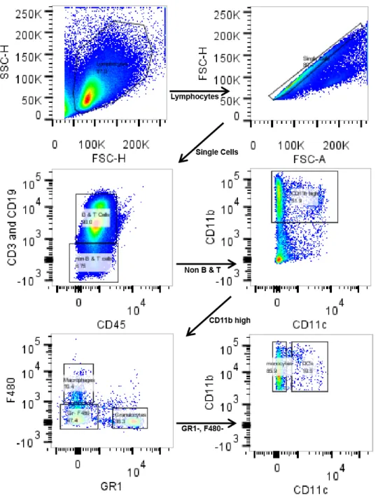
10. Representative gating scheme from BALB/c mouse spleen….……... 45
11. Flow cytometry of peripheral blood leukocytes and splenocytes shows dramatically higher uptake of particles by granulocytes in BALB/c mice...46
12. Depletion of granulocytes from mice using rat anti-mouse GR-1 antibodies………... 48
13. Depletion of granulocytes significantly increases particle exposure to blood in BALB/c mice but not C57BL/6 mice………...… 49
14. Depletion of granulocytes significantly increases particle exposure to blood in BALB/c mice but not C57BL/6 mice…...……...……...…..… 51
15. Polarized M2 macrophages take up more particles than M1 macrophages in mice and humans……….…….. 55
1
CHAPTER ONE: INTRODUCTION
Nanoparticles in medicine
Nanoparticles have been investigated for medicinal applications for over 40 years (Chakraborty et al., 2013). Nanoparticle use in medicine can be broadly grouped into two categories, particles used for therapeutic applications in which the particle serves as therapeutic vehicle, and particles used for diagnostic applications. For the majority of nanoparticle therapeutics the principle is to immobilize a therapeutic agent within the matrix of a nanoparticle and thereby alter the pharmacokinetics (PK) of the therapeutic agent. The guiding principle behind this is that therapeutic agents that are normally toxic, unstable, or
insoluble can be sequestered within the particle until they can be released at an appropriate time and place. In most systems the particle utilizes a targeting agent such as an antibody or a DNA aptamer to direct the particle to a particular physiological destination (Beech et al., 2013; Fokong et al., 2012; Lammers et al., 2012; Li et al., 2013b; Shin et al., 2013). Once present in the desired location in the body the particle either degrades naturally or is triggered to degrade by altered physiological conditions such as low pH, reducing environment, or
2
physiological interest and limiting off target effects (Morachis et al., 2012b). For diagnostic applications particles are functionalized with materials that can be detected with medical technology such as radiolabels for PET/SPECT imaging, MRI contrast agents, or fluorophores for fluorescent imaging (Feng et al., 2013; Gu et al., 2013; Wadajkar et al., 2013). Again in most circumstances diagnostic nanoparticles are conjugated with targeting agents to direct them to a site of interest in the body to aid physicians in the diagnosis and assessment of pathological conditions. Despite decades of work and thousands of papers published on the topic of nanomedicine, only two nanoformulations have been approved for clinical use in the USA. Abraxane which is a naoparticle formulation of albumin and paclitaxel, and doxil a liposomal formulation of doxorubicin
(Coleman et al., 2006; Gradishar, 2006a; Krown et al., 2004). While there is still significant hope that nanoparticles will play a major future role in the clinic it has become clear that several major hurdles still must be overcome before
nanotherapies will be widely efficacious. Here we will examine issues related to the response of the immune system to nanoparticles in blood.
Classes of Nanoparticles
3
medical applications. They can be made out of a very wide variety of materials with varying properties, can be easily designed to hold drugs inside the particle, can be functionalized with surface ligands of choice, and benefit from a huge amount of chemistry and materials knowledge of polymers and their properties. However it is challenging to make polymer particles smaller than 80-100nm, and they often show a wide range of sizes within one batch (Feng et al., 2013;
Morachis et al., 2012a; Zhang et al., 2013). Polymeric micells (Fig 1 B) are formed by generating a polymer shell with one surface (typically the interior) being hydrophobic and the other being hydrophilic. The interior of the particle is typically filled with an oil droplet that can dissolve large amounts of hydrophobic drugs in the hydrophobic interior. The hydrophilic exterior can then be
4
first nanodrug to get full approval in US for use (Doxil) (Biganzoli et al., 2007; Coleman et al., 2006). They are created by making a lipid bilayer typically
~100nm in size that contain a water droplet on the inside that can be loaded with hydrophilic therapeutics. The outer surface of the liposome can then be
functionalized with targeting agents or stealthing ligands. Liposomes are easy to make and very biologically compatible with low toxicology and good PK, but like polymeric micelles they are unstable and difficult to control drug release from (Chattopadhyay, 2013; Jolck et al., 2010; Naik et al., 2013; Qian et al., 2012). In recent years some labs have started referring to viruses as nanoparticles (Fig 1 E), which is a technically correct definition. This has caused virus mediated genetic therapies to become incorporated into nanomedicine. In addition some effort has been made to formulate viral particles that contain a drug in the interior instead of genetic material. Viruses have the advantages of excellent natural targeting, cell penetrating, and controlled delivery of cargoes. However. viruses often illicit an immune response which limits their efficacy in vivo and can be dangerous to the patient. It is also difficult to produce large amounts of virus (Lentz et al., 2012; Manjila et al., 2013; Sun et al., 2013b; Usme-Ciro et al., 2013). Finally carbon nanotubes (Fig 1 F), have shown some potential as
diagnostic agents due to their unusual optical and magnetic properties that make them easy to detect in vivo. However it is difficult to get nanotubes to
demonstrate good PK, and difficult to target them to sites of interest for
5
(Li et al., 2013a; Reynolds et al., 2013; Sun et al., 2013a; Tan et al., 2011; Yildirimer et al., 2011).
The PRINT® System for Nanoparticle Synthesis.
Recently a new technique known as particle replication in non-wetting templates (PRINT) has been developed for the generation of hydrogel
nanoparticles with a high level of control over the size and shape of the particles (Rolland et al., 2005). This allows for more precise experiments comparing effects of size and shape on particle PK (Gratton et al., 2007), cell entry (Gratton et al., 2008a; Gratton et al., 2008c), targeting (Wang et al., 2010), PEG stealthing (Perry et al., 2012) and novel classes of microparticles (Merkel et al., 2011). In all cases by taking advantage of the nature of the PRINT system particles with limited diversity of size and shape can be compared much more rigorously than more heterogeneous particle populations, which allows for more detailed
7 The MPS system
The arm of the immune system that is most commonly associated with nanoparticles is the reticuloendothelial system (RES)/mononuclear phagocyte system (MPS). The MPS system broadly consists of tissue macrophages and dendritic cells (DCs) found in the spleen, liver, and lungs as well as monocytes in the spleen and blood (Miyata and van Eeden, 2011; Moghimi et al., 2012; Wynn et al., 2013). In addition some authors have recently suggested that neutrophils in the blood should also be considered part of the MPS due to their similar
function and behavior to other MPS cells, thoughtraditionally neutrophils have not been considered a part of the MPS system. The MPS system has been well characterized as functioning as the innate immune system’s effecter arm in the blood (Fitrolaki et al., 2013; Kono et al., 2012; Ogiku et al., 2011). Additionally in pathological contexts the MPS system, especially the DCs, function as the main antigen presenting cells in the spleen for activation of adaptive T and B cell responses. This particular function will not be considered further here, but has been extensively reviewed recently (Blum et al., 2013; Fehres et al., 2013; Guery and Hugues, 2013).
8
into the lumen of the blood vessel. When phagocytic receptors on the filopodia engage microbial surfaces the microbes are bound and rapidly internalized by the kuppfer cell, this process prevents bacteria and other microbes that
commonly enter the blood in the intestines from passing into the rest of the body and causing sepsis (Gregory et al., 2002; Gregory and Wing, 2002; Shih-Ching et al., 2004; Wong et al., 2010). Kuppfer cells have also been implicated in a wide range of pathological conditions including liver fibrosis (Frasinariu et al., 2013; Purohit and Brenner, 2006; Tomita et al., 2013), fatty liver disease (Koek et al., 2011; Sawada et al., 2013), hepatic cancer (Bortolami et al., 2002; Van den Eynden et al., 2013), malaria (Frevert et al., 2008; Tavares et al., 2013a; Tavares et al., 2013b), and alcoholic liver disease(Bala et al., 2012; Petrasek et al., 2012; Szabo et al., 2012; Wan et al., 2013). If microbes are introduced directly into the blood, it is well known that the majority of microbes will be cleared by the action of kuppfer cells in the liver (Shou et al., 1994). In the case of genetic or
pharmacolgical treatments which significantly blunt the activity of kuppfer cells there is a significantly increased risk of death during sepsis (Knoferl et al., 2002).
After the liver the second largest collection of MPS cells that access the blood is found in the spleen, where at least eight different distinct populations of macrophages can be found (den Haan and Kraal, 2012). The exact role and nature of all of the subpopulations of splenic macrophages is poorly understood, but most of the minor populations are believed to be important in antigen
9
implies, are located in the red pulp of the spleen. Under normal physiological conditions they remove aged red blood cells from blood as it passes through the spleen (Ganz, 2012). This action plays an important role in preventing anemia and inflammation from the lysis of aged red blood cells in inappropriate locations in the body (Kovtunovych et al., 2010). In the case of sepsis red pulp
macrophages will bind and phagocytose microbes in the blood as it passes through the spleen. In the case of a normally functioning immune system the spleen will clear the second largest amount of microbes during sepsis after the liver, with the red pulp macrophage being the dominant player in this process.
The activity of macrophages in the lung is highly inconsistent from one species to another. All mammals contain large numbers of macrophages in the lungs,
however in some species such as rodents (Fels and Cohn, 1986) the
macrophages are polarized into the airways and do not respond to the presence of microbes in the blood of the lungs and therefore clear only small numbers of microbes during sepsis (Fels and Cohn, 1986). However in some larger
mammals such as dogs, macrophages in the lungs appear to respond both to microbes in the airways and the blood allowing them to clear microbes in the case of sepsis (Merrill, 1990). Human lung macrophages appear to respond both to pathogens in blood and in the airways making the lung the third most prevalent site of clearance in humans after liver and spleen (Merrill, 1990).
10
vessels under normal conditions where they appear to perform a patrolling surveillance function. In the case of injury CX3CR1 high monocytes quickly migrate into the site of injury where they first release cytokines and chemokines to draw other immune cells to the site of injury, then differentiate into tissue macrophages to assist in the clearance of invading microbes/cellular debris (Auffray et al., 2007; Geissmann et al., 2010). CX3CR1 low monocytes are normally resident in the spleen, but upon injury the chemokines during injury induces CX3CR1 monocytes to migrate from the spleen into the blood. After circulation in the blood they then enter the site of injury where they differentiate into macrophages (Swirski et al., 2009).
11
MPS system likely due to their different life cycle and microbial killing activities. However, recently some authors have suggested that due to their lineage, motility and phagocytic capacity they should more properly be classified as an MPS cell (Rabinovitch, 1995; Silva and Correia-Neves, 2012). Surprisingly despite they abundance and phagocytic capacity few nanoparticle studies have examined their role in particle clearance. It has been shown however that certain particle types can be found in neutrophils after intravenous injection (Leuschner et al., 2011). In addition it has been shown that in vitro nanoparticles can be phagocytosed by neutrophils and induce neutrophil netting, a process in which the nuclear contents of the neutrophils are mixed with the granular contents and then exocytosed to create a DNA, granular, extracellular, anti-microbial net (Bartneck et al., 2010).
MPS clearance of nanoparticles
Early studies of the PK of nanoparticles injected intravenously showed surprisingly fast clearance of particles from the blood (Fernandez-Urrusuno et al., 1996). Upon further examination of the biodistribution (BD) of the particles it was shown that the majority of the particles were located within the kuppfer cells of the liver and the red pulp cells of the spleen (Illum and Davis, 1984; Illum et al., 1984). This result has subsequently been repeated with a wide variety of
12
indentify key factors involved in particle binding and clearance by macrophages. No study to date has been able to truly prevent all binding and clearance of particles, but several physical and chemical characteristics such as
hydrophobicity, strong charge, rigidity, size, and the rate at which serum proteins bind to a particle have been identified as playing a negative role (Moselhy et al., 2000). From this work a general consensus has emerged that particles less than 500nm, that are flexible, hydrophilic and uncharged will demonstrate the best PK in vivo. Other studies have attempted to determine which specific receptors on
macrophages bind to particles to mediate clearance. These studies have been primarily in vitro using immortalized cell lines, and often non-specific inhibitors with many characterized off target effects. These caveats make drawing any strong conclusions about important receptors difficult, but they do provide an initial list of suspects including scavenger receptor A (Patel et al., 2010), Fc gamma receptor three (Yang et al., 2010), compliment receptor 3 (Sahay et al., 2010), among others.
The most commonly used strategy to extend the circulation time of nanoparticles in vivo has been to graft a thick coating of high molecular weight polyethylene
13
completely prevent clearance of particles by MPS cells, instead it slows the kinetics. In addition there are reasons to believe that PEG is a less than ideal solution for clinical applications. First it appears that PEGylation typically
competes with targeting strategies as targeting molecules must protrude past the PEG layer and be present in sufficient quantities to result in targeting. This has a tendency to disrupt the coordinated shell of water around the particle, and
creates the possibility of MPS cells binding directly to the targeting ligand,
resulting in clearance (Pardeshi et al., 2012), or for serum proteins to bind to the particle opsinizing it. It has also been shown that a large number of patients treated with PEGylated agents will develop short term anti-PEG IgM antibodies (Ishihara et al., 2009; Shimizu et al., 2012), this response is transient, typically lasting for only a few weeks, but it significantly limits the flexibility of dosing
14 Nanoparticle PK assays
The study of nanoparticle PK has typically utilized a blood draw assay in which nanoparticles are injected into a large number of animals at time zero. At various time points after injection the animals are sacrificed and bled. The
collected blood can then be assayed for concentrations of nanoparticles by using reporters such as fluorescence or radiation (Zamboni et al., 2012). In the case of inorganic nanoparticles the concentration can be directly assayed by detecting the presence of elements in the particles not found in biological samples such as gold, in the case of gold nanoparticles or cadmium and selenium in the case of quantum dots (Dreaden et al., 2012a; Dreaden et al., 2012b). These assays are advantageous as they are simple to perform; the data can be applied to existing pharmacokinetic models for small molecule PK, and can provide direct
concentrations of nanoparticles in blood and/or tissue. There are however several disadvantages to these assays: First this technique requires the use of large numbers of animals and therefore large amounts of nanoparticles, as a group of animals is required at every time point. Second these assays offer limited temporal resolution, it is difficult to get data points for less than 1-2min after injection of particles making the baseline value unclear, and every time point assayed requires the use of additional animals and particles. Third it is common for these types of experiments to result in high errors as it is difficult to sacrifice animals and bleed them in a controlled, repeatable way. These
15
that large numbers of often difficult to manufacture experimental particles or expensive/rare animals must be used. An alternate assay that allows for PK data from a small number of animals usingfewer particles and higher temporal
resolution is critically needed by the field to allow for rapid screening of particles and animal models.
Intravital microscopy
Intravital microscopy is an emerging technique in biological sciences that allows for the direct imaging of cells, molecules, tissues, etc. in living animals. It is highly advantageous over traditional in vitro tissue culture based imaging for the obvious reason of being performed in a physiologically relevant system. Intravital microscopy is most commonly performed using a multiphoton
microscope (Niesner and Hauser, 2011). This type of scope uses two carefully aligned infrared laser lines that cross at a precisely defined position. At the point where the two lasers cross their energy is roughly doubled allowing them to excite photophores that typically are excited by visible light (Benninger et al., 2008). This is advantageous as infrared light exhibits far greater tissue penetration than visible light allowing for the stimulation of photophores that would otherwise be too deep in tissue to be imaged (Bakalova, 2007). However, for imaging in tissues that are shallower such as the epidermis, it is often
advantageous to use traditional laser scanning confocal microscopy as tissue penetration is less important and greater resolution can be achieved with
16
study diverse biological processes including cancer invasion (Uchugonova et al., 2013), development (Canaria and Lansford, 2010), immune function (Sumen et al., 2004), neurobiology (Garaschuk et al., 2006), as well as many others. In nanomedicine intravital microscopy is slowly gaining traction and has recently been used to assay the effect of size and shape on tumor extravasation of particles (Smith et al., 2012), targeting of particles to neovasculature (Smith et al., 2008), the circulation of gold nanoparticles (Tong et al., 2009b), and the uptake of particles by tumors (Smith et al., 2010). By combining the knowledge and resources for intravital imaging of the immune system with intravital imaging of nanoparticles, we may be able to better understand the clearance of
nanoparticles by the immune system. In addition, by directly imaging particle circulation in blood it is possible to use intravital microscopy as an alternative method for assaying nanoparticle circulation times that avoids many of the traditional complication of nanoparticle PK assays.
Th1/Th2 immunology
17
CD4+ T cells take on one of at least four different classes: the T helper cell, the Th1 T cell, the Th2 T cell, and the Th17 T cell (Boswell et al., 2011; Muranski and Restifo, 2013; Stadecker et al., 2004; Tundup et al., 2012). These populations are defined by the types of cytokines and chemokines that they secrete, and thereby inform other immune cell behavior. T helper cells are considered to be tolerogenic; they react to autoantigens and suppress the activity of CD8 killer T cells, and B cells that recognize the self antigen (Schmitt and Williams, 2013). They are characterized to secrete the cytokines interleukin 9 (IL-9), interleukin 10 (IL-10), and tissue growth factor beta (TGF-beta). Th1 T cells are typically
elucidated by bacterial infections, they release interferon gamma (IFNg) as their primary effecter molecule (Cope et al., 2011). IFNg functions primarily to prime and activate cells of the innate immune system to fight microbial pathogens, by upregulating molecules required for the killing of bacteria and yeast. In addition, Th1 T cells contribute to inflammatory conditions in infected tissue, which allow for the entry of immune cells, the slowing of bacterial growth, and the influx of plasma carrying immune molecules such as antibodies and complement
(Paulnock, 1992; Wynn et al., 1992). Macrophages activated by Th1 T cells are termed M1 or classically activated macrophages (Novak and Koh, 2013). Th2 cells are classically thought of to be induced by parasitic infections (Pearce and Reiner, 1995). They release TGF-beta, IL-4, IL-7 and IL-13 as effector
18
Macrophages activated by Th2 T cells are termed M2 macrophages (Mantovani et al., 2002). Th17 T cells are most classically thought of as being elucidated by viral infections (Romagnani, 2008), and have also been shown to mediate many autoimmune diseases (Brembilla and Chizzolini, 2012). IL-17 is the primary cytokine of Th17 cells. IL-17 strongly induces the actively of CD8 killer T cells to fight viral pathogens.
It is known that in humans exposure to certain types of infections early in life will have a tendency to polarize the immune system more towards one of these types of CD4+ T cell programs. This polarization will, in turn, cause an individual to be better protected from some types of pathologies and be more vulnerable to others (Grogan and Locksley, 2002). Despite the fact that these types of immune priming events are critical for immune function, nothing is currently known as to how CD4+ T cell priming affects the clearance of
nanoparticles by the immune system. Fortunately, this is a tractable problem due to the fact that several laboratory mouse strains are known to naturally polarize towards one type of immune response or another. For instance, the strain C57BL/6 has been shown to always have a Th1 type immune response, while the strain BALB/c always has a Th2 type response (Mills et al., 2000).
M1/M2 Macrophages
19
cells, primarily IFNg have long be known to induce a state of macrophage activation that is now known as classical or M1 activation (Nakano et al., 2001). In this case, macrophages are prepared for defense of tissue from invading microbes such as bacteria and fungi. To accomplish this, macrophages alter their morphology to become mesenchymal and upregulate enzymes involved in the formation of reactive oxygen species (ROS) such as nitric oxide synthase (iNOS) and microbial killing enzymes such as granzyme (Mills et al., 2000). Studies have shown that M1 polarized macrophages are more successful at both
phagocytosing bacteria, and killing both phagocytosed bacteria and bacteria in surrounding tissue. M1 macrophages also secrete pro-inflammatory cytokines and chemokines to draw other immune cells to site of infection and activate them (Martinez et al., 2006). These effects render M1 macrophages highly effective at the elimination of invading microbes, but are also highly destructive to the
20
arginine to NO. In addition, a wide range of scavenger and lectin receptors are upregulated likely to help with the removal of microbial and cellular debris (Noel et al., 2004). Likely due to the upregulation of these receptors, M2 macrophages have been shown to more readily phagocytose oxidized low density lipoproteins, apoptotic bodies, and latex beads (Chinetti-Gbaguidi et al., 2011; Durafourt et al., 2012). Finally, M2 macrophages have been shown to secrete anti-inflammatory cytokines and induce division of tissue cells for the repair of injury (Cudejko et al., 2011).
The current model of infection suggests that macrophage function is a two-step process. Initially, macrophages present at the site of infection or monocytes recruited to the site of infection differentiate and/or polarize into M1 macrophages to fight microbes present at the infection. However, as runaway inflammation is highly damaging to the tissue after a certain length of time, M1 macrophages either repolarize to or are replaced by M2 macrophages. The M2 macrophages then function to remove cellular and microbial debris and to promote tissue growth and repair (Biswas et al., 2012).
Despite the marked difference in macrophage behavior between M1 and M2 macrophages and the fact that macrophages are a key player in the
21
CHAPTER TWO: AN INTRAVITAL MICROSCOPY ASSAY FOR ASSESING THE RELATIVE PHARMACOKINETICS OF NANOPARTICLES IN VIVO
As discussed in chapter 1, the majority of PK assays for nanoparticles rely on a system of blood draws from multiple mice at certain defined timepoints to generate concentration of particles in blood over time. These assays are limited in their temporal resolution, error measurements, and repeatability due to the large number of mice required. For instance, using a fairly minimal number of time points for PK (control, 5min, 15min, 30min, 1hr, 2hr, 6hr, 12hr, 24hr) and a minimal number of mice (3 per time point), a blood draw PK experiment requires 27 mice for completion. Using a low dose of particles (15mg/kg) in an average weight young female mouse (20g) ends up requiring ~8mg of particles to be synthesized. For a better dataset that is more likely to reveal significant
differences in particle PK, mouse and particle numbers will need to increase by at least 50%. This high requirement for numbers of mice and particles becomes extremely demanding on resources and time, especially when using difficult-to-synthesize experimental particles and/or difficult to maintain mouse models. Therefore, traditional blood draw PK assays are an unappealing method for screening various formulations of nanoparticles and/or mouse models of
22
frame. This chapter will be devoted to describing the development of a novel intravital microscopy assay for nanoparticle circulation times in vivo that
addresses the above needs. A description of this assay has been published in PNAS by Merkel et. al. (REF).
Detailed protocol: These assays were performed using an Olympus IV100 laser scanning intravital microscope. However, in principle this assay could be performed with limited modifications with any fluorescence microscope capable of supporting a heating pad during imaging.
Mouse preparation: Experiments were performed on young healthy female BALB/c mice with an average weight of 20g. Mice were prepared by
anesthetization with inhalable isoflurane gas at 2% in O2. Once mice were fully
anesthetized, a pediatrics 27 gauge catheter was inserted into the tail vein and immobilized with superglue. The ear of the mouse was prepared by application of men’s Nair to the ear for 1min, followed by washing the ear 4X with room temp water to remove all Nair and clean the ear. The mouse’s ear was than observed for ~2min for any signs of irritation or inflammation from the Nair such as
23
placing the outer edge of the ear on the tape, and then gently rolling a finger over the ear from the outer edge towards the head to flatten the ear. It is important to spread the ear from the outer edge in as going the other direction can result in burst capillaries at the outer edge of the ear and inflammation. If at any point in the process inflammation is observed in the ear, the experiment should be stopped as inflammation results in particles accumulating in the site of the inflammation and, therefore. gives false PK data.
Imaging setup: Blood vessels were located in the mouse’s ear by
illuminating the ear with white light and then imaging with the green fluorescence channel (excitation 488nm, short pass filter 506-540nm). The 506-540nm
24
In initial experiments the location of vessels was confirmed by injection of
vascular contrast agents such as BSA conjugated to Alexa647 dyes (Figure 1 B). After blood vessels were located, the scope was set to image under the same protocol that would be used for particles (example, 1 frame/sec, 70% 633 laser, PMT gain 700, offset 0, dwell time 10µs/pixel, 320X320 pixels) for 15 min. After 15 min the movie was examined for evidence of significant drift in the XY or Z dimensions. If drift was observed the ear would be removed from the block, and a fresh piece of tape placed on the block followed by retaping the ear and another 15min imaging. When the ear was shown to be stable we proceeded to the particle imaging.
Particle imaging: For initial experiments developing this intravital
25
prior to particle injection in the far red channel to serve as a baseline reading for particle intensity. 500µg of particles were then injected followed by flushing the catheter with 40µl of heparin lock solution. The particles quickly filled the blood vessel and were visualized by the scope (Fig 2 B and C). Throughout the course of the imaging the number of circulating particles decreased as expected (Fig 2 D). To examine if the assay showed differences in circulation times for
microparticles of different stiffness, as would be expected, we dosed mice with four different particle types with Young’s modulus’s ranging from 63.9kPa to 7.8kPa.
Image analysis: Movies were exported as TIF files to ImageJ for analysis. In ImageJ individual TIFs were complied together into stacks. Each stack was condensed by averaging four frames together using the intensity projection plugin. This resulted in 360 frames over 2hrs, or one frame for every 20sec. A straight line region of interest (ROI) was drawn in the main vein present in each movie. The average fluorescent intensity in the ROI was calculated for each frame of the movie. This data was exported to Microsoft Excel or GraphPad Prism for further analysis. Analysis included calculating lines and area under the curve to compare circulation times between particles of different modulus, or deriving PK values such as particle T1 and T2 half-lives and elimination volumes (Table 1). Analysis revealed that as expected decrees in particle modulus
26 TABLE 1.
Modulus values and compartmental analysis of RBCM particles from intravital microscopy experiments % Cross-linker Modulus of bulk material, kPa Distribution half-life, h Elimination half-life, h AUC,
fluoresence* h
Average R2
10% 63.9 ± 15.7 0.038 ±
0.0012 2.88 ± 0.92 0.65 ± 0.14 0.8966 5% 39.6 ± 10.4 0.066 ± 0.036 5.12 ± 2.17 0.76 ± 0.57 0.9029 2% 16.9 ± 1.7 0.15 ± 0.025 7.12 ± 0.82 1.35 ± 0.26 0.9468 1% 7.8 ± 1.0 0.35 ± 0.13 93.29 ± 31.09 15.44 ± 15.63 0.9330
*
Ranges given represent one standard deviation. Values were derived from scans of three mice.
Confirmation of intravital results: To confirm that the new intravital PK assay was delivering valid results, we re-tested the results from with the 1% crosslinker using a traditional blood draw PK. A large number of mice were injected with 500µg particles at the beginning of the experiment. Four mice were sacrificed at 5min, 30min, 1hr, 2hrs, 12hrs, 24hrs, 48hr, 138hrs post injection. Mice were sacrificed by cardiac puncture and the blood and organs were collected. The fluorescence intensity in the blood was calculated for each time point and the PK values for the micro particles were derived from these
27 TABLE 2.
Pharmacokinetic parameters for 1% cross-linked RBCMs calculated from blood draws taken out to 5 d postinjection
A, mg/mL
B, mg/mL
α, h-1 β, h-1 αt 1/2,
h
βt1/2, h VC,
mL
AUC, mg h/mL
CLT,
mL/h
Vdβ,
28
CHAPTER THREE: THE EFFECT OF TH1 AND TH2 IMMUNITY AND MOUSE STRAIN ON NANOPARTICLE CLEARANCE
INTRODUCTION:
The potential clinical applications of nanoparticles and nanoformulations have been investigated for more than 30 years. Nanoparticle approaches have the potential to revolutionize drug delivery by allowing for the encapsulation of drugs with poor solubility or stability in a stable carrier particle. In addition, targeting nanoparticles to specific pathological sites may allow increased effective dose of drug at the needed site while decreasing systemic drug exposure, and therefore side effects. However, to date, only two
nanoformulations for cancer treatment have been approved for clinical use (Doxil and Abraxane) (Coleman et al., 2006; Gradishar, 2006b; Krown et al., 2004). One major obstacle for the use of nanoparticles in vivo is rapid clearance by the cells of the reticuloendothelial system (RES)/ mononuclear phagocyte system (MPS) (Alexis et al., 2008; Leuschner et al., 2011; Nel et al., 2009; Zamboni et al., 2012). In addition to rapid clearance, variable activity of the MPS between patients leads to widely variable pharmacokinetics of nanoformulations in the clinic, reducing the efficacy of both approved and future experimental
29
particles clearly extends their circulation time in vivo (Papahadjopoulos et al., 1991; Zamboni et al., 2012); however, up to 25% of patients exhibit circulating anti-PEG antibodies prior to treatment, or develop anti-PEG antibodies after the first administration of PEGylated particles (Garay et al., 2012; Shimizu et al., 2012). These factors limit the utility of PEGylation of nanoparticles in the clinic and suggest that a better understanding of the biomolecular interactions of nanoparticles and the MPS is critical for the development of alternative methods to PEGylation for the extension of nanoparticle circulation times in vivo.
The MPS is comprised of the macrophages and dendritic cells located in the liver and spleen, as well as monocytes and other phagocytic cells in the blood and spleen. When nanoparticles are injected intravenously and begin to circulate in the blood, they make direct contact with these MPS cells. Once a particle is in contact with MPS cells, receptors on the cell surface either directly recognize the particle, or recognize opsonizing serum proteins that have become attached to the particle. This leads to internalization of the particle and
sequestration in the MPS cells (Yoo et al., 2010). Extensive work has been done to understand particle uptake at the cellular level, with various studies implicating scavenger receptors (Patel et al., 2010), complement (Yang et al., 2010), and Fc receptors (Sahay et al., 2010). The majority of these studies have been
30
Another relevant factor that has received little attention in the nanoparticle field is the effect that global immune status, such as the balance of Th1/Th2 cytokines and M1/M2 macrophages, has on the clearance process. During immune responses, helper T cells adopt distinct Th1 or Th2 identities, leading to the secretion of specific combinations of cytokines and chemokines that instruct a wide variety of immune cells, including macrophages [as reviewed in Murphy et. al. (Murphy and Reiner, 2002)]. The presence of Th1 cytokines has a tendency to polarize macrophages towards a pro-inflammatory M1 phenotype (Mills et al., 2000). Conversely, Th2 immune responses can induce macrophage polarization towards an anti-inflammatory M2 phenotype that promotes wound healing and the resolution of inflammation, yet is known to contribute to diseases such as allergies and asthma (Gordon and Martinez, 2010; Mills et al., 2000). In addition, M1 and M2 macrophages express different repertoires of phagocytic receptors, and may show differential efficiency of endocytosis and phagocytosis (Gordon and Martinez, 2010; Mills et al., 2000). It appears the effect of
macrophage polarization on phagocytosis is target-dependent: M1 macrophages show enhanced phagocytosis of S. aureus (Krysko et al., 2011), while M2
31
background could affect clearance of nanoparticles via changes in the relative number of M1 vs. M2 macrophages (Murphy and Reiner, 2002).
Current techniques for assaying quantitative or relative nanoparticle pharmacokinetics (PK) have significant drawbacks for screening multiple particle types and/or animal models due to issues with time, expense, sensitivity, and temporal resolution (see Discussion). Intravital microscopy (IVM) is an appealing alternative for assaying relative PK of nanoparticles in vivo. IVM has been used to assay the accumulation of targeted particles in tumors (Smith et al., 2008), tissue (Hak et al., 2010), the circulation time of gold nanorods (Tong et al., 2009a), the accumulation of particles in the liver (Cheng et al., 2012), the effect of size and shape on tumor extravasation (Smith et al., 2012) and the circulation of hydrogel microparticles (Merkel et al., 2011). In order to determine the roles of particle parameters and global immune status on nanoparticle PK, a calibration quality nanoparticle that has low batch-to-batch variability, with very low
polydispersity is required. To this end, we used the Particle Replication in Non Wetting Templates (PRINT®) technique to generate particles of varying sizes. PRINT provides superior control over particle geometry and physical properties by taking advantage of a novel soft lithography technique for particle fabrication. This results in low batch-to-batch variability while having very low polydispersity values (Rolland et al., 2005) (Gratton et al., 2008b). Recent work has
32
reproducibly calibrated by adjusting the density of PEG chains present on the surface (Perry et al., 2012).
In this study, we observed striking differences in nanoparticle clearance kinetics between commonly used ‘wild-type’ mouse strains using intravital
33 RESULTS:
Intravital microscopy (IVM) allows for rapid screening of nanoparticle
clearance.
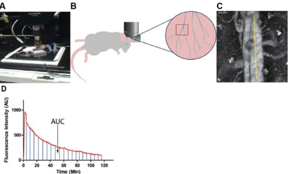
The ability to easily and inexpensively screen different formulations of nanoparticles in animal models is an unmet need in the field of nanomedicine. To address this need, we have modified an intravital microscopy technique that we recently developed to measure the pharmacokinetics of red blood cell mimetic microparticles (RBCM) (Merkel et al., 2011). This technique allows for screening the relative nanoparticle resident times in the blood using as few as four animals per condition. In order to reduce variation in nanoparticles for these experiments, we have employed the PRINT technique to generate monodisperse 300 nm cylindrical PEG hydrogel nanoparticles containing far-red fluorescent dyes for in vivo and in vitro imaging (Fig. 5); these particles are similar in size and
composition to those used in a recent study by our group (Perry et al., 2012). When injected intravenously (IV), these particles produce bright fluorescence in the vasculature of a mouse and can be easily imaged in the ear (Fig. 5A-C). A time vs. fluorescence intensity profile can then be generated, and the area under the curve (AUC) can be calculated (Fig. 5D). By comparing the AUC from
34
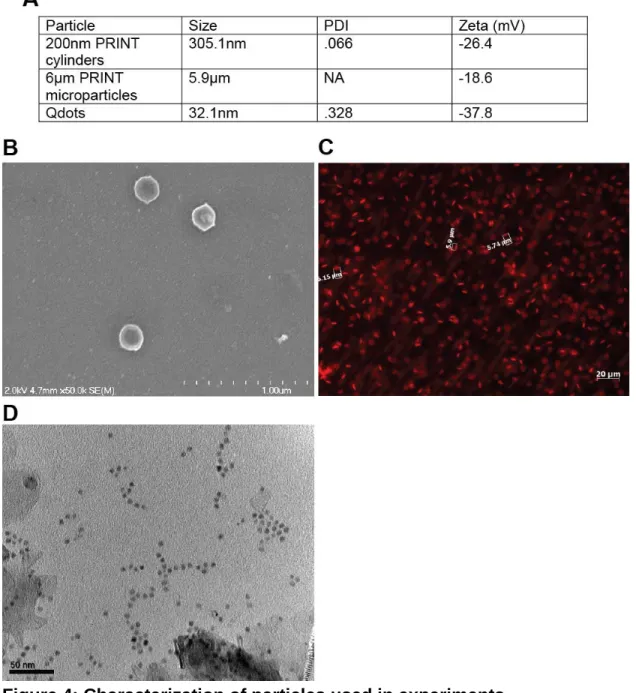
Figure 4: Characterization of particles used in experiments.
(A) Size, PDI, and Zeta potential for all particles used. Size was determined by dynamic light scattering for 200nm PRINT particles and Qdots, fluorescence microscopy was used to determine size of microparticles.
(B) Scanning electron micrograph of 200nm PRINT particles.
35
Figure 5: An Intravital Microscopy Based Assay for Screening Nanoparticle Clearance Rates in Live Animals
(A) A blood vessel is positioned in the center of the scan area.
(B) A cartoon showing the orientation of the mouse, the positioning of the objective, and the region of the mouse ear imaged.
(C) Movies are analyzed in ImageJ by selecting a straight line ROI in the large vein and average fluorescence intensity for each time point is calculated. Scale bar equals 50µm.
36
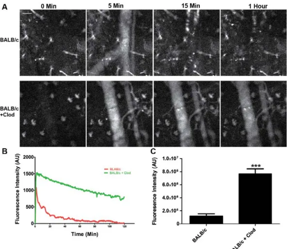
As the clearance of nanoparticles is mediated by the phagocytic cells of the mononuclear phagocyte system (MPS), we depleted these cells from female BALB/c mice using liposomal clodronate (Clod) (Camilleri et al., 1995) and continuously measured the blood fluorescence over a two hour period in treated and untreated mice (Fig. 6A). As expected, plots of average fluorescence
37
Figure 6: Treatment of BALB/c mice with liposomal clodronate increases the circulation of nanoparticles in vivo.
(A) Still images from a BALB/c mouse and a BALB/c mouse pre-treated with liposomal clodronate show significant differences in blood fluorescence from nanoparticles. Scale bar equals 50µm.
(B) A plot of mean fluorescence intensity versus time shows the difference in particle clearance rates between the two conditions (N=4).
(C) Plotting area under the curve shows a significant (p<0.003, t test) increase in fluorescence intensity in blood with clodronate pretreatment (N=4).
38
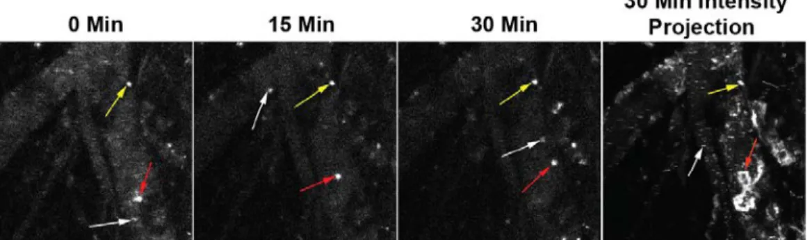
concentrated particles became visible. Upon careful observation, it became clear that these visible areas of fluorescence were cells and not extracellular
aggregates of particles, as some of them moved in a random manner including some periods of persistent migration against the blood flow (Fig. 7). Based on observations from others, it is likely that these cells are a combination of neutrophils and peripheral monocytes (Auffray et al., 2007). This uptake of particles in the peripheral vasculature could explain the common finding across many nanoparticle studies that a significant fraction of the injected particle dose is not recovered from the exsanguinated blood and major organs (Vasquez et al., 2011). Since a substantial proportion of blood vessel surface is left behind in the carcass and skin, particle clearance in the peripheral vasculature may account for some of the missing dose.
Figure 7: Peripheral immune cells clear nanoparticles in the vasculature.
Still images from representative movie (Sup. Movie 3) show the presence of three different types of cells made fluorescent by the uptake of labeled
39
Different mouse strains clear nanoparticles with different kinetics.
40
due to the MPS. As peripheral immune cells containing particles were consistently observed to a greater extent in BALB/c mice compared to other strains tested, it is possible that peripheral clearance is stronger in BALB/c mice resulting in a significant amount of particles being cleared in the first pass through the circulatory system.
Figure 8: Th1 mouse strains clear nanoparticles slower than Th2 strains.
(A) Still images from supplemental movies 1-6 show the differences in particle circulation between the four mouse strains as well as liposomal clodronate pre-treated BALB/c and C57BL/6 mice.
(B) Plots of fluorescence intensity versus time for all four mouse strains and clodronate-treated mice (N=4 mice per condition).
41
To further address differences in clearance, we performed a biodistribution study by injecting BALB/c and C57BL/6 mice with particles and then sacrificing mice at 5min, 30min, 2hr and 24hrs post injection, followed by measuring
42
Figure 9: Biodistribution of 300nm PRINT hydrogel particles in Balb/c and C57BL/6 mice.
(A) Distribution of particles in the lungs, heart, and kidneys of Balb/c and C57BL/6 mice. C57BL/6 mice showed significantly higher amounts of particle in heart and kidneys then Balb/c mice at 30min (P<.05 t-test), and 2hrs
(P<.004 t-test) (N=4).
43
(C) Relative amounts of particle present in Balb/c and C57BL/6 whole blood, C57BL/6 had significantly higher levels of particles in blood at 5min, 30min and 2hrs compared to Balb/C mice (P<.05 t-test) (N=4).
(D) Relative amounts of particle present in Balb/c and C57BL/6 plasma, C57BL/6 had significantly higher levels of particles in blood at 5min, 30min and 2hrs compared to Balb/C mice (P<.05 t-test) (N=4).
(E) Relative amounts of particle present in Balb/c and C57BL/6 blood cell fraction of whole blood, C57BL/6 had significantly higher lower of particles in blood at 5min, and 30min compared to Balb/C mice (P<.05 t-test) (N=4). Some of the differential clearance of particles is due to uptake by
granulocytes in Th2 prone strains.
44
45
46
Figure 11: Flow cytometry of peripheral blood leukocytes and splenocytes shows dramatically higher uptake of particles by granulocytes in BALB/c mice.
47
(B) Flow cytometry of splenocytes shows significantly higher uptake of particles by DC’s (P< .05, t test) and granulocytes (p<0.003, t-test) in BALB/c mice while monocytes take up significantly more particles in C57BL/6 mice (p<0.05, t-test) (N=4).
To address this idea directly, we depleted granulocytes from both strains by administering anti-Ly6-G antibodies, which has previously been shown to selectively deplete granulocytes in vivo (Arnold et al., 2010). Flow analysis confirmed that after antibody depletion, granulocyte numbers were reduced to below detection in both strains (Fig. 12). IVM was performed on both strains after granulocyte depletion to determine the effect on particle blood exposure. After granulocyte depletion, BALB/c mice showed equivalent peak fluorescence intensity compared to C57BL/6 mice (Fig. 13A), and a significant increase in particle blood exposure compared to untreated BALB/c mice (Fig. 13B). Conversely, C57BL/6 mice after granulocyte depletion showed no significant increase in peak fluorescence or particle blood exposure when compared to untreated C57BL/6 mice (Fig. 13B). These data indicate that some of the lower particle blood exposure (i.e., faster clearance) and lower peak particle
concentration in BALB/c mice during the early passage through circulation is due to particle uptake by granulocytes in the blood.
To determine the fate of particles in the blood and spleen in the absence of granulocytes, we assessed particle uptake by immune populations in
48
increased particle uptake by blood monocytes was observed in granulocyte depleted BALB/c mice, but not in C57BL/6 mice (Fig. 13C). In the spleen, granulocyte depletion did not affect monocyte uptake, but did increase macrophage uptake, with a greater increase in the BALB/c mice (Fig. 13D). Together, these data indicate that granulocytes account for some of the
increased clearance seen in the BALB/c mice, but that in the absence of these cells, BALB/c mice still clear nanoparticles faster than C57BL/6 mice.
49
Figure 13: Depletion of granulocytes significantly increases particle exposure to blood in BALB/c mice but not C57BL/6 mice.
(A) Plots of fluorescence intensity versus time for four BALB/c and C57BL/6 mice with and without monocyte depletion (N=4 mice per condition).
(B) Plots of area under the curve show a significant increase in particle exposure to blood in BALB/c mice (p<0.01, One-way ANOVA, Dunnett post-test) but not C57BL/6 mice (N=4 mice per condition).
(C) Flow cytometry analysis of changes in PBL particle distribution in BALB/c and C57BL/6 mice following granulocyte depletion. BALB/c monocytes take up
50
(D) Flow cytometry analysis of splenocytes after granulocyte depletion in BALB/c and C57BL/6 mice. Macrophages in spleen take up significantly more particles after granulocyte depletion in both strains (p<0.05, t test) (N=3 mice for
granulocyte depleted conditions and N=4 mice for controls).
Differential clearance of particles in Th1 vs. Th2 strains is dependent on
particle type.
As a wide variety of nanoformulations are being investigated for potential clinical use and often demonstrate very different properties in vivo, we tested whether the differential clearance observed in various mouse strains is specific to the PRINT particles (300 nm, PEG hydrogel). We tested the blood exposure of quantum dots (Qdots) in BALB/c and C57BL/6 mice. The quantum dots were polymer coated with a carboxylated polymer that prevents aggregation in aqueous environments. The Qdots tested are a different class of nanoparticle (inorganic nanocrystal VS hydrogel) with a different surface (proprietary carboxilated polymer vs. PEG), a different shape (sphere vs. cylinder) and an order of magnitude smaller (avg size 30nm Qdots vs. 300nm cylindrical PRINT hydrogels (Fig. 4A-C). Upon IV injection, the Qdots were more rapidly removed from blood by both BALB/c and C57BL/6 mice than PRINT particles. However, the Th1 vs. Th2 biased strain clearance difference was still present (Fig. 14A, B), including a lower peak fluorescence intensity with Qdots, suggesting that a large number of Qdots are cleared in the periphery of BALB/c mice. To test if
51
filtration in the lungs (Merkel et al., 2011). Interestingly, BALB/c and C57BL/6 mice demonstrated the same microparticle exposure to blood (Fig 14C, D), suggesting that rapid removal of nanoparticles in Th2 prone strains is not observed with larger particles requiring phagocytic clearance.
Figure 14: Depletion of granulocytes significantly increases particle exposure to blood in BALB/c mice but not C57BL/6 mice.
(A) Plots of fluorescence intensity versus time for quantum dots in blood of BALB/c and C57BL/6 mice show a similar trend to what was observed with 300 nm PRINT hydrogel particles (N=4).
(B) Area under the curve shows a significant (p<0.05, t-test) increase in blood fluorescence from quantum dots in C57BL/6 mice VS BALB/c mice.
(C) Plot of fluorescence intensity in blood versus time for BALB/c and C57BL/6 mice show similar clearance rates for microparticles.
52
Murine bone marrow derived macrophages and primary human peripheral
blood monocyte-derived macrophages take up more particles with M2
polarization.
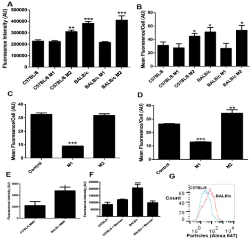
We examined if differential clearance by Th1 and Th2 prone strains can be reproduced in vitro by culturing primary bone marrow derived macrophages (BMM) from both strains and testing particle uptake by BMMs. We chose to use BMMs as opposed to granulocytes due to the fact that BMMs can be maintained in culture for longer time periods, thus allowing treatment with Th1 and Th2 cytokine mixtures to polarize these macrophages towards M1 or M2 phenotypes, respectively. We imaged the accumulation of particles inside the macrophages and quantified the integrated fluorescence intensity of particles per cell. We also processed cells for analysis by flow cytometry. Consistent with our in vivo
observations, untreated BALB/c macrophages take up significantly more particles than untreated C57BL/6 macrophages in vitro as assayed by both confocal
53
level as C57BL/6 macrophages, while M2 polarization showed no significant difference compared to BALB/c control (Fig. 15A, B).
As significant differences are known to exist between mouse and human macrophages (Mestas and Hughes, 2004),we repeated particle uptake
experiments with human monocyte-derived macrophages from two healthy human volunteers. Volunteer A’s macrophages showed high uptake of particles with no stimulation, a significant reduction in uptake with M1 polarization, and no difference with M2 polarization (Fig. 15C). Volunteer B’s macrophages showed intermediate uptake of particles with no stimulation, a significant decrease in uptake with M1 polarization, and a significant increase in uptake with M2 polarization (Fig. 15D). These results confirm that immune polarization fundamentally impacts cellular interactions with nanoparticles in human cells.
One significant difference between M1 and M2 macrophages is that M2 macrophages are known to express higher levels of many different scavenger and lectin receptors than M1 macrophages. Flow cytometric analysis of revealed increased MMR (macrophage manose receptor) expression on the surface of BALB/c macrophages (BMMs) compared to C57BL/6 macrophages (Fig. 15E), which is consistent with previously published transcriptional profiling data (35,38). To test whether the differential uptake of particles by BALB/c and C57BL/6
54
55
Figure 15: M2 polarized macrophages take up more particles than M1 macrophages in mice and humans.
(A) Average integrated fluorescence per cell showed a significant (p<0.0001, One-way ANOVA Dunnett’s post-test) increase in particle uptake by BALB/c untreated, BALB/c Th2-treated, and C57BL/6 Th2-treated VS C57BL/6 untreated cells. C57BL/6 M1-treated and BALB/c M1-treated showed no significant
difference vs. C57BL/6 untreated Cells (N=4).
(B) Flow cytometry analysis of uptake showed a significant (P<0.05, One-way ANOVA Dunnett’s post-test) increase in uptake by BALB/c untreated, BALB/c Th2-treated, and C57BL/6 Th2-treated VS C57BL/6 untreated cells. C57BL/6 M1-treated and BALB/c M1-treated showed no significant difference vs. C57BL/6 untreated cells (N=4).
(C) Flow cytometry analysis of uptake by human macrophages from volunteer A. M1 macrophages took up significantly less particles than control or M2
56
(D) Flow cytometry analysis of uptake by human macrophages from volunteer B. M1 macrophages took up significantly less than control macrophages (P<0.0001, One-way ANOVA Dunnett’s post-test). M2 macrophages took up significantly more than control macrophages (P<0.001, One-way ANOVA Dunnett’s post-test) (N=4).
(E) Flow cytometry analysis of surface MMR expression on Balb/c and C57BL/6 mice. Balb/c mice showed significantly higher surface expression of MMR (P<0.05, t-test) (N=4).
(F) Microscopy analysis of uptake by BMMs after mannan blocking. Addition of Mannan reduced uptake by Balb/c macrophages to the same levels as C57BL/6 controls (P<0.0001, One-way ANOVA Dunnett’s post-test)(N=4).
57
58 DISCUSSION
In this study, we have identified a striking difference in nanoparticle clearance in different strains of mice that arises due to differences in global immune status differences. Th1-prone strains such as C57BL/6 clear particles more slowly and have a higher blood exposure compared to the Th2-prone BALB/c strain that demonstrated rapid particle clearance. Both
monocytes/macrophages and granulocytes in the peripheral vasculature and spleen are responsible for clearance differences between the strains.
Interestingly, the differences in clearance were recapitulated in vitro using mouse and human macrophages treated with either Th1 or Th2
chemokine/cytokine mixtures. This effect may be due, in part, to differences in surface expression of scavenger receptors such as the macrophage mannose receptor (MMR).
In most studies nanoparticle PK is determined by blood draws at specific time points post injection, combined with appropriate assays for determining the particle concentration in blood (Chu et al., 2012; Kulkarni and Feng, 2013). This method has the advantage of directly measuring the particle itself rather than a reporter probe and provides absolute particle concentration in the blood.
59
needed for each condition tested. This makes blood draw assays an
unattractive option for screening multiple types of animals and/or particle types due to the high time and resource investment required. In addition, blood draw PK tends to give very limited temporal resolution, usually seven or fewer points per 24hr experiment (Caron et al., 2012).
Intravital microscopy (IVM) has a number of advantages for measuring nanoparticle clearance. With IVM, the relative amount of particles in the bloodstream is measured with a wide dynamic range allowing for
measurements spanning pre-injection to peak particle concentration through clearance in a single animal. Fluorescent probes can be directly incorporated into hydrogel nanoparticles during synthesis, therefore eliminating the need for extensive post-fabrication modifications. In addition, this technique is very fast, allowing for real time or near real time measurement of relative particle
concentrations. Finally, IVM can be multiplexed with different fluorescent dyes coupled to different particle types or formulations to measure differential clearance in the same animal. However, IVM is limited by the fact that only relative numbers are readily available from this type of experiment, the particle must be fluorescent, and it is difficult to run experiments longer than 3hrs due to the stress on the animal from extended anesthesia.
60
unaware of any previous study that has directly compared the effect of mouse strain on nanoparticle circulation times. Our results indicate mouse strain background is a critical factor for nanoparticle clearance and is likely important for the interpretation of all results of nanomedicine studies. In fact, the changes in circulation times observed between Th1 and Th2 prone mouse strains is equivalent to the changes that can be produced by heavily PEGylating particle surfaces (Perry et al., 2012). This demonstrates that changes in the immune status of patients and experimental animals may affect nanoparticle PK to at least the same degree as the material properties of particles. However, far less work has been done studying the effects of biology on nanoparticle PK than the effects of material properties on nanoparticle PK. It is perhaps not surprising that we see different results in different mouse strains for clearance, as mouse strain background has significant impact on biological processes ranging from immune function (Kastrukoff et al., 2012), pain sensation (Mogil et al., 1999) and cancer (Mesquita et al., 2012). Outbred, wild mice show a large degree of heterogeneity in many measures of immune function, while inbred lab strains of mice show very little intrastrain variation (Abolins et al., 2011). In the future, it would be beneficial for researchers in the nanoparticle field to begin explicitly considering mouse strain immunology when designing experiments and interpreting data.
61
vs. Th2 status. Numerous studies have established that Th1 and Th2 cytokines can induce macrophage differentiation into the corresponding M1 and M2 phenotypes (Gordon and Martinez, 2010). The result that Th1 prone mice (eg. C57BL/6) clear nanoparticles slower is somewhat surprising as M1
macrophages are thought to be more inflammatory, involved in the destruction of pathogens, and have been shown to be more phagocytic towards S. aureus (Krysko et al., 2011). The M2 macrophages prevalent in the fast clearing Th2 stains are generally thought to be anti-inflammatory, with involvement in wound healing and potentially less phagocytic (Gordon and Martinez, 2010). However, M2 macrophages are thought to have higher levels of endocytosis and may thereby take up small particles rapidly. In addition, M2 macrophages are known to express higher levels of scavenger and lectin receptors that could be
responsible for the increased clearance (Martinez et al., 2006). Our data on the elevated surface expression of MMR on M2 macrophages and the reduced nanoparticle uptake upon mannan treatment support this notion.
While the addition of mannan blocks the enhanced uptake of PEG
hydrogel particles in M2 macrophages in vitro, a baseline uptake of particles still occurs with treatment. Indeed, clearance in vivo likely involves multiple
62
C57BL/6 strain. Since treatment of the slow-clearing C57BL/6 mice with clodronate liposomes (removing all MPS cells) did not show a statistically significant increase in nanoparticle circulation times in a 2hr window, it is unlikely that the genetic loss of any single candidate clearance receptor would produce a measurable decrease in nanoparticle clearance. In the future, it will be essential to backcross these KO’s into the BALB/c background where these experiments are likely to produce more informative results.
It is also worth considering the role of granulocytes (primarily neutrophils) in nanoparticle clearance. Although this cell type is rarely discussed as an MPS cell (Rabinovitch, 1995; Silva and Correia-Neves, 2012), our data clearly
indicates that granulocytes play a significant role in the differential nanoparticle clearance observed between mouse strains. Previous reports indicate that BALB/c mice have greater numbers of circulating granulocytes than C57BL/6 mice (Petkova et al., 2008). Our data indicate that mere differences in
granulocyte numbers are unlikely to account for the clearance differences between strains, as a smaller percentage of C57BL/6 granulocytes take up particles than BALB/c granulocytes. Since neutrophils constitute a large fraction of circulating white blood cells in humans (Bishton and Chopra, 2004) and neutropenia is a common side effect of clinical treatments such as
63
Similar to our results with mice, the pharmacokinetics of nanoparticles is highly variable in human patients (Caron et al., 2012). Previous studies
demonstrated that monocyte function, as assayed by phagocytic capacity and oxidative burst, can be used to predict the clearance of liposomal Doxil (Caron et al., 2012). Since both the phagocytic capacity and oxidative burst are partially controlled by the patient’s global immune status (Mills et al., 2000), it is likely that the Th1/Th2 balance is an important factor in human nanoparticle
clearance as well. This is supported by our results with human macrophages that showed a significant decrease in particle uptake from both volunteers’ macrophages following M1 polarization, and one volunteer’s macrophages showing an increase in uptake with M2 polarization. Future studies are needed to explore the role of previous immune priming events such as infections or allergies on nanoparticle clearance. Our data suggest that a more
comprehensive understanding of how global immune regulation affects
64
CHAPTER FOUR: FUTURE DIRECTIONS
The nature of the interactions between nanoparticles and the immune system still requires significant study to be fully understood, appreciated and manipulated. The work presented here suggests promising avenues to be pursued and provides several valuable tools to use in future work. In this chapter I will address some, although certainly not all, possible future work that can derived from the foundation of this body of work.
Genetic Factors Controlling Th1/Th2 Polarization and Nanoparticle
Clearance
65
after identical treatment with L-media and serum which contain the same base line of cytokines. This suggests that there is some type of basal genetic
difference between BALB/c and C57BL/6 macrophages that, while correlated with Th1/Th2 prone states, exists independent of cytokine stimulation. Finally, the dramatic difference observed in neutrophil activity towards particles in BALB/c vs C57BL/6 suggests that differences between Th1 and Th2 cytokine activity is not the entire story, as short lived neutrophils in healthy mice will have seen very little in the way of cytokines during their life cycle. These three data points suggest that there is some upstream genetic difference causing the Th1/Th2 polarization, the differences in particle uptake by unstimulated macrophages, and the differences in particle uptake by neutrophils.
Determining what this driving genetic difference is could both reveal important aspects of how the immune system interacts with nanoparticles, as well as important genetic regulation of Th1/Th2 immunity and its relationship to innate immunity.
66
slow and fast nanoparticle clearance by determining which collaborative cross strains clear faster or slower, and grouping these datasets. This can then be followed by in depth genome and transcriptome analysis to determine which alleles are consistently altered in fast and slow clearing mice. Determining which genetic pathways are consistently changed in fast vs slow clearing mice may allow us to both predict candidate receptors on macrophages and
neutrophils which could be blocked to extend clearance, or pathways which could be drugged to extend particle circulation.
Determination of Gene Products Controlled by Th1/Th2 Cytokines That
Clear Nanoparticles
Ultimately the most likely way to extend the circulation time of
nanoparticles in vivo is to block the receptors on macrophages that bind and clear particles. This is preferential to drug treatments that inactivate phagocytes as it will have fewer off-target effects and leave patients less immune
suppressed. However in order to generate function blocking antibodies or FAB fragments against the relevant phagocytic receptors we must first positively identify which receptors mediate nanoparticle clearance. We anticipate that this can be accomplished in mice using two approaches. First we will need to isolate some combination of macrophages, neutrophils and monocytes from BALB/c, DBA2, C57BL/5 and B10D2 mice. From these cells RNA can be extracted and analyzed by RNA deep sequencing technology to generate an accurate