REGULATION AND FUNCTION OF THE CEREBRAL CAVERNOUS MALFORMATION 2 PROTEIN
Lisa Eileen Stalheim Crose
A dissertation submitted to the faculty of the University of North Carolina at Chapel Hill in partial fulfillment of the requirements for the degree of Doctor of
Philosophy in the Department of Pharmacology.
Chapel Hill 2009
ABSTRACT
LISA EILEEN STALHEIM CROSE: Regulation and Function of the Cerebral Cavernous Malformation 2 Protein
(Under the direction of Gary L. Johnson, Ph.D.)
Cerebral cavernous malformations (CCM) are vascular lesions of the central nervous system characterized as clusters of dilated, thin-walled blood vessels. CCM lesions are fragile and prone to vascular leakiness and rupture, leading to hemorrhages that cause seizure and stroke. Familial CCM has been shown to be genetically linked to three genes: CCM1, CCM2, and CCM3. The
proteins encoded by these genes have no apparent catalytic activity, suggesting they are scaffolds to organize and localize functional protein complexes in cells. This scaffolding function has been appreciated for CCM2, which encodes
Osmosensing Scaffold for MEKK3 (OSM). CCM2 (OSM) coordinates a signaling complex that consists of Rac1, MEKK3, and MKK3 to activate p38 in response to osmotic stimuli. The studies described here analyze the function of CCM2 in the context of cerebral cavernous malformations. Using proteomic, biochemical, and
in vivo models, we characterize CCM2 as a critical regulator of endothelial cell
co-iii
immunoprecipitation and fluorescence resonance energy transfer (FRET) between CCM1 and CCM2, implicating a common genetic and molecular pathway in CCM pathogenesis. We also characterize CCM2 as a Smurf1 binding partner. Through a novel CCM2 PTB domain – Smurf1 HECT domain interaction, CCM2 recruits Smurf1 to specific locations at the plasma membrane where it specifically degrades RhoA. Knockdown of CCM2 in brain endothelial cells leads to increased RhoA protein levels and ROCK signaling. Functionally, this leads to deficiencies in cell migration, tube formation, and maintenance of a permeability barrier. To determine the role of CCM2 in vivo, we used Danio rerio
as a model for vertebrate development. Loss of CCM2 expression leads to decreased blood flow due to restrictions and abnormalities of the aortic arch. The findings presented here indicate that CCM2 regulates protein complexes and signaling pathways important in endothelial cell function and provide insight into the molecular mechanisms involved in cerebral cavernous malformation
v
ACKNOWLEDGEMENTS
This work would not have been possible without the outstanding mentorship of Dr. Gary Johnson. Gary has provided all the tools, both experimental and intellectual, to ensure the success of this project and my success as a scientist. I am also indebted to the members of the Johnson Lab for their scientific training and support. I would especially like to thank Dr. Amy Abell who always made time to teach me new techniques, troubleshoot
experiments, discuss ideas, and be my “go-to” person for all things science. I feel very fortunate to have been surrounded by such an outstanding and creative group of scientists.
I would also like to thank the members of my thesis committee, Drs. Leslie Parise, Lee Graves, Rudy Juliano, and Adrienne Cox for their scientific guidance and assistance in writing my dissertation.
I am grateful for funding from the Integrated Vascular Biology Program, which is coordinated by Dr. Nobuyo Maeda, and a predoctoral fellowship from the American Heart Association.
I would like to thank Drs. Mark Vitha and Ron Torry at Drake University and Drs. Peter Anderson and Craig Moneypenny at University of Florida for being my first scientific mentors and for encouraging me to go to graduate school. If not for this early guidance and support I would have never considered graduate school as an option. I would also like to thank Drs. Rob Nicholas and Lee Graves for making sure I did my graduate work at UNC.
Finally, I would like to thank my Mom and Dad for teaching me all the skills I needed to be successful, Julie and Amy for always encouraging me and
vii
TABLE OF CONTENTS
LIST OF TABLES... x
LIST OF FIGURES ... xi
LIST OF ABBREVIATIONS ...xiii
CHAPTERS I. Introduction ...1
CCM1...1
CCM2...5
CCM3...7
Animal models of CCM ...8
Mus musculus...8
Caenorhabditis elegans... 10
CCM cell of origin... 11
The two-hit hypothesis ... 14
CCM2, structure and function... 15
Objectives of this project ... 16
CCM signaling pathways... 16
CCM2 protein function in vivo... 17
II. Materials and Methods ... 22
Chapter 3 ... 22
Chapter 4 ... 26
III. CCM1 and CCM2 are binding partners; implications for a
common signaling pathway in CCM ... 35 Introduction ... 35 Results ... 36
CCM2+/- cells have perturbed MAPK activation in
response to hyperosmotic shock... 36 A functional CCM2 PTB domain is necessary for
interaction with CCM1... 38 CCM1/CCM2 interaction is not dependent on the
same CCM1 NPxY sequence critical for ICAP1
interaction... 40 CCM1 contains a functional nuclear localization
sequence... 41 CCM1 localization is influenced by association with
CCM2... 42 ICAP1 influences the subcellular localization of
CCM1... 43
Discussion... 43 IV. CCM2 regulates aortic arch morphogenesis in Danio
rerio ... 52 Introduction ... 52 Results ... 53
The model organism Danio rerio has a CCM2
homolog... 53 CCM2-specific morpholinos knock down CCM2
expression... 54 CCM2 knockdown leads to decreased embryonic
blood flow... 54 Morphant embryos have normal vascular patterning
ix
CCM2 morphants have developmental defects of
the head and trunk... 57
Discussion... 58
V. CCM2 acts as a negative regulator of ROCK signaling by promoting Smurf1-mediated degradation of RhoA ... 71
Introduction ... 71
Results ... 72
CCM2 binds Smurf1... 72
CCM2 binds Smurf1 via a PTB domain - HECT domain interaction... 74
CCM2 is not a Smurf1 substrate, nor does it affect Smurf1 catalytic activity... 75
Co-expression of CCM2 and Smurf1 leads to cell rounding... 76
CCM2 localizes Smurf1 by binding the HECT domain... 77
CCM2 regulates RhoA degradation... 77
Knockdown of CCM2 in brain microvascular endothelial cells leads to dysregulation of the actin cytoskeleton... 78
CCM2 regulates endothelial cell migration... 79
Endothelial tubule formation and maintenance of a permeability barrier requires CCM2... 80
Discussion... 81
VI. Conclusion ... 97
Recent advancements ... 97
Future directions ... 100
LIST OF TABLES TABLE
3.1. FRET values for CCM1/2 interactions ... 48 5.1: The NPXY motif in the Smurf1/2 HECT domain is
xi
LIST OF FIGURES FIGURE
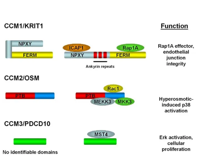
1.1: Domain structure, interacting proteins, and known function
of CCM1, CCM2, and CCM3 ... 19
1.2: Osmotic signaling to p38 via OSM/MEKK3 in mammals is analogous to signaling to Hog1 via STE50/STE11 in yeast. ... 20
1.3: The neurovascular unit... 21
3.1: MEFs heterozygous for a CCM2 gene trap allele have impaired p38 activation upon hyperosmotic stress ... 46
3.2: CCM1 associates with CCM2... 47
3.3: The CCM1/2 interaction is dependent on the CCM2 PTB domain ... 49
3.4: CCM1 subcellular localization is influenced by CCM2 and by a CCM1 NLS... 50
3.5: ICAP1 associates with CCM1 and CCM2 and sequesters CCM1 in the nucleus... 51
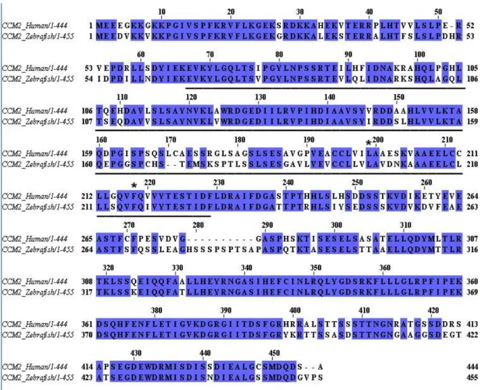
4.1: The Danio rerio CCM2 homolog... 63
4.2: CCM2 knockdown strategy ... 64
4.3: CCM2 morphant phenotype ... 65
4.4: CCM2 (OSM) morphants have decreased blood flow in comparison to wild type embryos while blood vessel diameter is unaffected... 66
4.5: CCM2 morphant embryos have normal vascular patterning and bulbous arteriosus development ... 67
4.6: Aortic arch malformation in CCM2 morphant embryos... 68
4.7: CCM2 morpholino-treated embryos have abnormal neural tube closure ... 69
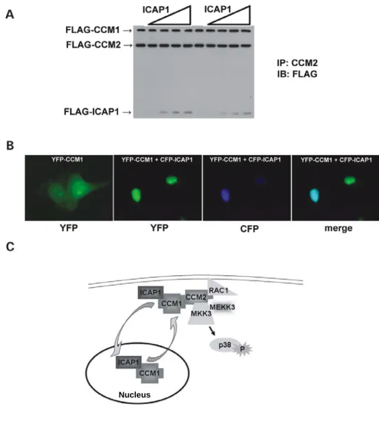
5.1: Smurf1 binds CCM2, MEKK2, and MEKK3... 86 5.2: CCM2 interacts with Smurf1 via a novel PTB domain –
HECT domain interaction... 87 5.3: CCM2 is not a Smurf1 substrate nor does it impact Smurf1
catalytic activity... 88 5.4. Expression of Smurf1 WT and CCM2 leads to cell
morphology changes ... 89 5.5: CCM2 regulates Smurf1-medated RhoA abundance ... 90 5.6: CCM2 knockdown leads to cytoskeletal changes in brain
endothelial cells ... 91 5.7: Increased association of phospho-MLC2 with stress fibers
in CCM2 knockdown cells is abrogated with treatment with
the ROCK inhibitor Y-27632 ... 92 5.8: Loss of CCM2 expression impairs directed cell migration ... 93 5.9: CCM2 is necessary for endothelial barrier function and
endothelial tubule formation... 94 5.10: Model of CCM2-Smurf1 function ... 96 6.1: Model of CCM protein signaling complexes in endothelial
xiii
LIST OF ABBREVIATIONS BAEC: Bovine aortic endothelial cell
bEND.3: Mouse brain endothelioma cell
C2: Calcium-dependent phospholipid binding domain CCM: Cerebral cavernous malformations
CCM1: Cerebral cavernous malformation 1 gene
CCM1: Protein encoded by cerebral cavernous malformation 1 gene, also known as KRIT1
CCM2: Cerebral cavernous malformation 2 gene
CCM2: Protein encoded by cerebral cavernous malformation 2 gene; also known as OSM, malcavernin
CCM3: Cerebral cavernous malformation 3 gene
CCM3: Protein encoded by cerebral cavernous malformation 3 gene; also known as PDCD10
CFP: Cyan fluorescent protein Dab: Disabled homolog 1
DAF-16: abnormal DAuer Formation-16; Caenorhabditis elegans homolog of
FOXO-1
DAF2-DA: 4,5-diaminofluorescein diacetate DAPI: 4′,6′-diamidino-2-phenylindole
DIC: Differential interference contrast E1: Ubiquitin-activating enzyme E2: Ubiquitin-conjugating enzyme E3: Ubiquitin ligase
EGTA: ethylene glycol tetraacetic acid
FRET: Fluorescence resonance energy transfer
FRETNC: Fluorescence resonance energy transfer normalized and corrected GFP: Green fluorescent protein
GST: glutathione S-transferase HA: Hemagglutinin polypeptide tag
HECT: Homologous to the E6-AP carboxyl terminus Hpf: Hours post fertilization
HUVEC: Human umbilical vein endothelial cell ICAP1: Integrin cytoplasmic adapter protein-1
KRI-1: Caenorhabditis elegans homolog of KRIT1/CCM1
KRIT1: Krev Interaction Trapped 1 MAPK: Mitogen activated protein kinase
MAP2K: Mitogen activated protein kinase kinase
MAP3K: Mitogen activated protein kinase kinase kinase MAPKK: Mitogen activated protein kinase kinase
MAPKKK: Mitogen activated protein kinase kinase kinase MEEC: Mouse embryonic endothelial cell
MEF: Mouse embryonic fibroblast MLC2: Myosin light chain 2
MO: Morpholino
NEDD4: neural precursor cell expressed, developmentally down-regulated 4 NLS: Nuclear localization sequence
xv PTB: Phosphotyrosine binding
RNA: Ribonucleic acid RNAi: RNA interference
RT-PCR: Reverse transcription polymerase chain reaction
SDS-PAGE: sodium dodecyl sulfate polyacrylamide gel electrophoresis shRNA: short hairpin RNA
siRNA: small interfering RNA
Smurf1/2: Smad ubiquitination regulatory factor 1/2
WW: Proline-rich binding domain; named for critical tryptophan (W) residues within the domain
I. Introduction
Cerebral cavernous malformations (CCM) are vascular lesions of the central nervous system characterized as clusters of dilated, thin-walled blood vessels. These vascular “caverns” are surrounded by connective tissue,
preventing innervation of the surrounding neural parenchyma (Clatterbuck et al., 2001). CCM lesions are fragile and prone to vascular leakiness and rupture, leading to hemorrhages that cause seizure and stroke (Marchuk et al., 2003; Plummer et al., 2005). In the general population, it is believed that 0.5 – 1.0% individuals will be affected by CCM in their lifetime.
Cavernous malformations (also known as cavernous angiomas) can occur sporadically or through autosomal dominant inheritance of mutations in one of three genes: CCM1, CCM2, and CCM3. Although there is incomplete
penetrance of the CCM phenotype, this familial form accounts for over 20% of CCM cases. Therefore, understanding the roles of CCM1, CCM2, and CCM3 in
cell physiology is critical for our understanding of CCM pathogenesis.
CCM1
Mutations in CCM1 lead to truncations of the protein Krev Interaction
2
al., 2002; Chen et al., 2002; Couteulx et al., 2002; Davenport et al., 2001; Eerola et al., 2001; Laberge-le Couteulx et al., 1999; Laurans et al., 2003; Lucas et al., 2001; Marini et al., 2003; Marini et al., 2004; Musunuru et al., 2003; Sahoo et al., 2001; Sahoo et al., 1999; Verlaan et al., 2002; Xu et al., 2003; Zhang et al., 2000b). It is widely believed that these mutations in CCM1 lead to loss of
function of this gene.
CCM1/KRIT1 had previously been identified in a yeast two-hybrid screen for proteins that interact with the Ras family GTPase Krev1/Rap1A. Initial characterization of CCM1 found that the carboxy- terminus of CCM1 shared a weak homology to the Band 4.1 protein. This region of homology would later be characterized as a FERM domain, based on the domains of Band four-point-one, Ezrin, Radixin, and Moesin. CCM1 also contains three ankyrin domains in the central region of the protein, assumed to be for additional protein interactions. The FERM domain of CCM1 was determined to be necessary for interaction with Krev1/Rap1A and CCM1 itself did not bind strongly to Ras. It was speculated that because of its binding to Krev/Rap1A and not Ras, CCM1 could be specifically regulating Krev/Rap1A signaling (Serebriiskii et al., 1997).
Following the initial study of CCM1 as a Rap1A binding protein, this data was contested when additional groups could not confirm this association. In these studies, a longer CCM1 bait was used. This was due to the identification of additional CCM1 exons at the 5’ end of the sequence, extending CCM1 by 207
that either the amino-terminus of CCM1 masks the Krev1/Rap1A binding site, or previous data using the shorter CCM1 needed further confirmation (Zawistowski et al., 2002; Zhang et al., 2001). The CCM1-Rap1A interaction was therefore re-examined. Using multiple biochemical assays, CCM1 was confirmed as a Rap1A binding protein, but binding was regulated by a CCM1 conformational change (Sophie Béraud-Dufour, 2007). CCM1 was found to exist in an open or closed conformation, and this conformational change regulated Rap1A binding. This work confirmed the CCM1-Rap1A interaction, and resolved the discrepancy associated with previous studies of CCM1-Rap1A interactions.
The CCM1-Rap1A interaction was determined to function as a mechanism to recruit CCM1 to endothelial cell junctions. Endogenous CCM1 localized at endothelial cell junctions, as determined by co-localization with β-catenin. Endogenous β-catenin also associated with endogenous CCM1 by
co-immunoprecipitation. Expression of an active form of Rap1A (Rap1A-G12V) increased association of CCM1 with β-catenin, while expression of an inhibitor of Rap1A activity (Rap1GAP) decreased association of CCM1 with β-catenin. Knockdown of CCM1 expression led to loss of β-catenin at cell junctions, increased stress fiber formation, and increased endothelial cell permeability. Further, expression of exogenous CCM1 was able to reverse increased
permeability induced by expression of Rap1GAP. Taken together, CCM1 plays an important role in endothelial permeability through Rap1A-mediated
4
A second binding partner for CCM1 is Integrin Cytoplasmic Adapter Protein-1 (ICAP1) (Zawistowski et al., 2002; Zhang et al., 2001). ICAP1 is a negative regulator of β1 integrin signaling implicated in osteoblast proliferation and differentiation (Bouvard et al., 2007). ICAP1 contains a Phosphotyrosine Binding (PTB) domain, which is responsible for its association with the β1 tail through a conserved Asparagine – Proline – any amino acid – Tyrosine (NPxY) motif (Chang et al., 1997; Zhang and Hemler, 1999). An NPxY motif was also found within the amino acid sequence of CCM1 (N192-Y195), suggesting that the ICAP1 PTB domain could interact with this CCM1 NPxY motif. Mutation of the CCM1 NPxY motif led to a decrease in ICAP1 binding demonstrated by GST pulldown and by liquid beta-galactosidase assay (Zawistowski et al., 2002). Because of the critical role of β1 integrins in endothelial cell function, it was hypothesized that loss of CCM1 may lead to defects in endothelial cell adhesion and migration through ICAP1 binding.
The importance of the CCM1-ICAP1 interaction was defined when ICAP1 was found to regulate the open/closed conformation of CCM1 (Sophie Béraud-Dufour, 2007). The CCM1 intermolecular interaction was shown to be between CCM1 NPxY motifs and the CCM1 FERM domain. Presumably, this was occurring between the CCM1 NPxY motifs and the F3 subdomain of the CCM1 FERM domain, which shares structural similarity to PTB domains (Calderwood et al., 2002). The interaction between the CCM1 NPxY motif and CCM1 FERM domain was lost upon addition of ICAP1. Furthermore, in GST-pulldown
complex. Based on these data, it was suggested that ICAP1 could induce the open conformation of CCM1 by binding CCM1 NPxY motifs. In the open
conformation, CCM1 would then be available to interact with Rap1A. Based on our current knowledge of CCM1 as a Rap1A effector, this would imply that ICAP1 could act to regulate the availability CCM1 as a Rap1A effector to regulate cell junctions (Figure 1.1).
CCM2
MGC4607 was identified as the CCM2 gene in 2003. In nine families
analyzed, eight unique mutations, predominantly deletions leading to frameshift changes, were found within the CCM2 gene. Based on the amino acid sequence
of CCM2, a PTB domain was identified in the amino-terminus of CCM2.
Interestingly, the amino-terminal end of CCM2, including the PTB domain,
harbored all of the mutations identified in the cohort (Liquori et al., 2003). Further analysis identified more pronounced genetic lesions in CCM2 patients, including
large deletions of almost the entire CCM2 gene (Liquori et al., 2007).
Concomitant with the discovery of MGC4607 as the CCM2 gene, the mouse homolog of CCM2 was characterized as Osmosensing Scaffold for
MEKK3 (OSM). Although there are many upstream MAPKKKs that activate p38, MEKK3 was found to be specifically involved in hypertonic activation of this pathway (Uhlik et al., 2003). MEKK3 knockdown cells were found to be deficient in their activation of p38 in response to sorbitol. OSM was found to be an
6
MEKK3 as bait, OSM was found to associate with MEKK3 and MKK3 by immunoprecipitation. Expression of OSM with MEKK3 was found to
synergistically increase MKK3 phosphorylation. Further, joint knockdown of OSM with MEKK3 synergistically decreased activation of p38 in response to sorbitol treatment. Endogenous OSM was found to associate with actin ruffles, and sorbitol treatment of COS-7 cells induced OSM and MEKK3 association in live cells by fluorescence resonance energy transfer (FRET). The GTPase Rac1 was also found to associate with OSM, and this association was induced by sorbitol treatment in live cells. Therefore, it was hypothesized that the signaling module of Rac1-OSM-MEKK3-MKK3 to activate p38 was analogous to the Hog1 osmotic signaling pathway in Saccharomyces cerevisiae (Uhlik et al., 2003).
In response to high osmotic conditions, cells rapidly lose water and shrink. Yeast counter this loss of water by accumulating intracellular glycerol through closure of glycerol channels and up-regulation of genes for glycerol synthesis. This is in part coordinated by the Hog1p mitogen activated protein kinase
20% of these genes are fully Hog1p-dependent, based on Hog1-deficient cells. Yet, the genes that are fully Hog1p-dependent include genes that are the most strongly upregulated in response to hyperosmotic stress (Hohmann et al., 2007; Willem H Mager, 2002). Therefore, the Hog1p pathway is necessary for osmotic adaptation, and this pathway is coordinated by Ste50p, the OSM homolog
(Figure 1.2).
Since OSM is the mouse homolog of CCM2, the MEKK3-p38 signaling pathway could therefore be involved in CCM pathology. Indeed, MEKK3 and p38 knockout animals have defects in vascularization in utero (Adams et al., 2000; Yang et al., 2000). However, if deficiencies in p38 signaling lead to CCM is not known.
CCM3
The CCM3 gene was documented in 2005 by two independent groups
(Bergametti et al., 2005; Guclu et al., 2005). Similar to CCM1 and CCM2, most
mutations in the CCM3 gene led to premature stop codons or gene deletions.
The CCM3 gene encodes the protein Programmed Cell Death protein 10.
CCM3/PDCD10 was first identified in a screen for genes upregulated in the human myeloid cell line TF-1 upon apoptotic induction. Analysis of the primary structure of CCM3 does not identify any functional domains. CCM3 is
ubiquitously expressed and is highly conserved, from Caenorhabditis elegans to
8
CCM3 is a binding partner for Mammalian Sterile Twenty-like 4 (MST4). MST4 is a Ste20-like kinase capable of activating the ERK MAPK pathway and promoting cell proliferation (Lin et al., 2001). CCM3 and MST4 co-localize in distinct perinuclear regions in HeLa cells. Expression of exogenous CCM3 in PC-3 cells demonstrated that CCM3 could also induce cellular proliferation. RNAi-medated knockdown of CCM3 inhibits normal cellular proliferation as well as MST4-induced proliferation. CCM3 expression increased phosphorylation of ERK, which could be abrogated by treatment with MST4 siRNA. Therefore, CCM3 and MST4 could act in a common pathway to activate ERK signaling (Ma et al., 2007). Although these studies demonstrate a clear role for CCM3 and MST4 in ERK signaling and cell proliferation, to date there has been no examination of CCM3 in endothelial cells.
Animal models of CCM
Mus musculus
In order to understand the role of the CCM genes in development and angiogenesis, gene knockout models were generated for CCM1. The first mouse
CCM1 knock-out was a CCM1 gene-trap model, generated by insertion of the
beta-galactosidase gene in CCM1 exons 6-7. The resulting CCM1-null
differences in proliferation rates of the endothelial cells in this region. Furthermore, these vessels were defective in their ability to recruit smooth muscle cells and expressed significantly less EphrinB2, Dll4, and Notch4, which
are arterial markers. It was suggested that CCM1 was necessary for arterial identity and morphogenesis (Whitehead et al., 2004).
These studies were the first to examine the physiological importance of
CCM1 in vascular development. However, this model was not adequate for
modeling CCM pathogenesis. Human CCM patients are not homozygous for mutations in CCM genes. For all CCM genes identified, patients are
heterozygous for familial mutations in CCM genes. Therefore, in order to
determine if the CCM1 knock-out mouse was a proper animal model for CCM, it
was important to determine if mice that were heterozygous for the CCM1 gene
had an increased chance of CCM formation. In 20 adult mice heterozygous for
CCM1 (aged 8 weeks to 14 months), no brain lesions were seen by gross
dissection. This suggested that a “two hit” genetic model (described below) may be needed to generate a proper mouse model for CCM. With this in mind, a
CCM1+/- Trp53-/- double knock-out mouse line was generated. Because of the
increased rate of mutation in a p53-null background, it was believed that this could induce somatic muations and increase the prevalence of CCM lesions. This rationale led to brain vascular malformations in five of the nine
10
patients (Plummer et al., 2004). This suggested that a second mutation, a somatic mutation, was necessary to model CCM1 pathogenesis in mice.
Interestingly, CCM2 heterozygous gene-trap mice displayed cerebral
hemorrhage without knockout of Trp53. The penetrance of such lesions was low
(about 10% of adult animals), but the morphology of the lesions was similar to those seen in CCM1+/-Trp53-/- knockout mice. Similar to CCM1, the CCM2
knockout was generated by insertion of a gene-trap vector into the CCM2 gene.
The insertion of βGeo into exon 6 of CCM2 caused a deletion of 45 base pairs of
this exon leading to a transcript that contained the amino terminus of CCM2 through the PTB domain, but truncation of the carboxy terminus. Similar to
CCM1 knockout mice, CCM2 homozygous null mice died during gestation. Due
to the low penetrance of lesions it was hypothesized that a two-hit model was also likely for a mouse model of CCM2 (Plummer et al., 2006). To date, no mouse models of CCM3 have been published. These mouse models show that CCM can be modeled in mice; however the molecular events that trigger CCM lesion formation are not known.
Caenorhabditis elegans
The C. elegans homologue of CCM1, KRI-1, was identified in a screen for
FOXO1. The FOXO1 homolog in C. elegans is DAF-16. Phosphorylation of
DAF-16 sequesters it in the cytoplasm, preventing its ability to regulate gene expression. Loss of KRI-1 expression in the sterile longevity model suppressed longevity. Examination of intestinal DAF-16 showed that in KRI-1 RNAi treated animals DAF-16 had reduced nuclear localization. This nuclear localization defect was independent of DAF-16 phosphorylation, as the mislocalization could be overcome by expression of a DAF-16 whose Akt phosphorylation sites were mutated to alanine. This suggested a role for KRI-1 in DAF-16 localization, either by preventing Akt phosphorylation of DAF-16, or through an independent
localization function of KRI-1 (Berman and Kenyon, 2006). However, the mechanism of this regulation has not been determined, either in C. elegans or
mammalian systems.
CCM cell of origin
CCM lesions are predominantly in the central nervous system. What causes lesions to specifically occur in the CNS? As neural, astrocytic, and pericytic innervations regulate the cerebral microvasculature (Figure 1.3), the cell of origin for CCM has been relatively controversial. An initial study of the CCM1
expression pattern in the mouse embryo suggested that CCM1 was
predominantly expressed in neural cells but not endothelial cells. Using in situ hybridization, CCM1 mRNA was ubiquitously detected in E7.5-E9.5 mouse
embryos. Later term embryos displayed predominant nervous system
12
The tissues of the cardiovascular system showed embryonic expression of
CCM1 in the heart and aorta, but not in capillaries. Furthermore, in adult human
and mouse tissues, CCM1 was expressed in the neuronal layers of the cerebral
cortex, the brainstem, and epithelial tissues. CCM1 expression was not detected
in the adult vasculature (Denier et al., 2002). Similar expression patterns were observed for CCM2 RNA. In adult murine brain tissue, CCM2 expression was
strong in neuronal populations but undetectable in brain vascular endothelium (Plummer et al., 2006). This suggested that cells other than endothelium may be involved in the pathogenesis of CCM.
A more complex view of CCM gene expression in the mouse was observed by Petit and colleagues in 2005. By in situ hybridization, strong
expression of CCM1, CCM2, and CCM3 was observed in the developing nervous
system, including the ventricular zone, intermediate zone, cortical plate and ganglionic eminence in the brain, as well as the spinal cord. Similarly, neuronal cell layers at P8 and P19 displayed strong expression of the 3 CCM genes as well as the motor neurons of the grey matter of the adult spinal cord. In the cardiovascular system, CCM1, CCM2, and CCM3 were expressed in the heart,
aorta, and cardinal vein at E10.5, and at E14.5 this expression persisted in the superior vena cava and aorta, internal carotid, and vertebral and basilar arteries. Examination at P0 showed loss of expression in these regions. In the cerebral vasculature, weak or no expression was observed prenatally, but postnatally weak expression was observed for CCM2 and CCM3 at P0 and P8. This
and transient expression of CCM genes in the cerebral vasculature and predominant expression of these genes in neuronal cells. In regards to CCM pathogenesis, a prominent role for perenchymal cells and perenchymal-endothelial cell interactions could not be discounted (Petit et al., 2006).
CCM is believed to be due to endothelial defects, based on ultrastructural data that shows defective cell to cell contacts within CCM lesions (Clatterbuck et al., 2001). This is supported by the CCM1 knockout mouse, which displayed
vascular abnormalities but normal brain development (Whitehead et al., 2004). However, mRNA expression data implies a potential role for CCM genes in neuronal cells. The cellular interactions within the cerebral vasculature are complex, including a prominent role for neuronal, astrocytic, and pericytic innervation in the microvasculature of the brain (Figure 1.3). The importance of these supporting cells has become clearer through use of conditional knockout animals. One example of the importance of the cerebrovascular support cells is with the αvβ8 integrin. Knockout of the αv integrin leads to embryonic (E10.5-E12.5) and perinatal lethality due to intracranial and intestinal hemorrhage. The αv-null embryos displayed normal vascular patterning and vascularization of the
14
knockout of the αv integrin in endothelial cells (using Tie2-Cre) did not lead to intracerebral hemorrhage, but knockout of the αv integrin in astrocytes (using GFAP-Cre) or neurons and astrocytes (using Nestin-Cre) caused intracerebral hemorrhage (McCarty et al., 2005). Further, the endothelial conditional knockout of β8 integrin displayed no cerebral hemorrhages with but did display cerebral hemorrhages in a neural-astrocytic conditional knockout model. To further elucidate what cell type is involved in the cerebral hemorrhages, a β8 integrin conditional knockout for postmitotic neurons (using Nex-Cre) was used, which did not cause cerebral hemorrhage (Proctor et al., 2005). Taken together, these studies suggested that the astrocytic interactions with endothelial cells were the cause of cerebral hemorrhage. Indeed, conditional β8 integrin knock out mice with hemorrhages had disorganized cortical glia and endothelial cells, and αvβ8 integrin was found to be localized on glial cell processes in wild type mice
(McCarty et al., 2005; Proctor et al., 2005). As CCM is a disease predominantly manifested in the blood vessels of the central nervous system, similar neural, glial, and endothelial interactions may prove important in the pathogenesis of the disease. Ideally, similar conditional knockout studies in mice will be performed with CCM1, CCM2, and CCM3 to elucidate these mechanisms.
The two-hit hypothesis
near absence of lesions in animals that are heterozygous for CCM1 or CCM2 expression. When crossed into a Trp53-/- background to induce genetic
instability and somatic mutations, mice heterozygous for the CCM1 gene-trap allele had significantly more CCM lesions than those without Trp53 knockout
(Plummer et al., 2004). Therefore it was hypothesized that there must be a second genetic hit associated with CCM lesion formation, analogous to what is observed in retinoblastoma (Goodrich and Lee, 1993).
Recent studies definitively prove the “two hit” hypothesis for induction of CCM. A recent study was able to demonstrate that bi-allelic mutations in CCM genes existed in endothelial cells from CCM lesions. These somatic mutations were not seen in cells surrounding the lesion (Akers et al., 2009). Further
support of the two hit hypothesis was a study of CCM protein immunoreactivity in CCM lesions. Loss of immunoreactivity of CCM1, CCM2, or CCM3 was
observed in endothelial cells from CCM lesions with the corresponding mutations in CCM1, CCM2, or CCM3, but not adjacent vascular beds (Pagenstecher et al., 2009). These studies show that in human CCM, mutation of both alleles and subsequent loss of expression of CCM1, CCM2, or CCM3 are critical steps in CCM pathogenesis.
CCM2, structure and function
16
superfold, which is a structural feature common to all PTB domains. As the name implies, the PH domain superfold is similar to the prototypical
phospholipid-binding PH domain. Indeed, numerous PTB domains have been shown to bind to phospholipids, likely regulating their cellular localization.
A distinguishing feature of Dab-like PTB domains is their peptide binding specificity. The canonical binding motif for PTB domains is Asn-Pro-X-Tyr (NPxY), which forms a β turn. The prototypical PTB domains of Shc and IRS-1 bind to these NPxY motifs when the tyrosine is phosphorylated. Dab-like PTB domains bind to unphosphorylated tyrosines in this motif, and in some cases prefer phenylalanine (NPxF). Therefore, interactions between Dab-like PTB domains and their interacting peptides are due to extensive hydrophobic contacts and hydrogen bonds within and surrounding the NPxY or NPxF motif.
Interestingly, the CCM2 PTB domain is most similar to the ICAP1 PTB domain, which binds to CCM1 (Uhlik et al., 2005). Thus, there is the potential that CCM2 could be utilizing its PTB domain to bind to CCM1. This would link CCM1 and CCM2 genetically and molecularly, suggesting that a common signaling pathway is involved in CCM pathogenesis.
Objectives of this project
CCM signaling pathways
Examination of the primary sequence of CCM1, CCM2, and CCM3 illustrates that these three proteins do not contain any identifiable catalytic
hypothesize that CCM pathogenesis could be caused by improper protein scaffolding and subsequent cell signaling abnormalities. Since mutations in
CCM1, CCM2, or CCM3 all lead to the same disease, there is the potential that
CCM1, CCM2, and CCM3 are involved in a common signaling pathway. This sole pathway may be responsible for CCM. It is also possible that individual pathways mediated by CCM1, CCM2, or CCM3 could lead to similar phenotypes. In order to understand these signaling pathways, we would like to determine proteins that interact with CCM2. We can then begin to elucidate the functions of these proteins both in normal cell physiology and how loss of these proteins could lead to CCM. We plan to utilize proteomic strategies to find binding partners for CCM2.
CCM proteins also need to be examined in the context of cerebral microvascular cells. To date, studies that have utilized endothelial cell culture systems have used either HUVECs or BAECs. These are good models for primary endothelial cells, but different models are needed for study of CCM. The endothelial cells of the brain are specialized to maintain the blood brain barrier, and their physiology is quite different from other endothelial cells. Because cerebral cavernous malformations lesions frequently occur in the central nervous system, the most logical cell system to use would be cerebral endothelial cells. Therefore, we plan to examine role of endogenous CCM2 in brain endothelial cells using RNAi.
18
Loss of CCM2 expression in a mouse gene-trap model leads to embryonic lethality at E9.5. Such early embryonic lethalities are difficult to study, and
mouse colony breeding and maintenance for examination of embryonic lethalities are costly and time consuming. Since CCM2 is necessary for embryonic
development, we plan to utilize a common model for vertebrate development,
Danio rerio. The zebrafish developmental model has many advantages when
20
Figure 1.2: Osmotic signaling to p38 viaOSM/MEKK3 in mammals is analogous to signaling to Hog1 via STE50/STE11 in yeast. Rac1 and OSM associate at the plasma membrane. Upon hyperosmotic stimulus, OSM coordinates a signaling complex of MEKK3 and MKK3 to activate p38. Yeast utilizes a signaling
Figure 1.3: The neurovascular unit. The microvasculature of the central nervous system contains endothelial cells supported by pericyte and astrocytic
innervations. Endothelial cells maintain tight junctions with each other as well as complex interactions with supporting cells. The cerebral vasculature also
II. Materials and Methods
Chapter 3
Plasmids and cell culture
Full-length cDNAs for human CCM1 and ICAP1α were isolated byRT– PCR from HUVEC RNA, N-terminally FLAG-tagged by PCRand subsequently cloned into the mammalian expression vector pRK5. Full-length cDNA for CCM2 was N-terminally FLAG-taggedby PCR using human IMAGE Consortium clone 3501896 was used astemplate and subsequently cloned into pRK5. For CFP and YPF fusion proteins, full-length human CCM1 cDNA was inserted in-frameto the coding sequence of EYFP of pEYFPC1 (Clontech) and full-lengthhuman ICAP1α cDNA was inserted in-frame to the coding sequence of ECFPof
pECFPC1 (Clontech). CFP-CCM2 and hemagglutinin (HA)-MEKK3have been described previously (Uhlik et al., 2003). Missense mutations weremade using QuickChangeTM (Stratagene) site-directed mutagenesiskit and confirmed by sequencing.
Immunoprecipitation and western blotting
For co-immunoprecipitation assays, 293T cells were transfectedwith the constructs listed using FuGene6TM reagent (Roche). Transfected 293T cells or MEFs were lysed ina buffer containing 0.6% Triton X-100, 137 mM NaCl, 10% glycerol and 20 mM Tris pH 7.4 supplemented with CompleteTM protease inhibitor tablets (Roche). About 500 µgof cell lysate was incubated with a rabbit
polyclonal CCM2 (OSM)antibody (Uhlik et al., 2003) with gentle rocking at 4°C for 2 hours followed by incubationwith protein G-sepharose 4B beads
(Amersham) at 4°C for 1 hour. The beads werewashed five times with lysis buffer, boiled for 5 min inSDS-loading buffer and resolved by SDS–PAGE. For westernblotting, the following antibodies were used: anti-FLAG antibody (M2, Sigma), anti-HA antibody (Sigma), and affinity-purifiedanti-CCM1 antibody (Bethyl Laboratories, Inc.).
Fluorescence resonance energy transfer
For live-cell fluorescence microscopy and CFP-YFP FRET experiments, COS-7 cells were grown on 25 mm round glass coverslips. Cells were
24
2000). Briefly, images for YFP excitation/YFP emission,CFP excitation/CFP emission and CFP excitation/YFP emission(raw FRET) were obtained using an Axiovert 200M microscope from Zeiss (Germany) and imaging software from Intelligent Imaging Innovations (Denver, CO). Background-subtractedYFP and CFP images were then fractionally subtracted from raw FRET images on the basis of measurements for CFP bleed-through(CCM2=0.55 and CFP-CCM2 F217A=0.52) and YFP cross-excitation(CCM1=0.021 and YFP-CCM1 N192A=0.024). This fractional subtractiongenerated corrected FRET images (FRETC) that function as visual representationsof interacting proteins.
MEF isolation
E10.5 primary MEF cultures were established from wild type embryos and littermate embryos heterozygousfor a LacZ gene trap insertion in the mouse
Ccm2 gene (cellline RRG051, Bay Genomics, allele designation
Ccm2Gt(pGt0Lxf)1Dmar). Cells were maintained in Iscove's modified Dulbecco's
medium(IMDM, Invitrogen)/10% FBS (Invitrogen) and supplemented with penicillin/streptomycin(Invitrogen) and amphotericin B (GensiaSicor Pharmaceuticals)at the manufacturers' recommended concentrations.
Established MEF cell lines were genotyped by PCR using the following primers: CATCCCTGTCTGGGAACCTA-3' (CCM2 intron 6, reverse),
MAPK activation assay
Spontaneously immortalized wild type and CCM2+/– MEFs were
serum-starved in OptiMEMTM (Invitrogen) overnight. Cells were then treated with 0.2 M sorbitol or 10 µg/ml anisomycin for the indicated times and harvestedin lysis buffer containing 0.6% Triton X-100, 137 mM NaCl,10% glycerol and 20 mM Tris pH 7.4 supplemented with CompleteTM protease inhibitor tablets (Roche). Fifty µg of cell lysate was resolved by SDS–PAGE. For westernblotting, anti-p38 MAP kinase (Santa Cruz Biotechnology, sc-7149) and anti-phospho-p38 MAP kinase (Thr180/Tyr182, Cell Signaling, #9211) were used. Phosphorylatedp38 was quantified using densitometry and normalized to levelsof total p38 to yield an activation index. Fold inductionwas calculated as the activation index of the sample dividedby the activation index of the untreated sample. For add-back experiments, CCM2+/– MEFs weretransfected with pRK5-FLAG-CCM2 using the
Amaxa NucleofectorTM. Cells were used 24 hours post-transfection for p38 activation experiments.
Immunofluorescence
COS-7 cells were grown and transfected on fibronectin-coated coverslips (Becton Dickinson). Cells werewashed with phosphate-buffered saline (PBS) and subsequently fixed for 14 minutesin 3% paraformaldehyde/3% sucrose/PBS, washed three times withPBS and permeabilized for 6 min in 0.1% Triton
26
minutes in a blocking solution consisting of DMEM containing 10% goat serum. Coverslipswere then incubated for 2 hours at room temperature with anti-FLAG antibody (M2, Sigma) diluted in blocking solution. After washing withPBS, coverslips were incubated for 1 hour at room temperaturewith goat anti-mouse antibody conjugated to AlexaFluorTM 594 (1:1000, MolecularProbesTM,
Invitrogen). After final washes with PBS, coverslipswere mounted with ProLong GoldTM antifade reagent (MolecularProbesTM, Invitrogen).
Chapter 4
Zebrafish husbandry
Wild-type AB* Danio rerio were handled in accordance with IACUC
approved protocols. Embryos were obtained from natural spawning. Embryos were raised in 30% Danieau solution (17 mM NaCl, 0.21 mM KCl, 0.12 mM MgSO4, 0.18 mM Ca(NO3)2, 1.5 mM HEPES pH 7.6) and incubated at 28.5º C.
Morpholino injection:
Morpholinos were purchased from Gene Tools (Philomath, OR).
Morpholinos were designed to the translational start site of the Danio rerio CCM2
gene. Two CCM2-specific morpholinos (MO) were used: MO #1: ATCTAATACAGCGAAAATGAAGAGC, MO #2:
injection (57 mM NaCl, 0.7 mM KCl, 0.4 mM MgSO4, 0.6 mM Ca(NO3)2, 5 mM HEPES pH 7.6). Embryos were injected between the 1- and 4- cell stages.
DAF2-DA labeling
The fluorescent nitric oxide (NO) indicator 4,5-diaminofluorescein
diacetate (DAF-2DA) was a gift from Dr. Margaret Kirby (Duke University). Live embryos were incubated in 10 μM DAF-2DA for 4 hours in the dark at 28.5º C. After incubation, embryos were then euthanized and imaged for DAF2-DA fluorescence.
Blood flow measurements
In silico microangiography was performed as previously described (Malone et al., 2007).
Chapter 5
Cell culture
HEK293 and COS-7 cells were maintained in Dulbecco’s modified Eagle medium (DMEM, Gibco) supplemented with 10% fetal bovine serum (FBS, Gibco) and penicillin/streptomycin (Gibco). Microvascular brain endothelial cells (bEND.3) cells were purchased from ATCC and also cultured in DMEM
28
Lipofectamine (Invitrogen) according to the manufacturer’s instructions. Human umbilical vein endothelial cells (HUVEC) and EBM-2 growth medium were purchased from Lonza. HUVECs were used below passage five.
Antibodies
Western blotting was performed using the following antibodies: polyclonal FLAG (F7425, Sigma), monoclonal M5 FLAG (Sigma), polyclonal anti-HA (sc-805, Santa Cruz), monoclonal anti-anti-HA (12CA5), anti-MEKK3 (#1673-1, Epitomics), anti-CCM2 (described in (Uhlik et al., 2003)), anti-GFP (sc-8334, Santa Cruz), anti-Smurf1 (#2174, Cell Signaling), anti-Erk2 (sc-154, Santa Cruz), anti-ubiquitin (sc-8017, Santa Cruz), anti-MEKK2 (described in (Kesavan et al., 2004)), anti-RhoA (#05-822, Millipore), anti-Rac1 (#05-389, Millipore), donkey anti-Rabbit (Jackson Immunoresearch), and sheep anti-Mouse (GE). For immunofluorescence, the following reagents were used: anti-phospho (Ser19)-myosin light chain 2 (#3671, Cell Signaling), rhodamine-phalloidin (Molecular Probes), FITC Donkey anti-rabbit (Molecular Probes), and DAPI (4′,6′ -diamidino-2-phenylindole; 0.04 ng/ml).
Plasmids
Research Institute, Mount Sinai Hospital). pCMV Myc-RhoA was a gift from Dr. Keith Burridge (University of North Carolina). pLL5.0 was a gift from Dr. Jim Bear (University of North Carolina). pCMV5B Flag-Smurf2 C716A and
pGEX4T1-Smurf1 C699A were purchased from Addgene (Addgene plasmids 11747 and 13503, respectively).
pcDNA Flag-Smurf1ΔHECT and pcDNA Flag-Smurf1 HECT were
constructed by amplifying Smurf1 (bp 28-1104 for Smurf1ΔHECT, bp 1105-2274 for Smurf1 HECT) by PCR from pCMV5 Flag-Smurf1 using Smurf1-specific primers engineered with flanking HindIII restriction sites. The resulting PCR products were digested with HindIII (New England Biolabs) and ligated into the HindIII site of pcDNA3. Smurf1 missense mutants were generated using the Quikchange Mutagenesis Kit (Stratagene) according to the manufacturer’s instructions. Plasmids were confirmed by DNA sequencing (UNC-CH Genome Analysis Facility).
RNAi
30
concentration) was added before applying the supernatant to bEND.3 or MEEC cells set in 6-well plates. At 24 hours after the final infection, cells were either used for experiments or selected with 2 ug/mL puromycin for 2 days.
FLAG-CCM2 pulldown
Flag-CCM2 pulldown experiments were done exactly as described in (Hilder et al., 2007).
Immunoprecipitation and Western blotting
Cells were washed once with PBS and subsequently lysed on ice in 1% NP-40, 20 mM Tris at pH = 7.5, 100 mM NaCl, 10% glycerol, 0.1 mM EDTA, 0.1 mM EGTA 10 μg/mL leupeptin, 2 μg/mL aprotinin, 1 mM Na3VO4, 1 mM
phenylmethylsulfonyl fluoride. Cell lysates were cleared by centrifugation for 10 minutes at 4º C. Proteins were resolved by SDS-PAGE on either 8% or 12% polyacrylamide gels and subsequently transferred to nitrocellulose.
For immunoprecipitation, 300-500 μg of protein lysate was incubated with 3-5 μg of either anti-FLAG (F7425, Sigma) or anti-HA (sc-805, Santa Cruz) for 2 hours at 4º C with gentle rocking. 40 μL of a 1:1 dilution of Protein G Separose (Invitrogen) was added and incubated for another hour at 4º C. Sepharose beads were then washed five times with lysis buffer and boiled for five minutes in SDS loading buffer before resolving the immunoprecipitated proteins by SDS-PAGE.
COS-7 cells were grown and transfected on square coverslips. bEND.3 cells were grown on Matrigel-coated glass coverslips (1:50, BD Biosciences) and subsequently washed with PBS and fixed for 14 minutes with 3%
paraformaldehyde/3% sucrose in PBS. Cells were then permeabilized for 6 minutes with 0.1% Triton X-100 in PBS. After blocking in 10% donkey
serum/DMEM, coverslips were incubated in primary antibody diluted in blocking buffer for 2 hours at room temperature. Coverslips were washed with PBS and incubated with secondary antibody and DAPI for 1 hour at room temperature. After washes in PBS, coverslips were mounted in 90% glycerol/OPDA and sealed with nail polish. Imaging was performed using an Axiovert 200M
microscope from Zeiss (Germany) and imaging software from Intelligent Imaging Innovations (Denver, CO). To determine phospho-MLC2 association with stress fibers, five representative images for each experimental condition were masked for rhodamine-phalloidin intensity and the mean intensity of phospho-MLC2 was measured on this mask.
In vitro ubiquitination assay
The in vitro ubiquitination assay was adapted from (Wang et al., 2006).
32
for 15 minutes at room temperature before ending the reaction with 2X SDS-PAGE loading buffer and boiling for 5 minutes. The reaction products were then analyzed by Western blot for ubiquitinated species.
In vitro wound healing assay
bEND.3 cells stably expressing pLKO.1 or CCM2 shRNAs were transduced with pLL5.0 for expression of GFP. Cells were grown in 24-well dishes to a monolayer. Using a P200 pipet tip, a wound across the cell
monolayer was made across the center of the well. The cells were then washed once with growth medium and incubated at 37º in the BD Pathway High Content Imager. Wounds were imaged on GFP fluorescence every 15 minutes for 16 hours on a 10X objective. Percent wound healing was determined as the area of the wound remaining after 16 hours divided by the initial area of the wound (ImageJ).
Real-time RT-PCR
Twenty-four hours after the final infection of bEND.3 cells or twenty-four hours following puromycin selection, total RNA was isolated using the RNeasy Mini Kit (Qiagen) according to the manufacturer’s directions. From this
(Mm00524581_m1) and Mus musculus beta-actin Taqman probe were
purchased from Applied Biosystems. Data were collected on the Applied
Biosystems 7500 Fast System. Samples were run in triplicate and standardized to beta-actin.
GST-pulldown assay
GST-Smurf1 C699A was produced exactly as described in (Wang et al., 2006). His-CCM2 and His-MEKK3 were purified as described previously (Uhlik et al., 2003). GST-pulldown experiments were done as described in (Nakamura et al., 2006).
In vitro tube formation assay
In a 24 well plate, 350 uL of growth factor-reduced Matrigel (BD
Biosciences) was polymerized at 37º for 30 minutes. After polymerization, 1.69 x 10⁵ cells/well were placed on top of the Matrigel and incubated at 37º for 16 hours.
HUVEC Permeability Assay
For permeability assays, 12-mm, 0.4-μm pore Transwells (Corning) were coated in Matrigel (BD Biosciences; 1:100 dilution in PBS) for 1 hour. During this time, HUVECs were trypsinized and 5 x 10⁵ cells per condition were
ON-34
TARGETplus SMARTpool siRNA (Dharmacon) was added. The suspension was electorporated using program M-003 on the Nucleofector Device (Amaxa). The cells were removed from the cuvette, added to 900 μL of growth medium, and allowed to recover at 37ºC for 10 minutes. The cell suspension was then split between two Matrigel-coated Transwells; 500 μL of cells were plated in the upper chamber with 1.5 mL of growth medium in the lower chamber. The growth
medium was changed after 24 hours. Forty-eight hours post-electroporation, the medium from both chambers was removed and replaced with serum- and growth factor-free EBM-2. FITC-dextran (40 kD molecular weight; Invitrogen) was added to the upper chamber at a final concentration of 0.625 mg/mL. As a control to induce permeability, EGTA at a final concentration of 4 mM was added to one set of control cells to disrupt the Ca2+-dependent cell-cell adhesion
III. CCM1 and CCM2 are binding partners; implications for a common signaling pathway in CCM
Introduction
Three genes have been identified in the autosomal dominant form of cerebral cavernous malformations: CCM1, CCM2, and CCM3 (Bergametti et al.,
2005; Denier et al., 2004; Laberge-le Couteulx et al., 1999; Liquori et al., 2003; Sahoo et al., 1999). The cellular functions of the CCM1, CCM2, and CCM3 proteins are largely uncharacterized, although CCM1 and CCM2 have both been implicated in cell signaling processes by the proteins that they bind. CCM1 binds the GTPase Rap1A and the integrin adaptor ICAP1 suggesting a role for CCM1 in integrin mediated adhesion and signaling (Serebriiskii et al., 1997; Zawistowski et al., 2002). The mouse homolog of CCM2 was characterized as Osmosensing Scaffold for MEKK3 (OSM). OSM binds Rac1, MEKK3, and MKK3 to activate p38 in response to osmotic stress (Uhlik et al., 2003). CCM3 binds a Ste20-like kinase and is associated with activation of Erk signaling (Ma et al., 2007). However, the molecular mechanism of how loss of CCM1, CCM2, or CCM3 leads to CCM is not known.
36
protein interaction domains. CCM1 contains a C-terminal FERM domain and multiple ankyrin repeats. CCM2 contains a PTB domain in its N-terminus. Mutations in CCM genes are mostly frameshift or nonsense mutations.
Therefore, mutations in CCM1, CCM2, or CCM3 could lead to truncated proteins
or loss of protein expression leading to loss of protein complex formation and impaired cell signaling.
In this study, we show that CCM1 and CCM2 are binding partners. The CCM2 PTB domain is necessary for a canonical interaction with NPxY motifs within CCM1. We provide evidence of co-immunoprecipitation and fluorescence resonance energy transfer (FRET) between CCM1 and CCM2, implicating a common genetic and molecular pathway in CCM pathogenesis.
The data presented in this chapter was done in collaboration with Drs. Jon Zawistowski and Mark Uhlik and was published in Human Molecular Genetics in 2005 (Zawistowski et al., 2005).
Results
CCM2+/- cells have perturbed MAPK activation in response to hyperosmotic
shock
CCM2 is an osmosensing scaffold for MEKK3-mediated p38 MAPK phosphorylation (Uhlik et al., 2003). We therefore assessed the role of
RRG051) has a LacZ insertion in exon 6 of CCM2 which is predicted to be a loss
of function mutation (Plummer et al., 2006). Heterozygous MEFs were used for these experiments because they are the genotypic equivalent of familial CCM patients. We first verified that there was reduced CCM2 protein in heterozygous MEFs by western blot with CCM2 antibody (Figure 3.1A). Cells heterozygous for the CCM2 mutation had significantly less activated p38 MAPK in response to a
sorbitol time course as assessed by p38 Thr180/Tyr182 phosphorylation (Figure 3.1A-B). This decrease of p38 phosphorylation was restored in experiments where exogenous FLAG-CCM2 was transfected into CCM2 +/- MEFs (CCM2 add-back, Figure 3.1B), demonstrating that the effect was specific to the
reduction of CCM2. These results are consistent with previous work implicating CCM2 in this signaling pathway (Uhlik et al., 2003).
CCM1 associates with the CCM2/MEKK3 protein complex
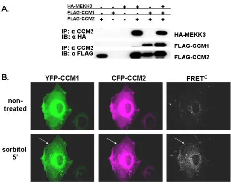
CCM2 acts as a scaffold for MEKK3-mediated p38 MAPK phosphorylation following sorbitol stimulation (Uhlik et al., 2003), but the role of CCM1 in this pathway is not known. To determine whether CCM1 is a member of the CCM2/MEKK3 scaffold complex, we expressed epitope-tagged CCM1, CCM2 and MEKK3, and examined associating proteins by immunoprecipitation with anti-CCM2 antibody. All three molecules were detected in the complex (Figure 3.2A).
38
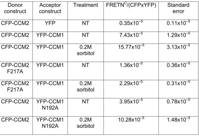
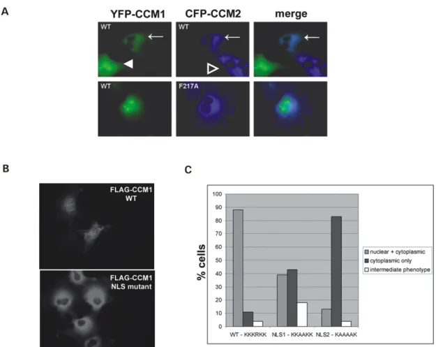
examine the potential for the CCM1 and CCM2 to associate, we performed YFP-FRET analysis in live COS-7 cells. Cells expressing YFP-CCM1 and CFP-CCM2 yield a FRETNC value of 7.43 x 10⁻⁵ (s.e.m. = 1.29 x 10⁻⁵) while control cells expressing YFP and CFP-CCM2 yield a FRETNC value of 0.35 x 10⁻⁵ (s.e.m. = 0.11 x 10⁻⁵ ) (Table 3.1). Visualization of the corrected FRET value revealed a diffuse interaction signal throughout the cytoplasm (Figure 3.2B).
We then determined if an osmotic stimulus would affect CCM1/CCM2 association. Cells expressing YFP-CCM1 and CFP-CCM2 treated with sorbitol displayed a two-fold increase in the FRETNC value over non-treated cells (Table 3.1). Localization of CCM1-CCM2 complexes shifted from diffusely cytoplasmic to the cell periphery as a consequence of sorbitol stimulation as revealed by visualization of the corrected FRET signal (Figure 3.2B). Re-localization and interaction of CCM1 and CCM2 may reflect a functional role for these proteins at the plasma membrane.
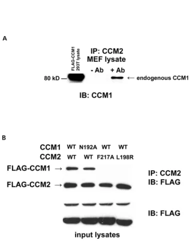
To confirm the interaction of epitope-tagged CCM1/2, we performed co-immunoprecipitation experiments with endogenous protein. Endogenous CCM1 was efficiently co-immunoprecipitated with CCM2 from MEF lysates (Figure 3.3A). Therefore, we conclude that CCM1 and CCM2 are binding partners in
vivo.
A functional CCM2 PTB domain is necessary for interaction with CCM1
we hypothesized the CCM1/CCM2 association was occurring through a
canonical NPxY/PTB domain interaction. The CCM2 PTB domain is most similar to the Dab-like PTB domains (Uhlik et al., 2005). Based on the structure and binding sites of these domains, we engineered a mutation into CCM2 PTB binding pocket that would be predicted to loose association with NPxY motifs. Point mutation of the corresponding residue in CCM2 (F217A) completely abrogated the ability of FLAG-CCM1 to co-immunoprecipitate with FLAG-CCM2 (Figure 3.3B), suggesting that PTB domain integrity is critical for the CCM1/2 interaction.
We confirmed the effect of the CCM2 F217A mutation on the CCM1/2 interaction using FRET. Consistent with immunoprecipitation, COS-7 cells co-expressing YFP-CCM1 and CFP-CCM2 F217A had reduced FRET values
(FRETNC= 1.355 X 10⁻⁵, s.e.m = 0.355 X 10⁻⁵) when compared to the FRET value for the wild type CCM1/2 proteins (FRETNC = 7.43 x 10⁻⁵, s.e.m. = 1.29 x 10⁻⁵) (Table 3.1). We observed a similar loss of FRET following sorbitol
stimulation (CCM2-F217A FRETNC = 2.29 x 10⁻⁵, s.e.m. = 0.307 x 10⁻⁵ ; WT FRETNC = 15.77 x 10⁻⁵ , s.e.m. = 3.13 x 10⁻⁵) (Table 3.1).
Germline mutations in the CCM1 and CCM2 genes have been exclusively
40
capable of association with CCM1 as determined by co-immunoprecipitation (Figure 3.3B). Loss of association of CCM1 with the patient mutation CCM2 L198R suggests that the CCM1-CCM2 interaction may be relevant in CCM pathogenesis.
CCM1/CCM2 interaction is not dependent on the same CCM1 NPxY sequence
critical for ICAP1 interaction
Previous work has shown that ICAP1, another PTB domain-containing protein, associates with CCM1 through an ICAP1 PTB domain – CCM1 NPxY motif interaction (Zawistowski et al., 2002). CCM1 contains 3 NPxY motifs. Therefore, we wanted to determine if CCM2 and ICAP1 were binding to the same site on CCM1, or if they could both associate with CCM1 at the same time. We tested this hypothesis by co-immunoprecipitation and found that mutagenesis of the critical asparagine (N192A) residue in the CCM1 NPxY motif critical for ICAP1 binding did not disrupt the CCM1/2 association (Figure 3.3B). FRET experiments found that CCM1-N192A and CCM2 interactions were diminished relative to wildtype CCM1 and CCM2, but interaction was not completely
abolished (Table 3.1). The FRET data indicate that the N192A CCM1 mutation has a slight effect on CCM1/2 binding, while in the context of
ICAP1. ICAP1 overexpression did not prevent CCM1 from associating with CCM2; instead, all three molecules were present in the complex precipitated with the CCM2 antibody (Figure 3.5A). This suggests that ICAP1 and CCM2 do not bind to the same CCM1 binding site and ICAP1, CCM2, and CCM1 can
associate as a protein complex.
CCM1 contains a functional nuclear localization sequence
The interaction of CCM1/2 prompted us to compare the subcellular localization of CCM1 with CCM2. Endogenous CCM2 localizes to actin-rich membrane ruffles (Uhlik et al., 2003). In contrast, we observed a whole cell distribution for YFP-CCM1 in COS-7 cells. We hypothesized that the nuclear localization of the CCM1 molecule may be due to a nuclear localization sequence (NLS). Examination of the primary sequence of CCM1 identified a putative NLS using the PSORT II program (EXPASY). A “pat4” four-residue pattern, similar to the NLS of the SV40 large T antigen, was predicted at residues 46, 47 and 48 in the unique N-terminal region of CCM1 (corresponding to 46-KKKRKK-51). In addition, a “pat4” sequence was detected at residue 569 within the FERM domain of CCM1 (corresponding to KKHK).
42
NLS mutant 1 was reduced to ~ 40% from the ~ 90% of wild-type FLAG-CCM1 cells displaying nuclear localization, while nuclear localization for the more severe FLAG-CCM1 NLS mutant 2 was reduced to ~ 10% of the cells (Figure 3.4C). These data suggest that the putative N-terminal NLS in CCM1 is functional, and suggest that CCM1 may have nuclear-cytoplasmic shuttling capability.
CCM1 localization is influenced by association with CCM2
In addition to contributions of the CCM1 NLS to CCM1 subcellular
localization, we hypothesized that association with CCM2 could influence CCM1 subcellular localization. In COS-7 cells, CCM1 exhibits whole cell localization, while CCM2 exhibits exclusively cytoplasmic localization. In cells co-expressing YFP-CCM1 and CFP-CCM2, YFP-CCM1 nuclear localization is lost and
experiments suggesting a nuclear-cytoplasmic shuttling role for CCM1. We conclude that CCM1 can act as a nuclear-cytoplasmic shuttling protein and this shuttling of CCM1 can be regulated by CCM2.
ICAP1 influences the subcellular localization of CCM1
We have demonstrated that CCM2 is capable of influencing the
localization of CCM1. ICAP1, which also binds CCM1, may similarly influence CCM1 localization. When expressed alone, CFP-ICAP1 exhibits predominantly nuclear localization. When co-expressed with CFP-ICAP1, YFP-CCM1
localization shifts from total-cell to exclusively nuclear (Figure 3.5B). Thus, through PTB-domain interactions, ICAP1 and CCM2 are capable of influencing the localization (and presumably function) of CCM1.
Discussion
CCM1 and CCM2 have been implicated in the disease cerebral cavernous
malformations. Although mutation of these genes leads to the same disorder, there has been insufficient data to understand the mechanism of the
pathogenesis of CCM. In this work, we provide evidence that CCM1 and CCM2 are binding partners, placing them in the same molecular pathway. This
interaction is abrogated by a patient mutation in CCM2 (CCM2 L198R)
44
The characterization of CCM2 as Osmosensing Scaffold for MEKK3 (OSM) has shed new light on the potential signaling mechanisms that are impacted by loss of CCM2 expression. OSM was found to coordinate osmotic stress-induced p38 activation via Rac1, MEKK3, and MKK3. Indeed, MEKK3 and p38 knockout mice have shown that these signaling pathways are necessary for vascular development and integrity (Adams et al., 2000; Deng et al., 2007; Yang et al., 2000). In this work we have shown that CCM2 heterozygous MEFs are defective in their ability to activate p38 in response to osmotic stress. Importantly, exogenous expression of CCM2 led to a complete recovery of this phenotype. Further, in this work we demonstrated that CCM1 can associate with the CCM2-MEKK3 protein complex, suggesting a role for CCM1 in MEKK3 mediated signaling pathways. Impaired p38 activation may influence
downstream p38-specific transcriptional activation involved in the maturation of new vessels and for the maintenance of the existing vessel architecture.
Therefore the loss of p38 activation may ultimately contribute to the formation of the misshapen vascular beds of the cerebral cavernous malformation.
Until now, the localization of CCM1 had not been examined. Our
demonstration of CCM1 as a nuclear-cytoplasmic shuttling protein opens a new avenue of study of CCM1 function and regulation. We also demonstrate that the CCM1 binding partners ICAP1 and CCM2 play a role in the localization of CCM1. ICAP1 appears to recruit CCM1 to the nucleus, while CCM2 sequesters CCM1 in the cytoplasm. ICAP1 and CCM2 both utilize their PTB domains in their
CCM2 may dictate the cellular localization of CCM1, or there may be a more complex signaling event associated with changes in CCM1 localization. These CCM1-ICAP1 and CCM1-CCM2 complexes clearly warrant further study to determine the nuclear and cytoplasmic roles of CCM1.
ICAP1 is known to regulate integrin-mediated signaling events. Activation of β1 integrins has been shown to recruit ICAP1 to the cytoplasm where it acts as an adaptor protein. ICAP1 is also capable of redistributing to the nucleus after integrin engagement. Therefore, the shuttling of ICAP1-CCM1 complex could be regulated by integrin signaling. In the cytoplasm, CCM1 could associate with CCM2 to control MAPK activation in response to integrin-mediated signaling. In this way a CCM1-CCM2 complex would be necessary to bridge signaling
between integrins and MAPK pathways. It would then be reasonable to believe that loss of expression of CCM1 or CCM2, as is predicted in CCM patients, would lead to abhorrent integrin-mediated signaling. Indeed, endothelial cells in sections of CCM lesions appear to have loss of cell-to-cell contact (Clatterbuck et al., 2001), suggesting loss of adhesion.
46
Figure 3.1: MEFs heterozygous for a CCM2 gene trap allele have impaired p38 activation upon hyperosmotic stress. (A) Western blotting with anti-CCM2
Figure 3.2: CCM1 associates with CCM2. (A) CCM1 is identified in a ternary complex with CCM2 and MEKK3. 293T cells were transfected with the indicated constructs, immunoprecipitated with anti-CCM2 antibody and subjected to western blotting with the indicated antibodies. In addition to HA-MEKK3 and CCM1 singly co-immunoprecipitating with CCM2, a FLAG-CCM1/FLAG-CCM2/HA-MEKK3 ternary complex is detected. Approximate molecular weights are FLAG-CCM1: 85 kDa and FLAG-CCM2: 60 kDa. (B) CCM1/CCM2 interact in live cells and this interaction is enhanced upon
hyperosmotic stress. COS-7 cells transfected with CFP-CCM2 and YFP-CCM1 were visualized at room temperature and analyzed by FRET. CFP fluorescence, YFP fluorescence and corrected FRET (FRETC) were examined for non-treated cells (top) and cells were treated with 0.2 M sorbitol for 5 min (bottom). Non-treated cells yield a diffuse FRETC signal throughout the cytoplasm. Cells treated with sorbitol display a re-distribution of CFP-CCM2 and YFP-CCM1 to the cell
48 Donor
construct construct Acceptor Treatment FRETN
C/(CFPxYFP) Standard error
CFP-CCM2 YFP NT 0.35x10⁻⁵ 0.11x10⁻⁵
CFP-CCM2 YFP-CCM1 NT 7.43x10⁻⁵ 1.29x10⁻⁵
CFP-CCM2 YFP-CCM1 0.2M
sorbitol 15.77x10
–5 3.13x10⁻⁵
CFP-CCM2 F217A
YFP-CCM1 NT 1.36x10⁻⁵ 0.36x10⁻⁵
CFP-CCM2 F217A
YFP-CCM1 0.2M sorbitol
2.29x10⁻⁵ 0.31x10⁻⁵
CFP-CCM2 YFP-CCM1 N192A
NT 3.95x10⁻⁵ 0.78x10⁻⁵
CFP-CCM2 YFP-CCM1 N192A
0.2M sorbitol
10.28x10⁻⁵ 1.48x10⁻⁵
Table 3.1. FRET values for CCM1/2 interactions. COS-7 cells were treated for 5 min with 0.2 M sorbitol and compared with FRET interaction values of
non-treated (NT) cells. Multiple cells were visualized to obtain normalized
50
Figure 3.4: CCM1 subcellular localization is influenced by CCM2 and by a CCM1 NLS. (A) The CCM2 PTB domain regulates CCM1 localization. COS-7 cells were transfected with CFP-CCM2 or CFP-CCM2 F217A with YFP-CCM1 and fluorescence was examined in live cells at room temperature. YFP-CCM1
Figure 3.5: ICAP1 associates with CCM1 and CCM2 and sequesters CCM1 in thenucleus. (A) ICAP1 does not compete with CCM2 for binding to CCM1, but is a member of a ternary complex with CCM1 and CCM2. 293T cells were
transiently transfected with the indicated constructs. Cell extracts were analyzed by immunoprecipitation using a polyclonal antibody to CCM2 followed by western blotting with anti-FLAG antibody. (B) ICAP1 sequesters CCM1 in the nucleus. COS-7 cells were transfected with YFP-CCM1 alone and co-transfected with CFP-ICAP1. Live COS-7 cells transfected with YFP-CCM1 alone exhibit whole-cell fluorescence, whereas YFP-CCM1 is sequestered in the nucleus when co-expressed with CFP-ICAP1. (C) Molecular model for CCM protein function. CCM1 can act as nuclear-cytoplasmic shuttling protein. In the presence of ICAP1, CCM1 is predominantly nuclear. In the presence of CCM2, CCM1 is predominantly cytoplasmic, where it can associate with the MEKK3 signaling