THE NUCLEUS IN CELL POLARIZATION, MIGRATION, AND MECHANOTRANSDUCTION
David Michael Graham
A dissertation submitted to the faculty at the University of North Carolina at Chapel Hill in partial fulfillment of the requirements for the degree of Doctorate of Philosophy in the Department of
Cell Biology and Physiology in the School of Medicine.
Chapel Hill 2017
ii © 2017
iii
ABSTRACT
David M. Graham: The Nucleus in Cell Polarization, Migration, and Mechanotransduction (Under the direction of Keith Burridge and James E. Bear)
Many signaling pathways converge on the nucleus to regulate critical nuclear events such as transcription, DNA replication and cell cycle progression. While the vast majority of research in this area has focused on signals generated in response to hormones or other soluble factors, the nucleus also responds to mechanical forces. During the past decade or so, much has been learned about how mechanical force can affect transcription, as well as the growth and differentiation of cells. Much has also been learned about how force is transmitted via the cytoskeleton to the nucleus and then across the nuclear envelope to the nuclear lamina and chromatin. The nucleus has long been postulated to play a critical physical role during cell polarization and migration, however, that role has not been defined or rigorously tested. Here, I enucleated cells to test the physical requirement of the nucleus during polarization and directed migration. Using enucleated mammalian fibroblasts (cytoplasts), I found that polarity
establishment and cell migration in 1D and 2D occur without the nucleus. Cytoplasts migrate toward soluble (chemotaxis) and surface-bound (haptotaxis) extracellular cues and migrate collectively in scratch-wound assays. Consistent with previous studies, migration in 3D environments was dependent upon the nucleus. In part, this may reflect the decreased force exerted by cytoplasts on mechanically compliant substrates. This response is mimicked in both cells with nuclear lamina defects, and upon inhibition of actomyosin-based contractility.
iv
v
ACKNOWLEDGEMENTS
The work described herein comes from the concerted efforts and patience of many. I thank Keith and Jim for providing guidance and support while also allowing sufficient freedom for me to explore. I thank the members of my committee for helpful suggestions and
vi
PREFACE
I have contributed as a co-author to other projects that have resulted in publications while a graduate student at UNC. These published works are not tangential to the subject or focus of my dissertation work and thus will not be presented within this thesis. In brief, these include, in chronological order, a research article published in Science Signaling, volume 8, issue 362, pages ra12, on 3 February 2015. Here, I cloned out endogenous FoxP1 isoforms from zebrafish, generated expression plasmids for the production of mRNA, and microinjected mRNA and morpholinos into zebrafish zygotes for expression studies. This work provided insight toward FOXP1 transcription factor function in potentiation of Wnt/β-catenin signaling during embryonic development. This work was performed in direct collaboration with Mathew P. Walker and Michael B. Major. I contributed to a research article published in Molecular Biology of the Cell, volume 26, issue 18, pages 3205-3214, on 15 September 2015. Here, I
characterized membrane surface expression and subcellular localization of a glycosylation mutant of the adhesion protein, JAM-A. This work was performed in direct collaboration with David W. Scott and Keith Burridge. I contributed to a research article published in Cancer Research, volume 76, issue 11, pages 3826-3838, on 23 May 2016. Here, I characterized
vii
the membrane. This work was performed in collaboration with Mark A. Messerli at South Dakota State University.
Previously published work is included in this thesis dissertation. Chapter 1 contains work published in a review in Current Opinions in Cell Biology, volume 40, pages 98-105, on 25 March 2016. This review was written by David Graham and Keith Burridge. Funding for this was provided by Keith Burridge. Chapter 2 contains work currently under review at the Journal of Cell Biology. David Graham designed and performed experiments, analyzed data, and prepared
viii
TABLE OF CONTENTS
ABSTRACT ... iii
ACKNOWLEDGEMENTS ... iv
PREFACE...vi
TABLE OF CONTENTS ... viii
LIST OF FIGURES ... x
LIST OF ABBREVIATIONS AND SYMBOLS ... xi
Chapter 1: Introduction ... 1
1.1 Overview ... 1
1.2 Introduction to mechanotransduction ... 1
1.3 The nucleus: linking structural form to function ... 2
1.4 LINC complexes and nuclear mechanotransduction ... 3
1.5 Nesprins ... 5
1.6 SUNs ... 7
1.7 Emerin ... 8
1.8 Addressing the nuclear lamina in prestress/tension ... 9
1.9 Figures...11
ix
2.1 Overview ... 14
2.2 Introduction ... 14
2.3 Results ... 16
2.4 Discussion ... 27
2.5 Materials and methods ... 34
2.7 Figures ... 51
Chapter 3: Concluding thoughts and future directions ... 77
3.1 The nucleus as a distal regulator of the molecular clutch ... 77
3.2 Future challenges for mechanobiology ... 81
3.3 The future of nuclear mechanobiology ... 83
x
LIST OF FIGURES
Figure 1.1. The nuclear lamina and cytoskeleton are highly interconnected...11
Figure 1.2. LINC complexes tether the nucleus ... 12
Figure 1.3. Force transduction to the nucleus and back ... 13
Figure 2.1. Cytoplast generation and characterization ... 51
Figure 2.2. Cell polarity ocurs in the absence of the nucleus ... 53
Figure 2.3. Directed cell migration occurs in the absence of the nucleus. ... 55
Figure 2.4. The nucleus is dispensable for directed migration in the scratch-wound assay. ... 57
Figure 2.5. The nucleus is dispensable for migration in 1D but not 3D ... 59
Figure 2.6. The nucleus regulates cell contractility and migration in response to substratum rigidity ... 61
Figure 2.7. The LINC complex and lamin A regulate cell contractility and migration in response to substratum rigidity ... 63
Figure 2.8. The nucleus is an integral component of the molecular clutch ... 65
Figure 2.9. Characterization of enucleation and cytoplasts ... 67
Figure 2.10. Continued characterization of cytoplasts ... 69
Figure 2.11.Proper centrosome and Golgi positioning occurs in the absence of the nucleus ... 71
Figure 2.12. 1D and 2D cell migration occur in the absence of the nucleus ... 73
xi
LIST OF ABBREVIATIONS AND SYMBOLS
FN – Fibronectin h – Hour
min – Minute s – Second kPa – Kilopascal
LINC – Linker of nucleoskeleton and cytoskeleton PDGF – Platelet-derived growth factor
1
CHAPTER 1: INTRODUCTION1
1.1 Overview
Many signaling pathways converge on the nucleus to regulate crucial nuclear events such as transcription, DNA replication and cell cycle progression. Although the vast majority of research in this area has focused on signals generated in response to hormones or other soluble factors, the nucleus also responds to mechanical forces. During the past decade or so, much has been learned about how mechanical force can affect transcription, as well as the growth and differentiation of cells. Much has also been learned about how force is transmitted via the cytoskeleton to the nucleus and then across the nuclear envelope to the nuclear lamina and chromatin. In this introduction, we focus on some of the key proteins that transmit
mechanical signals across the nuclear envelope.
1.2 Introduction to mechanotransduction
Cells respond to mechanical forces in their environment (Discher et al., 2005; Janmey et al., 2013). Forces influence cell division, differentiation, and migration, ultimately affecting processes from morphogenesis to tissue repair. Mechanotransduction - the process by which mechanical stimuli generate cellular signaling events - occurs in all eukaryotic cells and is attributed partly to the structural qualities of the cytoskeleton which behaves as a conductive and viscoelastic material. In this way, the cytoskeleton transmits force and propagates stress within and between cells. Characterizing the elements that sense, transduce, and respond to
1 This chapter previously appeared as a review in Current Opinions in Cell Biology. The original
2
physical force has implicated adhesion receptors, cytoskeletal elements, and organelles in a structurally integrated network (Hoffman et al., 2011; Wang et al., 2009).
Morphological changes to the nucleus in response to force were observed over 80-years ago (Chambers and Fell, 1931; Sauer, 1935). Later work showed that forces applied to integrins can lead to rapid (seconds) force transmission to the nucleus (Maniotis et al., 1997), resulting in positional and morphological changes to the nucleus itself. The effects of mechanical force on nuclear positioning (Starr, 2009; Lombardi et al., 2011), nuclear morphology (Guilak et al., 2000; Pajerowski et al., 2007), and gene activity (e.g. c-fos, egr-1, iex-1, c-myc) (Gieni and Hendzel, 2008; Lombardi et al., 2011; Lammerding, 2005) have also been observed in other contexts. Immediate nuclear responses to force (<30 minutes), such as physical changes to the nuclear lamina (Pajerowski et al., 2007), repositioning of intranuclear markers (Booth-Gauthier et al., 2012), and nuclear localization of mechanical response mediators (Dupont et al., 2011; Driscoll et al., 2015), suggest that a cell’s mechanotransduction pathways coordinate and communicate with the nucleus. On longer time scales (hours-days), the nucleus can alter its stiffness to reflect the stiffness of the cellular microenvironment (Swift et al., 2013). Changes in matrix stiffness activates genetic programs to direct development (Mammoto et al., 2013), tumorigenesis (Levental et al., 2009), and stem cell fate (Engler et al., 2006). These findings indicate that the nucleus is a critical component of the cell mechanoresponse and provides, at the very least, long term cellular adaptation to force through transcriptional regulation. But how is this
accomplished and what effect do mechanical forces have on nuclear function? To understand this further, we examine recent literature regarding the role of the LINC complex in
mechanotransduction and nuclear function with emphasis on Nesprin, SUN, and emerin proteins.
1.3 The nucleus: linking structural form to function
3
and outer nuclear membranes connect via nuclear pores that mediate communication between the cytoplasmic and nucleoplasmic compartments. The inner nuclear membrane is mechanically supported by the nuclear lamina which consists of filamentous lamin proteins (lamins A, B, and C), and several integral membrane proteins, including LEM-domain containing members, LAP2, emerin, and MAN1 (Barton et al., 2015). The nuclear lamina is a dynamic structure that
associates with chromatin domains and regulates the global organization of chromatin and gene expression (Zullo et al., 2012; Solovei et al., 2013). Multiple severe pathologies, known as laminopathies (Isermann and Lammerding, 2013), are associated with defects to proteins of the nuclear lamina, underscoring its structural importance to physiology.
Early biochemistry and electron microscopy studies contributed to the notion that the cytoskeleton interconnects with the nuclear lamina (Capco et al., 1982; Fey et al., 1984; Lehto and Virtanen, 1978; Berezney and Coffey, 1974) (Figure 1.1) but our current molecular
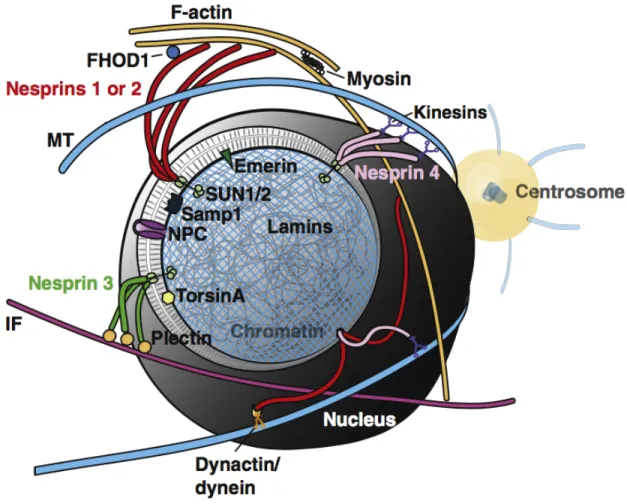
understanding stems from studies on nuclear migration (Dupin and Etienne-Manneville, 2011). Characterization of two distinct families of proteins that co-localize to the nuclear envelope, namely the SYNE/Nesprin-family (Apel et al., 2000; Zhang et al., 2001) and SUN-family (Malone et al., 1999; Lee et al., 2002), were shown to connect the cytoskeleton and nuclear lamina. Seminal work by Starr and colleagues in C. elegans mutants, anc-1 and unc-84, demonstrated that ANC-1 (homologue of Nesprin 2) associates with actin at its N-terminus while UNC-84 (SUN1/2 homologue) associates at its C-terminus (Starr and Han, 2002). Furthermore, UNC-84 localizes to the nuclear envelope in a lamin-dependent manner (Lee et al., 2002). Thus, a molecular bridge linking the nuclear lamina to the cytoskeleton was defined and shown to be critical for nuclear movement. The term LINC (linker of nucleoskeleton and cytoskeleton) complex was coined for these structures (Crisp et al., 2006) (Figure 1.2) and later work revealed homologues for its core components from yeast to human.
1.4 LINC complexes and nuclear mechanotransduction
4
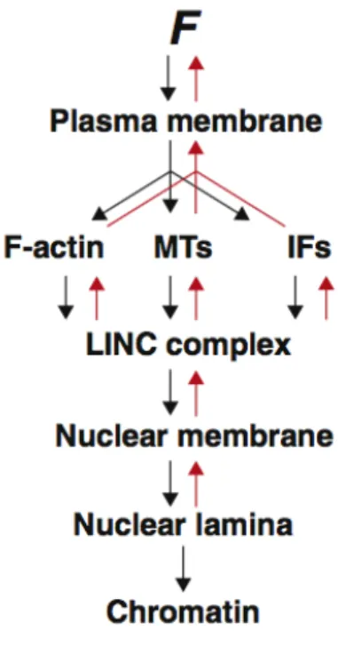
suggesting that sensory, transducing, and responding functions exist within the nucleus itself. Similar to whole cells which have adhesion receptors bridging the extracellular environment to the cytoskeleton, the LINC complex connects across the nuclear membrane, associating filamentous systems on each side. The structural similarity between whole cells and nuclei raises the possibility that force-sensitive signal amplification which in focal adhesions involves proteins like talin and vinculin, may similarly occur at the nuclear membrane. We speculate that the spectrin-repeats of Nesprins may cooperatively unfold under tension, exposing binding sites that promote Nesprin dimerization and recruitment of additional factors, facilitating complex stability and rigidity. In this light, LINC complexes could act as force-sensitive signaling hubs for cytoplasmic proteins and fine-tune nuclear responses to various mechanosensory inputs. A force-sensing mechanism on Nesprins could be locally amplified by the structural changes that occur within the nuclear lamina (Guilluy et al., 2014a).
LINC complexes are thought to be the primary structure controlling nuclear
mechanotransduction but how does nuclear mechanotransduction affect cell function? Driscoll and colleagues recently showed that the LINC complex contributes to the pre-stress state of the cell using mesenchymal stem cells (Driscoll et al., 2015). (Pre-stress derives from stresses generated within and experienced by cells in their environment.) This suggests that LINC complexes regulate cell-wide tension as well as strain transfer to the nucleus. This balance of internal cellular tension is a key component of the cellular tensegrity model proposed by Donald Ingber (Ingber, 1997; Wang et al., 2009). The major elements that regulate the pre-stress state of the cell (adhesion receptors, actomyosin contractility, and the cytoskeleton) are also
5
of the nucleus (Guilluy and Burridge, 2015) (Figure 1.3). This can have broader and longer lasting consequences to the pre-stress state of the cell as seen during stem cell differentiation on substrates of different rigidity (Engler et al., 2006).
1.5 Nesprins
Mammals have five Nesprin genes (SYNE 1-4, KASH5) that share a conserved C-terminal KASH (Klarsicht/ANC-1/Syne-1 homology) domain that interacts with SUN proteins. Cytoskeletal force is transduced to LINC complexes via specific Nesprins which bind different cytoskeletal systems. For example, Nesprin 1 and 2 bind actin (Starr and Han, 2002) and connect to microtubules via dynein/dynactin (Zhang et al., 2009) and kinesins (Zhang et al., 2009; Fan and Beck, 2004); Nesprin 3 binds to intermediate filaments through plectin
(Wilhelmsen, 2005); Nesprin 4 binds to microtubules through kinesin (Roux et al., 2009); and KASH5 binds to microtubules through dynein/dynactin (Morimoto et al., 2012). Importantly, Nesprins are complex as they reveal multiple splice-variants which add to their functional repertoire; have adaptable expression patterns making depletion studies difficult; and are thought to interact both physically and functionally with each other (Zhang et al., 2001; Lu et al., 2012; Rajgor et al., 2012; Duong et al., 2014). It is likely that Nesprins 1-3 predominantly
mediate force transduction to the nucleus in most cells as they are widely expressed relative to Nesprin 4 and KASH5. Expression of Nesprin 4 appears restricted to sensory hair and secretory epithelial cells (mammary, salivary, pancreas) (Roux et al., 2009; Horn et al., 2013) while
KASH5 is restricted to reproductive organs (Morimoto et al., 2012).
6
depletion of Nesprin 1 causes failure to align in response to uniaxial stretch (Chancellor et al., 2010), while depletion of Nesprin 3 causes a failure in centrosome reorientation in response to fluid shear (Morgan et al., 2011). Use of dominant-negative approaches that recapitulate loss of Nesprin-SUN complexes demonstrate force transmission from the cytoskeleton to nucleus is reduced (Lombardi et al., 2011). It was recently shown that nuclear localization of the
mechanically responsive transcriptional cofactor, YAP, is dependent upon Nesprin 1G in response to stretch (Driscoll et al., 2015). The LINC complex is also important for NFκB activity in response to stretch (Brosig et al., 2010). Together, these findings suggest that LINC
complexes may regulate other mechanoresponsive transactivators, such as β-catenin (Avvisato et al., 2007) and Twist (Wei et al., 2015). Nesprin 2 has already been shown to regulate Wnt-ligand induced nuclear translocation of β-catenin (Neumann et al., 2010).
Maintaining nuclear positioning requires force transmission from the cytoskeleton to the nucleus (Friedl et al., 2011; Gundersen and Worman, 2013) and Nesprin loss results in defects to this critical process in many systems (Zhang et al., 2007b; Starr and Han, 2002; Tsujikawa et al., 2007; Mosley-Bishop et al., 1999; Postel et al., 2011; Schoenenberger et al., 2011). It is difficult to separate nuclear migration defects, which affect cell polarity, migration and other processes, from defects ascribed to mechanotransduction. However, the deregulation of these processes may be attributed to the latter. For example, dorsal actin stress fiber structures that traverse the apical side of the nucleus have been implicated in force transmission through the LINC complex to the nucleus (Khatau et al., 2009) (Luxton et al., 2010). Photo-ablation of Nesprin-positive stress fibers over the nucleus causes local deformation of the underlying nucleus and nuclear displacement (Nagayama et al., 2013, 2014), suggesting the LINC complex regulates nuclear position by maintaining tension between the cytoskeleton and nucleus.
7
in 3D (Petrie et al., 2014). In this work, actomyosin contractile force is transmitted to the nucleus via vimentin and Nesprin 3. Depletion of Nesprin 3 caused a concomitant loss of nuclear
positioning and intracellular pressure asymmetry during 3D migration.
The importance of Nesprin function in mechanotransduction can also be recognized in human diseases. Patients with Emery-Dreifuss muscular dystrophy (EDMD) exhibit late-onset neuromuscular disorders with mutations in emerin (X-linked form), lamin A/C (autosomal dominant form), or Nesprin 1 and 2 (Zhang et al., 2007a). EDMD leads to increased nuclear fragility and defective mechanosensitive gene responses in highly contractile skeletal and cardiac muscle. In mice, deletion of Nesprin 1 and 2 results in cardiomyopathy as well as impaired gene expression in response to mechanical stimuli (Banerjee et al., 2014).
Additionally, mutations in Nesprin 4 have been identified from families that exhibited hereditary hearing-loss (Horn et al., 2013). nesprin-4-/- mice showed gradual degradation of the highly mechanosensory outer-hair cells within the cochlear organ. Nuclear positioning defects were concomitant with hair cell degradation.
1.6 SUNs
8
regulation and function have been seen elsewhere and are discussed below.
Cells can respond to low magnitude vibrations and Uzer and colleagues have shown that the nucleus is critical for detecting this type of mechanical stimulus (Uzer et al., 2015). Working with mesenchymal stem cells, they found activation of FAK and Akt pathways by vibration induced RhoA signaling, F-actin remodeling, and repression of adipogenic gene expression. SUN1/2 co-depletion, as well as expression of the DN-KASH domain, disrupted vibration-induced responses (Uzer et al., 2015). In C. elegans, UNC-84 (SUN1/2 homologue) interacts with lamin to transfer cytoplasmic forces to the nucleus during nuclear migration (Bone et al., 2014). Co-depletion of SUN1/2 also blocks nuclear stiffening in response to forces applied to isolated nuclei via Nesprin 1 (Guilluy et al., 2014b), suggesting that SUN1 and SUN2 can operate separately and may be functionally redundant. This is consistent with SUN1/2 null mice in which Nesprin 1 localization is disrupted but not in either SUN1 or SUN2 expressing cells (Lei et al., 2009). Conversely, functional differences have also been proposed. Despite similar affinities to the KASH domain of mini-Nesprin 2G, SUN1 has been shown to be dispensable for Nesprin 2 anchoring while SUN2 was necessary (Ostlund et al., 2009). In C. elegans, UNC-84 may recruit UNC-83 (KASH-domain containing protein) at the nuclear envelope where they mediate force transmission during nuclear migration (Starr et al., 2001). UNC-84 is required for proper nuclear envelope architecture in high force-bearing cells (Cain et al., 2014a), consistent with its role as a force transducer in the LINC complex. Lastly, SUN1 protein levels increase in lamin null cells as a result of reduced protein turnover, whereas SUN2 remains unchanged (Chen et al., 2012). This suggests that different protein degradation pathways and
compensation mechanisms may regulate SUN1 and 2 and could provide insight into how SUN1 contributes to lamin pathologies.
1.7 Emerin
9
al., 2007; Mislow et al., 2002) and other proteins (Berk et al., 2013). Emerin mutations in EDMD (Bione et al., 1995) and emerin-null fibroblasts exhibit defects in mechanotransduction (Rowat et al., 2006; Lammerding, 2005). Emerin becomes tyrosine phosphorylated by Src kinase in response to tension applied to isolated nuclei via Nesprin 1 (Guilluy et al., 2014a). This rapid phosphorylation coincides with accumulation of lamin A/C and nuclear reinforcement. Emerin promotes actin polymerization (Holaska et al., 2004), potentially increasing nuclear rigidity as a result of actin polymerization at the nuclear lamina in some situations. Interestingly, emerin phosphorylation increases on substrata of increasing stiffness (Guilluy et al., 2014a) and this is blocked after decreasing whole cell actomyosin contractility through inhibiting myosin-II. This suggests that cellular pre-stress can regulate emerin phosphorylation and nuclear signaling. Furthermore, Emerin regulates mechanoresponsive transcription factors such as Lmo7 and MKL1 (Ho et al., 2013) and thus may be important in relaying mechanical signals that affect longer term adaptation. MKL1 dissociates from G-actin and translocates to the nucleus upon mitogen and mechanical stimuli (Vartiainen et al., 2007). Aberrant MKL1-SRF signaling can be rescued in lamin null and mutant cells by addition of emerin (Ho et al., 2013). Taken together, these findings demonstrate emerin’s ability to regulate rapid nuclear stiffening, actin cytoskeletal polymerization, and gene activation, though, how emerin function is regulated during these processes is unclear.
1.8 Addressing the nuclear lamina in prestress/tension
Overwhelming evidence demonstrates that the nucleus is integral to
10
evident in cells that experience high mechanical strain, such as cardiac and skeletal myocytes (Cho et al., 2017). As the LINC complex regulates the pre-stress state in multiple ways, these cell-types may be particularly prone to defects in the LINC complex. Strong evidence for this was recently provided by Cain and colleagues in unc-84 mutants in which nuclear envelope architecture was irregular only in cells that experience high mechanical strain (Cain et al., 2014a). It is important to remember that dynamic cellular adaptation to mechanical stress is critical for cell homeostasis and is well defined for bone and soft tissue (Wolff’s law and Davis’ law) and has also been seen in other cell-types (DuFort et al., 2011). As we continue to explore the role of nuclear mechanotransduction, it will be valuable to address the individual
11
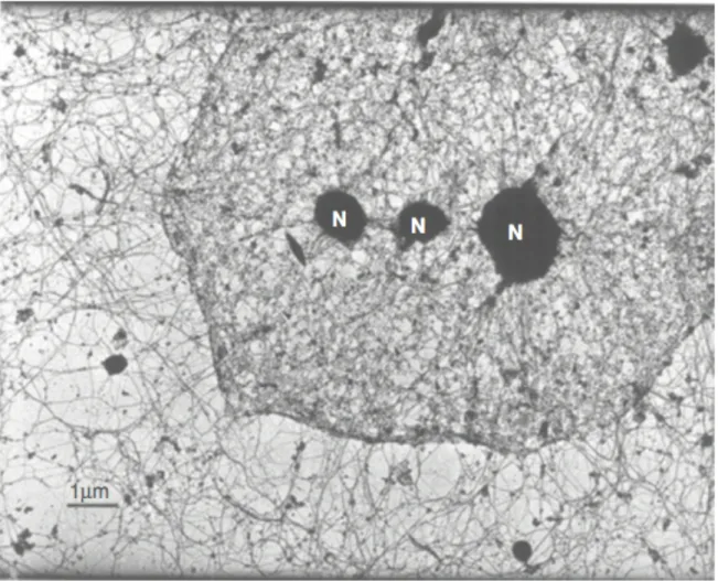
Figure 1.1 The nuclear lamina and cytoskeleton are highly interconnected. Transmission
electron micrograph of a HeLa cell, after removal of membranes and nucleic acids, showing nuclear filaments interconnecting with the cytoskeleton. Reprinted from Cell, vol. 29, Capco DG, Wan KM, Penman S, “The nuclear matrix: three-dimensional architecture and protein
12
Figure 1.2 LINC complexes tether the nucleus. The nuclear envelope is cut-away to expose
13
Figure 1.3 Force transduction to the nucleus and back. Schematic demonstrating the flow of
14
CHAPTER 2: THE ROLE OF THE NUCLEUS IN CELL MIGRATION, POLARITY AND MECHANOTRANSDUCTION
2.1 Overview
The nucleus has long been postulated to play a critical physical role during cell
polarization and migration. However, that role has not been defined or rigorously tested. Here, we enucleated cells to test the physical necessity of the nucleus during polarization and directed migration. Using enucleated mammalian cells (cytoplasts), we found that polarity establishment and cell migration in 1D and 2D occur without the nucleus. Cytoplasts directionally migrate toward soluble (chemotaxis) and surface-bound (haptotaxis) extracellular cues and migrate collectively in scratch-wound assays. Consistent with previous studies, migration in 3D
environments was dependent upon the nucleus. In part, this likely reflects the decreased force exerted by cytoplasts on mechanically compliant substrates. This response is mimicked both in cells with nucleo-cytoskeletal defects, and upon inhibition of actomyosin-based contractility. Together, our observations reveal that the nucleus is dispensable for polarization and migration in 1D and 2D but critical for proper cell mechanical responses.
2.2 Introduction
15
nucleus can lead to developmental defects (Zhang et al., 2009), impair cellular function
(Metzger et al., 2012), and is seen in several human diseases (Gundersen and Worman, 2013). A more recent and equally important physical role of the nucleus has been ascribed to
mechanical signaling within the cell. Here, the degree of structural integration of the nucleus within the cell is postulated to be crucial for regulating how cells sense and respond to force.
During polarity establishment and cell migration, the nucleus is actively positioned in many cell-types. For example, in fibroblasts, rearward nuclear movement allows anterior orientation of the centrosome and promotes anterior-posterior polarity of the cell in 2D (Gomes et al., 2005). In cells migrating in 3D which exhibit unidirectional polarity, the nucleus can be actively repositioned to act as an intracellular piston to facilitate migration (Petrie et al., 2014). Molecular motors, cytoskeletal elements, and cell adhesions are structurally connected within the cytoskeletal system as a whole and it is though that each contributes to tensional
homeostasis of the cell (DuFort et al., 2011). In light of this, aberrant force transmission between the cytoskeleton and nucleus has been suggested as the underlying cause for
defective nuclear positioning. However, it is unclear how the position of the nucleus conversely regulates mechanical signaling within the cell to collectively affect these processes. How would removal of the nucleus affect force transmission within the cell?
Recent work has dramatically expanded our understanding of the molecular
underpinnings of the mechanical linkages that connect the nucleus to cytoskeletal elements of the cytoplasm. Forces are transmitted through the linker of nucleoskeleton and cytoskeleton (LINC) complex (Crisp et al., 2006) where inner nuclear membrane proteins, Sun1 and Sun2, directly bind with outer nuclear membrane Nesprin proteins in the lumen of the nuclear
envelope. Nesprin proteins span the outer nuclear membrane to associate with the cytoskeleton and associated motors, whereas Sun proteins associate with lamin A/C, nuclear pore
16
strain-stiffening of the nucleus in response to extrinsic force (Guilluy et al., 2014b). In addition to applied forces, intrinsic cell-derived forces can transmit through dorsal actin stress fibers to the LINC complex, allowing posterior positioning of the nucleus via actin retrograde flow (Luxton et al., 2010). Because cell-derived forces are highly dependent upon the mechanical properties of the microenvironment, the LINC complex likely plays an important role in regulating the
response of the cell to environmental rigidity. This was shown for rigidity-dependent nuclear localization of YAP (Elosegui-Artola et al., 2017). Together, these and many other recent studies (Graham and Burridge, 2016) demonstrate the intricate network of molecular connections that help position the nucleus and make it sensitive to mechanical cues.
Several studies have reported defects in cell polarity, migration, and
mechanotransduction upon disruption of nucleo-skeletal connections. It is unclear what role the nucleus plays during these processes and how they are affected by nuclear-loss as opposed to aberrant nuclear positioning. Cellular enucleation is an older approach that has been used to explore migration in the absence of the nucleus (Goldman et al., 1973; Shaw and Bray, 1977; Euteneuer and Schliwa, 1984, 1992; Verkhovsky et al., 1999). We revisited this technique to study the role of the nucleus in cell polarity and distinct forms of migration (e.g. in 1, 2 and 3D) and sought to understand what role the nucleus plays as cells respond to guidance cues, particularly mechanical cues. Few studies have directly measured the effect of nucleo-skeletal disruption on cell behavior in response to mechanical properties of the environment. This is important as the nucleus is integral to cellular responses to force (Wang et al., 2009). In the current study, we have used cellular enucleation to examine how the presence or absence of a nucleus affects cell polarization, cell migration, and mechanical signaling within cells.
2.3 Results
Generating cytoplasts
17
reproducibly generate high purity cytoplasts (cells without nuclei) from large populations (~2 x 10⁷/gradient). We used both rat embryonic fibroblasts (REF52) and human umbilical vein endothelial cells (HUVECs) to generate cytoplasts. Cells were incubated in the presence of cytochalasin and centrifuged at high speed through a density gradient (Fig. 2.1A). This resulted in formation of three distinct strata within the gradient. Fluorescence analysis of fractions from REF52 cells showed fraction 1 contained mostly cellular debris; fraction 2 contained cytoplasts; and fraction 3 contained nucleoplasts (nuclei surrounded by cytosol and encased in plasma membrane) (Fig. 2.1B). Similar fractionation strata and composition were observed with HUVECs (Fig. 2.9A). Based on morphological observations, enucleation appears to occur through repositioning of the nucleus through the cell body, leading to hyper-elongation of the cell parallel to the direction of the g-force vector (Fig. 2.9B). Toward the tail end of the cytoplast (opposite end of nuclear exit), small fragments separate, generating the constituents of fraction 1. Enucleation occurs in the presence of g-force alone but efficiency is increased with
actomyosin destabilization (Fig. 2.9C). Enucleation of cells expressing nuclear localized-tdTomato led to localized-tdTomato fluorescence in cytoplasts with decreased presence in nucleoplasts (Fig. 2.9D). This result is consistent with nuclear envelope rupture (Denais et al., 2016; Raab et al., 2016) which likely occurs during nuclear exit from the cell.
18
intact K1/2 = 23.0 ± 2.0; p>0.05), including typical radial morphology during spreading, were
found between intact cells and cytoplasts (Fig. 2.9H).
We analyzed cytoplasts for the presence of nuclear proteins, major organelles, and the cytoskeleton. Cytoplasts were devoid of nuclei and most nuclear-associated proteins (Fig. 2.1E; Fig. 2.10A), contained cytoskeletal networks for filamentous actin, vimentin, and microtubules, and formed vinculin-containing focal adhesions (Fig. 2.1F). Cytoplasts retained endoplasmic reticulum, Golgi, mitochondria, and centrosomes (Fig. 2.1G). Immunofluorescent staining of nucleoplasts revealed nuclear-associated proteins, organelles, and cytoskeletal systems (Fig. 2.10B). We measured cytoplast survival with viability dyes and found REF52 cytoplasts to be stable for 48 h (Fig. 2.1H). HUVEC cytoplasts had decreased viability compared to REF52 cells, showing a significant decrease at 6 h with loss of half the population at ~18 h post enucleation (Fig. 2.10C). We did not observe obvious decreases in protein levels for Src, nonmuscle myosin IIA, vinculin, and other proteins over 24 h in REF52 cytoplasts (Fig. 2.10D). To reduce possible effects attributed to cytoplast degeneration, we used REF52 cytoplasts for most experiments as they exhibited increased survival over HUVEC cytoplasts. These experiments were performed <27 h post enucleation with most performed <19 h post enucleation. HUVEC cytoplasts were used for shorter experiments (<8 h) with 18 h experiments being the longest. Together, these data demonstrate the ability to generate cytoplasts at both high-purity and quantity.
The nucleus is not required for establishing anterior-posterior polarity
The position of organelles, including the nucleus and centrosomes, are hallmarks of cell polarity. We assessed polarity establishment in the absence of the nucleus to understand if the nucleus is necessary for proper localization of centrosomes and the Golgi apparatus.
19
organelle positioning relative to the cell centroid from Y-coordinate values, as significant differences from the cell centroid were not found for X-coordinate values for all patterns tested (Fig. 2.11C). Centrosomes, which normally position at the cell centroid, were indeed found near the cell centroid for REF52 intact cells and cytoplasts (Fig. 2.2A, Fig. 2.11D). The mean
centrosome position for intact cells and cytoplasts on crossbows was -1.1 ± 0.4 and -1.2 ± 0.4 µm, respectively, from the cell centroid (Fig. 2.2B). Similar to centrosomes, the Golgi positions between the nucleus and the cell leading edge, thus we measured Golgi positioning on patterns. Consistent with centrosome localization, the Golgi was found near the cell centroid for intact cells and cytoplasts for all patterns tested (Fig. 2.2C, Fig. 2.11E) with the mean Golgi position not differing between intact cells and cytoplasts (Fig. 2.2D). Next, we measured centrosome localization in HUVEC cytoplasts. HUVEC cytoplasts are smaller than REF52 cytoplasts and rarely occupied the full area of the circle micropattern (largest area of the patterns used), preventing us from considering this particular shape. Thus, we used triangle patterns instead. Centrosomes were positioned at the cell centroid for HUVEC intact cells and cytoplasts on crossbow and triangle micropatterns (Fig. 2.2E, 2.2F). These data demonstrate that normal cell polarization, as evidenced by cell morphology and predicted organelle positioning, occurs independently of the nucleus.
The nucleus is not essential for random and directed 2D migration
Cell migration is intrinsically a polarity-driven process (Ridley et al., 2003), thus, we analyzed 2D random migration in cytoplasts. We found REF52 and HUVEC cytoplasts were migratory, exhibiting anterior-posterior polarity, dynamic lamellipodial extension, and rear
20
with increasing FN concentration. We measured the relative amount of FN on glass to
determine if concentrations above 100 µg/ml were capable of binding and, as such, sensed by cells. Detectable increases in FN up to at least 400 µg/ml were measured, suggesting that 100 µg/ml is not saturating and that higher concentrations can influence migration behavior (Fig. 2.11G). We measured surface expression levels of β1 and β3 integrins in REF52 intact cells and cytoplasts to see if reduced integrin levels, as a result of enucleation, might explain this response. Using flow cytometry and live-cell labeling with fluorescent-conjugated antibodies, we detected reduced levels of β1 and β3 integrins in cytoplasts as compared to intact cells (Fig. 2.3D). However, when normalized to cell size (from flow cytometer forward-scatter metrics), the relative β1 and β3 integrin levels were not different between cytoplasts and intact cells.
Furthermore, integrin localization was consistent with adhesion complexes. Lastly, we sought to gauge the effects of cytoplast degeneration on migration velocity as this would influence
migration efficiency over time. We found a decreased rate of -0.12 ± 0.02 µm/h over 24 h for REF52 cytoplasts (Fig. 2.3E) demonstrating that cytoplast migration velocity is slightly affected over time.
Next, we examined directional migration in REF52 cytoplasts to determine what role the nucleus plays as cells respond to guidance cues. We measured directional migration via a microfluidic-based approach, as previously described (Wu et al., 2012). Migration was monitored in gradients of either platelet-derived growth factor (PDGF; for chemotaxis) or
surface-bound FN (for haptotaxis). Directional fidelity is shown as forward migration index (FMI) which describes the directional persistence of a cell toward (positive FMI) or away from
(negative FMI) an extracellular cue. Cytoplasts showed positive chemotactic (Fig. 2.3F) and haptotactic (Fig. 2.3G) responses, similar to intact cells (Fig. 2.3H). These data demonstrate that the nucleus is dispensable for directional migration in response to PDGF and FN.
21
margin has been implicated in this migratory response (Gomes et al., 2005; Luxton and Gundersen, 2011), thus we assessed the physical necessity of the nucleus in this form of migration. Cytoplast monolayers from REF52 cells were mostly free of nuclei-containing cells (Fig. 2.12A), however, purity decreased over time as any intact cells proliferated. We measured the density of nuclei at the end of all scratch experiments (~16-20 h post scratch) and found nuclear densities at 15.6 ± 0.4 nuclei for intact cells and 1.3 ± 0.1 nuclei for cytoplasts in a 100 µm2 area. At these densities, effects from intact cells within the cytoplast monolayer are unlikely
to affect cytoplast-driven scratch closure. REF52 cytoplasts were capable of scratch closure which occurred between 4-7 h for intact cells and 7-16 h for cytoplasts (Fig. 2.4A). The average closure time for intact cells was 5.4 h. At this time, cytoplasts closed 80% of the scratch. On average, cytoplasts closed 95.6% of the scratch over 16 h. To reduce scratch closure effects driven by cell proliferation, we inhibited cell division with mitomycin C pre-treatment (Fig. 2.4B, 2.4D). The average time for cytoplast scratch closure was unchanged from untreated, however, for intact cells it increased from ~5 h to 8 h. Although the initial rates of scratch closure were similar for both intact cells and cytoplasts, total closure took longer for REF52 cytoplasts (Fig. 2.12B). This difference may reflect the slight time-dependent decrease in migration velocity in cytoplasts. Despite this difference, the time to close half of the scratch (t1/2) was not different
between intact cells and cytoplasts for all treatments tested (Fig. 2.4C). HUVEC cytoplasts were also capable of scratch closure (Fig. 2.4E, 2.4F). A narrower scratch was used for these
22
The nucleus is dispensable for migration in 1D but not 3D environments:
Recent work has shown that the nucleus performs specialized physical functions during 3D migration (Petrie et al., 2014; Denais et al., 2016; Raab et al., 2016). We explored cytoplast migration in collagen gels of two different porosities to gauge cell migration efficacy in
environments of different physical constraint and ligand density (Fig. 2.5A). Collagen matrices were prepared with different gelation temperatures, producing loose reticular (LR) and highly reticular (HR) matrices, as described (Doyle et al., 2015). Cytoplasts migrated slower than intact cells for both LR and HR matrices in both 2D (on top of the gel; Fig. 2.5B) and 3D (inside the gel; Fig. 2.5C). Transitioning from 2D to 3D environments caused a decrease in cell velocity for both intact cells and cytoplasts. Interestingly, cytoplast 3D velocity did not change between LR and HR matrices whereas intact cells did. Moreover, cytoplasts showed a less pronounced uniaxial morphology in 3D than did intact cells (Fig. 2.5D). Compared to intact cells, which showed an average accumulated distance over 8 h of 91.4 ± 4.6 µm, cytoplasts were largely immotile with an average accumulated distance of 28.0 ± 1.2 µm (Fig. 2.5E). The low velocity and low accumulated distance for REF52 cytoplasts was also observed for HUVEC cytoplasts (Fig. 2.5F). No difference in 3D cell velocity was measured between REF52 and HUVEC cytoplasts (3.0 ± 0.1 µm/h and 3.3 ± 0.3 µm/h, respectively; p>0.05). This non-migratory
23
Next, we explored two mutually non-exclusive explanations for the impaired 3D
migration of cytoplasts: 1) migration in 3D environments uniquely requires the physical presence of the nucleus (Petrie et al., 2016) or 2) the low rigidity collagen matrices differentially affect cytoplasts versus intact cells. The first explanation is difficult to assess and might be addressed with nuclear addback experiments in 3D gels as a way to rescue cytoplast migration in situ, or the use of alternative 3D matrices that might confer migration to cytoplasts. Nuclear addback is technically difficult and was not attempted. The use of different matrix materials was not
supported as cytoplasts from primary human fibroblasts were shown to slowly migrate (~4 µm/h) inside cell-derived matrices (Petrie et al., 2014), closely matching the low cell velocities we observed in collagen. Alternatively, the effect of the presence of the nucleus on 3D migration could be determined with 1D migration being used as a surrogate for 3D, since these two forms of migration share several principles (Doyle et al., 2009). Thus, we turned to using
micropatterned 1D lines (Fig. 2.12H) where we found REF52 and HUVEC cytoplasts exhibited a uniaxial morphology and polarity, similar to intact cells. Contrary to our expectations, cytoplasts migrated in 1D (Fig. 2.12I) with velocities on 5 µm lines of 11.5 ± 0.6 µm/h for REF52 cells and 38.3 ± 1.5 µm/h for HUVECs (Fig. 2.5G, 5H). Cytoplasts were also migratory on lines coated with either FN or collagen (Fig. 2.12J). These data demonstrate that the nucleus is dispensable for migration in 1D but not 3D environments.
The nucleus regulates cell contractility and the sensitivity of the cell to mechanical cues
24
environments in a cell-compatible manner is not trivial, causing concomitant changes to ligand density and pore geometry. Consequently, we tested the effect of microenvironment stiffness on 2D migration by measuring migration in 2D on FN coated substrata of known stiffness.
Using a range of hydrogels at 0.2, 0.5, 1, 8, 25, and 50 kPa, and glass, we found intact cells and cytoplasts showed pronounced, biphasic responses in migration velocity with relation to substrata stiffness (Fig. 2.6A). REF52 intact cells showed a peak migration velocity on 8 kPa substratum whereas cytoplasts showed an unexpected peak migration velocity on 25 kPa substratum. When plotted together, a shift in peak of the biphasic response was evident, with the maximum cytoplast velocity significantly shifted toward stiffer substrata (Fig. 2.6B). This trend was not repeated upon inhibition of transcription or translation (Fig. 2.13A). These data show cell migration velocity is dependent upon substrate stiffness; a property observed in other cells (Sunyer et al., 2016; Plotnikov et al., 2012; Peyton and Putnam, 2005). Because
mechanosensing depends upon both environmental forces and cell-generated forces (Janmey et al., 2009), and cell-generated forces are largely regulated by actomyosin-based contractility (Pelham and Wang, 1997; Raab et al., 2012; Pathak and Kumar, 2012), we surmised the shift in the optimum stiffness for migration could be a product of reduced whole-cell contractility and a reduction in mechanosensitive signaling on account of loss of the nucleus. For instance, if cell contractility is reduced, a higher substratum rigidity would be necessary to activate mechanically sensitive pathways that regulate migration. To gain insight into this, we tested baseline
25
We used traction-force microscopy to measure the contractile energy (a whole cell measure showing the mechanical effort used by the cell in substrate deformation; also known as strain energy) and traction stress (a per area unit measure of the mechanical effort used by the cell in substrate deformation). We found that cytoplasts from REF52 cells had significantly reduced contractile energy and traction stress, as compared to intact cells (Fig. 2.6C, 2.6D). This does not appear to be cell specific as HUVEC cytoplasts also exhibited a similar shift in peak of the biphasic response toward more rigid substrata (Fig. 2.6E) and showed decreased contractile energy and traction stress (Fig. 2.6F; Fig. 2.13C). These data suggest that the nucleus regulates cell contractility and controls the sensitivity of the cell to mechanical cues.
The LINC complex and lamin A also regulate cell contractility and the sensitivity of the
cell to mechanical cues
We sought to understand if cell contractility and traction stress could be similarly
26
Lamin A/C is an important mechanosensitive nuclear protein (Swift et al., 2013) and is non-essential for LINC complex anchorage (Crisp et al., 2006; Haque et al., 2006; Padmakumar et al., 2005). We tested the extent this structural protein of the nuclear lamina has on
mechanoresponse. Using Lmna-/- mouse embryonic fibroblasts (MEFs), we found a pronounced shift in peak of the biphasic response toward 25 and 50 kPa substrata, as compared to a peak at 8 kPa substratum for Lmna+/+ cells (Fig. 2.7D). Consistent with this, traction-force
measurements revealed a decreased contractile energy in Lmna-/- cells (Fig. 2.7E; Fig. 2.13F),
however, traction stress was not different between Lmna-/- and Lmna+/+ MEFs. Using Lmna -/-cells rescued with lamin A only (Fig. 2.13G), the peak migration velocity on different rigidities was shifted toward less rigid substrata (peak at 8 kPa substratum) in rescued but not mock rescued cells (peak at 8 and 25 kPa substrata) (Fig. 2.7G). Remarkably, nearly complete restoration of contractile energy was measured in lamin A rescued cells (Fig. 2.7H). Similar to Lmna-/- and Lmna+/+ MEFs however, traction stress was not different between lamin A and mock
rescued Lmna-/- MEFs (Fig. 2.7I; Fig. 2.13H). These data demonstrate a similar nuclear-based
modulation of cell migration and contractile energy to that observed in cytoplasts and cells bearing loss of the LINC complex. However, unlike enucleation or depletion of Sun1/Sun2 proteins, the presence of lamin A/C does not affect traction stress.
Lastly, we directly tested the role of contractility on regulating migration velocity on different rigidity substrata. Intact REF52 cells were treated with either 15 µM or 50 µM blebbistatin (bleb) to reduce actomyosin-based contractility, and cell migration velocity on different rigidity substrata was measured. Consistent with our earlier measurements, cells showed a shift in peak of the biphasic response from 8 kPa substratum, observed in untreated and 15 µM bleb treated, to 25 kPa substratum with 50 µM bleb treatment (Fig. 2.7J). The shift in peak migration that was measured upon nuclear loss, loss of connectivity between the
27
response to environments of different rigidity. These data suggest that the nucleus can regulate the sensitivity of the cell to mechanical cues via modulation of whole cell contractility; a role consistent with the nucleus playing a role in an integrated molecular clutch.
2.4 Discussion
Cell biologists have investigated the physical role of the nucleus in establishing cell polarity and in cell migration for many decades, with a more recent focus on its role in
mechanotransduction. Based on our data using both fibroblasts and endothelial cells, we show that the nucleus is not necessary for establishing polarity or directional cell migration but is important for regulating the sensitivity of the cell to mechanical cues. Our data support a working model whereby the nucleus is a critical component of an integrated molecular clutch encompassing focal adhesions, actin stress fibers, and the nucleus.
The nucleus, cell polarity and 2D cell migration:
A relationship between the positions of the centrosome (MTOC) and nucleus has long been recognized in many cells (Luxton and Gundersen, 2011). This relationship has been studied extensively, particularly in the context of cells in culture migrating into a scratch wound. For many migrating cells, there is an orientation of the centrosomal-nuclear axis such that the centrosome is located in front of the nucleus and the axis corresponds to the direction of migration. It has been shown that rearward nuclear movement reorients the position of the centrosome and that nuclear repositioning establishes cell polarity (Gomes et al., 2005). These and other observations (Lee et al., 2007) have led to the view that the nucleus is critical for anterior-posterior cell polarity. Earlier work, however, suggested the opposite (Chambers and Fell, 1931; Goldstein et al., 1960; Goldman et al., 1973; Piel et al., 2000). We find that
28
nucleus, consistent with the notion that the nucleus is not strictly necessary for proper positioning of these organelles.
Similarly, we show that the nucleus is not essential for 2D migration under random and directed conditions. Although cytoplasts migrate more slowly on conventional FN concentrations (10 µg/ml) than control cells, similar migration velocities between cytoplasts and intact cells are found at higher FN concentrations. This suggests that the nucleus is not necessary for migration since changing FN density (which changes adhesion strength) can greatly modulate migration velocity. Given that the establishment of the centrosomal-nuclear axis has been implicated in directed migration, it is striking that cytoplast migration is little affected by the loss of the nucleus. Our directed migration data show cytoplasts chemotax and haptotax at similar efficiencies to intact cells. This indicates that the nucleus is not essential for sensing and
responding to these extracellular cues or in establishing and maintaining the polarity required for directional migration. Furthermore, despite showing differences in total scratch closure time, our scratch assay data show similar rates of closure for half the scratch area between cytoplasts and intact cells. Several factors could potentially explain the total scratch closure time-lag in cytoplasts, such as time-dependent cytoplast degradation or decreased FN density from the scratch margin to the scratch center. However, our data indicate that a nucleus is not needed for the polarized positioning of the centrosome and Golgi, and also is not needed for directed cell migration.
29
abrogates these defects. Though it is not known how an improperly positioned nucleus hinders cell polarity and migration in all contexts, it most likely involves the role of the nucleus in maintaining cytoskeletal organization and, through this, proper coordination of intra- and intercellular forces. The LINC complex directly mediates force transmission between the nucleus and cytoskeleton (Lombardi et al., 2011; Alam et al., 2015; Arsenovic et al., 2016; Stewart et al., 2015). Aberrant force transmission between the cytoskeleton and the nucleus could differentially affect force-sensitive signaling pathways that regulate polarity establishment and maintenance, as well as cell migration.
The nucleus and 3D cell migration:
Although cytoplast migration on 2D surfaces was comparatively normal, it was greatly impaired in 3D collagen gels. At the outset of this study we were uncertain what effect removing the nucleus would have on a cell’s migration in 3D. This is because the nucleus has been reported to both facilitate and impede migration efficiency in constrained spaces. For example, lobopodial migration is driven by a nuclear-piston mechanism that allows cells to move in 3D (Petrie et al., 2014). It is worth noting that this mechanism of migration does not explain our cytoplast 3D data as the nuclear-piston mechanism was shown in cell-derived matrices and does not operate in collagen matrices. Cytoplasts from that study, however, did show low migration velocity (~ 4 µm/h) in cell-derived matrix, consistent with poor migration of cytoplasts in 3D environments.
30
HR matrices (which differ in pore size) are different in intact cells but not cytoplasts, suggesting pore size is not hindering migration in the absence of the nucleus. Because cytoplasts can signal on collagen, exert force on collagen fibers, signal in response to mechanical load on collagen, degrade and remodel matrix, these factors are unlikely to explain the impaired
migration of cytoplasts in 3D. One other possible explanation for the poor migration of cytoplasts in 3D is a failure to polarize in this environment without a nucleus. Despite our efforts, we
cannot confidently support or refute the ability of cytoplasts to polarize in 3D gels. Very little work has addressed markers of polarity for cells in 3D settings, making it difficult to identify a reliable polarity indicator, particularly in the absence of a nucleus and this will have to be re-visited when better approaches have been developed.
So why do cytoplasts migrate so poorly in 3D environments? We considered two
explanations: dimensionality and the low rigidity of the matrix used in our studies. We observed a general decrease in migration velocity upon changing between 2D and 3D collagen for both intact cells and cytoplasts. Because migration of cells along narrow lines of ECM (1D migration) is thought to be similar to 3D migration (Doyle et al., 2009), we examined how cytoplasts
migrate on 1D fibronectin-coated lines. Cytoplasts showed robust migration on these lines. However, these 1D matrix-coated lines were generated on rigid (glass) substrates, similar to the 2D random and directed migration studies described above. Consistent with this idea that rigidity may be critical, cytoplasts exhibit a relatively low migration velocity on the 2D top surface of 3D collagen gels. Ideally, we would have liked to test this notion in a 3D environment,
31
Cell migration and the integrated molecular clutch:
The velocity of cell migration depends on both the density of the matrix molecules (e.g. fibronectin) on the substrate and on the rigidity of this surface. With both increasing matrix density and substratum rigidity, most cells demonstrate a biphasic migration velocity response (Peyton and Putnam, 2005; Pathak and Kumar, 2012; Lauffenburger and Horwitz, 1996). One of the striking results emerging out of our work is that the presence or absence of a nucleus (or connections to the nucleus) affects this response both to matrix density and substrate rigidity. With fibroblasts and endothelial cells, removal of the nucleus shifted the peak velocity to higher matrix densities and to higher substrate rigidities. For intact cells, the biphasic velocity response to increasing fibronectin concentrations was generally interpreted as the result of too little adhesion being insufficient to generate optimal traction force, whereas too strong adhesion prevents detachment of adhesions, thereby retarding migration. However, agents that inhibit myosin activity or promote it were previously observed to shift the peak velocity to either faster or slower speeds depending on the fibronectin concentration, indicating that the velocity profile could not be explained simply based on differences in adhesion strength (Gupton and
Waterman-Storer, 2006). It was concluded that migration velocity reflects the interplay of many interdependent factors, including adhesion strength but also myosin II activity and actin
dynamics (Gupton and Waterman-Storer, 2006). A large body of work has shown that migration velocity depends on nonmuscle myosin II and retrograde actin flow generating traction, as well as on “molecular clutches” (the sites of adhesion involving integrins, often clustered in focal adhesions) transmitting this traction to the substratum (Case and Waterman, 2015).
32
Building on this idea, we postulate that the LINC complex and nuclear lamina serve as a critical part of an extended and integrated molecular clutch that includes focal adhesions, contractile actin stress fibers, and the nucleus (Fig. 2.8). Actomyosin contractility regulates how cells sense and respond to force. In previous work, it was shown that inhibiting myosin also could increase the velocity of cells moving on soft substrates (Pathak and Kumar, 2012). Similarly, in our work here we observed that inhibiting myosin II with blebbistatin shifted the peak migration velocity of intact cells to higher rigidity substrata. This effect of blebbistatin on migration speed was mimicked by enucleation, consistent with this latter shift being due to changes in the mechanotransduction properties of the cytoplasts. Also, consistent with
cytoplasts having decreased contractility, they demonstrated reduced collagen gel contraction, decreased contractile energies and decreased traction stresses. Similar results were obtained when we broke the cytoskeletal connections to the nucleus by disrupting the LINC complex through Sun1/Sun2 depletion. Again, this shifted the peak migration velocity to more rigid substrata and decreased contractile energy and traction stress. This suggests that nucleo-cytoskeletal connections regulate cell contractility and cell behavior in a manner similar to regulating actomyosin function. Because of the known structural connections between the nucleus and the actin cytoskeleton, a functional interdependence likely exists. Recent efforts have begun to dissect the signaling pathways regulating the LINC complex and actin
cytoskeleton, revealing transcription-independent functions that involve regulation of RhoA activity (Thakar et al., 2016). Our data support the role of nucleo-skeletal connections in regulating cell contractility, however, the extent to which these connections affect cell behavior likely encompasses other processes such as actin retrograde flow rates and adhesion
dynamics. Adequately addressing these phenomena will require in-depth studies and will be pursued in future work.
33
components affect cytoskeletal organization, cell migration, and physical properties of the cells. In particular, previous work with Lmna-/- cells has shown decreases in stress fiber organization, actin dynamics, focal adhesion area, RhoA activity, nuclear stiffness, mechanically induced nuclear signaling, and more recently, contractility itself (Broers, 2004; Hale et al., 2008;
Lammerding et al., 2006; Khatau et al., 2009; Ho et al., 2013; van Loosdregt et al., 2017). In our work, we have found that the Lmna-/- cells also show a shift in the peak of their migration
velocity to higher rigidity substrata. Consistent with the above results, they also show decreased contractile energy but unexpectedly we did not detect a decrease in traction stress. The reason for this is currently unclear and we have not been able to determine whether this reflects the difference between a soft nucleus that is still attached via the LINC complex to the cytoskeleton as opposed to a disconnected nucleus. Alternatively, the Lmna-/- cells may be affecting other
signaling pathways or experimental parameters, such as changes in the polarization of the traction forces, which in turn, affect traction stress and cell migration (Jurado et al., 2005; Meili et al., 2010; Bastounis et al., 2014).
One important aspect of our integrated molecular clutch model (Fig. 2.8) is the
bidirectional nature of force in the model. Force on the molecular clutch arises from retrograde actin flow, driving forward protrusion of the leading edge, and also from actomyosin contractility pulling the nucleus and rest of the cell body forward. Myosin-based contractility develops tension between the clutch and the nucleus because of the inter-connections between the cytoskeleton and the nuclear envelope mediated by the LINC complex. The tension developed between these two structures (the adhesions and the nucleus) will be diminished by decreasing the rigidity of the substratum or by enucleation or disrupting the connections to the nucleus. The reduced tension transmitted to the clutch will, in turn, alter the cell’s migratory response to both matrix rigidity and matrix density. Depleting lamin A, however, has an intermediate effect
34
continuing importance of understanding the cytoskeletal-nuclear interconnections and also the molecular details of the molecular clutch. During the past few years, much has been learned about how tension exerted on the clutch affects the properties and interactions of components mediating adhesion (Elosegui-Artola et al., 2016). Much still remains to be learned about these interactions and also how the signaling events generated by mechanical tension feeds back to impact cell behavior. Ultimately, this information should lead to a better understanding of how cells respond not only to the composition of their environment but also to its physical properties.
2.5 Materials and methods
Reagents and materials
Commercial antibodies used for Western blotting and immunohistochemistry were purchased from Cell Signaling Technology (rabbit EEA1, rabbit NUP98, rabbit anti-LMNA/C, mouse anti-histone H3, rabbit anti-MHC2A, rabbit anti-RLC (myosin regulatory light chain), rabbit anti-FAK, rabbit anti-FAK (Y397), rabbit anti-paxillin (Y118), rabbit anti-vinculin, rabbit anti-vimentin, rabbit anti-Src, rabbit anti-AMPKα, rabbit anti-GAPDH), EMD Millipore (rabbit anti-Sun2, mouse anti-actin), Abcam (rabbit anti-emerin, rabbit-anti-Sun1), Sigma (mouse anti-α-tubulin, mouse anti-γ-tubulin), BD (mouse anti-GM130), BioLegend (Alexa Fluor 488-conjugated anti-β1 and anti-β3 integrins), and Thermo Fisher Scientific (HRP-conjugated goat anti-mouse and goat anti-rabbit; Alexa Fluor 488, 568, and 633 goat anti–mouse and goat anti-rabbit). Phalloidin (Alexa Fluor 488, 568, 633), ER-Tracker Red (BODIPY), MitoTracker Green FM, CellTracker Green CMFDA, CellTracker Red CMTPX, calcein-AM, Hoechst 33342, Vybrant Dye-Cycle Green nuclear stain, and Trypan Blue were purchased from Thermo Fisher Scientific. Human fibronectin used to conjugate to Cy5 was purchased from BD. Cy5
conjugation to fibronectin was performed as previously described (Wu et al., 2012). Fibronectin used for all other experiments was purified from human plasma, as previously described
35
Ficoll-400 was purchased from Fisher (BP525). Polyacrylamide hydrogels were purchased from Matrigen. Mitomycin C, GM6001, actinomycin D, cycloheximide, and SU6656 were purchased from Tocris. Cytochalasin B was purchased from Enzo Life Sciences. (-)-blebbistatin was purchased from Sigma.
Cell culture, expression vectors, and RNAi experiments
REF52 cells were grown in high-glucose DMEM (Invitrogen, Carlsbad, CA) containing 10% fetal bovine serum (FBS; Sigma-Aldrich) and 100 U/ml Pen-Strep (Invitrogen). Plasmid transfections were performed with Lipofectamine 2000 reagent (Invitrogen), based on
manufacturer’s protocol. Stable REF52 lines were generated by transfecting cells with specified constructs and sorting for fluorescence via successive rounds of flow-cytometry. These lines include a nuclear localization sequence (NLS) tdTomato chimera expressing line that was generated with the pQC-NLS-tdTomato construct, courtesy of Connie Cepko’s lab. A Golgi-EGFP expressing line was used for all micropattern work, and was generated with the pLL-5.5-GIX (Uetrecht and Bear, 2009) construct. This construct encodes a human β
-1,4-galactosyltransferase-EGFP chimera. A centrin-EGFP expressing line was generated with the p3XGFP-centrin construct. HUVECs were purchased from Lonza and cultured in EBM-2 endothelial growth basal medium (EBM-2). All lamin A/C mouse embryonic fibroblast lines, including Lmna+/+, Lmna-/-, Lmna-/- rescued with lamin A or mock rescued, were generously provided by Jan Lammerding’s lab at Cornell University.
RNAi-mediated depletion of Sun1 and Sun2 were performed using siRNA duplexes purchased from Dharmacon. Two separate siRNA pairs were used for Sun1 and Sun2. These were siSun1/Sun2 pair 1: Sun1 GUAUAUACCAAGACGCCAU-3’), Sun2
GAGACUUACGAGACGAAGA-3’) and siSun1/Sun2 pair 2: Sun1
36
siRNA duplexes was performed with Mirus siQUEST reagent according to manufacturer. Cells were used for experiments beginning at 48 h post-transfection. Validation of RNAi-mediated depletion was monitored after each experiment via Western blot. Quantification of protein knockdown was measured using ImageJ.
Western blotting
Cells were lysed in either radio immunoprecipitation assay (RIPA) lysis buffer (150mM NaCl, 50mM Tris-HCl, 1mM EDTA, 0.24% sodium deoxycholate,1% Igepal (pH 7.5)) or 2X Laemmli sample buffer (120 mM Tris-HCL (pH 6.8), 4% SDS, 20% glycerol, 0.02%
bromophenol blue). All lysis buffers contained 100 nM aprotinin, 50 µM leupeptin, 10 µM pepstatin A, and 50 mM sodium orthovanadate. Lysates were run on SDS-PAGE gels and transferred to polyvinylidene fluoride membranes (Immobilon-P; EMD Millipore). Membranes were blocked with either 5% (w/v) milk or bovine serum albumin for 1 h at ambient temperature before being incubated with primary antibodies overnight at 4oC. Following primary antibody incubation, blots were washed and incubated with horseradish peroxidase-conjugated secondary antibody at ambient temperature for 1 h. Western blots were developed with SuperSignal West Pico or Femto Chemiluminescent Substrate (Thermo Fisher Scientific) and either scanned on a ChemiDoc MP System (Bio-Rad laboratories) or developed on film.
Cellular enucleation
37
U/ml Pen-Strep). The stock Ficoll solution was sterile filtered (0.4 µm) and stored at 4oC. The refractive index of the stock was measured on a refractometer. For the REF52 cell line and cells of similar volume/size (e.g. HUVEC, HeLa), the optimal refractive index of 1.373 produced good purity cytoplasts. Discontinuous iso-osmotic density gradients were poured from freshly
prepared stocks of 30%, 20%, 18%, and 15% Ficoll-DMEM containing 10 µg/ml cytochalasin B (dissolved in 100% ethanol), and 0.2% DMSO. Next, 2 ml each of the 30%, 20%, and 18% solutions were layered into a 13.2 ml (14 X 89 mm, Beckman Coulter) cellulose nitrate centrifuge tube, with the greatest density starting at the bottom of the tube. Lastly, 1 ml of the 15% solution was added to the top. The remaining 15% solution was stored at 4oC. Prepared gradients were covered in Parafilm and left to equilibrate overnight in a tissue culture incubator. The SW41 Ti rotor buckets were incubated at 37oC overnight. The next morning, up to 2 x 10⁷ cells/gradient were lifted from tissue culture dishes by either divalent-free PBS containing 5 mM EDTA or with 0.05% trypsin-EDTA solution. Cells were pelleted, washed, and resuspended in 1 ml of pre-warmed 15% Ficoll-DMEM. Resuspended cells were then layered on the top of the gradient. Lastly, the gradient was topped off with standard tissue culture media, filling the tube to the top, then loaded into the pre-warmed SW41 Ti rotor bucket and incubated in a tissue-culture incubator for 45 minutes. The gradient was then centrifuged in a Beckman Coulter Optima LE-80K ultracentrifuge at a RCF max of 125,000 X g (27,000 RPM) for 1 hr at 30oC and stopped at minimal braking. Fractions were collected from the gradient and washed twice in PBS and twice in DMEM. Cell density and purity were measured on a Cellometer cell counter (Nexcelom) after staining fractions with the Vybrant Dye-Cycle Green nuclear stain.
Flow cytometry
38
on ice. Stained populations were individually profiled in a Bio-Rad S3 flow cytometer. For population analyses, approximately 50,000 cells were profiled per sample. In addition to nuclear dye detection, cytoplasts were also identified from intact cells based on distinct side-scatter profiles. Periodically, this was used to assess cytoplast population purity. FlowJo (v10.1r5) software was used for graphic visualization of population distributions and extraction of statistical values. All fluorescence threshold values were designated based on unlabeled and labeled cells. Values reporting percent enucleation efficiency are based on seven independent enucleation runs.
Surface expression of integrins
Cell surface expression of β1 and β3 integrins was performed by staining adherent cells that had been seeded on 10 µg/ml fibronectin for 3 h under tissue culture conditions. Cells were stained with Alexa Fluor 488-conjugated antibodies against β1 or β3 integrin (Bio-Legend) for 15 min in serum-containing medium in a tissue-culture incubator per supplier’s
39
Microscopy and image analysis
Cells were fixed with 4% paraformaldehyde in Krebs S-buffer and permeabilized in 0.2% Triton X-100 in PBS for 10 min at room temperature. Cells were blocked for 30 min in PBS containing 5% BSA. Primary antibodies in PBS containing 1% BSA were stained overnight at 4°C followed by extensive washes in PBS. Dyes such as ER-Tracker Red (BODIPY),
MitoTracker Green FM, and calcein-AM require living cells for staining and were used per manufacturer’s recommendation. Fluorescent dye–conjugated secondary antibodies were diluted to 1:1000-1:3000 in 1% BSA in PBS and applied for 1 h at ambient temperature followed by extensive washes in PBS. For nucleoplast stains, nucleoplasts were seeded onto fibronectin (20 µg/ml) coated glass coverslips and, when appropriate, fixed after 30 min. Glass coverslips coated with poly-L-lysine resulted in higher retention of nucleoplasts. Nucleoplasts were
permeabilized and stained as described above. Fluoromount-G (Electron Microscopy Sciences) was used as the mounting medium for fixed cells on coverslips. Fluorescent images were acquired on either a Zeiss Axiovert 200M microscope using 20× or 40× objectives or on a Olympus FV1000 using a 40× objective.
Cell viability