BRL3 and AtRGS1 cooperate to fine tune
growth inhibition and ROS activation
Meral Tunc-Ozdemir1, Alan M. Jones1,2 *
1 Department of Biology, University of North Carolina at Chapel Hill, Chapel Hill, NC, United States of
America, 2 Department of Pharmacology, University of North Carolina at Chapel Hill, Chapel Hill, NC, United States of America
Abstract
Plasma membrane-localized leucine-rich repeat receptor-like kinases directly activates G protein complex via interaction with seven transmembrane domain Regulator of G-protein Signaling 1 (AtRGS1) protein. Brassinosteroid insensitive 1 (BRI1) LIKE3 (BRL3) phosphor-ylates AtRGS1 in vitro. FRET analysis showed that BRL3 and AtRGS1 interaction dynamics change in response to glucose and flg22. Both BRL3 and AtRGS1 function in glucose sens-ing and brl3 and rgs1-2 ssens-ingle mutants are hyposensitive to high glucose as well as the brl3/
rgs1 double mutant. BRL3 and AtRGS1 function in the same pathway linked to high glucose
sensing. Hypocotyl elongation, another sugar-mediated pathway, is also implicated to be partially mediated by BRL3 and AtRGS1 because rgs1-2, brl3-2 and brl3-2/ rgs1-2 mutants share the long hypocotyl phenotype. BRL3 and AtRGS1 modulate the flg22-induced ROS burst and block one or more components positively regulating ROS production because the
brl3/rgs1 double mutant has ~60% more ROS production than wild type while rgs1-2 has a
partial ROS burst impairment and brl3 has slightly more ROS production. Here, we pro-posed a simple model where both BRL3 and AtRGS1 are part of a fine-tuning mechanism sensing glucose and flg22 to prevent excess ROS burst and control growth inhibition.
Introduction
The Arabidopsis genome encodes more than 200 leucine-rich repeat receptor-like kinases (LRR RLK) and many are known to regulate developmental and defense-related processes including cell proliferation, stem cell maintenance, hormone perception, host-specific as well as non-host-specific defense responses, wounding response, and symbiosis [1]. Consider-able indirect evidence links RLKs to G protein signaling pathways which prompted us to pro-pose a previously-unknown mechanism. Specifically, we propro-posed direct activation of the G protein complex through a seven-transmembrane domain Regulator of G-protein Signaling 1 (AtRGS1) protein phosphorylation by RLKs in what we termed the “mix and match” model for signal discrimination [2]. A screen of 70 active, recombinant arginine-aspartate type LRR RLKs discovered several RLKs that phosphorylate AtRGS1in vitroand one of them is is related to brassinosteroid insensitive1 (BRI1) and is designated BRI1-LIKE3 (BRL3) [3]. BRL3 is one a1111111111 a1111111111 a1111111111 a1111111111 a1111111111 OPEN ACCESS
Citation: Tunc-Ozdemir M, Jones AM (2017) BRL3
and AtRGS1 cooperate to fine tune growth inhibition and ROS activation. PLoS ONE 12(5): e0177400.https://doi.org/10.1371/journal. pone.0177400
Editor: Diane Bassham, Iowa State University,
UNITED STATES
Received: March 28, 2017
Accepted: April 26, 2017
Published: May 18, 2017
Copyright:©2017 Tunc-Ozdemir, Jones. This is an open access article distributed under the terms of theCreative Commons Attribution License, which permits unrestricted use, distribution, and reproduction in any medium, provided the original author and source are credited.
Data Availability Statement: All relevant data are
within the paper and its Supporting Information files.
Funding: This work was supported by grants from
the NIGMS (R01GM065989) and NSF (MCB-0718202) to A.M.J.
Competing interests: The authors have declared
of four members of the plant hormone brassinosteroid (BR) receptor family in Arabidopsis [4–7].
Although BRL3 phosphorylates AtRGS1in vitro, the consequences of this interaction are not known. Heterotrimeric G proteins control growth, cell proliferation, abiotic and biotic stress and hormone responses and glucose sensing [8]. Brassinosteroid (BR) and glucose regu-late various common responses in plants [9]. Therefore, we hypothesized that BRL3 and AtRGS1 may function in BR and glucose crosstalk. However,rgs1mutants have wild type sen-sitivity towards BR [9]. Therefore, we investigated a possible role for BRL3 in AtRGS1-me-diated glucose sensing independent of BR. The G protein pathway represents one of the mechanisms in plants to detect and respond to changes in sugar dose and duration [10–12]. AtRGS1 internalizes in response to D-glucose upon phosphorylation by AtWNK8, which is one of 11 WNK (WITH NO LYSINE) family Ser/Thr kinases in Arabidopsis [13].rgs1-2 mutant plants are hyposensitive to D-glucose [14] whereasgpa1-3 is hypersensitive to D-glu-cose [15]. G protein signaling is also directly activated by BAK1, the binding partner to BRL3 [5], in response to flg22, a 22-amino acid bacterial flagellin peptide [3]. Therefore, we also con-sidered that BRL3 and AtRGS1 function together in flg22 responses.
Loss of function alleles for BRL3 and AtRGS1 do not have gross developmental phenotypes [4,16] and therefore due to a lack of pleotrophy, they are useful for studies on signaling. BRL3 binds brasinolide but has otherwise not yet been implicated in a signaling pathway other than for brassinosteroid.
Here, we report high glucose and flg22 responses ofbrl3mutants and thebrl3/rgs1double mutant and dynamics of BRL3/AtRGS1 complex in response to these ligands.brl3andbrl3/
rgs1mutants were hyposensitive to high glucose similar torgs1-2 mutants. These results sug-gest a direct role for G-protein/BRL3 complex in sugar signaling. The flg22-induced ROS burst is slightly higher inbrl3mutants and vice versa in thergs1mutants. On the other hand, thebrl3/rgs1double mutant produces ~60% more ROS in response to flg22. This suggests that in the absence of BRL3 and AtRGS1, there is release of inhibition of an unknown component that positively regulates flg22 induced ROS burst. In brief, BRL3 and AtRGS1 work together to fine tune growth inhibition and ROS production.
Materials and methods
Plant materials and growth conditions
Arabidopsis thaliana(Arabidopsis) Col-0 and T-DNA insertion null mutantsrgs1-2[17] and
brl3-1 and brl3-2(SALK_079612CandSALK_006024C(with no full length coding sequence, transcript or genomic DNA as shown onS1A and S1B Fig) respectively),brl1 (SALK_005982),
fls2 (SAIL_691_C4)[18],bak1-4 (SALK_116202) [19], andbak1-5 [20]. No full-length coding sequence, transcript, or genomic DNA was detected inbrl3-2as shown onS1A and S1B Fig. The AtRGS1 open reading frame (At3g26090) behind the CaMV 35S promoter with a C-ter-minal YFP tag in pEG101 Gateway1-compatible destination vector was overexpressed in Ara-bidopsis thaliana(Arabidopsis) Col-0 wild type [13] orbrl3-2plants and labeled wt/OEand
Imaging
Transient expression inN.benthamianafor Fo¨rster Resonance Energy Transfer (FRET) was performed as previously described [12,21]. Briefly,Agrobacteriumcarrying a binary plasmid encoding either AtRGS1-YFP, BRL3-CFP, or P19 (viral RNA silencing suppressor (Shamloul et al., 2014)) were infiltrated in 10 mM MgCl2, 10 mM 2-(N-morpholino) ethanesulfonic acid
and 200μM acetosyringone buffer into the abaxial sides of 4–5 week-oldN.benthamianaleaves with a needleless 1-mL syringe. The mitochondrial RFP marker Mt-rk, obtained from the Ara-bidopsis Biological Resource Center (CD3-991), was used as an internal positive transforma-tion control for BiFC along with nYFP- and cYFP-tagged proteins. On the 4th to 6th day post-agro infiltration, confocal imaging of leaf discs incubated in 1μM flg22 or different doses of glucose for various time points was performed using a Zeiss LSM710 confocal laser scanning microscope equipped with a C-Apochromat 40X/1.20NA water immersion objective. A 489-nm and a 561-489-nm diode laser was tuned to excite YFP and mcherry, respectively. Emission was detected at 526–563 nm (YFP) and 583–622 nm (mcherry) by a photomultiplier tube detector. For acceptor photobleaching, a 514-nm and a 458-nm argon lasers was tuned to excite YFP (acceptor) and CFP (donor), respectively. Emissions were detected within the range of 516–596 (YFP) and 460–517 (CFP) nm. Region of interests were photobleached by scanning for 100 iterations with a 514-nm argon laser line at 100% power with a pinhole diam-eter set to 1.00 airy units. Acceptor photobleaching decreased YFP channel intensity to ~20– 30% of its initial value. FRET efficiency % was then calculated via Zen Software (http://www. zeiss.com/microscopy/en_de/downloads/zen.html).
ROS burst assay
The flg22-induced ROS burst was measured according to Chung and coworkers [22] using 16–24 leaf discs from 6- to 8-week-old Col-0,fls2, andbrl3mutant plants. Reaction mix (100μl of 17μg/ml of luminol [Sigma; A8511], 10μg/ml of horseradish peroxidase [Sigma; P6782], and 1μM flg22 (ChinaPeptides) was added to leaf discs incubated with water overnight in a 96-well plate. Luminescence was measured immediately with 1 s integration at 2 min intervals over 48 min using a SpectraMax luminometer (Molecular Devices).
Results
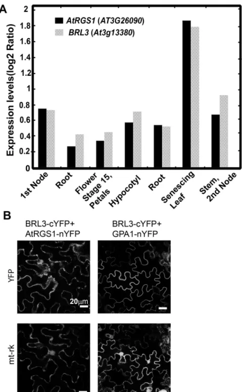
BRL3 interacts with AtRGS1 in vivo
Because BRL3 phosphorylates AtRGS1 [3] and potentially regulates G protein signaling and becauseAtRGS1andBRL3are both expressed in the 1stnode, root, hypocotyl, flower stage 15/ petals, stem/2ndnode, and senescing leaf (Fig 1A), we investigated its functional interaction with AtRGS1. First, we checked if AtRGS1 physically interacts with BRL3in vivoby bimolecu-lar fluorescence complementation (BiFC). BRL3 interacted with AtRGS1in vivo(Fig 1B) whereas a negative control membrane protein, AtHIR2 did not (S2 Fig). AtRGS1-nYFP showed no signal when it was expressed alone inN.benthamiana. The tagged Gαsubunit, GPA1-nYFP, interacted with BRL3-cYFP (Fig 1B) while GPA1-nYFP by itself did not comple-ment flouresence (S2 Fig).
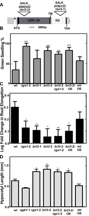
BRL3 and AtRGS1 modulate glucose induced growth inhibition
mutants (Fig 2A),brl3-1 (SALK_079612C; highly reduced expression) andbrl3-2 (SALK_ 006024C; no full length transcript detected) (Fig 2AandS1A Fig) and tested for high glucose
Fig 1. BRL3 interacts with AtRGS1 in vivo. (A) BRL3 and AtRGS1 are co-expressed in several tissues.
Arabidopsis eFP Browser [38] available athttp://www.bar.utoronto.ca/was used for exploring Arabidopsis microarray data to visualize BRL3 and AtGRS1 gene expression profile.(B) BRL3 complements fluorescence by AtRGS1 and GPA1 in BiFC. Confocal images of N. benthamiana cells expressing BRL3-cYFP and AtRGS1-nYFP or BRL3-cYFP and GPA1-nYFP pairs. The upper row indicates the YFP complementation results and the lower row is a transformation reporter, mitochondria protein tagged with mCherry (mt-rk).
Fig 2. BRL3 and AtRGS1 controls glucose responses. (A) T-DNA insertion position in brl3-1 and brl3-2 on
responses (Fig 2B and 2C). First, we used the standardized green seedling assay described by Moore and coworkers [23] to understand the role of BRL3 in glucose signaling. The average percentage of seedlings growing on 6% glucose and showing green cotyledons was determined. Bothrgs1-2 andbrl3mutants were less responsive to 6% glucose than wild type seedlings (P
value<0.0011) as shown inFig 2B.
In the absence of glucose the singlebrl3mutants andbrl3/rgs1double mutant had shorter roots (S3A and S3B Fig) suggesting a positive regulatory function for BRL3 and AtRGS1 in root growth. Inbri1 brl1 brl3mutants, reduced cell expansion was suggested to be responsible for the overall short root phenotype of BR signaling mutants [7]. Thebrl3/rgs1short root phe-notype is consistent with this report of impaired cell expansion in thebri1 brl1 brl3triple mu-tant. However in the presence of glucose, thebrl3roots were not shorter than wild type (S2D Fig). Glucose reduced elongation of 5-day-old wild type root by 2 fold whereas it decreased elongation by ~1.4 fold inrgs1-2,brl3-1,brl3-2 andbrl3-2/rgs1-2 roots (Pvalue<0.05 to 0.001) (Fig 2C). Overexpression of AtRGS1 did not compensate for the loss of BRL3 function in either the green seedling assay or the root growth assay. A nontoxic nonmetabolized sugar, mannitol, was used as an osmotic control [24]. The mannitol effect on root growth was slightly more pronounced than that of glucose (~3.3 fold reduction; 0.5 in log scale). Consistent with a previous report onbrl3mutants and mannitol [25], the osmotic control mannitol decreased the root elongation equally for all genotypes tested (S3C Fig).
We tested early hypocotyl growth of thergs1-2,agb1-2 (Gβsubunit),brl3-2 single andbrl3 -2/rgs1-2 double mutants because this development is partially mediated by G protein-mediated sugar signaling [9,26,27]. Hypocotyl length of seedlings grown on½ ×MS plates with 1% sucrose for 2 d in darkness was measured (Fig 2D). Bothrgs1-2 andbrl3-2 mutants had longer hypoctyls (Pvalue<0.05 to 0.001) and the similar hypocotyl phenotype ofbrl3-2/rgs1-2 dou-ble mutants to single mutants suggested that BRL3 and AtRGS1 function in the same pathway regulating hypocotyl growth. Because AtRGS1 is phosphorylated by BRL3 [3] and overexpres-sion of AtRGS1 inbrl3-2 transgenic lines conferred no effect on the hypocotyl phenotype of
brl3-2, we speculated that BRL3 regulates hypocotyl growth via AtRGS1.
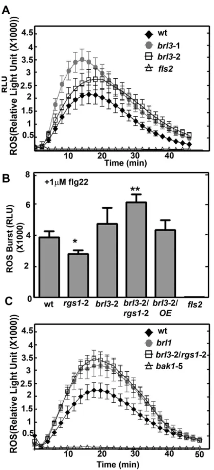
BRL3 and AtRGS1 affect flg22 induced ROS burst
Because plants with anrgs1null mutation showed a slight decrease in flg22-induced ROS pro-duction [28], we quantitated this flg22-induced ROS response in thebrl3mutants. The peak of ROS production at 1μM flg22 induction was slightly but not significantly increased inbrl3leaf disks (Fig 3A). This slight increase was reproducibly observed in three replicate experiments. Peak ROS production quantitated at the 18thminute in response to 1μM flg22 was plotted for repeats (LRR), transmembrane (TM, dark gray region) and kinase domains (KD, indicated in white) are labeled accordingly. The 5’ and 3’ UTRs are highlighted in light gray and the single intron is indicated with a carat (^). Both T-DNA insertions are in the single exon. (B) BRL3 is required for glucose perception. % of green cotyledons of 8-day-old Arabidopsis seedlings grown on¼MS media with 6% glucose under continuous dim-light (20–40μEinstein/m2/s). The averages are from 4 biological replicates with 8 to 30 seedlings for each genotype. Error bars represent standard deviations. One-way ANOVA followed by Tukey tests were used to compare each genotype to wildtype (wt) (**,***P value<0.001, 0.0001) (C) BRL3 and AtRGS1 regulate root growth elengation response induced by high glucose. Log-transformed fold change in root growth of Arabidopsis seedlings grown on¼MS media with 6% glucose at continuous dim light between the 4thand 5thday. This experiment (n = 30 to 100) was repeated three times with similar results. Images were captured and ImageJ was used to quantitate lengths OE means overexpression of AtRGS1 in the indicated wild type (Col-0) or brl3-2 mutant. Student’s t-test was used to compare each genotype to wt (*,**P value<0.05, 0.01). (D) Seedlings were grown on½ ×MS plates with 1% sucrose for 2 d in darkness. Length of hypocotyls was measured and quantified by image J. Data are mean±SEM. One-way ANOVA followed by Tukey tests were used to compare each genotype to wt (*,**P value<0.05, 0.001).
wild type plants,brl3-2 andrgs1-2 single andbrl3-2/rgs1-2 double mutants (Fig 3B). TheS3D Fig
provides the data for the entire time course. There was ~ 30% less ROS production inrgs1-2 (Pvalue<0.05) while ~23% more ROS was produced inbrl3-2 mutants compared to wild type plants. Remarkablybrl3-2/rgs1-2 had ~ 60% more ROS produced than wild type (Pvalue< 0.001). Overexpressing AtRGS1 inbrl3-2 did not change the higher ROS phenotype ofbrl3-2 and did not have a major affect on this mutant (data not shown). Both AtRGS1 or BRL3 supress a positive ROS burst regulator. Their absence relieves this suppression and leads to ~ 60% more ROS production. We also tested another brassinosteroid insensitive1 (BRI1) homolog, BRI1-L-IKE1 (BRL1), which also phosphorylates AtRGS1in vitro[3] to explore its role in the ROS burst. Similar to what we observed withbrl3mutants,brl1also has a large ROS burst in response to flg22 (Fig 3C). This suggests that BRL1 and BRL3 may act together to inhibit ROS production.
BRL3 and AtRGS1 interaction dynamics change with glucose and flg22
BRL3 phosphorylates AtRGS1in vitro[3] and it interacts with AtRGS1 in BiFC assays (Fig 1B), however the dynamics of this interaction are unknown. Because BRL3 is involved in glu-cose and flg22 responses, we tested the physical movements of the BRL3/AtRGS1 complex over time after ligand application.
AtRGS1 changes its interaction with WNK kinases in a glucose dose- and time-dependent manner [12]. In brief, high concentrations of D-glucose (6% for 30 min) rapidly signal through AtWNK8 and AtWNK10 phosphorylation of AtRGS1, whereas low, sustained sugar concen-tration (2% for 5 hr) slowly activates the pathway through phosphorylation of AtRGS1 by AtWNK1, allowing the cells to respond similarly to transient, high-intensity signals and sus-tained, low-intensity signals. Because AtRGS1 is set in motion by glucose, the interaction dynamics between AtRGS1-YFP and BRL3-CFP in response to high and low levels of sugar over time were quantitated through FRET analyses (Fig 4). Glucose (6%) rapidly (10 min) decreased (Pvalue<0.0073) FRET Efficiency (Fig 4A) and this effect took much longer at a lower glucose dose (Pvalue<0.0034) (Fig 4B) indicating a change in distance or orientation between the two fluorophores in response to glucose. As a negative control, AtRGS1-YFP and BRI1-CFP (another brassinosteroid hormone receptor) did not respond to glucose (5 hr 2%,
Fig 4C) indicating the specificity of BRL3.
AtRGS1 and AtGPA1 interaction is affected by flg22 within 5 min [3]. Therefore, we ana-lyzed the dynamics of the AtRGS1-YFP and BRL3-CFP interaction in response to flg22 over time. AtRGS1-YFP and BRL3-CFP showed a FRET efficiency decrease within the first 5 min-ute of 1μM flg22 (Pvalue<0.05) (Fig 4D) indicating a change in distance or orientation between these two proteins.
Discussion
While it is established that AtRGS1 modulates flg22-induced ROS production [29] and that brasinosteroids inhibit flg22-triggered immune signaling [30], the study here provides genetic (within 2–4 minutes) and transient oxidative burst in leaf discs obtained from 6- to 8-week-old Col-0 (wt) but not in fls2 mutants, where the receptor for flg22, FLS2, is genetically ablated. The ROS burst in brl3 mutant plants were elevated slightly but not significantly. (B) ROS burst is fine tuned by BRL3 and AtRGS1. Max ROS burst peak (18thmin) in response to 1μM flg22 in leaf discs of brl3-2/rgs1-2 mutants are significantly higher than wild type (n = 16 to 24). (C) ROS burst is negatively regulated by another member of the BRI1 family, BRL1. brl1 mutants show significantly higher ROS burst than wild type (n = 16 to 24). The point mutation in flg22 co-receptor, BAK1, leads to dimished ROS burst in bak1-5 mutants thus these plants serve as negative control for ROS assay. Measurement of the ROS burst was carried out using the luminol-based luminescence protocol described in Materials and Methods.
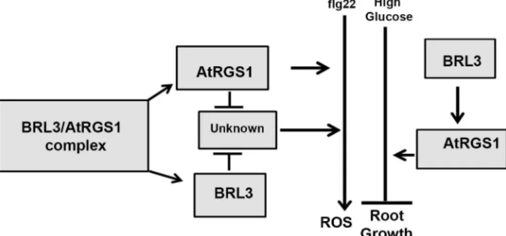
evidence for the interaction of BRL3 and AtRGS1 andin vivoevidence on the dynamics of this RLK/G protein complex in response to glucose and flg22. As shown inFig 5, the genetic inter-actions revealed here provide a complex relationship between BRL3 and AtRGS1 in repression of the ROS burst and growth. Both of the proteins contribute to glucose sensing and root growth. Because loss of either AtRGS1 and BRL3 does not completely abrogate glucose or flg22 responses, these membrane proteins are not absolutely essential for these responses. Rather, we conclude that the function of BRL3 and AtRGS1 is to fine tune the main pathway. Thebrl3andrgs1-2 single and thebrl3/rgs1double mutants are hyposensitive to high glucose (Fig 2B and 2C). We speculate on what else could contribute to glucose- induced fine-tuning pathway mediated by AtRGS1 and BRL3. Arabidopsis EXORDIUM-LIKE1, a BR-regulated gene that is involved in the carbon starvation response [31] and an AtRGS1 interactor as well [32], may also be involved. Given that EXO overexpression increased vegetative growth in comparison to wild-type plants [33] similar torgs1-2 mutants [14,34] and it is required for adaptation to carbon- and energy-limiting growth conditions, it is worthy of investigation of a possible role in the model (Fig 5) in the future. Genetic analysis of thebrl1 brl3 bak1-3triple mutants showed that BAK1, BRL1, and BRL3 modulate root growth and development [5]. All of these RLKs phosphorylate AtRGS1in vitro[3]. This phosphorylation event may activate G protein subunits that are involved in modulation of cell proliferation, root growth and archi-tecture [35]. Because BRL3 interacts with AtGPA1in vivo(Fig 1A), the functional relevance of this interaction in context related to cell proliferation, root growth and architecture should be fully investigated.
Although AtRGS1 and BRL3 also have independent minor functions in the ROS burst as indicated by the phenotypes of the single mutants, they share the same type of inhibitory func-tion on the unknown positive regulator of ROS burst component (Fig 5). The maximum ROS burst inrgs1-2 is slightly reduced (Pvalue<0.05) whereas it is slightly increased inbrl3-2. Therefore, AtRGS1 is a positive regulator of the ROS burst whereas BRL3 is a negative regula-tor. Because brassinosteroids inhibit pathogen-associated molecular pattern-triggered immune signaling including the oxidative burst and defense gene expression [30], a slight increase in the ROS burst in thebrl3-2 mutant is expected. If AtRGS1 and BRL3 are functioning indepen-dently,brl3-2/rgs1-2 is expected to show wild type response. However 60% more ROS than wild type (Pvalue<0.001) is produced in the absence of BRL3 and AtRGS1 (Fig 3C). This indicates that an unknown positive regulator of ROS is freed inbrl3-2/rgs1-2. Thus, we con-clude an inhibitory function for BRL3 and AtRGS1 on this unknown component in the flg22 induced ROS burst (Fig 5). This released component starts a domino effect on ROS and triggers other pathway(s) that also positively regulate ROS. Therefore analyzing BRL3 and AtRGS1 function in ROS is just looking at the tip of an iceberg. There are other components waiting to be discovered in ROS burst regulation. To explain the complex genetic interaction between AtRGS1 and BRL3 in the flg22 response, we speculate that both proteins dissociate to interact with the unknown component given that the FRET Efficiency % between this pair drops in response to flg22 (Fig 4D). If so, what are the nature of these components? This unknown would be an interacting partner to both BRL3 and AtRGS1 and a positive regulator
Fig 4. Glucose and flg22 cause conformational change between AtRGS1 and BRL3. (A) Glucose (6%)
increases the physical distance or orientation between BRL3 and AtRGS1 within 3 to 10 min. FRET analysis was carried out using the acceptor bleaching method in leaf epidermis cells of N. benthamiana expressing AtRGS1-YFP and BRL3-CFP transiently as described in Material and Methods. (B) 2% Glucose for 5 hr also increases the physical distance or orientation between BRL3 and AtRGS1. (C) 2% Glucose does not change FRET Efficiency % between another brassinosteroid receptor kinase, BRI1, and AtRGS1. (D) FRET analysis using the acceptor bleaching method in leaf epidermis cells of N. benthamiana expressing AtRGS1-YFP and BRL3-CFP transiently and treated with 1μM flg22.
of ROS burst. Because the time frame AtRGS1 and BRL3 dissociates is within the period it associates with WNKs and BAK1 in response to glucose and flg22 respectively [12,29], this unknown partner could be WNKs. So it is noteworthy to investigate the links between BRL3 and WNKs and also the competition between AtRGS1 and BRL3 for BAK1 interaction. Given that BRL1 functions similar to BRL3 in the ROS burst (Fig 3C), phosphorylates AtRGS1 and interacts with BAK1 [5], exploring role of BRL1 along with BRL3 is also essential.
Because BRL3 binds BL with high affinity [4] and is redundant to BRL1 and BRI1 in brassi-nosteroid perception [5], we investigated if AtRGS1 functions along with BRL3 in this path-way. Dose–response curve of exogenous BL treatments (0, 0.1, and 1 nM) of 6-d-oldrgs1-2
seedlings showed similar root length as wild-type andbrl3mutants (S4A Fig). In addition, AtRGS1 endocytosis was not induced either with BL or BRZ (S4B Fig). FRET assays provided no evidence for a dynamic change in AtRGS1 and BRL3 interaction inN.benthamianacells treated with 1 or 100μM BL for 2–3 hours (S4C Fig). Therefore, we have not identified a clear role for AtRGS1 in brassinosteroid perception although GPA1 is involved in this response [36,37].
In summary, our analyses revealed the importance of the BRL3 and AtRGS1 in fine tuning growth inhibition and ROS production. Through genetic analysis, we found that BRL3 and AtRGS1 both sense glucose and flg22. They both inhibit growth in response to high glucose. They similarly function in flg22 induced ROS burst by negatively controlling ROS production. Thus, they make small adjustments in these pathways in order to achieve the optimum growth inhibition and ROS burst through dynamic interactions.
Supporting information
S1 Fig. Genotyping new mutants. (A) Semi quantitative PCR measurement ofbrl3transcript levels inbrl3-1 (SALK_079612C) andbrl3-2 (SALK_006024C). Whole plant tissue from
Fig 5. Proposed model for BRL3 and AtRGS1 functions in glucose and flg22 reponses. Partially
reduced ROS burst in rgs1-2 (P value<0.05) and slightly increased in brl3-2 indicate that AtRGS1 is a positive regulator of ROS burst whereas BRL3 is a negative regulator. 60% more ROS than wild type (P value<0.001) is produced in brl3-2/rgs1-2 (Fig 3B). Absence of AtRGS1 and BRL3 removes the inhibitory effect on an unknown positive regulator of ROS. Removal of BRL3 and AtRGS1 inhibition on this unknown component does not only activates it but also generates a cumulative event by triggering other pathway(s) positively regulating ROS burst.Glucose induced inhibitory effect on root elongation is impaired in plants without AtRGS1 or BRL3. Diminished glucose effect on the single rgs1-2 and brl3-2 mutants is similar to what is observed with brl3-2/rgs1-2 double mutants (Fig 2C). This proves that AtRGS1 and BRL3 function in the same glucose response pathway. Because AtRGS1 overexpression in brl3-2 mutants results in same phenotype as brl3-2, it is concluded that BRL3 acts upstream of AtRGS1.
seedlings grown in fresh 1/2×MS liquid media for 9 days was harvested by flash freezing in liquid N2. The mRNA and cDNA were prepared with RNAeasyTM(Qiagen) and Superscript III (Invitrogen), respectively, according to the manufacturer’s instructions. The PCR amplifi-cation protocol with Taq polymerase consists of an initial denaturation step at 95 ˚C for 5 min, followed by 30 amplification cycles at 94 ˚C for 30 s, 57 ˚C for 1 min,72 ˚C for 90 s and 4 ˚C for 1 min. Forward full-lengthbrl3coding sequence primer:ATGAAACAACAATGGCAGTTCT TGA; Reverse Full length brl3 coding sequence primer: TTGTAGACATCTCC AAATCCACCTG(B)Genotyping of twobrl3-2/rgs1-2 plants using the pri-mers and protocol above. Pripri-mers used arebrl3-2 Left genomic primer: CCAGTGAACTCGTTTGAGCTC;brl3-2 Right genomic primer: TTTATCGAACACT TTGTGGGC;rgs1-2 Left genomic primer: TGTTGATGAAAAGCCTTAGCG;rgs1-2 Right genomic primer: TAGCTGCTACGCTGGAGAAAC; and T-DNA left border primer: TGG TTC ACG TAG TGG GCC ATC.
(JPG)
S2 Fig. Negative controls for BiFC. The negative control AtHIR2-nYFP does not complement
BRL3-cYFP. Neither AtRGS1-nYFP nor GPA1-nYFP produces fluorescence by itself. (JPG)
S3 Fig. Negative control for the sugar response. (A) Top: 96 hr- old wt,brl3-2,rgs1-2,brl3-2/ rgs1-2 mutants grown on¼MS media under continuous dim-light (20–40μEinstein/m2/s) vertically. Bottom: 8-day-old Arabidopsis seedlings grown on¼MS media under continuous dim-light (20–40μEinstein/m2/s) horizontally. (B) Root Elongation. (C) BRL3 and AtRGS1 are not involved in high mannitol response. (D) ROS burst in response to 1μM flg22 in leaf discs (n = 16 to 24) including all the time points from 0 to 48 min.
(TIF)
S4 Fig. Brassinolide (BL) assay. (A)rgs1-2 mutants show similar root growth inhibition response with BL to wt. (B) AtRGS1 internalization is not affected by BRZ or BL. (C) No change is detected in AtRGS1 and BRL3 interaction dynamics in response to BL. (JPG)
Author Contributions
Conceptualization: MT-O.
Data curation: MT-O.
Formal analysis: MT-O.
Funding acquisition: AMJ.
Investigation: MT-O.
Methodology: MT-O.
Project administration: AMJ.
Resources: AMJ.
Software: MT-O.
Supervision: MT-O.
Visualization: AMJ MT-O.
Writing – original draft: AMJ MT-O.
Writing – review & editing: AMJ MT-O.
References
1. Torii KU. Leucine-rich repeat receptor kinases in plants: structure, function, and signal transduction pathways. Int Rev Cytol. 2004; 234: 1–46.https://doi.org/10.1016/S0074-7696(04)34001-5PMID:
15066372
2. Urano D, Jones AM. Heterotrimeric G protein-coupled signaling in plants. Annu Rev Plant Biol. 2014; 65: 365–84.https://doi.org/10.1146/annurev-arplant-050213-040133PMID:24313842
3. Tunc-Ozdemir M, Urano D, Jaiswal DK, Clouse SD, Jones AM. Direct Modulation of a Heterotrimeric G protein-coupled Signaling by a Receptor Kinase Complex. J Biol Chem. 2016;
4. Caño-Delgado A, Yin Y, Yu C, Vafeados D, Mora-Garcı´a S, Cheng J-C, et al. BRL1 and BRL3 are novel brassinosteroid receptors that function in vascular differentiation in Arabidopsis. Development. 2004; 131: 5341–51.https://doi.org/10.1242/dev.01403PMID:15486337
5. Fàbregas N, Li N, Boeren S, Nash TE, Goshe MB, Clouse SD, et al. The brassinosteroid insensitive1-like3 signalosome complex regulates Arabidopsis root development. Plant Cell. 2013; 25: 3377–88.
https://doi.org/10.1105/tpc.113.114462PMID:24064770
6. Salazar-Henao JE, Lehner R, Betego´n-Putze I, Vilarrasa-Blasi J, Caño-Delgado AI. BES1 regulates the localization of the brassinosteroid receptor BRL3 within the provascular tissue of the Arabidopsis pri-mary root. J Exp Bot. 2016; 67: 4951–61.https://doi.org/10.1093/jxb/erw258PMID:27511026 7. Kang YH, Breda A, Hardtke CS. Brassinosteroid signaling directs formative cell divisions and
proto-phloem differentiation in Arabidopsis root meristems. Development. 2017; 144: 272–280.https://doi. org/10.1242/dev.145623PMID:28096215
8. Urano D, Chen J-G, Botella JR, Jones AM. Heterotrimeric G protein signalling in the plant kingdom. Open Biol. 2013; 3: 120186.https://doi.org/10.1098/rsob.120186PMID:23536550
9. Gupta A, Singh M, Laxmi A. Multiple Interactions between Glucose and Brassinosteroid Signal Trans-duction Pathways in Arabidopsis Are Uncovered by Whole-Genome Transcriptional Profiling. Plant Physiol. 2015; 168: 1091–105.https://doi.org/10.1104/pp.15.00495PMID:26034265
10. Grigston JC, Osuna D, Scheible W-R, Liu C, Stitt M, Jones AM. D-Glucose sensing by a plasma mem-brane regulator of G signaling protein, AtRGS1. FEBS Lett. 2008; 582: 3577–84.https://doi.org/10. 1016/j.febslet.2008.08.038PMID:18817773
11. Liao K-L, Jones RD, McCarter P, Tunc-Ozdemir M, Draper JA, Elston TC, et al. A shadow detector for photosynthesis efficiency. J Theor Biol. 2017; 414: 231–244.https://doi.org/10.1016/j.jtbi.2016.11.027
PMID:27923735
12. Fu Y, Lim S, Urano D, Tunc-Ozdemir M, Phan NG, Elston TC, et al. Reciprocal encoding of signal inten-sity and duration in a glucose-sensing circuit. Cell. 2014; 156: 1084–95.https://doi.org/10.1016/j.cell. 2014.01.013PMID:24581502
13. Urano D, Phan N, Jones JC, Yang J, Huang J, Grigston J, et al. Endocytosis of the seven-transmem-brane RGS1 protein activates G-protein-coupled signalling in Arabidopsis. Nat Cell Biol. 2012; 14: 1079–88.https://doi.org/10.1038/ncb2568PMID:22940907
14. Huang J-P, Tunc-Ozdemir M, Chang Y, Jones AM. Cooperative control between AtRGS1 and AtHXK1 in a WD40-repeat protein pathway in Arabidopsis thaliana. Front Plant Sci. 2015; 6: 851.https://doi.org/ 10.3389/fpls.2015.00851PMID:26528314
15. Chen Y, Ji F, Xie H, Liang J, Zhang J. The regulator of G-protein signaling proteins involved in sugar and abscisic acid signaling in Arabidopsis seed germination. Plant Physiol. 2006; 140: 302–10.https:// doi.org/10.1104/pp.105.069872PMID:16361523
16. Urano D, Maruta N, Trusov Y, Stoian R, Wu Q, Liang Y, et al. Saltational evolution of the heterotrimeric G protein signaling mechanisms in the plant kingdom. Sci Signal. 2016; 9: ra93.https://doi.org/10.1126/ scisignal.aaf9558PMID:27649740
17. Chen J-G, Willard FS, Huang J, Liang J, Chasse SA, Jones AM, et al. A seven-transmembrane RGS protein that modulates plant cell proliferation. Science. 2003; 301: 1728–31.https://doi.org/10.1126/ science.1087790PMID:14500984
18. Zipfel C, Robatzek S, Navarro L, Oakeley EJ, Jones JDG, Felix G, et al. Bacterial disease resistance in Arabidopsis through flagellin perception. Nature. 2004; 428: 764–7.https://doi.org/10.1038/
19. Roux M, Schwessinger B, Albrecht C, Chinchilla D, Jones A, Holton N, et al. The Arabidopsis leucine-rich repeat receptor-like kinases BAK1/SERK3 and BKK1/SERK4 are required for innate immunity to hemibiotrophic and biotrophic pathogens. Plant Cell. 2011; 23: 2440–55.https://doi.org/10.1105/tpc. 111.084301PMID:21693696
20. Schwessinger B, Roux M, Kadota Y, Ntoukakis V, Sklenar J, Jones A, et al. Phosphorylation-dependent differential regulation of plant growth, cell death, and innate immunity by the regulatory receptor-like kinase BAK1. PLoS Genet. 2011; 7: e1002046.https://doi.org/10.1371/journal.pgen.1002046PMID:
21593986
21. Tunc-Ozdemir M, Fu Y, Jones AM. Cautions in Measuring In Vivo Interactions Using FRET and BiFC in Nicotiana benthamiana. Plant Signal Transduction: Methods and Protocols (Methods in Molecular Biol-ogy). 2nd ed. Humana Press; 2016.
22. Chung E-H, El-Kasmi F, He Y, Loehr A, Dangl JL. A plant phosphoswitch platform repeatedly targeted by type III effector proteins regulates the output of both tiers of plant immune receptors. Cell Host Microbe. 2014; 16: 484–94.https://doi.org/10.1016/j.chom.2014.09.004PMID:25299334
23. Moore B, Zhou L, Rolland F, Hall Q, Cheng W-H, Liu Y-X, et al. Role of the Arabidopsis glucose sensor HXK1 in nutrient, light, and hormonal signaling. Science. 2003; 300: 332–6.https://doi.org/10.1126/ science.1080585PMID:12690200
24. Li Y, Lee KK, Walsh S, Smith C, Hadingham S, Sorefan K, et al. Establishing glucose- and ABA-regu-lated transcription networks in Arabidopsis by microarray analysis and promoter classification using a Relevance Vector Machine. Genome Res. 2006; 16: 414–27.https://doi.org/10.1101/gr.4237406
PMID:16424108
25. ten Hove CA, Bochdanovits Z, Jansweijer VMA, Koning FG, Berke L, Sanchez-Perez GF, et al. Probing the roles of LRR RLK genes in Arabidopsis thaliana roots using a custom T-DNA insertion set. Plant Mol Biol. 2011; 76: 69–83.https://doi.org/10.1007/s11103-011-9769-xPMID:21431781
26. Singh M, Gupta A, Laxmi A. Glucose control of root growth direction in Arabidopsis thaliana. J Exp Bot. 2014; 65: 2981–93.https://doi.org/10.1093/jxb/eru146PMID:24719453
27. Ullah H, Chen JG, Young JC, Im KH, Sussman MR, Jones AM. Modulation of cell proliferation by het-erotrimeric G protein in Arabidopsis. Science. 2001; 292: 2066–9.https://doi.org/10.1126/science. 1059040PMID:11408654
28. Torres MA, Morales J, Sa´nchez-Rodrı´guez C, Molina A, Dangl J. Functional interplay between Arabi-dopsis NADPH oxidases and heterotrimeric G protein. Mol Plant Microbe Interact. 2013;
29. Tunc-Ozdemir M, Jones AM. Ligand-induced dynamics of heterotrimeric G protein-coupled receptor-like kinase complexes. Chang I-F, editor. PLoS One. 2017; 12: e0171854.https://doi.org/10.1371/ journal.pone.0171854PMID:28187200
30. Albrecht C, Boutrot F, Segonzac C, Schwessinger B, Gimenez-Ibanez S, Chinchilla D, et al. Brassinos-teroids inhibit pathogen-associated molecular pattern-triggered immune signaling independent of the receptor kinase BAK1. Proc Natl Acad Sci U S A. 2012; 109: 303–8.https://doi.org/10.1073/pnas. 1109921108PMID:22087006
31. Schro¨der F, Lisso J, Mu¨ssig C. EXORDIUM-LIKE1 promotes growth during low carbon availability in Ara-bidopsis. Plant Physiol. 2011; 156: 1620–30.https://doi.org/10.1104/pp.111.177204PMID:21543728 32. Klopffleisch K, Phan N, Augustin K, Bayne RS, Booker KS, Botella JR, et al. Arabidopsis G-protein
inter-actome reveals connections to cell wall carbohydrates and morphogenesis. Mol Syst Biol. 2011; 7: 532.
https://doi.org/10.1038/msb.2011.66PMID:21952135
33. Coll-Garcia D, Mazuch J, Altmann T, Mu¨ssig C. EXORDIUM regulates brassinosteroid-responsive genes. FEBS Lett. 2004; 563: 82–6.https://doi.org/10.1016/S0014-5793(04)00255-8PMID:15063727 34. Colaneri AC, Tunc-Ozdemir M, Huang JP, Jones AM. Growth attenuation under saline stress is
medi-ated by the heterotrimeric G protein complex. BMC Plant Biol. 2014; 14: 129.https://doi.org/10.1186/ 1471-2229-14-129PMID:24884438
35. Chen J-G, Gao Y, Jones AM. Differential roles of Arabidopsis heterotrimeric G-protein subunits in mod-ulating cell division in roots. Plant Physiol. 2006; 141: 887–97.https://doi.org/10.1104/pp.106.079202
PMID:16679415
36. Gao Y, Wang S, Asami T, Chen J-G. Loss-of-function mutations in the Arabidopsis heterotrimeric G-protein alpha subunit enhance the developmental defects of brassinosteroid signaling and biosynthesis mutants. Plant Cell Physiol. 2008; 49: 1013–24.https://doi.org/10.1093/pcp/pcn078PMID:18499742 37. Ullah H, Chen J-G, Wang S, Jones AM. Role of a heterotrimeric G protein in regulation of Arabidopsis seed
germination. Plant Physiol. 2002; 129: 897–907.https://doi.org/10.1104/pp.005017PMID:12068128 38. Winter D, Vinegar B, Nahal H, Ammar R, Wilson G V, Provart NJ. An “Electronic Fluorescent
Picto-graph” browser for exploring and analyzing large-scale biological data sets. PLoS One. 2007; 2: e718.