Microorganisms in the human placenta are
associated with altered CpG methylation of
immune and inflammation-related genes
Martha Scott Tomlinson1, Paige A. Bommarito1, Elizabeth M. Martin1, Lisa Smeester1, Raina N. Fichorova2, Andrew B. Onderdonk3, Karl C. K. Kuban4, T. Michael O’Shea5, Rebecca C. Fry1*
1 Department of Environmental Sciences and Engineering, Gillings School of Global Public Health, University of North Carolina, Chapel Hill, North Carolina, United States of America, 2 Laboratory of Genital Tract Biology, Department of Obstetrics and Gynecology, Harvard Medical School and Brigham and Women’s Hospital, Boston, Massachusetts, United States of America, 3 Department of Pathology, Harvard Medical School and Brigham and Women’s Hospital, Boston, Massachusetts, United States of America, 4 Division of Pediatric Neurology, Department of Pediatrics, Boston Medical Center, Boston, Massachusetts, United States of America, 5 Department of Pediatrics, School of Medicine, University of North Carolina, Chapel Hill, North Carolina, United States of America
Abstract
Microorganisms in the placenta have been linked to adverse pregnancy outcomes as well as neonatal illness. Inflammation in the placenta has been identified as a contributing factor in this association, but the underlying biological mechanisms are not yet fully understood. The placental epigenome may serve as an intermediate between placental microbes and inflammation, contributing to adverse outcomes in the offspring. In the present study, genome-wide DNA methylation (n = 486,428 CpG sites) of 84 placentas was analyzed in relation to 16 species of placental microorganisms using samples collected from the Extremely Low Gestation Age Newborns (ELGAN) cohort. A total of n = 1,789 CpG sites, corresponding to n = 1,079 genes, displayed differential methylation (q<0.1) in relation to microorganisms. The altered genes encode for proteins that are involved in immune/inflam-matory responses, specifically the NF-κB signaling pathway. These data support bacteria-dependent epigenetic patterning in the placenta and provide potential insight into mecha-nisms that associate the presence of microorgamecha-nisms in the placenta to pregnancy and neonatal outcomes. This study lays the foundation for investigations of the placental micro-biome and its role in placental function.
Introduction
The placenta is a critical regulator of the prenatal environment and is essential for a healthy pregnancy and fetal development. It transports nutrients from mother to fetus and produces hormones necessary to maintain pregnancy and support the fetus [1]. The placenta can also harbor bacterial communities that, depending on their composition, affect pregnancy
a1111111111 a1111111111 a1111111111 a1111111111 a1111111111
OPEN ACCESS
Citation: Tomlinson MS, Bommarito PA, Martin EM, Smeester L, Fichorova RN, Onderdonk AB, et al. (2017) Microorganisms in the human placenta are associated with altered CpG methylation of immune and inflammation-related genes. PLoS ONE 12(12): e0188664.https://doi.org/10.1371/ journal.pone.0188664
Editor: Amr H Sawalha, University of Michigan, UNITED STATES
Received: August 24, 2017
Accepted: November 10, 2017
Published: December 14, 2017
Copyright:©2017 Tomlinson et al. This is an open access article distributed under the terms of the Creative Commons Attribution License, which permits unrestricted use, distribution, and reproduction in any medium, provided the original author and source are credited.
Data Availability Statement: All methylation files are available from the GEO database (accession number GSE106089).
Funding: This research was supported by grants from the National Institutes of Health (http://www. nih.gov): R01 ES019315, P42ES005948, T32ES007018, 1UG3OD023348-01,
outcomes and fetal health [2,3]. Previously it was thought that all bacteria in the placenta orig-inate from infections of the lower genital tract [3], however, a number of studies have found that bacteria in the placenta are derived from vaginotropic non-infectious microflora [4,5]. In addition, bacteria derived from other tissues may contribute to the placental microbiome, such as the oral cavity [6]. For example, it has been proposed that oral bacteria can translocate to the placenta by hematogenous transmission [7].
Importantly, certain bacteria in the placenta have been associated with deleterious preg-nancy outcomes, such as preterm birth [3]. Among these placental bacteria areUreplasma urealyticum,Mycoplasma hominis,Gardnerella vaginalis, andPeptostreptococcussp., which are all associated with bacterial vaginosis, a disruption in the vaginal microbiota. Other placental bacteria, Group BStreptococcusandEscherichia coli, are associated with chorioamnionitis and fetal infection [3]. Little is known about the molecular responses to microorganisms in the pla-centa that potentially alter pregnancy and fetal outcomes.
The placental epigenome, particularly DNA methylation, is a potential mechanism that could explain this association and has not been explored. Epigenetic changes, such as DNA methylation, have been associated with bacteria in other tissues includingHelicobacter pylori
in the gastric mucosa [8–10], uropathogenicEscherichia coliin uroepithelial cells [11] and
Campylobacter rectusin murine placental tissue [12]. In human placental cell culture, the pres-ence of bacteria has been associated with both pro- and anti- inflammatory responses medi-ated by cytokines [13]. With an intriguing potential for sustained inflammation, our team showed in the Extremely Low Gestation Age Newborns (ELGAN) cohort that vaginotropic placental bacteria were associated with distinct inflammatory protein profiles in newborn blood withLactobacillussp. being anti-inflammatory and bacterial vaginosis-associated bacte-ria being pro-inflammatory [14]. These varying inflammatory responses induced by placental bacteria might be driven by CpG methylation in the placenta which could impact both placen-tal function and feplacen-tal well-being.
To our knowledge, this is among the first studies to assess placental CpG methylation in relation to placenta bacteria. Here we investigated whether placental microbes were associated with altered placental DNA (CpG) methylation patterns in the ELGAN cohort. The goal of these analyses is to provide insights into how microorganimsms in the placenta alter the pla-cental methylome and could thereby influence pregnancy and neonatal health outcomes.
Methods
ELGANs study subject recruitment
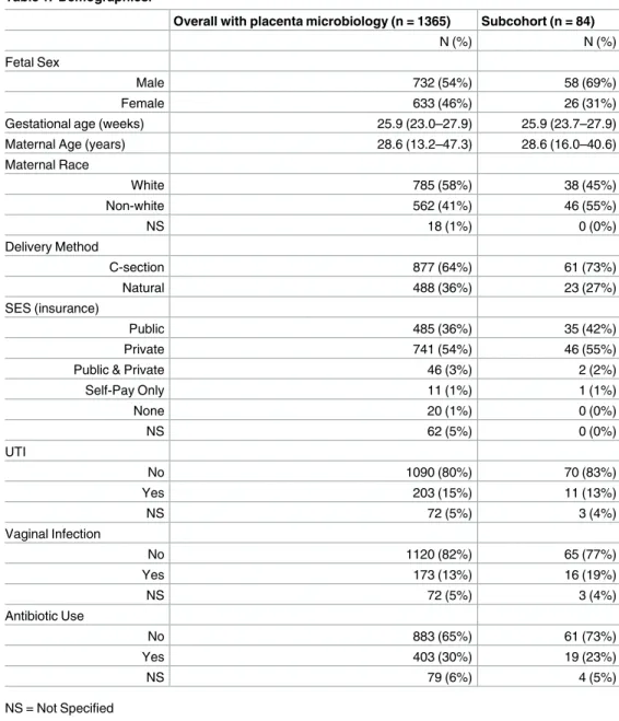
The recruitment process for the ELGAN study has been described in detail [15]. Briefly, between 2002 and 2004, we invited women who gave birth before 28 weeks gestational age at one of the 14 hospitals in 5 states in the United States to participate in the study. A total of 1,249 mothers and 1,506 infants enrolled in the study of which 1,365 placentas were collected and analyzed for microorganisms. A subcohort of 84 mother/infant pairs with similar average gestational and maternal age as the overall cohort were investigated in the present study (Table 1).
Ethics statement
The Institutional Review Boards at Baystate Medical Center in Springfield, MA, Beth Israel Deaconess Medical Center in Boston, MA, Brigham and Women’s Hospital in Boston, MA, Children’s Hospital in Boston, MA, Massachusetts General Hospital in Boston, MA, Tufts New England Medical Center in Boston, MA, UMass memorial Medical Center in Worcester, MA, Yale-New Haven Hospital, New Haven, CT, Forsyth Hospital, Baptist Medical Center in collection and analysis, decision to publish, or
preparation of the manuscript.
Winston-Salem, NC, University Health Systems of Eastern Carolina in Greenville, NC, North Carolina Children’s Hospital in Chapel Hill, NC, DeVos Children’s Hospital in Grand Rapids, MI, Sparrow Hospital in Lansing, MI, University of Chicago Hospital in Chicago, IL, and Wil-liam Beaumont Hospital in Royal Oak, MI approved all procedures. Informed, written consent was provided within a few days of delivery, either before or after. The mother’s consent cov-ered both her and the child’s participation in the study.
Placenta sample collection
Women participating in the ELGANs study were asked to provide their placentas for analysis. The technique used to collect the placentas is as follows: delivered placentas were placed in a sterile exam basin and transported to a sampling room where they were biopsied at the mid-point of the longest distance between the cord insertion and the edge of the placental disk. Table 1. Demographics.
Overall with placenta microbiology (n = 1365) Subcohort (n = 84)
N (%) N (%)
Fetal Sex
Male 732 (54%) 58 (69%)
Female 633 (46%) 26 (31%)
Gestational age (weeks) 25.9 (23.0–27.9) 25.9 (23.7–27.9)
Maternal Age (years) 28.6 (13.2–47.3) 28.6 (16.0–40.6)
Maternal Race
White 785 (58%) 38 (45%)
Non-white 562 (41%) 46 (55%)
NS 18 (1%) 0 (0%)
Delivery Method
C-section 877 (64%) 61 (73%)
Natural 488 (36%) 23 (27%)
SES (insurance)
Public 485 (36%) 35 (42%)
Private 741 (54%) 46 (55%)
Public & Private 46 (3%) 2 (2%)
Self-Pay Only 11 (1%) 1 (1%)
None 20 (1%) 0 (0%)
NS 62 (5%) 0 (0%)
UTI
No 1090 (80%) 70 (83%)
Yes 203 (15%) 11 (13%)
NS 72 (5%) 3 (4%)
Vaginal Infection
No 1120 (82%) 65 (77%)
Yes 173 (13%) 16 (19%)
NS 72 (5%) 3 (4%)
Antibiotic Use
No 883 (65%) 61 (73%)
Yes 403 (30%) 19 (23%)
NS 79 (6%) 4 (5%)
NS = Not Specified
Using sterile technique, the amnion was pulled back to expose the chorion. Traction was applied to the chorion and the underlying trophoblast tissue and a piece of tissue was removed. The tissue was placed into a cryo-vial and immediately immersed into liquid nitrogen. Speci-mens were stored until processing at minus 80˚C [4].
Bacterial analysis of placenta
The placentas were biopsied as soon as possible after delivery and were assessed for microor-ganisms as described [4]. Briefly, a section of each placental specimen was removed using a sterile scalpel and homogenized in a phosphate buffered saline solution (PBS). Serial dilutions of the homogenate were made in PBS and aliquots of the original homogenate and the dilu-tions were plated onto selective and nonselective bacteriologic media, which included: prere-duced Brucella base agar, tryptic soy agar, chocolate agar, and A-7 agar. Following incubation the various colony types were enumerated, isolated, and identified at the Brigham and Wom-en’s Microbiology Laboratory using estimated criteria [16]. Since we have determined the con-stituents of the chorion parenchyma in the ELGANs prevent the reliable detection of bacterial DNA by PCR techniques, this study assessed only placental colonization patterns obtained by culture techniques.
DNA extraction and assessment of DNA methylation
DNA extraction and assessment of DNA methylation by taking a 0.2 g subsection of placental tissue was cut from the frozen biopsy of dry ice, washed with sterile 1X PBS to remove residual blood, and homogenized with B-mercaptoethanol in Buffer RLT (Qiagen, Valencia, CA). DNA and RNA sequences with 18 or more nucleotides in length were collected using the All-Prep DNA/RNA/miRNA Universal Kit (Qiagen, Valencia, CA) following the manufacturer’s instructions. CpG methylation was assessed using the Illumina Human Methylation450 Bead-Chip©array (Illumina, Inc., San Diego, CA). This asses the DNA methylation levels of 486,428 individual probes at single nucleotide resolution. Isolated DNA was bisulfate-converted using the EZ DNA methylation kit (Zymo Research, Irvine, CA) and converted DNA was hybridized onto the array. The DNA methylation data was collected at Expression Analysis, Inc. (Durham, NC;www.expressionanalysis.com).
Methylation levels were calculated and expressed asβvalues (β= intensity of the methylated allele (M) / (intensity of the unmethylated allele (U) + intensity of the methylated allele (M) + 100) [17]. Batch effect was not a significant source of variation, as determined by principle component analysis (PCA). Based on manufacturer recommendations, probes that had high detection P-values (P>0.1) were removed from analysis (n = 24,591). Following data filtration, data were normalized using the beta-mixture quantile (BMIQ) normalization method using the wateRmelon package (version 1.11.0) in R (version 3.2.3). After normalization, probes that were annotated as single nucleotide polymorphisms (SNPs) by Illumina were removed from further analysis (n = 84,121), leaving a total of 377,716 probes, representing 20,418 genes. The rationale for this exclusion is that SNP variation can lead to false readings of DNA methylation signals that may be attributed to genomic variation rather alterations in actual methylation lev-els [18].
Statistical analysis
In order to determine whether the placental microbiome is associated with differences in DNA methylation patterning of the placenta, Analysis of Covariance (ANCOVA) was per-formed. There were 16 bacterial species assessed including:Lactobacillussp.,Prevotella bivia,
alpha-hemolyticStreptococcus,Ureaplasma urealyticum,Mycoplasmasp.,Staphyloccocussp., Propio-nibacteriumsp.,Actinomycessp.,Corynebacteriumsp.,Staphylococcus aureus,Streptococcus
Group B, andStreptococcusGroup D. Each model examined whether presence or absence of an individual bacterial species or bacterial type was associated with differential placental meth-ylation at any of the 377,716 probes. In the present study, variables were classified as con-founding if they displayed an association with both the exposure, placental microorganisms, and the outcome, DNA methylation. Models were adjusted based on several dichotomous var-iables including fetal-sex, maternal race, whether the mother took an antibiotic during preg-nancy, whether the mother experienced a vaginal infection during pregpreg-nancy, whether the mother experienced a urinary tract infection (UTI) during pregnancy, and whether the birth occurred via C-section. As there is no evidence that maternal age is associated with the pres-ence of microorganisms in the placenta, it was not included as a confounding variable. Bacte-rial species were considered to be significantly associated with methylation ifp-value<0.05 and the false discovery rate-correctedp-value<0.1 for any given probe. Data analysis was car-ried out using Partek Genomic Suites 6.6.
Gene set-based analysis
The differentially methylated probes and corresponding genes were analyzed to determine if they were enriched for specific biological functions. Four gene sets were analyzed where gene content was established usingwww.uniprot.org. The four gene sets that were tested were selected for their known roles in fetal development: transport-related genes (n = 3,704) [19], immune-related genes (n = 1,622) [19–21], inflammation-related genes (n = 410) [20,22] and growth/transcription factor-related genes (n = 2030) [23–25]. Aχ2
test was used to identify enrichment of key processes. Once the enriched processes were identified a right-tailed Fisher Exact test (α= 0.05) was conducted on the significant genes in the enriched pathways to iden-tify enriched canonical pathways and transcription factors using Ingenuity Pathway Analysis software as described in Martinet al., 2015 [26].
Results
Study subject characteristics
The placentas of 84 subjects within the ELGAN cohort were analyzed for this study. The aver-age gestational aver-age and maternal aver-age of the subcohort of 84 are the same as the overall cohort (n = 1,365) (Table 1). There are some differences between the subcohort and the overall cohort, including maternal race and sex of the infant. The overall cohort is predominately white (58%) while the subcohort is only 45% white and 55% non-white. The majority of the infants in both the overall cohort and subcohort are males. However, the percentage of males in the subcohort (69%) is much larger than the percentage in the overall cohort (45%). Of the 84 infants, 23 (27%) were delivered vaginally, while 61 (73%) were delivered by Cesarean sec-tion. Of these, 37 (44%) were on public insurance and 46 (55%) were on private insurance. A total of 11 (13%) of the women had a urinary tract infection, 16 (19%) had a vaginal infection, and 19 (22%) used an antibiotic during pregnancy.
Identification of differentially methylated probes in ELGAN placentas
exposed to bacteria
Placental CpG methylation differences were analyzed within the ELGAN cohort between 84 individuals with or without placental microbes for 377,716 CpG probes representing 20,418 genes. Statistical significance was adjusted using false discovery rate-corrected p-values (q<0.1). Of the 16 microbial species all but two, namely anaerobicStreptococcusand
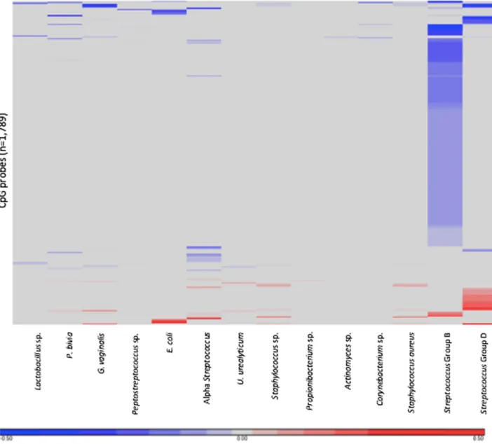
Mycoplasmasp., were associated with differentially methylated probes. A total of 1,789 probes, corresponding to 1,079 genes, were significantly differentially methylated between placentas that harbored bacteria and placentas that did not (S1 File). The probes that displayed differential methylation were unique depending on which bacteria was present (Fig 1).
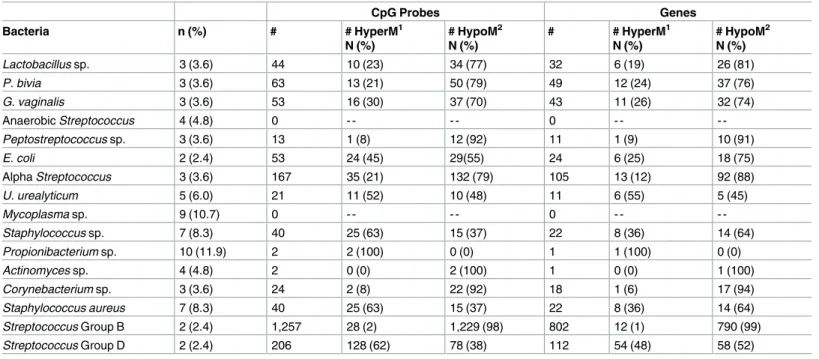
The microbial species that was associated with the greatest number of altered CpG sites wasStreptococcusGroup B with 1,257 differentially methylated CpG probes (n = 802 genes) (Table 2). The methylation pattern forStreptococcusGroup B leans heavily toward hypomethy-lation with 98% (1,232) of the sites being hypomethylated.
FollowingStreptococcusGroup B, the two microbial species that were associated with the most differentially methylated probes wereStreptococcusGroup D and alpha-hemolytic Strep-tococcuswith 206 and 167 CpG probes, respectively. Most of the probes altered in relation to
StreptococcusGroup D were hypermethylated (62%) while alpha-hemolyticStreptococcus -asso-ciated probes were mostly hypomethylated (79%). The CpG probes correspond to 112 genes forStreptococcusGroup D and 105 genes for alpha-hemolyticStretptococcus. The presence of
E.coliwas associated with 53 CpG probes representing 43 different genes. Of those probes 55% were hypomethylated.
Table 2. Presence of microorganisms in the placenta and methylation of CpG probes and associated genes.
CpG Probes Genes
Bacteria n (%) # # HyperM1
N (%)
# HypoM2
N (%)
# # HyperM1
N (%)
# HypoM2
N (%)
Lactobacillus sp. 3 (3.6) 44 10 (23) 34 (77) 32 6 (19) 26 (81)
P. bivia 3 (3.6) 63 13 (21) 50 (79) 49 12 (24) 37 (76)
G. vaginalis 3 (3.6) 53 16 (30) 37 (70) 43 11 (26) 32 (74)
Anaerobic Streptococcus 4 (4.8) 0 - - - - 0 - -
-Peptostreptococcus sp. 3 (3.6) 13 1 (8) 12 (92) 11 1 (9) 10 (91)
E. coli 2 (2.4) 53 24 (45) 29(55) 24 6 (25) 18 (75)
Alpha Streptococcus 3 (3.6) 167 35 (21) 132 (79) 105 13 (12) 92 (88)
U. urealyticum 5 (6.0) 21 11 (52) 10 (48) 11 6 (55) 5 (45)
Mycoplasma sp. 9 (10.7) 0 - - - - 0 - -
-Staphylococcus sp. 7 (8.3) 40 25 (63) 15 (37) 22 8 (36) 14 (64)
Propionibacterium sp. 10 (11.9) 2 2 (100) 0 (0) 1 1 (100) 0 (0)
Actinomyces sp. 4 (4.8) 2 0 (0) 2 (100) 1 0 (0) 1 (100)
Corynebacterium sp. 3 (3.6) 24 2 (8) 22 (92) 18 1 (6) 17 (94)
Staphylococcus aureus 7 (8.3) 40 25 (63) 15 (37) 22 8 (36) 14 (64)
Streptococcus Group B 2 (2.4) 1,257 28 (2) 1,229 (98) 802 12 (1) 790 (99)
Streptococcus Group D 2 (2.4) 206 128 (62) 78 (38) 112 54 (48) 58 (52)
1
HyperM = Hypermethylated
2
HypoM = Hypomethylated
Location of differentially methylated probes
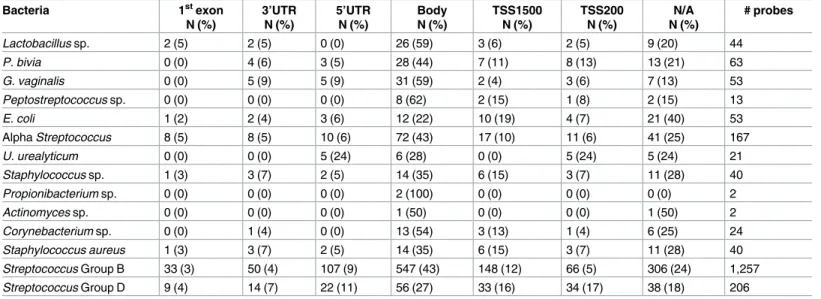
The majority of the significant CpG probes for all 14 bacterial species were located in the body of the gene (Table 3). Both of the probes associated withPropionibacteriumsp. were located in the gene body.Peptostreptococcussp. had the next highest percentage of differentially methylated probes in the gene body at 62%, whileActinomycessp. had the lowest percentage at 22%. There were also a large percentage of probes that did not correspond with a gene and therefore do not have a gene location. The remaining probes fell into the 1stexon, 3’UTR, 5’UTR, TSS1500, and TSS200 locations.
Fig 1. Heatmap of differentially methylated CpG probes corresponding to each of the microorganisms. The heatmap displays the 1,789 differentially methylated probes in relation to 17 microorganisms. Red represents increased methylation and blue represents decreased methylation.
Enrichment of biological functions and pathways among the differentially
methylated gene sets
We further analyzed whether these differentially methylated probes in the placenta were enriched for four specific biological functions: inflammation, growth/transcription factors, transport, and/or immune response, selected for their known critical functions for fetal devel-opment. For this analysis, the unique genes that corresponded with differentially methylated probes were considered (n = 1,080). A Yates correctedχ2
test was used in conjunction with a right-tailed Fisher Exact test (α= 0.05) to determine that immune-related proteins (n = 50, p = 0.000075), transcription/growth factors (n = 134, p = 0.00173), and inflammatory response (n = 35, p = 1.27E-09) were enriched (S1 Table).
Among the 50 immune-related genes, Nuclear Factor Kappa-light-chain-enhancer of acti-vated B Cells (NF-κB) signaling was the top canonical pathway (p = 5.98E-05) (S1 Table). Five of the immune-related genes are involved in NF-κB signaling including two kinases, Bone Mor-phogenetic Protein Receptor Type 1A (BMPR1A) and Protein Kinase C Zeta (PRKCZ), two enzymes, TNF Alpha Induced Protein 3 (TNFAIP3) and Ubiquitin Conjugating Enzyme E2 N (UBE2N), and one transmembrane receptor, Insulin Like Growth Factor 1 Receptor (IGF1R). The CpG probes corresponding to these five genes were all hypomethylated. All five of the genes had at least one of their hypomethylated probes altered whenStreptococcusGroup B bac-teria was present in the placenta. Alpha-hemolyticStreptococcuswas associated with a hypo-methylated probe forBMPR1AandPRKCZwhileIGF1Rhad probes altered with the presence ofP.bivia. Based on the transcription factor occupancy theory, which is a proposed mechanism of genome-wide patterning of DNA methylation [27], we identified the transcription factors of the immune-related genes that were enriched. The top five transcription factors were: Tripartite Motif Containing 24 (TRIM24)(p = 7.27E-05), RELA Proto-Oncogene NF-κB Subunit (RELA) (p = 2.54E-04), Tumor Protein P53 (TP53) (p = 3.18E-04), Core-binding Factor Beta Subunit (CBFB) (p = 6.58E-04), and POU Class 5 Homeobox 1 (POU5F1) (p = 7.82E-04) (S2 Table).
Discussion
The presence of microorganisms in the placenta has been associated with inflammation and negative birth outcomes, including preterm birth [3]. Intriguing data from mouse studies and Table 3. Gene-Specific location of CpG methylation.
Bacteria 1stexon
N (%)
3’UTR N (%)
5’UTR N (%)
Body N (%)
TSS1500 N (%)
TSS200 N (%)
N/A N (%)
# probes
Lactobacillus sp. 2 (5) 2 (5) 0 (0) 26 (59) 3 (6) 2 (5) 9 (20) 44
P. bivia 0 (0) 4 (6) 3 (5) 28 (44) 7 (11) 8 (13) 13 (21) 63
G. vaginalis 0 (0) 5 (9) 5 (9) 31 (59) 2 (4) 3 (6) 7 (13) 53
Peptostreptococcus sp. 0 (0) 0 (0) 0 (0) 8 (62) 2 (15) 1 (8) 2 (15) 13
E. coli 1 (2) 2 (4) 3 (6) 12 (22) 10 (19) 4 (7) 21 (40) 53
Alpha Streptococcus 8 (5) 8 (5) 10 (6) 72 (43) 17 (10) 11 (6) 41 (25) 167
U. urealyticum 0 (0) 0 (0) 5 (24) 6 (28) 0 (0) 5 (24) 5 (24) 21
Staphylococcus sp. 1 (3) 3 (7) 2 (5) 14 (35) 6 (15) 3 (7) 11 (28) 40
Propionibacterium sp. 0 (0) 0 (0) 0 (0) 2 (100) 0 (0) 0 (0) 0 (0) 2
Actinomyces sp. 0 (0) 0 (0) 0 (0) 1 (50) 0 (0) 0 (0) 1 (50) 2
Corynebacterium sp. 0 (0) 1 (4) 0 (0) 13 (54) 3 (13) 1 (4) 6 (25) 24
Staphylococcus aureus 1 (3) 3 (7) 2 (5) 14 (35) 6 (15) 3 (7) 11 (28) 40
Streptococcus Group B 33 (3) 50 (4) 107 (9) 547 (43) 148 (12) 66 (5) 306 (24) 1,257
Streptococcus Group D 9 (4) 14 (7) 22 (11) 56 (27) 33 (16) 34 (17) 38 (18) 206
cell culture suggest that bacteria in the placenta can alter CpG methylation [12,13]. To evalu-ate whether the presence of microorganisms is associevalu-ated with the human placental epigen-ome, we integrated data for 16 different microbial species in the placenta with genome-wide CpG methylation levels. A total of 1,789 probes, representing 1,079 genes, were identified that were differentially methylated (q<0.1) in relation to placental bacteria. Interestingly, each bac-teria type corresponded with a distinct CpG methylation pattern within the placenta. The genes that corresponded to the differentially methylated probes were enriched for their roles as growth/transcription factors, immune response proteins, and inflammatory response pro-teins and are active in the NF-κB pathway. Interestingly, the NF-κB pathway is critical during pregnancy and for fetal development [28,29]. Overall, our findings demonstrate that the pres-ence of microorganisms in the placenta is associated with differpres-ences in CpG methylation. The specific genes with altered methylation could represent etiologic factors that contribute to pla-cental function and fetal health.
The data from the study show that 14 of the bacteria species were associated with unique CpG probes, with not much overlap between the bacterial types. The threeStreptococcussp. displayed the most differentially methylated CpG probes. The presence ofStreptococcusGroup B was associated with the most differentially methylated probes, corresponding to 802 genes.
StreptococcusGroup D andα-Streptococcusfollowed with 206 probes (112 genes) and 167 probes (105 genes), respectively.Propionibacteriumsp. was the most prevalent bacteria present in 10 placentas. The majority of the differentially methylated probes for each placental bacte-rium, were hypomethylated, meaning they displayed decreased methylation levels at a specific probe in relation to microbes. The fact that there is little overlap between the CpG probes that are differentially methylated for each bacterium shows a diverse placental epigenetic response depending on which bacterial species is present.
In the present analysis, the NF-κB pathway was enriched among the microorganism-associ-ated differentially methylmicroorganism-associ-ated genes. The NF-κB pathway is a pro-inflammatory signaling pathway that is activated by pathogens or stressors [30]. It has been shown that bacterial plas-mid DNA activates a signaling cascade that leads to activation of the NF-κB pathway and expression of inflammatory genes [31]. There is also accumulating evidence that NF-κB activ-ity increases with the onset of labour [32,33] and is tied to children’s health later in life [34]. Of the five NF-κB genes that were differentially methylated, four are known to induce NF-κB signaling. For example,UBE2Nplays a role in activating the NF-κB pathway [35] andPRKCZ
is a component of the TNF/IL1βpathway that controls activation of the NF-κB pathway [36– 38] and induces contraction of myometrial tissue during late pregnancy [39].TNFAIP3is the only differentially methylated gene that inhibits NF-κB activation [40,41]. All probes associ-ated with the five NF-κB genes were hypomethylated, with at least one of the probes being altered in the presence ofStreptococcusGroup B. While there are exceptions, hypomethylation of CpG sites often leads to the upregulation of genes [42]. From this evidence, we conclude that the presence of microorganisms in the placenta, especiallyStreptococcusGroup B, is likely associated with activation of the NF-κB pathway. These findings are in agreement with the positive association between anaerobicStreptococcusand systemic inflammation in the ELGANs in early life, where these bacteria were associated with increased levels of five out of 16 tested inflammatory proteins that are upregulated by NF-κB activation including, IL-1β, IL-6, TNFR1, TNFR2, and E-selectin [14].
of the enriched transcription factors was NF-κB p65 (RelA), a transcriptional activator of the NF-κB pathway. Six of our 50 immune-related genes are transcribed by RelA and the CpG probes that are associated with these genes were all hypomethylated in the presence of Strep-tococcusGroup B bacteria. RelA is involved in the expression of IL-8, which is a chemokine that mediates an inflammatory response. An increase in IL-8 has been associated with pre-mature labor [43]. RelA has also been identified as a key regulator of the cytokine environ-ment that is required for a successful pregnancy. The suppression of RelA is critical for the shift towards Th2-type immune responses during pregnancy [44,45]. POU5F1 was associ-ated with all hypomethylassoci-ated genes and is of interest because it plays a crucial role in embryonic development and stem cell pluripotency [46]. These enriched transcription facts may influence CpG methylation in the placenta as well as pregnancy outcomes and fetal development.
There are multiple factors that should be considered when interpreting the results from this study. The sample size was relatively small (n = 84) and bacteria prevalence was low. Neverthe-less, 52% of the placentas in the study harbored at least one type of bacteria, which is similar to a prior study of 1,083 placentas in the ELGAN cohort that found 79% and 43% of preterm pla-centas at 23 weeks and 27 weeks, respectively, carried microorganisms [4]. It is important to note that the data on microbial presence represent live, functional bacteria detected as colony forming units rather than simply DNA, which may be derived from dead or non-functional microorganisms. While differential methylation of CpG sites in placentas with microorgan-isms was identified, gene expression was not a part of the analysis. As a proxy for gene expres-sion data the placental methylome data was integrated into existing genomics datasets to establish the functional epigenetics and biological pathways. Future research should have a larger sample size and incorporate mRNA and protein expression data along with the CpG methylation data.
This study is among the first to investigate the potential effect of placental bacteria on the methylome of the placenta. While outside the scope of the current study, future analysis could examine the molecular mechanism underlying microbe-CpG methylation, which could include altered DNA methyltransferase activity [11,47,48]. A major observation from the cur-rent study is that the CpG methylation patterning differs depending on microorganism pres-ence in the placenta. These differpres-ences might be associated with variation in fetal development, birth outcomes and later life disease of premature babies. The genes corresponding to the dif-ferentially methylated probes in this study are associated with immune and inflammatory responses, especially the NF-κB pathway. These epigenetic changes at the maternal-fetal inter-face could have long-lasting consequences for the health of the individual.
Supporting information
S1 File.β-differences of statistically significant differentially methylated probes for each bacterial species.
(XLS)
S1 Table. Genes associated with enriched biological functions and associated canonical pathways. Genes that were differentially methylated in the present study and were involved in the enriched biological functions are listed along with the top five enriched canonical pathways for each gene list and associated p-values.
(DOCX)
Author Contributions
Conceptualization: T. Michael O’Shea, Rebecca C. Fry.
Data curation: Elizabeth M. Martin.
Formal analysis: Paige A. Bommarito.
Funding acquisition: Karl C. K. Kuban, T. Michael O’Shea, Rebecca C. Fry.
Investigation: Lisa Smeester, Andrew B. Onderdonk.
Methodology: Elizabeth M. Martin.
Project administration: Karl C. K. Kuban, T. Michael O’Shea, Rebecca C. Fry.
Resources: Lisa Smeester.
Software: Elizabeth M. Martin.
Supervision: T. Michael O’Shea, Rebecca C. Fry.
Validation: Martha Scott Tomlinson.
Visualization: Martha Scott Tomlinson.
Writing – original draft: Martha Scott Tomlinson.
Writing – review & editing: Martha Scott Tomlinson, Raina N. Fichorova, Karl C. K. Kuban, T. Michael O’Shea, Rebecca C. Fry.
References
1. Godfrey KM. The role of the placenta in fetal programming-a review. Placenta. 2002; 23 Suppl A:S20– 7.
2. Stout MJ, Conlon B, Landeau M, Lee I, Bower C, Zhao Q, et al. Identification of intracellular bacteria in the basal plate of the human placenta in term and preterm gestations. Am J Obstet Gynecol. 2013; 208 (3):226 e1–7.
3. Goldenberg RL, Hauth JC, Andrews WW. Intrauterine infection and preterm delivery. N Engl J Med. 2000; 342(20):1500–7.https://doi.org/10.1056/NEJM200005183422007PMID:10816189
4. Onderdonk AB, Hecht JL, McElrath TF, Delaney ML, Allred EN, Leviton A, et al. Colonization of second-trimester placenta parenchyma. Am J Obstet Gynecol. 2008; 199(1):52 e1–e10.
5. Onderdonk AB, Delaney ML, DuBois AM, Allred EN, Leviton A, Extremely Low Gestational Age New-borns Study I. Detection of bacteria in placental tissues obtained from extremely low gestational age neonates. Am J Obstet Gynecol. 2008; 198(1):110 e1–7.
6. Aagaard K, Ma J, Antony KM, Ganu R, Petrosino J, Versalovic J. The placenta harbors a unique micro-biome. Sci Transl Med. 2014; 6(237):237ra65.https://doi.org/10.1126/scitranslmed.3008599PMID:
24848255
7. Fardini Y, Chung P, Dumm R, Joshi N, Han YW. Transmission of diverse oral bacteria to murine pla-centa: evidence for the oral microbiome as a potential source of intrauterine infection. Infect Immun. 2010; 78(4):1789–96.https://doi.org/10.1128/IAI.01395-09PMID:20123706
8. Maekita T, Nakazawa K, Mihara M, Nakajima T, Yanaoka K, Iguchi M, et al. High levels of aberrant DNA methylation in Helicobacter pylori-infected gastric mucosae and its possible association with gas-tric cancer risk. Clin Cancer Res. 2006; 12(3 Pt 1):989–95. https://doi.org/10.1158/1078-0432.CCR-05-2096PMID:16467114
9. Ding SZ, Goldberg JB, Hatakeyama M. Helicobacter pylori infection, oncogenic pathways and epige-netic mechanisms in gastric carcinogenesis. Future Oncol. 2010; 6(5):851–62.https://doi.org/10.2217/ fon.10.37PMID:20465395
10. Ushijima T, Hattori N. Molecular pathways: involvement of Helicobacter pylori-triggered inflammation in the formation of an epigenetic field defect, and its usefulness as cancer risk and exposure markers. Clin Cancer Res. 2012; 18(4):923–9.https://doi.org/10.1158/1078-0432.CCR-11-2011PMID:
11. Tolg C, Sabha N, Cortese R, Panchal T, Ahsan A, Soliman A, et al. Uropathogenic E. coli infection pro-vokes epigenetic downregulation of CDKN2A (p16INK4A) in uroepithelial cells. Lab Invest. 2011; 91 (6):825–36.https://doi.org/10.1038/labinvest.2010.197PMID:21242958
12. Bobetsis YA, Barros SP, Lin DM, Weidman JR, Dolinoy DC, Jirtle RL, et al. Bacterial infection promotes DNA hypermethylation. J Dent Res. 2007; 86(2):169–74.https://doi.org/10.1177/
154405910708600212PMID:17251518
13. Griesinger G, Saleh L, Bauer S, Husslein P, Knofler M. Production of pro- and anti-inflammatory cyto-kines of human placental trophoblasts in response to pathogenic bacteria. J Soc Gynecol Investig. 2001; 8(6):334–40. PMID:11750868
14. Fichorova RN, Onderdonk AB, Yamamoto H, Delaney ML, DuBois AM, Allred E, et al. Maternal microbe-specific modulation of inflammatory response in extremely low-gestational-age newborns. MBio. 2011; 2(1):e00280–10.https://doi.org/10.1128/mBio.00280-10PMID:21264056
15. O’Shea TM, Allred EN, Dammann O, Hirtz D, Kuban KC, Paneth N, et al. The ELGAN study of the brain and related disorders in extremely low gestational age newborns. Early Hum Dev. 2009; 85(11):719– 25.https://doi.org/10.1016/j.earlhumdev.2009.08.060PMID:19765918
16. Murray PR, Baron EJ, American Society for Microbiology. Manual of clinical microbiology. 8th ed. Washington, D.C.: ASM Press; 2003.
17. Pidsley R, CC YW, Volta M, Lunnon K, Mill J, Schalkwyk LC. A data-driven approach to preprocessing Illumina 450K methylation array data. BMC Genomics. 2013; 14:293. https://doi.org/10.1186/1471-2164-14-293PMID:23631413
18. Daca-Roszak P, Pfeifer A, Zebracka-Gala J, Rusinek D, Szybinska A, Jarzab B, et al. Impact of SNPs on methylation readouts by Illumina Infinium HumanMethylation450 BeadChip Array: implications for comparative population studies. BMC Genomics. 2015; 16:1003. https://doi.org/10.1186/s12864-015-2202-0PMID:26607064
19. Gheorghe CP, Goyal R, Mittal A, Longo LD. Gene expression in the placenta: maternal stress and epi-genetic responses. Int J Dev Biol. 2010; 54(2–3):507–23.https://doi.org/10.1387/ijdb.082770cgPMID:
19876832
20. Mor G, Cardenas I. The immune system in pregnancy: a unique complexity. Am J Reprod Immunol. 2010; 63(6):425–33.https://doi.org/10.1111/j.1600-0897.2010.00836.xPMID:20367629
21. Racicot K, Kwon JY, Aldo P, Silasi M, Mor G. Understanding the complexity of the immune system dur-ing pregnancy. Am J Reprod Immunol. 2014; 72(2):107–16.https://doi.org/10.1111/aji.12289PMID:
24995526
22. Wei SQ, Fraser W, Luo ZC. Inflammatory cytokines and spontaneous preterm birth in asymptomatic women: a systematic review. Obstet Gynecol. 2010; 116(2 Pt 1):393–401.https://doi.org/10.1097/ AOG.0b013e3181e6dbc0PMID:20664401
23. Lovicu FJ, McAvoy JW, de Iongh RU. Understanding the role of growth factors in embryonic develop-ment: insights from the lens. Philos Trans R Soc Lond B Biol Sci. 2011; 366(1568):1204–18.https://doi. org/10.1098/rstb.2010.0339PMID:21402581
24. Mendelson CR. Role of transcription factors in fetal lung development and surfactant protein gene expression. Annu Rev Physiol. 2000; 62:875–915.https://doi.org/10.1146/annurev.physiol.62.1.875
PMID:10845115
25. Thisse B, Thisse C. Functions and regulations of fibroblast growth factor signaling during embryonic development. Dev Biol. 2005; 287(2):390–402.https://doi.org/10.1016/j.ydbio.2005.09.011PMID:
16216232
26. Martin E, Ray PD, Smeester L, Grace MR, Boggess K, Fry RC. Epigenetics and Preeclampsia: Defining Functional Epimutations in the Preeclamptic Placenta Related to the TGF-beta Pathway. PLoS One. 2015; 10(10):e0141294.https://doi.org/10.1371/journal.pone.0141294PMID:26510177
27. Martin EM, Fry RC. A cross-study analysis of prenatal exposures to environmental contaminants and the epigenome: support for stress-responsive transcription factor occupancy as a mediator of gene-spe-cific CpG methylation patterning. Environ Epigenet. 2016; 2(1).
28. Cookson VJ, Chapman NR. NF-kappaB function in the human myometrium during pregnancy and par-turition. Histol Histopathol. 2010; 25(7):945–56.https://doi.org/10.14670/HH-25.945PMID:20503182
29. Espin-Palazon R, Traver D. The NF-kappaB family: Key players during embryonic development and HSC emergence. Exp Hematol. 2016; 44(7):519–27.https://doi.org/10.1016/j.exphem.2016.03.010
PMID:27132652
30. Baldwin AS, Jr. The NF-kappa B and I kappa B proteins: new discoveries and insights. Annu Rev Immu-nol. 1996; 14:649–83.https://doi.org/10.1146/annurev.immunol.14.1.649PMID:8717528
32. Lindstrom TM, Bennett PR. The role of nuclear factor kappa B in human labour. Reproduction. 2005; 130(5):569–81.https://doi.org/10.1530/rep.1.00197PMID:16264088
33. Lappas M, Rice GE. The role and regulation of the nuclear factor kappa B signalling pathway in human labour. Placenta. 2007; 28(5–6):543–56.https://doi.org/10.1016/j.placenta.2006.05.011PMID:
16843526
34. Leviton A, Gressens P, Wolkenhauer O, Dammann O. Systems approach to the study of brain damage in the very preterm newborn. Front Syst Neurosci. 2015; 9:58.https://doi.org/10.3389/fnsys.2015. 00058PMID:25926780
35. Deng L, Wang C, Spencer E, Yang L, Braun A, You J, et al. Activation of the IkappaB kinase complex by TRAF6 requires a dimeric ubiquitin-conjugating enzyme complex and a unique polyubiquitin chain. Cell. 2000; 103(2):351–61. PMID:11057907
36. Diaz-Meco MT, Dominguez I, Sanz L, Dent P, Lozano J, Municio MM, et al. zeta PKC induces phos-phorylation and inactivation of I kappa B-alpha in vitro. EMBO J. 1994; 13(12):2842–8. PMID:8026469
37. Ghosh S, Karin M. Missing pieces in the NF-kappaB puzzle. Cell. 2002; 109 Suppl:S81–96. 38. Leitges M, Sanz L, Martin P, Duran A, Braun U, Garcia JF, et al. Targeted disruption of the zetaPKC
gene results in the impairment of the NF-kappaB pathway. Mol Cell. 2001; 8(4):771–80. PMID:
11684013
39. Eude-Le Parco I, Dallot E, Breuiller-Fouche M. Protein kinase C and human uterine contractility. BMC Pregnancy Childbirth. 2007; 7 Suppl 1:S11.
40. Wertz IE, O’Rourke KM, Zhou H, Eby M, Aravind L, Seshagiri S, et al. De-ubiquitination and ubiquitin ligase domains of A20 downregulate NF-kappaB signalling. Nature. 2004; 430(7000):694–9.https://doi. org/10.1038/nature02794PMID:15258597
41. Boone DL, Turer EE, Lee EG, Ahmad RC, Wheeler MT, Tsui C, et al. The ubiquitin-modifying enzyme A20 is required for termination of Toll-like receptor responses. Nat Immunol. 2004; 5(10):1052–60.
https://doi.org/10.1038/ni1110PMID:15334086
42. Bird AP. CpG-rich islands and the function of DNA methylation. Nature. 1986; 321(6067):209–13.
https://doi.org/10.1038/321209a0PMID:2423876
43. Arntzen KJ, Kjollesdal AM, Halgunset J, Vatten L, Austgulen R. TNF, IL-1, IL-6, IL-8 and soluble TNF receptors in relation to chorioamnionitis and premature labor. J Perinat Med. 1998; 26(1):17–26. PMID:
9595363
44. Hadfield KA, McCracken SA, Ashton AW, Nguyen TG, Morris JM. Regulated suppression of NF-kap-paB throughout pregnancy maintains a favourable cytokine environment necessary for pregnancy suc-cess. J Reprod Immunol. 2011; 89(1):1–9.https://doi.org/10.1016/j.jri.2010.11.008PMID:21411157
45. McCracken SA, Gallery E, Morris JM. Pregnancy-specific down-regulation of NF-kappa B expression in T cells in humans is essential for the maintenance of the cytokine profile required for pregnancy suc-cess. J Immunol. 2004; 172(7):4583–91. PMID:15034076
46. Boyer LA, Lee TI, Cole MF, Johnstone SE, Levine SS, Zucker JP, et al. Core transcriptional regulatory circuitry in human embryonic stem cells. Cell. 2005; 122(6):947–56.https://doi.org/10.1016/j.cell.2005. 08.020PMID:16153702
47. Yin L, Chung WO. Epigenetic regulation of human beta-defensin 2 and CC chemokine ligand 20 expres-sion in gingival epithelial cells in response to oral bacteria. Mucosal Immunol. 2011; 4(4):409–19.
https://doi.org/10.1038/mi.2010.83PMID:21248725