646
ENTOMOPHAGA MAIMAIGA
IN ROMANIA AND FUTURE
POSSIBILITIES IN BIOLOGICAL CONTROL
OF
LYMANTRIA DISPAR
POPULATIONS
NETOIU CONSTANTIN1 TOMESCU ROMICA1 ILIESCU OVIDIU1 BUZATU ANDREI1 1
INCDS Marin Dracea e-mail: [email protected]
Key words: Lymantria dispar, Entomophaga maimaiga, epizootics, biological control
ABSTRACT
Once the pathogen Entomophaga maimaiga was introduced in Bulgaria (1999), they were reported Lymantria dispar larvae high mortality in Romania during 2004-2006 outbreak which developed in wet mixed forests of oak (Q. petraea) and beech (F. sylvatica)in the South-West of the country in Baile Herculane, Orsova and Modova Noua Forest Districts. Larvae mortality were reported at the beginning of the 2013-2015 Lymantria dispar outbreak in several Turkey oak (Q.cerris)and Hungaryan oak (Q. frainetto) forests in Southern Romania (Ghimpati, Slavesti, Costesti)and in the years 2013 and 2014, the pathogen acted over a large area,causing significant mortality. The first field and laboratory experiments regarding the possibilities of biological control of Lymantria dispar populations using Entomophaga maimaiga were installed in 2014.Using cadavers of Lymantria dispar larvae with symptoms ofthe disease caused by Entomophaga maimaiga collected from forests in Costesti area,various experiments were installed in the field, pursuing the possibility of spreading and establishing the pathogen. There have been used a design with two influencing factors:A the form of the dispersed material (powder -suspension);B – forest soil moisture (wet - dry). The spores were dispersed in the form of powder resulted from the grinding of dead larvae, and also in the form of water suspension, in wet soil and dry soil conditions.In the laboratory, experiments were performed on the persistence of spores in the soil and also in artificial growth on culture media of spores preserved in laboratory conditions. The results of the experiments are presented in the paper.
INTRODUCTION
Entomophaga maimaiga Humber,Shimazuand Soper 1988 is an enthomopathogen of Lymantria dispar L., 1758, one of the most harmful defoliators of broadleaves in Europe, Asia and North America.
647
The pathogen was introduced in North America in Massachussetts in 1910-1911 (Speare, Colley, 1912) and it was not reported until 1989, when it caused epizootics in seven North –Eastern States (Hajek et al., 1990).
A detailed review about biology, pathology, host specificity and epizootiology of Entomophaga maimaiga was published by Hajek (1999).
In 1999 introduction of the pathogen was conducted in Bulgaria in Karlovo region (Pilarska et al., 2000), followed in the same year and in 2000 by another introductions, in other regions of Bulgaria. In 2005 it was reported the first epizootics caused by Enthomophaga maimaiga in four regions located 30-70 kilometers from the introduction sites, in other regions of Bulgaria (Pilarska et al., 2006). Since 2011 Enthomophagamaimaiga has invaded several Lymantria dispar populations in Bulgaria (Georgiev et al.,2011).
In Central Serbia in two localities –Boracki-Gaj and Bogovada in 2011 where gypsy moth outbreaks were predicted, no considerable defoliation was observed, and high mortality of older instar larvae was reported. The mortality was between 85% and 92%, and in bodies of dead larvae it was revealed the presence of Enthomophaga maimaiga
spores. In 2012 a new epizootic caused by Enthomophaga maimaiga was reported in the
region of Avala hill near Belgrade. In 2013 and 2014 the pathogen was detected in over 100 localities in Serbia.The mortality rate was very high – between 86,7% and 98%,Enthomophaga maimaiga reducing the Lymantria dispar population density and preventing severeout breaks in Serbia(Tabacovic-Tosic,2012).
Also in 2011 in the European Turkey Enthomophaga maimaiga was found in two localities, Vize and Yalikoi, (Georgiev et al., 2012).
An epizootic occurred in 2005 in the region of Kremen (Southern Bulgaria), thus it was presumed that the pathogen crossed into Greece, the neighboring country. Microscopic analysis of larvae collected in the field and reared in laboratory revealed the presence of conidia and resting spores of Entomophaga maimaiga in Kidaris, Greece (Georgieva et al., 2013).
In 2012 Enthomophaga maimaiga was established in FYR Macedonia, causing larval mortality between 8,3% and 12,3% (Georgieva et al., 2013).
The Croatian foresters reported in early summer of 2012 a high incidence of dying gypsy moth larvae, in the Eastern part of Croatia (Hrasovek et al., 2013). In 2014 Enthomophaga maimaiga was confirmed further in the West, in the middle part of Croatia in forests around the city of Sisak.
In Bosnia and Herzegovina the pathogen was reported in the North-East part of the country, in 2013, at five localities near the Croatian sites where the fungus has been detected(Milotic et al., 2015).
High levels of mortality occurred in an outbreak of the gypsy moth population in Georgia in 2005 and molecular analyses of resting spores confirmed that the pathogen was Enthomophaga maimaiga (Kereselidze. 2011).
648
In Romania, the first mortality symptoms specific to Entomophaga
maimaiga,wereobserved in late summer 2006 by us, in populations of caterpillars of Lymantria dispar in the humid mixed thermophilic oak, beech and hornbeam forests South-West of the country (Herculane, Orşova, Berzeasca), when along with NPV, the pathogen stopped the outbreak (figure 1).
Figure 1. Lymantria dispar larvae killed by E. maimaiga and NPV. Baile Herculane, 2006.
Moreover, according to the field staffin that area, low mortality were observed as early as the late summer of 2005 but went unreported. Unfortunately, due to poor communications, mortality was not reported in 2006 until July, towards the end of the pathogen activity, so no detailed observation were made on Lymantria dispar larvae mortality(Tomescu, Neţoiu, 2006).
The next generation (2006-2007) of Lymantria dispar population entered into latency, and observations were impossible.
After 5 years, in 2011-2012 generation, in Southern Romania Lymantria dispar developed a new outbreak in the forests with mezoxerophile oaks, which until 2014 comprised the majority of oak forests in the plains and low hills of South and West Romania (figure 2).
649
In spring 2012 observations on populations of caterpillars in some of these forests Nebuna (Forest District Ghimpaţi), Zăvestreni (Forest District Slăveşti) and Turcului (Forest DistrictCosteşti) have revealed the emergence of caterpillars in the four instar with clear signs of Entomophaga maimaiga activity, without carrying out laboratory tests to identify the fungus.
In 2012/2013 and 2013/2014 generations, the outbreaks has grown both in surface and in intensity, comprising over 50 thousand hectares.
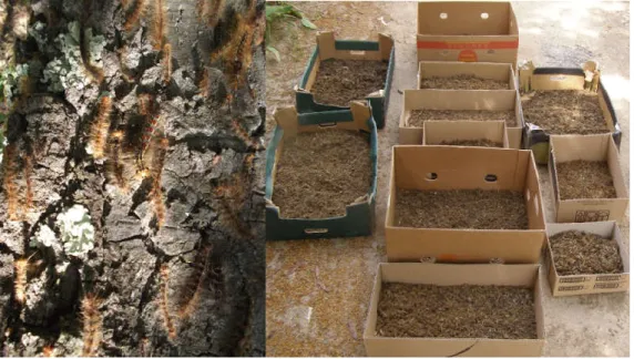
In spring 2013, under conditions of high infestations were applied aerial treatments with Dimilin SC 48 on an area of35 thous and hectares,and, amid a very wet spring, among Lymantria dispar caterpillars were recorded very high mortality due to the pathogen, which allowed collecting large quantities of cadavers (figure 3).
Figure 3.L.dispar larvae dead on tree trunck and collected cadavers, Balcesti, 2013.
In 2014, with a rainy spring and inoculation actions with fungal material collected in 2013, the epizootic manifested itself all over the range of the outbreak, as predicted, and the aerial treatments were cancelled.
MATHERIALS AND METHODS
After noting the appearance of symptoms of mortality specific to Entomophaga maimaiga among populations of Lymantria dispar larvae in 2012 and particularly in 2013 and 2014, when manifested in the form of an epizootic, they were made observations on the evolution of mortality and has been initiated an ample action with the participation of colleagues from Forest Administration for the collection of dehydrated larvae cadavers.
650
Because in the next generations were manifested epizootics caused by Entomophaga maimaiga over large areas of forests in Southern Romania, collecting cadavers of Lymantria dispar grew, resulting in the gathering of about 5 kilograms of dried fungal material, kept also refrigerated. The bulk of this fungal material was used in spring 2014(immediately after the melting of snow in the forest) in the inoculation of the fungus in many other forests infested with Lymantria dispar, where the pathogen had not acted. The results of pathogen inoculation in new areas have been encouraging, this being the object of another work. A small part of fungal material was stored refrigerated for future experiments, while the other was used in various experiments placed in the field and in the laboratory during the years 2014 and 2015.
The first experiment was placed in the laboratory with the objective of observing how the azygospores spread in suspension and the number of azygospores in water suspensions with different ratios of dispersion phase and dispersion medium, with the ultimate goal of finding new ways of introducing the pathogen in the forests.
To highlight the presence of azygospores and how the suspension settles, were carried out microscopic analysis (Kruss optical microscope, model MBLKW 2000) on samples taken from aqueous suspension with different rations of the disperse phase (fungal material) and the dispersion medium (water): 1/10, 1/50, 1/100, 1/200.
Counting azygospores per volume unit of the suspension was performed with counting chambers type Burker-Turk on 10 samples from each suspension, the ratio between the two parts of the suspension being 1/200, in various states of sedimentation.
Another experiment was placed in a laboratory, in order to follow the viability of azygospores while maintaining the fungal material refrigerated. Fungal material milled and stored 1,2 and 3 years was inoculated on various culture media, which were then put to incubate in favorable temperature(200C), relative humidity(80-90%), and light(8 hours/day) conditions,in the growth chamber type Binder KBW 240. As culture media were used wet filter paper, egg whites and artificial culture media Sabouraud CAN 1 and Sabouraud CAN 2. Observations were made daily for 7 days, macro and microscopic, following the formation of fungichyphaes (figure 4).
651
In order to highlight the spread and the survival time of azygospores in the soil of the forest, in fall 2015 soil samples were collected from an experimental area placed since spring 2013 (Balota Forest), were inoculations had been made with fungal material under different humidity conditions (humid and dry, spraying and dusting), and, in summer 2013 and 2014 occurred mortalities among Lymantria dispar larvae. The experiment is ongoing and will be the subject of an new article.
RESULTS AND DISCUSSIONS
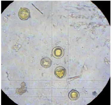
Highlighting the presence and number of fungal spores in water suspension Having the fungal material collected in previous years and kept in powder form refrigerated, by microscopic analysis on samples extracted from the aqueous suspension, it was revealed the presence of spores in all samples analyzed, regardless of the ratio of the dispersed phase and dispersion medium.
Dispersal of spores suspended in water differed greatly with the degree of sedimentation of suspension, which in turn depends on the ratio of the dispersed medium, the greater this ratio, the sedimentation rate is faster.
After testing several suspensions with different phases and reports between the two sides of dispersed systems, 1/200 report was shown to be the best for counting the resistant spores in the Burker-Turk counting chambers (figure 6).
Figure 6.Counting the resistant spores in the Burker-Turk chambers
652
The results of the number of spores in suspension varied widely depending on the state of sedimentation (table 1).
Given the great differences between the average number of azygospores per unit volume and the variation coefficients for the types of suspensions analyzed for future inoculations, it is recommended that the suspension to be homogenized by shaking throughout the application.
Variation of resistant spores average number depending on the state of sedimentation
Table 1
States bysedimentation
Number of samples
Resistant spores average number/ml
Standard deviation
Variation coefficients (%)
Homogenized1/200 10 1,5x10⁴ 0,70 47,14
Suspension
free1/200 10 3x10
3 0,45 152,75
Residue
1/200 10 14.8x10
4
2.71 18.33
To establish Entomophaga maimaiga in new areas using resistant spores requires a relatively small number of spores (6x10⁵ resting spores per 0.01 ha plot) collected from the field (Reardon, Hajek, 1998). At an average number of 1.5x10⁴azygospores per ml, we consider a quantity of 40 ml resistant spore suspensionper 0.01 ha plot (4 l/ha), stirred and uniformly distributed, effective to successfully spread the pathogen in the field.
Maintaining the viability of azygospores while held under refrigeration
Macro and microscopic observations performed daily on the evolution of the pathogen in the culture media tested, showed that on filter paper and egg white fungal hyphaes were not formed, probably due to lack of specific substances of the fatbody of Lymantria dispar larvae.
On the culture media Sabouraud CAN-1 and Sabouraud CAN-2,three days after inoculation, we have seen the development of fungal mycelia with diameters between 6 and 22 mm, higher on Sabouraud CAN-1 culture media (figure 7).
653
After seven days of inoculation on both culture media, the mycelia have a good growth, covering almost all of the entire surface of the culture dishes, and the free space in the dishes was colonized by a dark grey mold species (figure 8).
Figure 8.Culture media Sabouraud CAN-1 and Sabouraud CAN-2, seven days after inoculation.
After microscopic analysis of the micelles formed, it was observed abundant presence of conidiophores specific to Entomophaga maimaiga(figure 9).
Figure 9.Entomophagamaimaigahyphal bodies.
Conidiophores development on artificial culture media open future possibilities of pathogen multiplication in the laboratory and its subsequent release in nature.
CONCLUSIONS
654
cadavers infected with Entomophaga maimaiga, which allowed inoculation of the fungus in new areas in Romania, placement of experiments in order to track the spread of the fungus in space and time and find new possibilities of using the pathogen as a mycoinsecticid.
An average number of 1.5 x 10⁴ resistant spores per ml that we have achieved in water suspensions of fungal material dried and finely milled, revealed that an application rate of 40 ml suspension per 0.01 hectares would be sufficient to inoculate the fungus on that surface.
Laboratory tests on the viability of resistant spores in fungal material preserved one, two or tree years under refrigeration, showed that spores placed under favorable conditions on culture media(Sabouraud CAN-1 and Sabouraud CAN-2) forms
micelya,conidiophores and conidia, specific to Entomophaga maimaiga.
The results obtained in the laboratory opens future possibilities for use of the pathogen at a larger scale in the biological control of Lymantria dispar.
REFERECES
Georgiev, G., Mirchev, P.,Rossnev, B. Petkov, P. Georgieva, M.,Matova, M.Kitanova, S.,Pilarska, D.,Pilarski, P., Golemansky, V., Todorov, MHubenov, .,Z.,Takov, D.,2011: Introduction of Entomophaga maimaiga and control of Lymantria dispar calamitie sin Bulgaria, In: S. Kitanova (ed.), Proc. of Sci. Conf. ‘SustainableManagement of Oak Forests in Bulgaria’, Primorsko, 72–79.
Georgiev, G., P. Mirchev, M. Georgieva, B. Rossnev, P. Petkov,M. Matova, S. Kitanova, 2012a: First Record of Entomopathogenic Fungus Entomophaga maimaiga Humber, Shimazu and Soper (Entomophthorales: Entomophthoraceae) in Lymantria dispar(Linnaeus) (Lepidoptera: Lymantriidae) in Turkey, Acta zoological bulgarica, 64 (2): 123–127.
Georgiev, G., M. Tabaković-Tošić, D. Pilarska, P. Mirchev, M.Georgieva, P. Petkov, P. Pilarski, 2012b: Distribution of Entomophaga maimaiga Humber, Shimazu and Soper (Entomophthorales: Entomophthoraceae) on Balkan Peninsula, In: L. Rakonjac(ed.), Proc. of Int. Sci. Conf. "Forests in Future-Sustainable Use, Risks and Challenges", Belgrade,. 619–622.
Hajek, A.E., R.A. Humber, J.S. Elkinton, B. May, S.R.A. Walsh, J.C. Silver.
1990. Allozyme and RFLP analyses confirm Entomophaga maimaig responsible for 1989
epizootics in North American gypsy moth populations. Proceedings of the National Academy of Sciences, 87, 6979-6982.
Hajek, A. E. 1999: Pathology and epizootiology of Entomophaga maimaiga infections in forest Lepidoptera, Microbiology and Molecular Biology Reviews, 63: 814– 835.
Hrasovec, B., M. Pernek, I. Lukic, D. Diminic, M. Franjevic, A. Hajek, A. Linde, D. Pilarska. 2013. First record of the pathogenic fungus Entomophaga maimaiga Humber, Shimazu and Soper (Entomophtorales: Entomophtoraceae) within an outbreak populations of Lymantria dispar (Lepidoptera: Erebidae) in Croatia. Periodicum Biologorum, 115, 379-384.
Kereselidze, M., D. Pilarska, A. Hajek, A. B. Jensen, A. Linde,2011: First record
of Entomophaga maimaiga Humber, Shimazu&Soper (Entomophthorales:
655
Koraima,R.1954.Two epizootic diseases of the gypsy moth. Shirin-Boeki 27: 296-298.(In Japanese)
Milotić, M., O. Mujezinović, M. Dautbašić, T. Treštić, D. Pilarska, D. Diminić. 2015. First record ofentomopathogenic fungus Entomophaga maimaiga Humber, Shimazu& R.S. Soper (Entomophtorales: Entomophtoraceae) on gypsy moth (Lymantria disparL.) in Bosnia and Herzegovina. Šumarski List,1-2, 59-67.
Nielsen, C., Milgroom, M. G.,Hajek, A. E.,2005: Genetic diversityin the gypsy moth fungal pathogen Entomophaga maimaiga from founder populations in North America and source populationsin Asia, Mycological Research, 109: 941–950.
Pilarska, D., M. McManus, A. Hajek, F. Herard, F. Vega, P. Pilarski,G. Markova,
2000: Introduction of the entomopathogenic fungus Entomophaga maimaiga Hum., Shim.
& Sop. (Zygomycetes:Entomophtorales) to a Lymantriadispar(L.) (Lepidoptera: Lymantriidae) population in Bulgaria, Journal of Pest Science,73: 125–126
Reardon.R.C.,Hajek.A.E.,1998,The gypsy moth fungus Entomophaga maimaiga in North America.Forest Team Technology Enterprise Team, 97-11:16
Tabaković-Tošić, M., Georgiev, G.,.Mirchev, P., Tošić, D., ,Golubović-Ćurguz, V., 2012: Entomophaga maimaiga– new entomopathogenic fungus in the Republic of Serbia, African Journalof Biotechnology, 11: 8571–8577.y, 21: 589–596.
Tomescu, R.Netoiu, C.,2006.,Asistenţă tehnică pentru realizarea lucrărilor de combatere a defoliatorilor din pădurile de foioase. Manuscris I.C.A.S. (unpublished).