*Corresponding author: Naheed Mojgani, PhD, Biotech-nology Department, Razi Vaccine and Serum Research Institute, Agriculture Research Education and Extension Organization, Karaj, Iran.
Tel: +98 9121646063 Email: [email protected]
and
E. faecalis
isolated from human milk
Soodabeh Khalkhali1,2, Naheed Mojgani3*
1Department of Microbiology, Shiraz Branch, Islamic Azad University, Shiraz, Iran
2Department of Microbiology, Fars Research and Sciences Branch, Islamic Azad University, Marvdasht, Iran
3Biotechnology Department, Razi Vaccine and Serum Research Institute, Agriculture Research Education and Extension Organization, Karaj, Iran
Received: February 2017, Accepted: June 2017
ABSTRACT
Background and Objectives: Human milk is a continuous supply of Lactic Acid bacteria (LAB), including enterococci with probiotic potentials. The aim of this study was to analyze two Enterococcus species, isolated from human milk for their probiotic potential, bacteriocin producing ability and virulence traits.
Materials and Methods: Enterococcus faecium TA0033 and E. faecalis TA102 were tested for acid and bile tolerance, survival in simulated gastric and intestinal conditions. The antibacterial spectrum of the isolates was tested by agar well diffusion assay. The antagonistic agent was characterized by physico-chemical methods. The enterocin structural genes, virulence determinants, vancomycin resistance and biogenic amine genes, such as hdc1, hdc2, tdc, ldc and odc were also determined.
Results: The tested isolates survived acidic conditions, high bile salt (1%), simulated gastric and intestinal conditions. The
culture supernatant fluids of the two isolates inhibited the growth of Escherichia coli, Listeria monocytogenes, Salmonella typhi, Staphylococcus aureus, Shigella dysenteriae and Streptococcus agalactiae. The antagonistic activity was lost in the presence of proteolytic enzymes but tolerated the action of catalase, lysozyme and lipase. In contrast to enterocin TA102, enterocin TA0033 possessed bactericidal mode of action. Bacteriocin structural genes, entA and entB were present in the genome of the two isolates, while E. faecalis TA102 additionally harboured entP and bac31 genes. The phenotypic and geno-typic virulence assessment studies indicated hyaluronidase (hyl) production and vancomycin resistance in E. faecalis TA102 while, none of the isolates harboured the biogenic amine genes.
Conclusion: The presence of virulence genes in E. faecalis TA102 calls for careful monitoring of Enterococcus isolates for their safety parameters.
Keywords: Enterococcus, Bacteriocins, Biogenic amines, Human milk, Vancomycin, Virulence genes
ORIGINAL
AR
TICLE
INTRODUCTION
known health benefits of these bacteria they are used in a number of probiotic formulations. A significant character of Enterococcus species is their bacterio-cin produbacterio-cing ability, which is a useful biotechno-logical trait (2). To date, a number of bacteriocin producing enterococci has been isolated from breast milk, and their bacteriocins (enterocins) been
char-acterized (3). Enterocins are defined as small, ribo
-somally synthesized peptides displaying inhibitory actions, against the target bacteria by either dissipa-tion of proton motive force by pore formadissipa-tion, cell lysis, or interference with degradation and metab-olism of macromolecules (4). A number of reports have indicated the presence of virulence traits in this group of bacteria and their safety aspects are still un-der consiun-derations. All this calls for strict scrutinza-tions before considering them as a suitable probiotic candidate. Some of the significant virulence genes reported to date are; ace (collagen binding protein),
asa1 (aggregation substance), cpd (sex pheromone peptides), cylA (cytolysin activation), cylB (cytolysin transport), cylM (posttranslational modification of cytolysin), efaA (endocarditis antigen), esp (entero-coccal surface protein), gelE (Gelatinase), and hyl
(hyaluronidase) (5).
In the last few decades, vancomycin resistant En-terococci have emerged, causing major problems in infection control. Three phenotypic classes (VanA,
VanB and VanC) are defined by the level of resis
-tance to vancomycin and susceptibility or resis-tance to teicoplanin (6). Moreover, biogenic amines (BA) present in food are known to have deleterious health effects on humans, and thus selection of strains with no BA producing ability is highly recommended (7, 8). Hence, before we could exploit these bacte-ria in the food or biopharmaceutical industry, and or as a probiotic supplement, it is essential to evaluate their safety. In this study, we evaluated the bacte-riocinogenic and virulence characteristics of two
Enterococcus isolates by phenotypic and genotypic methods.
MATERIALS AND METHODS
Bacterial strains and cultivation conditions. E. faecium TA0033 and E. faecalis TA102, isolated from human milk samples of healthy young moth-ers in Tehran, capital of Iran, were identified by their carbohydrate fermentation pattern and 16S rRNA
sequencing (9, 10). The isolates were cultured on KF agar, supplemented with 1% TTC (2, 3, 5-Triph-enyl-Tetrazolium Chloride Solution, SIGMA, UK) and Kanamycin aesculin-Azide Agar (KAA, Oxoid, UK). All other Gram-positive and Gram-negative pathogens, used in the study were grown in BHI (Brain heart Infusion, Merck, USA) broth, at 37°C for 24 h in aerobic conditions. Stock-cultures were maintained in MRS broth supplemented with 20% glycerol and stored at -70°C.
Probiotic characterization of the isolates:Acid and bile tolerance. The isolates were screened for their acid and bile resistance, by culturing in MRS broth with different set pH values (2, 2.5, 3, 4, 5, 6) and bile concentrations (0.1, 0.5, 0.7, 1%). The re-sistance of the isolates at the tested pH values was recorded, by determining their growth (cfu/ml) at different time intervals. While, bile tolerance was es-timated, by calculating the Coefficient of inhibition (Cinh), according to the formula described by Gopal
et al. (11).
Cinh= ∆T8-T0 Control – ∆T8-T0 Treatment / ∆T8-T0
Con-trol
Where, ∆T8-T0 represents the difference in
absor-bance at time zero (T0) and after 8 h (T8). Cinh of less than 0.4 was considered significant, for the isolates to be considered as a suitable probiotic candidate.
Survival in simulated gastric and intestinal con-ditions. The survival of the selected isolates under simulated gastric and intestinal contents was stud-ied, by previously described method (12). The bac-terial survival under the tested conditions was cal-culated as:
R= Average of cells at 10min/ Average of cells at 0min
According to the formula, R =1 when no effect on the growth and survival of bacteria is seen, while a ratio of 0.5, indicated a loss of 50% of the viability. Ratios greater than 1 indicated bacterial growth.
wells were measured in millimeters, and based on these results the producer isolates were recorded as strong (≥20mm), moderate (≥16-19 mm) and weak (≤15 mm). The antibacterial activity demonstrated by the isolates was quantified, by slight modifications in the critical dilution method, described by Schillinger and Lucke (13). Two fold serial dilutions (100 µl) of NSF were poured in wells in agar plates seeded with the indicator strain, and incubated aerobical-ly at 37ºC for 24 h. The diameters of the inhibition zones were measured in millimeters, by subtracting the well diameter from the zone diameter. The results were expressed in arbitrary units per millimeter (AU/ ml), defined as the reciprocal of the highest dilution demonstrating zone of inhibition.
Physicochemical characterization of the antag-onistic agents. The antagonistic agents produced by the two isolates were physico-chemically character-ized. The effect of H2O2 for the antimicrobial activ-ity was excluded, by determining the antagonistic activity in the NSF after subjection to the enzyme catalase (1 mg/l). While, the chemical nature of the antagonistic agent was analyzed by treating the NSF of the producer strains with the enzymes like lipase, lysozyme, pepsin, pronase E, and proteinase K (Flu-ka, England), at a final concentration of 1 mg/l in phosphate buffer (10, 14).
Effect of variable pH (2.0, 4.0, 6.0, 8.0, and 10.0) and temperature ranges (60, 80, 100, 121ºC) on the NSF of the two producer isolates, at different time intervals of 15, 30, 60, 90 min, was studied.
The bactericidal or bacteriostatic activity of the studied bacteriocins was determined by critical dilu-tion assay using S. aureus as indicator strain. After 24 h, 5 μl of 10 mg/ml proteinase K solution (Sigma, USA) were spotted near each inhibition zone and the plates were again incubated at 37°C and observed for presence or absence of growth of indicator strain. Bactericidal mode of action of the bacteriocins was determined by the absence of indicator strain growth, after the destruction of the inhibitor by proteases. Absence of inhibition zones after enzy-matic treatment indicated a bacteriostatic mode of action.
Enterocin structural genes. The presence of bacteriocin structural genes in the producer isolates was studied, using a set of primers in a PCR assay. Multiple pairs of enterocin structural genes were
used, as described previously (14, 15). DNA template was prepared by suspending a loop full of bacterial colony in 10µl of lysis buffer (0.25% SDS/ 0.05 % NaOH), heated at 95°C for 5 min and centrifuged at 15 000 x g for 5 min. The samples were diluted in 90 μl of sterile distilled water, centrifuged as above and the supernatant used as template DNA. Cycling parameters included a 2 min initial denaturation at 94ºC, followed by 40 cycles of 45 s at 95ºC, 30 s at 56ºC for entP, bac31 and entL50A/B, 58ºC in the case of entA and 60ºC for entB, entQ, and cyl as annealing
temperature, and 60 s at 72ºC. Amplified PCR frag
-ments were resolved on 1% agarose gels, using a 100 bp ladder for size verification.
Biochemical virulence traits. The two isolates were screened for hemolytic activity, arginine hydro-lysis, gelatinase, lipase, DNase, lecithinase and hyal-uronidase production (16).
The ability of the mentioned Enterococcus isolates to produce biogenic amines (tyramine, histamine, putrescine, and cadaverine) was determined, using the decarboxylase broth and the method described by Bover-Cid and Holzapfel (17).
The phenotypic resistance of the selected isolates to vancomycin (30ug, Sigma, USA) was determined, by disc diffusion assay reported previously (18). Ac-cording to the recommendation of the Clinical and Laboratory Standards Institute (CLSI), the strain was considered to be resistant to antibiotics, if the
inhibition zone was ≤ 14 mm for the indicated anti
-biotic.
Genotypic virulence determinants. Virulence in
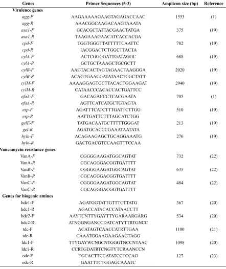
Enterococcus species was determined by PCR assay, using a set or primers targeting the virulence genes. Primer sequences and expected amplicons size are listed in Table 1. E. faecalis ATCC 29212; ATCC 51299 and E. faecium ATCC-BAA 2320; ATCC 19434 were used as positive control during this study. PCR parameters used, were according to the respective references (Table 1).
In order to determine Vancomycin resistance genes in the test isolates, three sets of primers name-ly VanA, VanBand VanC were used in a PCR reac-tion, as described earlier (19).
Table 1. Virulence genes and their sequences, used in the study
Reference
(1)
(19)
(19)
(19)
(19)
(19)
(1)
(19)
(19)
(19)
(22)
(22)
(22)
(20)
(20)
(21)
(20)
(23) Amplicon size (bp)
1553
375
782
688
2020
2940
705
510
213
276
732
635
484
367
534
1100
1098
127 Primer Sequences (5-3)
AAGAAAAAGAAGTAGAGACCAAC AAACGGCAAGACAAGTAAATA
GCACGCTATTACGAACTATGA TAAGAAAGAACATCACCACGA
TGGTGGGTTATTTTTCAATTC TACGGACTCTGGCTTACTA
ACTCGGGGATTGATAGGC GCTGCTAAAGCTGCGCTT AAGTACACTAGTAGAACTAAGGGA ACAGTGAACGATATAACTCGCTATT AAAAGGAGTGCTTACACTGGAAGAT
CATAACCCACACCACTGATTCC GACAGACCCTCACGAATA AGTTCATCATGCTGTAGTA AGATTTCATCTTTGATTCTTGG
AATTGATTCTTTAGCATCTGG TATGACAATGCTTTTTGGGAT AGATGCACCCGAAATAATATA ACAGAAGAGCTGCAGGAAATG
GACTGACGTCCAAGTTTCCAA
CGGGGAAGATGGCAGTAT CGCAGGGACGGTGATTTT CGGGGAAGATGGCAGTAT CGCAGGGACGGTGATTTT CGGGGAAGATGGCAGTAT CGCAGGGACGGTGATTTT
AGATGGTATTGTTTCTTATG AGACCATACACCATAACCTT AAYTCNTTYGAYTTYGARAARGARG ATNGGNGANCCDATCATYTTRTGNCC
ACATAGTCAACCATRTTGAA CAAATGGAAGAAGAAGTAGG TTYGAYWCNGCNTGGGTNCCNTAAC
CCRTGDATRTCNGTYTCRAANCCN TGCACTTCCATATCCTCCAG
GAATTTCTGGAGCAAATC Genes
Virulence genes agg-F agg-R asa1-F asa1-R cpd-F cpd-R cylA-F cylA-R cylB-F cylB-R cylM-F cylM-R efaA-F efaA-R esp-F esp-R gelE-F
gel-R hyln-F hyln-R
Vancomycin resistance genes VanA-F
VanA-R VanB-F VanB-R VanC-F VanC-R
Genes for biogenic amines hdc1-F
hdc1-R hdc2-F hdc2-R tdc-F tdc-R ldc1-F ldc1-R odc-F odc-R
(ornithine decarboxylase) were selected for studies. Primer sequences and PCR parameters were, as de-scribed previously (1, 6, 19-23).
Statistical Analyses. Differences in the preva-lence of virupreva-lence genes between the two Enterococ-cus species were compared, using the Chi-square test
with a p-value < 0.05, indicating statistical signifi
-cance.
RESULTS
isolat-ed from colostrum of healthy mothers in a previous study (NCBI Gene Bank with accession numbers KX158836.1 and KY009901.1, respectively), were evaluated for their probiotic potential and safety traits.
In vitro probiotic characterization of the isolates indicated the isolates to be resistant to acidic pH val-ues of 2.5 and above, while none could survive lower pH values of 2. Enhanced survival rate of the tested
Enterococcus isolates in the presence of 1% bile
con-centrations was observed, as their coefficient of in
-hibition (Cinh) values, appeared to be less than 0.4%. The survival rate of the selected isolates, in simu-lated gastric and intestinal content was indicated by their calculated R value. Fig. 1 shows survival of E. faecalis TA102 in simulated gastric conditions to be below 50 %, while in simulated intestinal conditions their survival percentage was significantly higher (R value > 2), compared to E. faecium TA0033.
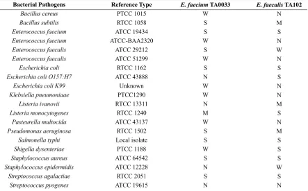
The cell free supernatant fluid (CFSF) of the
En-terococcus isolates demonstrated wide antibacteri-al spectrum, against a range of Gram-positive and Gam-negative pathogens. The CFSF of both the iso-lates were inhibitory towards the growth of E. fae-cium, E. coli, L. monocytogenes, S. typhi, S. aureus,
Sh. dysenteriae and S. agalactiae. However, none of the isolates could inhibit the growth of S. pyogenes
(Table 2). The spectrum of activity of E. faecium
TA0033 appeared significantly higher than E.
faeca-lis TA102 (P≤0.05).
The inhibitory activity demonstrated by the iso-lates, appeared unaffected by neutralization of their supernatant fluid and the action of enzyme catalase. These results indicated the absence of acid and H2O2 for the anti-bacterial activity (Fig. 2). The proteolyt-ic nature of the antagonistproteolyt-ic agent produced by both the isolates was confirmed, by their sensitivity to the tested proteolytic enzymes. In contrast, lipase and lysozyme had no effect on the inhibitory actions of the two isolates.
Effect of variable pH on the proteinaceous antag-onistic compound produced, by the Enterococcus isolates in study is illustrated in Fig. 3 Significant differences were recorded in the pH stability of the
studied enterocins (P≤0.05). According to the ob
-tained results, enterocins TA0033 was not able to resist the extreme acidic (pH 2.0) and alkaline (pH 10) conditions, whereas, enterocin TA102 retained its stability at pH 2.0 for 24 h. Both the enterocins lost their activity completely, at an alkaline pH 10 and
Fig. 2. Effect of neutral pH, catalase, lipase and lysozyme on the enterocin produced by E. faecium TA0033
Fig. 3. Acid resistance of enterocin TA0033 and enterocin TA102 at different pH within 24 h.
Table 2. Antibacterial spectrum of cell free supernatant fluids of Enterococcus species, against pathogens
E. faecalis TA102 N M S N W N S S N N M S N M S S S W S N
E. faecium TA0033 W S S W S W S N W W N M W S S W S N S N Reference Type PTCC 1015 RTCC 1058 ATCC 19434 ATCC-BAA2320 ATCC 29212 ATCC 51299 RTCC 1162 ATCC 43888 Unknown PTCC1290 RTCC 13311 RTCC 1240 ATCC 43137 RTCC 1502 Local isolate PTCC 1188 ATCC 64542 ATCC 12228 RTCC 2051 ATCC 19615 Bacterial Pathogens Bacillus cereus Bacillus subtilis Enterococcus faecium Enterococcus faecium Enterococcus faecalis Enterococcus faecalis Escherichia coli Escherichia coli O157:H7
Escherichia coli K99 Klebsiella pneumoniaae Listeria ivanovii Listeria monocytogenes Pasteurella multocida Pseudomonas aeruginosa Salmonella typhi Shigella dysenteriae Staphylococcus aureus Staphylococcus epidermidis Streptococcus agalactiae Streptococcus pyogenes
ATCC: American type culture collection; PTCC: Persian type culture collection; RTCC: Razi type culture collection.
S: Strong anti-bacterial activity (zone diameter ≥20mm) M: Moderate anti-bacterial activity (zone diameter ≥16- 19mm) W: Weak anti-bacterial activity (zone diameter ≤15mm)
N: No anti-bacterial activity (absence of zone of inhibition) All experiments performed in triplicate.
Table 3. Effect of heat treatment on the enterocin activity (AU/ml) of the Enterococcus isolates, at different time intervals
Time (min) 0 15 30 60 90
Enterocin TA0033 (AU/ml) Enterocin TA102 (AU/ml) 60ºC 25600 25600 12800 6400 6400 80ºC 25600 25600 6400 6400 1600 100ºC 25600 0 0 0 0 120ºC 25600 0 ND ND ND 60ºC 6400 6400 6400 6400 6400 80ºC 6400 6400 6400 6400 6400 100ºC 6400 6400 6400 1600 0 120ºC 6400 6400 0 ND ND ND: not determined
above. Maximum activity (AU/ml) was recorded at pH values of 2.0, 6.0 and 8.0 in both the isolates.
Thermal stability of enterocins TA0033 and TA102 is depicted in Table 3. Significant differences (p≤0.05) were recorded, in the thermal stability of the two enterocins. Enterocin TA102 appeared to be highly temperature resistant, compared to enterocins
TA0033, as it was able to resist 100ºC for 60 min and 121ºC for 15 minutes.
75% reduction in activity within 90 min. In contrast, no loss of activity of enterocin TA102 was seen at these temperatures, during the tested time intervals and no significant difference (p>0.05), in activity between treated and untreated supernatant were ob-served.
Enterocin TA0033 displayed bactericidal mode of action at 6, 400 AU/ml, whereas bacteriostatic activi-ty was detected at lower concentrations (200 AU/ml). Enterocin TA102 demonstrated bacteriostatic mode of action, as seen by the absence of inhibition zones after enzymatic treatment.
Multiple enterocin genes were detected in the test-ed isolates. The isolates possesstest-ed entA and entB, as indicated by a visible band of 126 and 159 bp, re-spectively. Whereas, entP and bac31, corresponding to DNA bond size of 121 bp and 248 bp, respectively, were detected only in E. faecalis TA102.
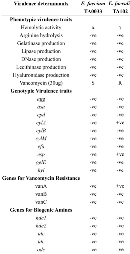
The phenotypic and genotypic virulence traits of the isolates are indicated in Table 4. During viru-lence trait characterizations, E. faecium TA0033 ap-peared non virulent as none of the tested virulence factors were observed in this isolate during biochem-ical and molecular genetic analysis. However, E. fae-calis TA102 showed the presence of cylA (688 bp, cytolysin), esp (510bp, Enterococcal surface protein) and vanA (732 bp, Vancomycin) (Fig. 4).
The genomic DNA of the two Enterococcal iso-lates was subjected to the genes coding for the en-zymes involved in biogenic amines (BA) production, including hdc1, hdc2, tdc, ldc, and odc. None of the
Fig. 4. Vancomycin resistance genes in E. faecium TA033 and E. faecalis TA102. Lanes M: Molecular weight marker; Lane 1, 2, 3: vanA, vanB and vanC in E. faecium TA0033; Lanes 4, 5, 6: vanA, vanB and vanC in E. faecalis TA102
respective genes appeared to be present in the ge-nome of the tested Enterococcus species and the two isolates were considered BA negative.
DISCUSSION
Human milk is a potential source of probiotic bacte-ria, including lactobacilli, streptococci, bifidobacteria and enterococci to the infant gut, affecting the overall composition of the neonate gut microbiota. Although enterococci are widely recognized as probiotic bac-teria, but opposed to other LAB genus they have yet Table 4. Phenotypic and genotypic virulent characters in Enterococcus isolates
Virulence determinants
Phenotypic virulence traits Hemolytic activity Arginine hydrolysis Gelatinase production
Lipase production DNase production Lecithinase production Hyaluronidase production
Vancomycin (30ug) Genotypic Virulence traits
agg asa cpd cylA cylB cylM efa esp gelE
hyl
Genes for Vancomycin Resistance vanA
vanB vanC
Genes for Biogenic Amines hdc1
hdc2 tdc ldc odc
E. faecium
TA0033
α
-ve -ve -ve -ve -ve -ve S
-ve -ve -ve -ve -ve -ve -ve -ve -ve -ve
-ve -ve -ve
-ve -ve -ve -ve -ve
E. faecalis TA102
γ
-ve -ve -ve -ve -ve -ve R
-ve -ve -ve +ve -ve -ve -ve +ve -ve -ve
+ve -ve -ve
not been assigned the GRAS (generally recognized as safe) status. E. faecium is considered a suitable probi-otic candidate for the modulation of immune respons-es against pathogens (24). In this study, we observed the probiotic properties of the two Enterococcus spe-cies, including their resistance in stress conditions like acidic environment, high bile salt concentrations, and simulated gastric and intestinal conditions. Another important characteristic of the isolates in the study was their wide antibacterial spectrum against a num-ber of Gram-positive and Gram-negative pathogens. In agreement with our findings, Ghrairi and colleagues (25), reported E. faecium MMT21 bacteriocin ability to inhibit not only closely related LAB, but also L. monocytogenes and S. aureus. Different spectrum of inhibitory action may be based on the bacteriocin pro-ducing strain, the indicator strain, and also the method used for bacteriocin detection. During physico-chem-ical characterizations of the antagonistic agent pro-duced by the two Enterococcus species in study, it was observed that the antibacterial actions exerted by the isolates were not due to acids or hydrogen perox-ide. Similar to other reports (26), enterocin TA0033 and TA102 appeared to be simple proteins rather than conjugated proteins linked to lipid or carbohydrate moiety. Temperature and pH stability of enterocins is considered an essential aspect, which may compro-mise their use in dairy products (27). Comparable to enterocin SE-K4, enterocin CRL 1826, mundticin KS and enterocin QU2 (28), Enterocin TA102 showed significant pH and thermal stability. The mentioned
enterocin TA102, showed significantly higher resis
-tance in these conditions, compared to other reported enterocins produced by E. faecium D081821, and E. faecium D081833.
Presence of more than one bacteriocin gene in En-terococcus species has been reported previously (4, 10, 12, 14). In agreement with these reports, we ob-served multiple enterocins genes in both of the pro-ducer isolates. Nevertheless, the incidence of numer-ous enterocin genes in enterococci is not necessarily associated with an enhanced bacteriocin activity, and not all enterocin genes should be expressed at the same time (29).
A microorganism considered, as a probiotic is es-sentially either of GRAS status or their safety param-eters are required to be investigated, before they could be considered a probiotic. Enterococcus species have been known to be responsible for nosocomial infec-tions, especially in neonates, and those who suffer
from underlying diseases (30). Therefore, the entero-coccal strain of clinical or industrial interest should be carefully and individually evaluated for their safe-ty and associated risk factors. In this study, we could demonstrate the safety of E. faceium TA0033, as none of the tested phenotypic or genotypic virulent traits were observed in this isolate. In contrast, E. faecalis
TA102 showed the presence of few pathogenic genes like cylA and esp. Our results are in accordance with
the previous findings, stating that pathogenic viru
-lence factors are more common in E. faecalis strains compared to E. faecium strains (31).
Antibiotic resistance is one of the major safety con-cerns inEnterococcistrains, particularly vancomycin resistance, as it is the drug of choice efficient against clinical infections by multidrug resistance pathogens. In our studies, E. faecalis TA102 appeared vancomy-cin resistant, during phenotypic and genotypic analy-sis. According to the reports, if the strain has either of the vancomycin resistance genes such as vanA, vanB and vanC, it is considered unsafe and could not be applied in the food or feeds (32). Another important safety concerns, are biogenic amine production by some probiotic isolates to be used in the dairy indus-try. Biogenic amines contained foods are known to have toxicological properties and are known to trig-ger health problems such as allergies, hypertension, hypotension, headaches, depressions, schizophrenia and Parkinson disorder (7) and thus should be avoided in food (33, 34). Absence of BA genes highlights the importance of a strain for use in starter and adjunct cultures.
In conclusion, results of this study determine the possible advantage of the indicated enterocins in dairy or biotechnology industry. The presence of couple of virulence genes in E. faecalis TA102 calls for careful monitoring of Enterococcus isolates, for their safety parameters.
ACKNOWLEDGEMENTS
A section of this work was financially supported by the Iran National Science Foundation (INSF), project No 92044469, during the year 2015-2017.
The authors declare no conflict of interest.
1. Eaton TJ, Gasson MJ. Molecular screening of Entero-coccus virulence determinants and potential for genet-ic exchange between food and medgenet-ical isolates. Appl Environ Microbiol 2001; 67: 1628-1635.
2. Franz CM, Huch M, Abriouel H, Holzapfel WH, Gal-vez A. Enterococci as probiotics and their implications in food safety. Int J Food Microbiol 2011; 151: 125-140. 3. Kozak K, Charbonneau D, Sanozky-Dawes R, Klae-hammer T. Characterization of bacterial isolates from the microbiota of mothers' breast milk and their infants. Gut Microbes 2015; 6: 341-351.
4. Ishibashi N, Himeno K, Fujita K, Masuda Y, Perez RH,
Zendo T, et al. Purification and characterization of mul -tiple bacteriocins and an inducing peptide produced by E. faecium NKR-5-3 from Thai fermented fish. Biosci Biotechnol Biochem 2012; 76: 947-953.
5. Carlos A, Semedo-Lemsaddek T, Barreto-Crespo M, Tenreiro R. Transcriptional analysis of virulence-relat-ed genes in enterococci from distinct origins. J Appl Microbiol 2010; 108: 1563-1575.
6. Miele A, Bandera M, Goldstien BH. Use of primers se-lective for vancomycin resistance genes to determine van genotype in enterococci and to study gene organi-zation in VanA isolates. Antimicrob Agents Chemoth 1995; 39: 1772-1778.
7. Elsanhoty RM, Ramadan MW. Genetic screening of biogenic amines production capacity from some lactic acid bacteria strains. Food Control 2016; 68: 220-228. 8. Laukova A, Szaboova R, Pleva P, Bunkova L,
Chrasti-nova L. Decarboxylase-positive Enterococcus faecium strains isolated from rabbit meat and their sensitivity to enterocins. Food Sci Nutr 2017; 5: 31-37.
9. Khalkhali S, Mojgani N. Characterization of candidate probionts isolated from human breast milk. Cell Mol Biol 2017; 82-88.
10. Mojgani N, Vaseji N, Khalkhali S, Naz Baloch M. Biochemical and molecular analysis of the antilisterial peptides produced by Enterococcus hirae strains iso-lated from raw ewe milk. J Adv Biol Biotech 2017;11: 1-11.
11. Gopal PK, Prasad J, Smart J, Gill HS. In vitro adher-ence properties of L. rhamnosus DR20 and Bifidobac -terium lactis DR10 strains and their antagonistic activ-ity against an enterotoxigenic Escherichia coli. Int J Food Microbiol 2001; 67:207-216.
12. Mojgani N, Hussaini F, Vaseji N. Characterization of indigenous Lactobacillus strains for probiotic proper-ties. Jundishapur J Microbiol 2015; 8(2): e17523. 13. Schillinger U, Lücke FK. Antibacterial activity of L.
sake isolated from meat. Appl Environ Microbiol 1989; 55: 1901-1906.
14. Aymerich T, Holo H, Håvarstein LS, Hugas M, Gar-riga M, Nes IF. Biochemical and genetic characteriza-tion of enterocin A from E. faecium, a new antilisterial
bacteriocin in the pediocin family of bacteriocins. Appl Environ Microbiol 1996; 62: 1676-1682.
15. Nami Y, Haghshenas B, Haghshenas M, Khosroushahi AD, Rosli R, Khosroushahi Y. Antimicrobial activity and the presence of virulence factors and bacteriocin structural genes in E. faecium CM33 isolated from ewe colostrum. Frontiers Microbiol 2015; 6:1-10.
16. de Azeredo LA, Leite SG, Freire DM, Benchetrit LC, Coelho R. Proteases from actinomycetes interfere in solid media plate assays of hyaluronidase activity. J Microbiol Methods 2001; 45: 207-212.
17. Bover-Cid S, Holzapfel WH. Improved screening pro-cedure for biogenic amine production by lactic acid bacteria. Int J Food Microbiol 1999; 53: 33-41.
18. Bauer A, Kirby WM, Sheris JKT. Antimicrobial sus-ceptibility testing by a standard single disc method. Am J Clin Pathol 1996; 45: 493-496.
19. Vankerckhoven V, Autgaerden T, Vael C, Lammens C, Chapelle S, Rossi R, Jabes D, Goossens H. Develop-ment of a multiplex PCR for the detection of asa1, gelE, cylA, esp, and hyl genes in enterococci and survey for virulence determinants among European hospital iso-lates of E. faecium. J Clin Microbiol 2004; 42: 4473-4479.
20. Reviriego C, Eaton T, Martín R, Jiménez E, Fernández L, Gasson MJ, Rodríguez JM. Screening of virulence determinants in E. faecium strains isolated from breast milk. J Hum Lact 2005; 21: 131-138
21. Semedo MA, Santos P, Martins MFS, Lopes JJ, Figue-iredo Marques R, Tenreiro MT, et al. Comparative study using type strains and clinical and food isolates to examine hemolytic activity and occurrence of the cyl operon in enterococci. J Clin Microbiol 2003; 41: 2569-2576.
22. Satake S, Clark N, Rimland D, Nolte FS, Tenover FC. Detection of vancomycin-resistant Enterococci in fecal samples by PCR. J Clin Microbiol 1997; 35:2325-2330. 23. Landete JM, de Las Rivas B, Marcobal A, Muñoz
R. Molecular methods for the detection of biogen-ic amine-producing bacteria on foods. Int J Food Microbiol 2007; 117(3):258-269.
24. Khalkhali S, Mojgani N. Enterococcus faecium; a suitable probiotic candidate for modulation of im-mune responses against pathogens. Int J Basic Sci Med 2017;2:77-82.
25. Ghrairi T, Frere J, Berjeaud JM, Manai M. Purification
and characterization of bacteriocins produced by E. faecium from Tunisian Rigouta cheese. Food Control 2008; 19:162-169.
26. Borzenkov V, Surovtsev V, Dyatlov I. Obtaining bacte-riocins by chromatographic methods. Adv Biosci Bio-technol 2014; 5: 446-451.
dochi-iso-lated E. faecium D081821 and D081833. Lett Appl Mi-crobiol 2007; 44: 320-325.
28. Zendo T, Eungruttanagorn N, Fujioka S, Tashiro Y, Nomura K, et al. Identification and production of a bac -teriocin from E. mundtii QU 2 isolated from soybean. J Appl Microbiol 2005; 99: 1181-1190.
29. Montel Mendoza G, Ale CE, Nader-Macías MEF, Pas-teris SE. Characterization of a bacteriocin produced by E. gallinarum CRL 1826 isolated from captive bull-frog: evaluation of its mode of action against L. mono-cytogenes and Gram-Negatives. J Bioprocess Biotech-nol 2015; 5: 250-256.
30. Casaus P, Nilsen T, Cintas L, Nes IF, Hernández PE, Holo H. Enterocin B, a new bacteriocin from E. fae-cium T136 which can act synergistically with enterocin
A. Microbiol 1997; 143: 2287-2294.
31. Trivedi K, Radmila S, Renata K. Bacteriocin activity of enterococci and presence of genes related to patho-genesis. Chez J food Scis 2012; 30: 330-335.
32. Garbutt JM, Ventrapragada M, Littenberg B, Mundy LM. Association between resistance to vancomycin and death in cases of E. faecium bacteremia. Clin Infect Dis 2000; 30: 466-472.
33. Ogier JC, Serror P. Safety assessment of dairy microor-ganisms: the Enterococcus genus. Int J Food Microbiol 2008; 126: 291-301.
34. Linares DM, del Rio B, Ladero V, Martinez N,
Fer-nandez M, Martin MC. Factors influencing biogenic