This is an open access journal, and articles are distributed under the terms of the Creative Commons Attribution-Non Commercial-ShareAlike 4.0 License, which allows others to remix, tweak, and build upon the work non-commercially, as long as appropriate credit is given and the new creations are licensed under the identical terms.
Focused extracorporeal versus Radial shock wave therapy in
treatment of chronic lateral epicondylitis (randomized control
trial)
Haidy Henry Fakhry Maher
1*, Ragia Mohamed Kamel
1, Hassan Husein Ahmed
2, Soheir Shehata
Rezk Allah
11Physical Therapy Department of Basic Sciences, Faculty of Physical Therapy, Cairo University, Cairo, Egypt, 2Orthopedic Surgery Department, Faculty of Medicine, Banha
University, Egypt.
Correspondence: Haidy Henry Fakhry Maher, Physical Therapy Department of Basic Sciences*, Faculty of Physical Therapy, Cairo University, Cairo, Egypt. E-mail: meladayad 2007 @ gmail.com
ABSTRACT
Objective: To investigate the therapeutic effect of FWST versus RSWT in patients with chronic lateral epicondylitis. Material and Methods: A total of 60 subjects of both genders were sought to participate in the current study, 60 patients (44females and 16males) with chronic lateral epicondylitis were assigned randomly into three equal groups. These individuals were eligible. Group A included 20 patients who received FSWT plus the conventional physical therapy while group B included 20 patients who received RSWT plus the conventional physical therapy, and group C included 20 who received the conventional physical therapy only. The treatment with shock wave for group A & B was applied only one session in the alternative week for the maximum of 3-5 sessions, and the conventional physical therapy for all groups were 12 sessions which was done three sessions per week. The participants’ ages ranged from 30 to 60. They were recruited from the orthopedic outpatient clinics. The exclusive criteria were subjects who had other co morbidities which would affect the results. Results: The statistical analysis using 3x4 multivariate design MANOVA showed that no significant impacts were recorded for the experimental group on the pressure pain test, pain at work and handheld dynamometer. In addition, no significant impacts were recorded in the measuring time in the pressure pain test, pain at work and handheld dynamometer. Also, the correlation among the treated groups and assessing phases was statistically non-significant. This tested group (first independent variable) on the pressure pain test, pain at work and handheld dynamometer was not influenced by the measuring periods (second independent variable) (F=0.84, P=0.647). Conclusion: Finally, it was concluded that: Radial shock wave therapy (B) had only effects on pain more than Focused shock wave therapy (A) but Focused shock wave therapy (A) had effects on muscle function and inflammation and tendon changes more than Radial shock wave therapy (B).
Keywords: Lateral epicondylitis- conventional physical therapy-Focused shock wave therapy-Radial shock wave therapy-pain.
Introduction
Generally, the inflammation in the tendons is called tendinitis or tendonitis and involves also inflammation in soft tissues surrounding the muscles beside the bones in the inflamed region particularly the bones in the elbow, shoulder, hip, wrist, ankle or knee. Histologically the tendon is composed of group or bands of flexible fibrous tissues that play an important role for connections of skeletal muscles to the bones [1].One of the most occurring myotendinosis type is the lateral epicondylitis (tennis elbow) which is accountable for severe pain and disability of movement of the affected limb. In addition, lateral epicondylitis (tennis elbow), is mostly triggered by the exaggerated practice of common extensor tendon (CET) that principally distresses the extensor carpi radialisbrevis [2]. Access this article online
Website: www.japer.in E-ISSN: 2249-3379
How to cite this article:Haidy Henry Fakhry Maher, Ragia Mohamed Kamel, Hassan Husein Ahmed, Soheir Shehata Rezk Allah. Focused extracorporeal versus Radial shock wave therapy in treatment of chronic lateral epicondylitis (randomized control trial). J Adv Pharm Edu Res 2018;8(3):68-76.
Commonly, lateral epicondylitis causes severe pains in the outer side of the elbow joint, which is accompanied with remarkable painfulness in the humeral bone particularly the lateral epicondylus. One of the main causes which are responsible for the exaggeration of the painful condition is the resistant extension of the fingers and wrist, and supination of the forearm. Some authors reported that the tennis elbow syndrome extremely interrupts the job of the arms [3].
However, tennis elbow is leading to a severe pain in the tendinous tissue of the muscle origins in the extensor muscles of the wrist at the lateral epicondyle of the humerus, subsequently leads to the disability of the affected limb. Hence, the tennis elbow has a great effect on the whole life of the affected subjects specially patients’ social and qualified life. Later, alepicondylitis is known as radiohumeral bursitis or tennis elbow and it affects on about 1% to 3% of the general inhabitants [4].
Tennis elbow syndrome was found to be affecting the subjects that their ages ranged between 30 and 60 years. Whereas, this syndrome is considered a painful condition, and may hinder the capability of the suffering individuals to accomplish their role in society. Although a high rate (approximately 40% to 50%) of tennis players have been diagnosed with the tennis elbow syndrome, several of those complained (15%) of labors at risky businesses, which usually need subjects to involve in duties necessitating over employment of the forearm, wrist and elbow
[5].
The main symptoms of the lateral epicondylitis are the severe pain above the lateral epicondyle of the humerous, generally resulted from the overuse of the forearm and wrist activities. The extensor carpi radialislongus and brevis in addition to the extensor digitorum and extensor carpi ulnaris originating beside the lateral epicondylar ridge of the humerous. The extensor extensor digitorum and carpi radialisbrevis are the main muscles that participate mainly in the manifestations of the disease. The usual cause of irritation of the extensor carpi radialis brevis tendon muscles is the over practices of elbow and wrist due to the irritation of the tendons of these muscles which leads to the irritation and inflammation, subsequently leading to soreness and swelling around the lateral side of the elbow. Wrist extension in some acute activities such as during playing tennis, may lead to intensify the symptoms. In addition, wrist motion is affected particularly by grasping and pinching, accordingly restraining the occupation of the hand [6].
This damage is very problematic due to the difficulty for the treatment, predisposed to return, and may extend for many weeks or even months, the typical episode may last for a duration of 6 months and two years [7].
The ideal diagnosis of the lateral epicondylitis mainly depends on the case history and clinical examinations. In individuals usually suffering from pain in the region of the lateral elbow joint, the symptoms are typically exacerbated during the wrist extension and digital resisting, also, the localized tenderness at the CET is apparent during clinical examination of the patients. One of the accurate methods for diagnosis of lateral epicondylitis has been through the application of Magnetic resonance imaging (MRI)
due to a good contrast resolution of the soft tissues and also the demonstrated adequate planes of the specificity, sensitivity, and precision [8].Other investigators reported that MRI has been a
valid and reliable tool for determining the harshness of the lateral epicondylitis, in addition, they found a positive correlation between the patient`s’ clinical signs and the degree of MR signal changes of CET [9].
The main purpose of the treatment of the lateral epicondylitis has depended on controlling the inflammation, starting with rest, periodic local corticosteroid injections and the application of non-steroidal anti inflammatory (NSAIDs), Exercise-based physical therapy and physical therapy program [phonophoresis, iontophoresis, transverse friction massage, ultrasound, and glyceryltrinitrate patches; low-level laser therapy; sclerotherapy; shock wave therapy; surgery, in addition to insulin growth factor (IGF) and the stem cell therapy] [10].
One of the most efficient methods used since 20 years ago for reliving the orthopedic and tendinopathy conditions is using extracorporeal shock wave therapy (ESWT), with the development of ESWT instruments for the lithotripsy wave (ESWL). ESWT has been considered as an tolerable and widespread non-invasive tool for controlling tendonitis and the musculoskeletal system disorders. Earlier researches on the management of tendinopathy reported that the extracorporeal shock wave therapy has been more effective than other methods of therapy [11]. Some of the chief reasons for the few applications
of ESWT referred to the misunderstanding of the apparatus or the lack of expertise to use the technique, in spite of its safety range and easy implementation and being supportive in some cases [12].
ESWT has been used as a therapy for many soft tissue damages such as lateral epicondylitis, plantar fasciitis, noncalcific and calcific inflammation of the tendons of the tendinopathy of the Achilles tendon and supraspinatus, but it has been recommended for the lateral epicondylitis and plantar fasciitis according to FDA. The mode of the action of ESWT for reliving the pain or healing has been in the healing of tendons [13]. The shock waves
can be classified into focused (FSWT) and radial (RSWT) shock wave therapies [14]. FSWT has focused on a pressure area
concentrated on a definite place, and can be adjusted at definite depths in the selected tissues, where the higher pressure is touched [11]. Focused type of shockwave is directed by the
reflection at definitive areas into the body of patients, and the waves are generated from a wide arc, hence the amount of energy discrete is minimal at the point of real wave generation [15].
Whereas, the radial shock wave therapy (RSWT) has been attributed to the scattering pressure zone of RSWT apparatus , which arrives at the source as the highest pressure, and didn`t require a certain distance in human body [16] and the omitted
waves radiate within the tissues, and influence a large area, but they do not deeply reach the thick tissues as that in focused wave therapy , therefore (RSWT) is effective for treating superficial lesions that assist the deep-tissue stimulation like backs and muscles[17]. In a study carried out during 2010 for comparing the
recalcitrant plantar fasciitis, they reported that FSWT was superior to RSWT for such conditions [18]. It has been indistinct
that the characteristics of waves either in focused or radial shock waves induce the therapeutic effects, it has been hard to correlate the physical differences between the two types of waves, and the clinical efficacy [17]. The available data reported no records on the
application of radial and focused shock wave therapy for the management of tennis elbow. The available data has been limited to only two articles dealing with their actions on the individuals suffering from such cases [19].
The wave properties that have been imperative for initiating the therapeutic actions of both FSWT and RSWT are unknown as well; hence, it has been hard to correlate the physical variations among FSWT and RSWT to their clinical efficacy. It has been even possible that the two methods would have various modes of actions. Limited data have been available on comparing the two methods of therapy from the point of clinical efficiency. In both researches, one dealing with patellar tendinopathy and the other dealing with plantar fasciitis, the differences in the clinical symptoms did not greatly differ between the used methods [20].
Materials and Methods
This study took place at Faculty of Physical Therapy, Cairo University, Cairo, Egypt and during the period between2017 to 2018.
Data collection: A total of 60 subjects of both genders were sought to participate in the current study, 60 patients (44females and 16males) with chronic lateral epicondylitis were assigned randomly into three equal groups, these individuals were eligible. Group A included 20 patients who received FSWT plus the conventional physical therapy, while group B included 20 patients who received RSWT plus the conventional physical therapy, and group C included 20 who received the conventional physical therapy only. The treatment with shock wave for group A & B was applied only one session in alternative week for the maximum of 3-5 sessions, and the conventional physical therapy for all groups were 12 sessions, three sessions per week. The participants’ ages ranged from 30 to 60 years. They were recruited from orthopedic outpatient clinics. The faculty of the School of Physical Therapy at Cairo University approval no. was 012/001432 and registered by Pan African Clinical Trial (Registry ID PACTR201805003234227). Each participant signed an informed consent.
Instrumentations:
Assessment instruments
Handheld dynamometer:
The hand held dynamometer has been developed to provide a detailed and objective measurement of the joints’ strength throughout its range of motion.
VAS: To assess the pain severity
Magnetic Resonance Imaging: MRI findings showed either focal fibrous degenerative tendon tissue or the microruptures of
collagenous fibres. The instrumentations for the treatment included Radial shock wave therapy, Focused shock wave therapy, and Ultrasound.
Data collection and statistical analysis:
1) Descriptive analysis:
It included:
• The mean (X): as an average describing the central tendency of the observation.
• The standard deviation (SD): as a measure of the dispersion of the result around the mean.
2) Inferential statistics:-
• MANOVA to identify the differences between groups.
• Pearson product-moment correlation coefficient to
investigate the association between the dependent variables.
The level of the significance was set at ≤0.05. interval of 95%.
Results
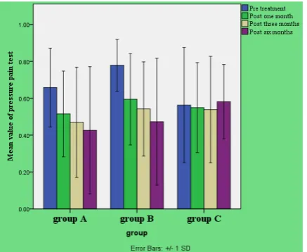
For Pressure Pain test:
In the present work, multiple pair wise assessment assays showed a non-significant variation recorded in the means of the "post three months” among (gr. A vs. B, p=0.99), (gr. A vs. C, p=0.94)and (gr. B vs. C, p=0.854) . Additionally, Multiple pair wise assessment tests revealed that there was no significant variation in the mean values of the "postsix months" among (gr. A vs. B, p=0.99), (gr. A vs. C, p=0.99), and (gr. B vs. C, p=0.298).
Figure 1:mean value of pressure pain test
For Pain at work:
Figure 2:Mean value of pain at work
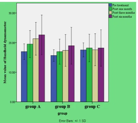
For handheld dynamometer
In the current work, multiple pairwise assessment assays demonstrated that statistically no important difference was found in the means of the "post three months" test within (gr.A vs. B , p=0.587), (group A vs. C, p=0.387), and (gr.B vs. C, p=0.99) . Additionally, multiple pairwise comparison assays (Post hoc tests) reported that no statistical significant variation was found in the means of the "post six months" test between (gr. A vs. B, p=0.99), (gr. A vs. C, p=0.726), and (gr. B vs. C, p=0.699).
Figure 3:Mean value of handheld dynamometer
Discussion
The aim of this work was conducted to
inspect:
The significant difference in pain level, muscle strength and MRI findings of CET after the application of FSWT in patients with chronic lateral epicondylitis.
There was a significant difference in the pain level, muscle strength and MRI findings of CET after the application of RSWT in patients with chronic lateral epicondylitis.
There was a significant difference in the efficacy of FSWT and RSWT on pain level, muscle strength and MRI findings of CET in patients with chronic lateral epicondylitis.
A total of 60 subjects of both genders were sought to participate in the current study, 60 patients (44females and 16males) with chronic lateral epicondylitis were assigned randomly into three equal groups, these individuals were eligible. The participants’ ages ranged from 30 to 60 years. They were recruited from orthopedic outpatient clinics.
Subjects with the ages range between 30 and 60 have been the most frequently subjected to be affected with the disease. Whereas the frequently apparent as an inflammatory disorder, the painful feeling of tennis elbow patients may hinder their capability to do their roles in the society. Whereas, about 40-50% of tennis players complain of this disease, several of the affected persons (15%) are labors in dangerous businesses, which usually need subjects to involve in works necessitating overdoing of forearm, the elbow, and wrist [5].
Regarding the pain scores (pressure pain test, pain at work); (within groups), radial shock wave therapy produced significant differences in pain scores in post one month, post three months and post six months.
The analgesic effect of the radial shock wave was explained by: The shock wave which arouse the nociceptors to spirit high-frequency nerve impulses (hyper-stimulation). The proliferation of nerve impulses was obstructed depending on the gate-control theory [21, 22].
Radial Shock waves either have altered the cell membrane partially or completely. The nociceptors could not build up a generator potential; thus pain sensation was circumvented [23].
Also, the radial shock waves might suppress the calcitonin gene-related peptide (CGRP) immunoreactivity in dorsal root ganglion neurons [24, 25].
Another possible effect of radial shock after the extracorporeal shock wave might be through the selective damage of unmyelinated nerve fibers in spite of the application in the musculoskeletal system [26].
As can be seen in the results in the current work, the extracorporeal shock wave therapy (ESWT) has been a talented physical tool for the treatment of the tennis elbow. Both primary and long-term results demonstrated a good tissue reaction. From the obtained results, significant effects on the relief of pain and progress in function of the affected regions of
the patients during the observation of patients along the follow-up were observed. The Radial shock wave can cover a large area and stimulate efficiently the affected tissues in comparison with the Focused shock wave which affects a narrow area in the latter
[16], this meant that the healed or treated area were also minimal.
The results of this study came in agreement with [27], in their
study 60 people were randomly divided into two groups of 30 patients: those treated by radial shockwave therapy and those treated by ultrasound therapy. Radial shockwave therapy consisted of 3 sessions at weekly intervals. Ultrasound therapy consisted of 10 treatments performed daily. The clinical status of the patients before the treatment, just after the treatment, and after 8 weeks was assessed with the Leitinen questionnaire and a visual analogue scale (VAS). Pain reduction, as assessed by VAS scores, was observed in both groups, who were not different at the baseline (p=0.807). While both therapies were effective (p<0.005), the mean pain intensity assessed by VAS scores in patients treated by the radial shockwave therapy was significantly lower compared to those treated by the ultrasound therapy; this relationship was present just after the treatment completion (p=0.001) and in 8 weeks after the treatment completion (p=0.002). Both radial shockwave and ultrasound therapies caused a reduction in the intensity and frequency of pain that persisted for at least 8 weeks, reducing the need for pain medication and improving the function of the treated upper limb. Ultrasound therapy was less effective than the radial shockwave therapy.
Also, [28] supported the current results. They carried a
prospective clinical study, including 16 patients (9 males, 7 females, with the age range of 47.2 ± 2.3 yrs) suffering from the lateral epicondylitis for 15.06 ± 4.06 months. The follow-up period was extended for 12 months divided into 5 phases. The obtained results revealed that there was a significant variation in the mean scores of pain at rest and on palpation. They concluded that RSWT was an effective method for managing the lateral epicondylitis over six months in case the disorder is refractory to further kinds of conservative therapy [28].
In contrast to the current study, [18] carried a pilot for treating 39
patients who were complaining of plantar fasciitis by using FSWT and radial ESWT. The results showed that FSWT was a reliable and effective method for the management of recalcitrant plantar fasciitis than RSWT.
Regarding the pain scores (pressure pain test and pain at work) between groups, in the current study, there were no significant differences of pain scores between the focused shock wave and the radial shock wave therapy, they were equal in affecting on the pain reduction despite the differences in physical properties. The wave features that were imperative for producing the therapeutic actions of F-SWT and R-PWT have not been known, consequently it was difficult to correlate the physical variations between the focused and radial shockwave to their clinical efficacy. It was even possible that the two described methods would have diverse mechanical reinforcements. In the previous two research studies dealing with the clinical effectiveness between F-SWT and R-PWT, one done on patellar tendinopathy and the other on plantar fasciitis, no main clinically pertinent variations between the two applied methods were recorded [20].
In some trials, where investigators reviewed the observation of individuals with tennis elbow, a shock wave (both focused and
radial) was also reported to diminish the intensity of pain over time [29, 30].
Comparing both types of shock wave therapy (FSWT, RSW) to the conventional physical therapy, in the current study, there were no significant differences between the pain scores (pressure pain test and pain at work).
These results agreed with those obtained by [31]. A randomized
controlled study was performed more recently where the shock wave therapy was compared with hot pack, ultrasound, and friction-massage therapies, and no significant differences were found within the intergroup, regarding the grip strength and pain scores. On the other hand, some researchers [32] found a
significant improvement in pain relief and functional activity in patients who were treated with focused extracorporeal shockwave therapy, the effect being persisted over a more extended follow-up.
Regarding the values of handheld dynamometer (within groups), the Focused shock wave therapy showed a significant difference in muscle function of hand grip (grip strength) post one month, but this difference didn’t change after three and six months post treatment.
NOTE: pain improvement was not the key for the improvement of hand grip.
Different postulations were supposed for illustrating the mode of action of shockwave as an effective tool for treating the muscular disorders. One of these ways was the separation of fixed actin-myosin links by the input of mechanical energy (spalling) as long as the force was vertical to the direction of muscle fibers [33, 34].
Another postulation was through the reduction of muscle tone via biological mechano-transduction. Authors reported that shockwave could stimulate the muscles mechanically. For induction of the muscular relaxation, the increase in the blood circulation and lymphatic drainage was required for exposing the affected area to 15 to 30 Hz shockwave which led to the breaking actin-myosin links, and the destruction of the damaged fibres [33, 34]. Also, it could influence the severity of pain at both the neural
and biological levels through improving the blood flow which subsequently led to reducing muscle tension/stiffness in the affected area [21].
The results of this study came in agreement with [35] in which they
reported significant improvements in pain, and grip strength within12 weeks of the follow-up period during the treatment with the shock wave therapy. Another research study [36]
recorded a significant reduction in pain provoking with the high activity after applying the shock wave therapy along 8 weeks of the follow-up period. The end results of all these studies were coordinated with the data obtained in this study, and the duration of follow-up showed that the efficiency of shock wave therapy was mostly shorter like in the current study.
Also, in some investigations in the extended follow-up times, the researchers observed diverse results. For instance, in a high quality study performed by [37] on 24 patients, they didn`t find
On the other hand, a study obtained by [38], they studied the
effectiveness of the ultrasound-guided focused shock wave therapy on the functional status, perception of pain, along short-, and long-terms. They could collect very few data assisting the use of shock wave therapy in the control of the lateral epicondylitis.
Regarding the values of handheld dynamometer (between groups), no differences were recorded between the two methods (FSWT and RSWT) in their effects on muscle function of hand (hand grip).
These results agreed with those obtained by [20] in which, they
treated 43 individuals (57 tendons) suffering from patellar tendinitis, who were assigned into 2 groups (FSWT and RSWT). Both groups gave three treatment sessions at 1w interval. All subjects were provided 2000 impulses per session (8 Hz, 2.4 bar (RSWT) and 4 Hz, 0.12 mJ/mm2 (FSWT), respectively). In addition, the patients contributed in an eccentric exercise program that started 2 weeks after the last shock wave therapy. The follow-up was accomplished from 1-14 weeks after the treatment, the results revealed no significant variation between the two methods (FSWT and RSWT) concerning their efficiency. The two methods inhibiting the degree of pains and improvement of hand grip were observed in the subjects’ condition.
Regarding Thomsen test results,(within groups),in the current, study it was found that Focused shock wave therapy had a significant effect on pain provoking test (Thomen test) after one month (further effects of Radial shock wave therapy were assessed, and post one month treatment found 13 subjects positive with the reported percentage of 65% , and 7 subjects negative with the reported percentage of 35% , post three months found 5 subjects with positive with the reported percentage of 25% , and 15 subjects negative with the reported percentage of 75% , and post six months found the same results as the post three months.
There have been some theories explaining the effect of FSWT on the subside inflammation, the high-energy focused shockwave produced an adequate amount of energy that could produce the controlled inflammation of the designated tissue. This inflammation has been shown to stimulate many mediators such as transforming growth factor beta I (TGF-β1) and insulin-like growth factor I (CGFI) and initiated the healing process [39].
Focused shock wave treatment (FSWT) increased the perfusion in ischemic tissues, stimulated the growth factors, decreased the inflammation, and accelerated the wound healing. It has been a safe technique classically used in urology and orthopedic surgery with success but there has been still limited literature regarding its use in the management of burns [40].
Regarding thomen test values (between groups), Radial shock wave therapy produced a late effect only post three months, 13 subjects were found positive with the reported percentage 65%, and 7 subjects were found negative with the reported percentage of 35%, these results were also found in this study meaning that FSWT was more effective than RSW in decreasing inflammation.
These results agreed with those obtained by [18], they reported
that the focused extracorporeal shock wave was superior than the radial extracorporeal shock wave therapy for the treatment of recalcitrant plantar fasciitis.
Regarding MRI finding grades, (within groups): It was found that in FSWT, there were more significant differences between MRI finding grades comparing to RSW.
MRI finding grades post one month of FSW treatment done on reported 12 subjects with the normal grades with the percentage of 60%, 5 subjects with the mild grade with the reported percentage of 25%, 2 subjects with the moderate grade with the reported percentage of 10%, and 1 subject with the severe grade with the reported percentage of 5%. Additionally, in the "post six months of treatment”, there were 14 subjects with the grade normal with the reported percentage of 70%, 4 subjects with the mild grade with the reported percentage of 20%, and 1 subject with the moderate grade with the reported percentage of 5%, and 1 subject with the severe grade with the reported percentage of 5%.
MRI finding grades post one month of RSW treatment were 4 subjects with the normal grade with the reported percentage of 20%, 10 subjects with the mild grade with the reported percentage of 50%, 3 subjects with the moderate grade with the reported percentage of 15%, and 3 subjects with the severe grade with the reported percentage of 15%. Additionally, in the "post six months of treatment", there were 9 subjects with the normal grade with the reported percentage of 45%, 6 subjects with the mild grade with the reported percentage of 30%, 2 subjects with the moderate grade with the reported percentage of 10%, and 3 subjects with the severe grade with the reported percentage of 15%.
MRI finding grades post one month of conventional therapy showed there was 1 subject with the normal grade with the reported percentage of 5%, 8 subjects with the mild grade with the reported percentage of 40%, 8 subjects with the moderate grade with the reported percentage of 40%, and 3 subjects with the severe grade with the reported percentage of 15%. Additionally, in the "post six months of treatment", there was 1 subject with the normal grade with the reported percentage of 5%, 7 subjects with the mild grade with the reported percentage of 35%, 9 subjects with the moderate grade with the reported percentage of 45% and 3 subjects with the severe grade with the reported percentage of 15%.
There was an overall improvement in tissue regeneration and tendon remolding by using FSWT rather than RSW.
studies, they established the heterogeneity of systems (F-SWT vs. R-PWT), the treatment programs, the study populations and they found some factors which might play a role in the judgment of the results and the firm recommendations concerning the most optimal SWT approach. The additional investigation was needed to govern the values of SWT for the control of tendinopathy [41].
The mode of action of extracorporeal shock wave therapy (FSW, RSW) has not yet been entirely elucidated. Experiments on laboratory animals on the Achilles tendon of rats and rabbits confirmed that it convinced the neovascularization with the early release of angiogenesis markers (the proliferating cell nuclear antigen PCNA and vascular endothelial growth factor VEGF) at the location of the tendonal insertion [42], where the low-intensity
shockwave treatment hastened the course of healing as TGF-β1 and insulin-like growth factor (IGF-I) played an significant part in the prompt of cell proliferation and tissue regeneration [43].
Regarding MRI finding grades (between the groups), in the current study, the maximal effect of the Focused shock wave were gained by the post one month, then the response was still fixed post 6 months.
Rather, the Radial shock wave had the accumulation response post 6 months not post one month.
It was concluded that FSWT was faster in their effectiveness (post one month) than RSW (post 6 months).
Also in this current study, it was found that both FSWT and RSW were effective in reducing pain, subsiding inflammation, and improving muscles’ function of hand (hand grip), but FSWT was superior on RSW based on its effects.
Moreover, the results of this study came in agreement with [18]
who carried a preliminary experiment to study the impacts of focused and radial shock wave therapy on 39 subjects complaining of plantar fasciitis. The study reported that FSWT was superior in the management of recalcitrant plantar fasciitis rather than the radial shockwave therapy.
In addition, many investigators found that FSWT and RWSWT were capable in relieving the pain over time in tennis elbow suffering individuals [29-31, 44], in spite of FSWT which could reach
to the deeper tissues than RSWT due to the concentrate of the higher energy on the defined spot point [45].
In contrast to the current study, a previous experiment was carried out in 2012 to illustrate the efficacy of FSWT with the varied intensity and RSWT for the control of plantar fasciitis. The results revealed that FSWT at the medium and high intensities achieved an efficient effect in reducing the pain, whereas, FSWT at the low intensity level and RSWT seemed to be low in it. Whereas, Radial shock wave therapy was a suitable alternative method due to its being low in coasts and may give a good response [46].
[21] carried out an experiment on 43 subjects complaining of
patellar tendinitis, who were treated with FSWT and RSWT. The results revealed that the efficiency of the two methods (FSWT and RSWT) was nearly equal without significant variations between them, and the both were helping in reducing the severity of pain and improving the health condition of patients.
A study was carried out in 2015 for the purpose of treatment of 25 tennis elbow patients by using FSWT and RSWT. The results revealed that after one week of treatment, there was an improvement in the muscle strength function, whereas, after the follow-up period, which extends from (3-12weeks) there was a gradual improvement in the pain of patients particularly, extensors and flexors of the wrist. They also reported that the two applied methods (FSWT and RSWT) were equally efficient in gradually relieving the pain and the function of extremity [19].
Conclusion
Finally, it is concluded that:
Radial shock wave therapy (B) only affected on pain more than Focused shock wave therapy (A), but Focused shock wave therapy (A) affected on the muscle function, inflammation and tendon changes more than Radial shock wave therapy (B). Both Focused and Radial shock wave therapies had effects on pain, function and inflammation, but Focused shock therapy had more and faster effects than Radial shock wave therapy.
References
1. Cappadaona JG, Pearee DA and Ciccotti MG. (2002):
Tennis and Golfer's elbow A2-sided challenge. The Journal of Musculoskeletal Medicine. 19: 330-339.
2. Cohen MS, Romeo AA, Hennigan SP, Gordon MJ. (2008). Shoulder Elbow Surg. Nov-Dec; 17(6):954-60.
3. Shiri R, Viikari-Juntura E. Lateral and Medial Epicondylitis (2011). Role of occupational factors. Best Pract Res ClinRheumatol. 25: 43-57.
4. Bisset, L, Paungmali, A, Vicenzino, B and Beller E. (2005): A systematic review and meta-analysis of clinical trials on physical interventions for lateral epicondylalgia. Br J Sports Med .39: 411-22.
5. Paoloni JA, Appleyard RC and Murrell GA. (2003). The orthopedic Research institute “Tennis Elbow Testing System; a modified chair pick-up test” interrater and interrater reliability and validity for monitoring lateral epicondylitis. The journal of shoulder and elbow Board of trustees. 31(6): 915 – 920.
6. Hall CM, Brody L. (1999). Musculotendinous disorders: lateral epicondylitis. In: LappiesP, ed. Therapeutic Exercise: Moving Toward Function. Philadelphia: Lippincott Williams& Wilkins.719–720.
7. Murtagh J. (1988). Tennis elbow. AustFam Physician.17: 90, 91, 94–95).
8. Kumar S, Stanley D, Burke NG, Mullett H (2011): Tennis elbow. Ann R CollSurg Engl. 93(6):432-6.
Common Extensor Tendon and the Patient’s Clinical Symptom?” Ed. Giancarlo Carli. Medicine .95:2681. 10. Selvier T and Wilson J (2000). Methods utilized in treating
lateral epicondylitis. Physical Therapy Reviews. 5: 117-124.
11. Speed C. (2014). A systematic review of shockwave
therapies in soft tissue conditions: focusing on the evidence. Br JSports Med. 48:1538–42.
12. Ioppolo F, Rompe JP, Cacchio (2014). A Clinical
application of shock wavetherapy(SWT) in musculoskeletal disorders. Eur J PhysRehabil MED .50(2): 217-230
13. Pan PJ, Chou CL, Chiou HJ, Ma HL, Lee HC, Chan RC.
(2003). Extracorporeal shock wave therapy for chronic calcific tendinitis of the shoulders: a functional and sonographic study. Arch Phys Med Rehabil.84:988–99
14. McClure S and Weinberger T. (2003). Extracorporeal
shock wave therapy: clinical applications and regulation. Clinical Techniques in Equine Practice; 2 (4): 358-367. 15. Sturtevant B (1996): Shock wave physics of lithotriptors.
In: Smith a, Badlani GH, Bagley DH, et aI, (eds.) Smith's textbook of endourology, St Louis: Quality Medical Publishing Inc; 5: 529¬-522.
16. Ogden JA, Toth-Kischkat A, Schultheiss R. (2001).
Principles of shock wave therapy. Clin Orthop Rel Res.; 387:8–17.
17. Cleveland R O, Chitnis P V, and McClure SR. (2007). Acoustic field of a ballistic shock wave therapy device, Ultrasound in Med. & Biol.; 33(8):1327–1335.
18. Lohrer H, Nauck T, Dorn-Lange NV, Scholl J, Vester JC (2010). Comparison of radial versus focused extracorporeal shock wave in plantar fasciitis using functional measures. Foot Ankle Int, 31(1): 1-9
19. Piotr Król1, Andrzej Franek, Jacekb Durmała, Edward
Błaszczak, Krzysztof Ficek, Barbara Król, EwaDetko, BartoszWnuk, Lidia Białek, Jakub Taradaj (2015): Focused
and Radial Shock Wave Therapy in the Treatment of Tennis Elbow: A Pilot Randomised Controlled Study, Journal of Human Kinetics .47:127-135.
20. Van der Worp H, Zwerver J, Hamstra M, van den Akker-Scheek I, Diercks RL (2014). No difference in effectiveness between focused and radial shockwave therapy for treating patellar tendinopathy: a randomized controlled trial. Knee Surg Sports Traumatol Arthrosc. 22: 2026-2032.
21. Zimmermann R, Cumpanas A, Midea F and Janetschek G
(2009). Extracorporeal shock wave therapy for the treatment of chronic pelvic pain syndrome in males: A randomized, Double-Blind Placebo-Controlled study. European Urology; 56 (3): 418-424.
22. Hausdorf, J, Sievers, B, Schmitt-Sody, M, Jansson, V, Maier, M & Mayer-Wagner, S. (2011). Stimulation of bone growth factor synthesis in human osteoblasts and fibroblasts after extracorporeal shock wave application. Arch Orthop Trauma Surg. 131, 303-309.
23. Wu, J, Ross, JP, Chiu, J-F. (2002). Reparable
sonoporation generated by microstreaming. J. Acoust. Soc. Am., 111, 1460-1464.
24. Kress, M, Guthmann, C, Averbeck, B and Reeh, PW
(1999). Calcitonin gene-related peptide and prostaglandin E2 but not substance P release induced by antidromic nerve stimulation from rat skin in vitro. Neuroscience. 89, 303– 310.
25. Takahashi, N, Wada, Y, Ohtori, S, Saisu, T & Moriya H. (2003). Application of shock wave to rat skin decreases calcitonin gene-related peptide immunoreactivity in dorsal root ganglion neurons. Auton Neurosci. 107, 81-94.
26. Krischek O, Hopf C, Nafe B, and Rompe JD. (1999).
Shock-wave therapy for tennis and golfer's elbow. Arch Orthop Trauma Surg; 119: 62-66.
27. Kubot A, Grzegorzewski A, Synder M, Szymczak W,
Kozłowski P. Ortop Traumatol Rehabi (2017). Radial
Extracorporeal Shockwave Therapy and Ultrasound Therapy in the Treatment of Tennis Elbow Syndrome. Ortop Traumatol Rehabil. 19(5):415-426.
28. Ilieva EM, Minchev RM, Petrova NS. (2012). Folia Med (Plovdiv): Radial shock wave therapy in patients with lateral epicondylitis.54 (3):35-41.
29. Lee SS, Kang S, Park NK, Lee CW, Song HS, Sohn MK,
Cho KH, and Kim JH. (2012). Effectiveness of initial extracorporeal shock wave therapy on the newly diagnosed lateral or medial epicondylitis. Ann Rehabil Med. 36(5): 681-687.
30. Rompe JD, Hope C, Kullmer K, Heine J and Burger R.
(1996). Analgesic effect of extracorporeal shock-wave therapy on chronic tennis elbow. J Bone Joint Surg Br .78(2):233-7.
31. Gündüz R, Malas FÜ, Borman P, Kocaoğlu S, and Özçakar L. (2012). Physical therapy, corticosteroid injection, and extracorporeal shock wave treatment in lateral epicondylitis. Clinical and ultrasonographical comparison. Clin Rheumatol .31:807-12.
32. Pettrone FA, McCall BR. (2005). Extracorporeal shock wave therapy without local anesthesia for chronic lateral epicondylitis. J Bone Joint Surg Am. 87(6):1297-304. 33. Shah, J P et al. (2008). Biochemicals associated with pain
and inflammation are elevated in sites near to and remote from active myofascial trigger points. Arch Phys Med Rehabil. 89 (1) 16-23.
34. Travell, J & Simmons, DG. (1983). Myofascial pain and dysfunction. The trigger point manual. The upper extremities (Volume 1). Baltimore: Williams & Wilkins. 35. Spacca G, Necozione S, Cacchio A. (2005). Radial shock
wave therapy for lateral epicondylitis: a prospective randomised controlled single-blind study. EuraMedicophys .41:17-25.
37. Mehra A, Zaman T, and Jenkin AI. (2003). The use of a mobile lithotripter in the treatment of tennis elbow and plantar fasciitis. Surgeon .1:290-2.
38. Staples MP, Forbes A, Ptasznik R, Gordon J, Buchbinder R. (2008). A randomized controlled trial of extracorporeal shock wave therapy for lateral epicondylitis (tennis elbow). J Rheumatol .35:2038-46.
39. Chuckpaiwang B, Berkson EM and Theodore GH. (2009). Extracorporeal shockwave for chronic proximal planter fasciitis: 225 patients with results and outcome predictors. The journal of foot and ankle surgery; 48 (2): 148-155.
40. Arno, A, Garcia, O, Hernan I, Sancho, J, Acosta A
andBarret JP (2010). Extra corporeal shock wave a new non-surgical method to treat severe burns. Burn; 80 (25): 119-129.
41. Johannes Zwerver, Charlotte Waugh, Henk van der Worp, Alex Scott (2016). Metabolic Influences on Risk for Tendon Disorders. Advances in Experimental Medicine and Biology.920:275-281.
42. Onose G, Chendreanu CD, Haras M, Spinu A, Andone I
(2011). Extracorporeal shock wave therapy – a new «wave» also in Physiatry. Practica Medicala 1(21):35-42. 43. Chen, YJ. et al. (2004). Recruitment of mesenchymal stem
cells and expression of TGF-P1 and VEGF in the early stage of shock wave-promoted bone regeneration of segmental defect in rats, Journal of Orthopaedic Research, 22 526-534.
44. Ko JY, Chen HS, Chen CM. (2001). Treatment of lateral epicondylitis of the elbow with shock wave. Clin Orthop .387:60-7.
45. Gerdesmeyer L, Maier M, Haake M, and Schmitz C.
(2002). Physical-technical principles of extracorporeal shockwave therapy (ESWT). Orthopade, 31(7):610–617. 46. Chang KV, Chen SY, Chen WS, Tu YK, Chien KL. (2012).