Cite as
Huang L, He K, Wang J, et al. Inhibition of eukaryotic initiation factor 3B suppresses proliferation and promotes apoptosis of chronic myeloid leukemia cells. Adv Clin Exp Med. 2019;28(12):1639–1645. doi:10.17219/acem/110323
DOI
10.17219/acem/110323
Copyright
© 2019 by Wroclaw Medical University This is an article distributed under the terms of the Creative Commons Attribution Non-Commercial License (http://creativecommons.org/licenses/by-nc-nd/4.0/)
Address for correspondence
Lili Sheng
E-mail: [email protected]
Funding sources
Natural Science Research Project of Colleges and Universities in Anhui Province, China; grant No. KJ2017A263.
Conflict of interest
None declared
Received on December 1, 2018 Reviewed on December 23, 2018 Accepted on June 27, 2019
Published online on November 28, 2019
Abstract
Background. Eukaryotic translation initiation factor 3B (eIF3b) has been reported to be overexpressed in colon, bladder and prostate cancers as well as in glioblastoma. However, there is no report on any cor-relation of eIF3b gene expression with cell proliferation and apoptosis in chronic myeloid leukemia (CML). Objectives. In this study, we evaluated the role of eIF3b in cell proliferation and apoptosis in CML. Material and methods. Samples from patients with CML and CML cell lines were used. Quantitative RT-PCR, siRNA transfection, flow cytometry, and western blot analysis were performed.
Results. Quantitative RT-PCR revealed that the expression of eIF3b mRNA in CML patients was higher than that in the non-malignant controls. The proliferation of CML cells decreased after transfection of the cells with siRNA. The proportion of cells in the G1 and S phases in the experimental group decreased after transfection, while the number of cells in the G2/M phase increased, as compared with the control group. The total cell apoptosis percentage in the sheIF3b group (transduction with lentivirus-anti-eIF3b in K562 cells) was higher than the shCtrl group (transduction with empty-vector lentivirus in K562 cells) after transfection. Caspase 3/7 activity was higher and the expression of anti-apoptotic protein BCL-2 was lower in the sheIF3b group than in the shCtrl group after transfection.
Conclusions. Our results suggest that downregulation of eIF3b expression inhibits proliferation and induces apoptosis in CML cells.
Key words: cell proliferation, eukaryotic initiation factor 3b, chronic myeloid leukemia
Inhibition of eukaryotic initiation factor 3B suppresses proliferation
and promotes apoptosis of chronic myeloid leukemia cells
Laiquan Huang
1,B,D, Kun He
1,B, Jianxin Wang
1,C, Jiawei Yan
1,C, Yizhi Jiang
1,C,
Zhongling Wei
1,E, Jun Zhang
1,E, Guangxi Li
1,E, Lili Sheng
2,A,F1 Department of Hematology, Yijishan Hospital, First Affiliated Hospital of Wannan Medical College, Wuhu, China 2 Department of Oncology, Yijishan Hospital, First Affiliated Hospital of Wannan Medical College, Wuhu, China
A – research concept and design; B – collection and/or assembly of data; C – data analysis and interpretation; D – writing the article; E – critical revision of the article; F – final approval of the article
Introduction
Chronic myeloid leukemia (CML) is a hematopoietic stem cell (HSC) malignancy that results from the forma-tion of the Philadelphia chromosome due to reciprocal chromosomal translocation t(9;22)(q34.12;q11.23), and it accounts for 15–20% of all leukemias.1 Patients with CML need to be treated for life; moreover, the progno-sis of patients with blastic transformation of CML is ex-tremely poor. Thus, there is an unmet need to find better treatments for CML.
Eukaryotic initiation factors (eIFs) are a large family of proteins that regulate the rate-limiting step of protein synthesis in the initiation stage.2 To date, numerous eIFs have been identified and several of them consist of multiple subunits that determine their functions.3,4 A growing num-ber of studies illustrate that various eIFs are anum-berrantly ex-pressed or activated in different types of human cancers.5,6 There is a growing interest in understanding the mecha-nisms of eIFs in modulating gene translation initiation and how they promote oncogenesis. Scientists have hypothesized that an increased rate of translation would affect the down-stream mRNA synthesis and that a high level of eIFs could enhance the translation of these mRNAs. Preclinical studies have shown that overexpression of eIFs resulted in cells trans-forming into those with cancerous characteristics, as dem-onstrated by pervasive growth and increased proliferation.7 The largest group of these eIFs, eukaryotic translation initiation factor 3 (eIF3), consists of 13 subunits (eIF3a through eIF3m) with a total molecular mass of 700 kDa, and plays an essential role in the initiation of translation.8 eIF3 mediates several steps of the translation initiation path-way, which includes recruiting the eIF2–GTP–MET–tRNA (iMet) ternary complex and other eIFs to the 40S ribosomal subunit to form the 43S preinitiation complex, mRNA re-cruitment and subsequent scanning of the 5’ untranslated region (UTR) and starting codon.9 eIF3b has been verified to be overexpressed in colon, bladder and prostate cancers as well as in glioblastoma due to its impact on the cellular activity of cancer cells.10–12
While several eIF proteins have been implicated in CML,6,7 there is no study in the literature exploring the correlation of eIF3b expression with cell proliferation and apoptosis in CML. Therefore, that is precisely the aim of the current study.
Material and methods
Sample collection
and eIF3b mRNA expression
A total of 16 CML patients were enrolled in this study at the Department of Hematology of Yijishan Hospital, First Affiliated Hospital of Wannan Medical College, Wuhu, China, from January 2015 to March 2017. As controls,
16 age- and gender-matched non-malignant patients who had undergone bone marrow biopsy (12 thrombocytopenic purpura, 3 infectious diseases and 1 healthy donor) were enrolled. During the bone marrow biopsy, 2 mL of bone marrow sample was obtained from each participant; mono-nuclear cells were then isolated. This was followed by total RNA extraction and eIF3b mRNA was detected with quan-titative real-time reverse transcription polymerase chain reaction (qRT-PCR). This study was approved by the Ethics Committee of Yijishan Hospital, First Affiliated Hospital of Wannan Medical College Hospital. The participants provided written informed consent.
eIF3b
expression in different
leukemia cell lines
TK-6, K562 and Jurkat cell lines were purchased from the Shanghai Institutes for Biological Science (Shanghai, China). After resuscitation, these cells were maintained in RPMI-1640 medium (Gibco, Carlsbad, USA) containing 10% fetal bovine serum (Gibco) and 1% penicillin/strepto-mycin (Corning, Lowell, USA) at 37°C with a humidified atmosphere of 5% CO2. Meanwhile, mononuclear cells were isolated from the bone marrow of 3 control sub-jects (patients without malignancies) and were defined as the control group. The expression of eIF3b mRNA was detected with qRT-PCR.
Detection of K562 cell transduction,
proliferation and apoptosis
K562 cells were transduced with the lentivirus-an-ti-eIF3b vector (LVpGCSIL-004PSC2749-1; Shanghai Gemma, Shanghai, China) and identified as the sheI-F3b group (the sequences of anti-eIas the sheI-F3b: 5′ -GCUACA-AGCUUGACAAGCAdTdT-3′ (F) and 5′- UGCUUGU-CAAGCUUGUAGCdTdT-3′ (R)); those transduced with an empty vector (psc3741, Shanghai Gemma) served as the control group (shCtrl group, irrelevant sequence). The level of fluorescence was observed under a fluores-cence microscope; all transduction rates were more than 90%. Briefly, qRT-PCR, western blotting, CCK-8 and fluo-rescence-activated cell sorting (FACS) were used to exam-ine the eIF3b mRNA level, protein level, cell proliferation, cell cycles, and apoptosis in the K562 cells. For western blot analysis, the total protein was adjusted into equal amounts and subjected to sodium dodecyl sulfate polyacrylamide gel electrophoresis (SDS-PAGE). Specific antibodies were used to detect each corresponding protein. The antibod-ies used for western blotting analysis are listed in Table 1.
RNA extraction and qRT-PCR analysis
was pretreated with RNase-free DNase, and 1 μg of RNA from each sample was used for cDNA synthesis with ran-dom hexamers (RNA to cDNA EcoDry™ Premix;
TAKA-RA, Kusatsu, Japan). The cDNA products were subjected to qRT-PCR with a SYBR FAST Mastermix kit (KAPA, Boston, USA). The following primer sequences were used: – eIF3b forward primer:
5’-CGGTGCCTTAGCGTTTGTG-3’; – eIF3b reverse primer:
5’-CGGTCCTTGTTGTTCTTCTGC-3’; – GAPDH forward primer:
5’-TGACTTCAACAGCGACACCCA-3’; – GAPDH reverse primer:
5’-CACCCTGTTGCTGTAGCCAAA-3’.
The PCR amplification was performed as follows: 95°C for 5 min, followed by 40 cycles of 95°C for 4 s and 61°C for 30 s. The PCR amplification was performed in tripli-cate. The qRT-PCR results were calculated with the 2–ΔΔCt method.
Western blot analysis
Western blotting was performed for the detection of eI-F3b and Bcl-2 expression. The total protein was extracted from cells lysed in RIPA buffer (Thermo Fisher Scientific, Waltham, USA) containing phosphatase and protease in-hibitors. Then, 20 μg of protein samples were subjected to SDS-PAGE and transferred onto polyvinylidene fluo-ride membranes (Merck Millipore, Bedford, USA). After blocking with 5% skim milk for 2 h, the membranes were incubated with the corresponding primary antibodies overnight at 4°C. Then, the membranes were incubated with the appropriate HRP-conjugated secondary antibod-ies for 1 h at room temperature. The bands were visu-alized using an enhanced chemiluminescence (ECL) kit (Merck Millipore), followed by exposure to X-ray film. All of the antibodies used are listed in Table 1.
CCK-8 assay
After transduction, cells coupled with CCK-8 dye were incubated with 5% CO2 at 37°C. The proliferation was evaluated at timepoints of 4 h, 24 h, 48 h, 72 h, and 96 h. The plates were tested and the fluorescence value was read
once a day with a microplate reader (Biotek, Winooski, USA). The fluorescence values of each of the 5 days were collected and analyzed to create a proliferation curve.
FACS analysis of cell cycle and apoptosis
For the analysis of the cell cycle, cells were collected 48 h after transduction. The cells were washed 3 times with phosphate-buffered saline (PBS) and fixed in 70% pre-chilled ethanol at 4°C for 1 h. The cells were then suspend-ed in 500 μL of PBS combinsuspend-ed with 50 μg/mL of propidium iodide (PI) and 100 μg/mL of ribonuclease, and incubated at 4°C for 30 min in the dark. A 300-mesh nylon net was used to filter and separate the aggregated cells. The cell cycle was analyzed with flow cytometry on a FACScan (BD Biosciences, East Rutheford, USA).
For the analysis of apoptosis, 48 h after transduction, the cells were collected and washed 3 times with PBS, then suspended in 100 μL of PBS; 1 μL of PI was added to the cell suspension, which was incubated at 4°C for 5 min in the dark. The cells stained with PI were detected using flow cytometry on a FACScan. The analysis was performed with a BD FACSCanto II flow cytometer and BD FACSDiva software v. 6.1.3 (BD Biosciences).
Caspase-3/7 activity analysis
Three days after transduction, cells were harvested for caspase-3/7 activity analysis using an Amplite™
Colori-metric Caspase 3/7 Assay Kit (AAT Bioquest, Sunnyvale, USA). Caspase-3/7 activity was measured using 30 μg of ly-sate by detecting the chromophore p-nitroaniline, which was cleaved from the labeled substrate N-acetyl-Asp-Glu-Val-Asp p-nitroanilide.
Statistical methods
The statistical analysis was performed using SPSS soft-ware v. 22.0 (IBM Corp., Armonk, USA). Data was pre-sented as mean ± standard deviation (SD). Comparisons between 2 groups were carried out with a t-test. A p-value <0.05 was considered statistically significant.
Results
Overexpression of eIF3b mRNA in CML
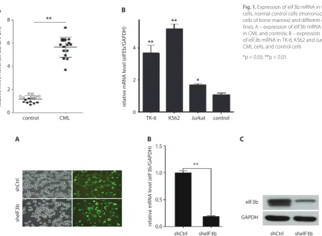
The results of qRT-PCR showed that the expression of eIF3b mRNA in the CML patients was higher than that of the control group (p < 0.01) (Fig. 1A). In addition, eIF3b
was found to be overexpressed in the TK-6 (p < 0.001), K562 (p < 0.001) and Jurkat (p < 0.05) CML cells when compared with normal control cells (mononuclear cells isolated from the bone marrow of healthy individuals, Fig. 1B).
Table 1. Details of the antibodies used
Antibody name Company Country Dilution ratio
Primary antibody
Mouse Anti-Flag Sigma USA 1:2000
Mouse anti-GAPDH Santa-Cruz USA 1:2000
Anti-BCL2 Abcam USA 1:2000
Anti-GAPDH Santa-Cruz USA 1:2000
Secondary antibody Goat Anti-Mouse IgG
shRNA inhibits eIF3b mRNA
and protein expression
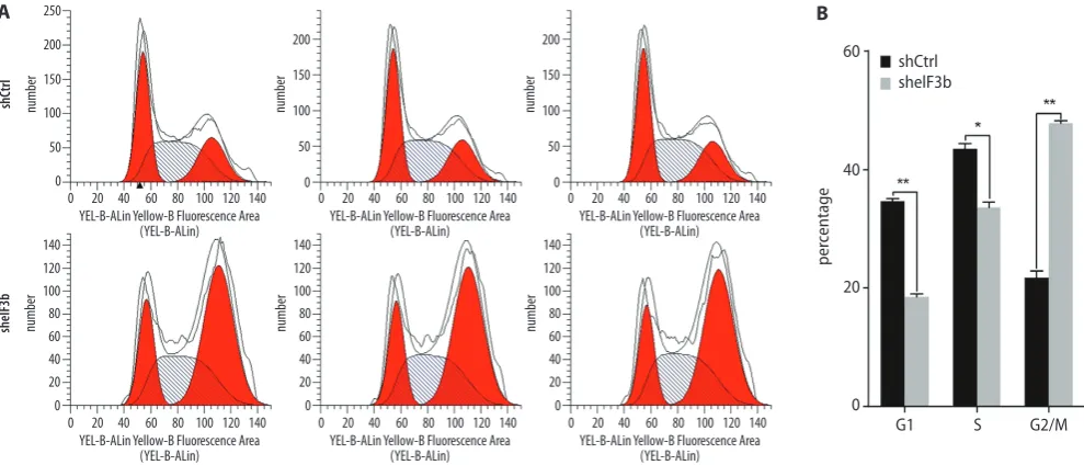
After transduction with lentivirus-anti-eIF3b or empty-vector lentivirus in the K562 cells for 3 days, the fluo-rescence expression was observed under a fluothe fluo-rescence microscope, which showed that the transduction efficiency of both was over 90% (Fig. 2A). After 3 days, the expression of eIF3b mRNA and protein had both decreased mark-edly in the sheIF3b group compared with the shCtrl group (Fig. 2B,C).
Downregulation of eIF3b mRNA-inhibited
proliferation of K562 cells
The CCK-8 assay showed no significant difference in baseline optical density (OD) between the sheIF3b and shCtrl groups. On days 3, 4 and 5 after transduction, the OD value of the sheIF3b group was lower than that of the shCtrl group (Fig. 3A,B), a finding which suggests that the inhibition of eIF3b mRNA suppresses the prolif-eration of K562 cells.
Fig. 1. Expression of eIF3b mRNA in CML
cells, normal control cells (mononuclear cells of bone marrow) and different cell lines; A – expression of eIF3b mRNA in CML and controls; B – expression of eIF3b mRNA in TK-6, K562 and Jurkat CML cells, and control cells
*p < 0.05; **p < 0.01.
Fig. 3. Effect of eIF3b mRNA downregulation on the proliferation of K562
cells; A – OD450 value in the shCtrl and sheIF3b groups 5 days after transduction; B – OD450 value in the shCtrl and sheIF3b groups 5 days after transduction
*p < 0.05; **p < 0.01.
Fig. 2. Transduction of shRNA and the expression of eIF3b mRNA and protein; A – transduction efficiency of shRNA in K562 cells under fluorescence
microscope; B – qRT-PCR of the relative mRNA level in the shCtrl and sheIF3b groups; C – western blotting of the level of eIF3b protein in the shCtrl and sheIF3b groups
**p < 0.01. control
relative mRNA level (elF3b/GAPDH) relative mRNA level (elFEb/GAPDH)
CML TK-6 control
A B
4
2
0 8
6
4
2
0
K562 Jurkat
relative mRNA level (elF3b/GAPDH)
shelF3b
shCtr
l
shelF3b shCtrl
shelF3b shCtrl
elF3b
GAPDH
A B C
1.5
1.0
0.5
0.0
day shCtrl shelF3b
shCtrl
OD
450
/F
old
OD
450
shelF3b
1
A B
2 3 4 5
day 1
3
2
1
0
10
8
6
4
2
0
Effect of
eIF3b
gene downregulation
on K562 cell cycles
Following transduction with shRNA lentivirus – on day 5 – the proportion of cells in the G1 (p < 0.01) and S phases (p < 0.05) of the sheIF3b group was lower than that of the shCtrl group, while the proportion of cells in the G2/M phase (p < 0.01) was higher than in the shC-trl group (Fig. 4A,B).
Effect of eIF3b mRNA downregulation
on the apoptosis of K562 cells
On day 5 after transduction, the total cell apoptosis percentage in the sheIF3b group was substantially higher
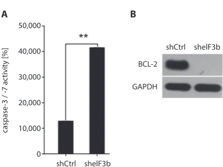
than that of the shCtrl group (p < 0.01, Fig. 5A,B). Caspase 3/7 activity in the sheIF3b group was remarkably high-er (p < 0.01,Fig. 6A) than that of the control group, and the expression of anti-apoptotic protein Bcl-2 was mark-edly lower in the sheIF3b group than in the shCrtl group (p < 0.01, Fig. 6B), suggesting that eIF3b downregulation promoted the apoptosis of K562 cells.
Discussion
Chronic myeloid leukemia is a clonal HSC disorder re-sulting from a reciprocal translocation between the long arms of ch9 and ch22, t (9;22), which leads to the fusion of the ABL1 proto-oncogene from ch9 with the BCR
Fig. 5. Effect of eIF3b gene downregulation on the apoptosis of K562 cells; A – cell apoptosis of K562 cells in the shCtrl and sheIF3b groups; B – percentage
of cell apoptosis in the shCtrl and sheIF3b groups **p < 0.01.
Fig. 4. Effect of eIF3b gene downregulation on the cell cycles of K562 cells; A – proportion of K562 cells in different phases in the shCtrl and sheIF3b groups;
B – proportion of K562 cells in G1, S, and G2/M phases in the shCtrl and sheIF3b groups *p < 0.01; **p < 0.01.
YEL-B-ALin Yellow-B Fluorescence Area (YEL-B-ALin) 0 number 250 200 150 100 50 0
20 40 60 80 100 120 140
YEL-B-ALin Yellow-B Fluorescence Area (YEL-B-ALin) 0 number shCtrl sheIF3b 100 80 60 40 20 0 120 140
20 40 60 80 100 120 140
YEL-B-ALin Yellow-B Fluorescence Area (YEL-B-ALin) 0 number 100 80 60 40 20 0 120 140
20 40 60 80 100 120 140
YEL-B-ALin Yellow-B Fluorescence Area (YEL-B-ALin) 0 0 20 40 60 B A per centage shelF3b shCtrl
G1 S G2/M
number 100 80 60 40 20 0 120 140
20 40 60 80 100 120 140 YEL-B-ALin Yellow-B Fluorescence Area
(YEL-B-ALin) 0 number 200 150 100 50 0
20 40 60 80 100 120 140
YEL-B-ALin Yellow-B Fluorescence Area (YEL-B-ALin) 0 number 200 150 100 50 0
20 40 60 80 100 120 140
4 6 0 2 8 10 peccentage shCtrl shelF3b
Red-R Fluorescence (RED-R-Hlog) Plot P03, gated on P01.R1
coun t 250 300 200 150 100 R3 50 0 Red-R Fluorescence (RED-R-Hlog)
Plot P03, gated on P01.R1
coun t 250 300 200 150 100 R3 50 0 Red-R Fluorescence (RED-R-Hlog)
Plot P03, gated on P01.R1
100 101 102 103 104 105 100 101 102 103 104 105 100 101 102 103 104 105
100 101 102 103 104 105 100 101 102 103 104 105 100 101 102 103 104 105
coun t 250 300 200 150 100 R3 50 0
Red-R Fluorescence (RED-R-Hlog) Plot P03, gated on P01.R1
coun t 250 300 200 150 100 R3 50 0 Red-R Fluorescence (RED-R-Hlog)
Plot P03, gated on P01.R1
coun t 250 300 200 150 100 R3 50 0 Red-R Fluorescence (RED-R-Hlog)
Plot P03, gated on P01.R1
housekeeping gene on ch22 to produce the BCR-ABL
gene.13 This fusion gene is transcribed into BCR-ABL1 mRNA and translated into the BCR-ABL protein, which initiates the progression of CML.14,15
Dysregulation of mRNA translation leads to the aberrant activation of cellular pathways that promote cell prolifera-tion, invasion and progression of leukemia. The main func-tion of eIFs is in the interacfunc-tion between the ribosome and mRNA, which takes part in the initial process of protein synthesis, affecting cell cycle, growth and apoptosis.16,17
eIF3b, a member of the eIF3 complex, is reported to be overexpressed in various tumor cells and acts as an onco-gene. A study indicated that eIF3b mRNA is abundantly expressed in colon cancer cells, and that the downregula-tion of eIF3b inhibits cell proliferation, reduces the number of cells in the G1 phase and increases the number of cells in the S/G2 phases, as well as promotes cell apoptosis.8 Another study investigating the role of eIF3b in esophageal squamous cell carcinoma (ESCC) also revealed that eIF3b
expression is much higher in ESCC tissues and ESCC cell lines, while a reduction in eIF3b suppresses cell prolifera-tion and stimulates cell apoptosis through the regulaprolifera-tion of the β-catenin signaling pathway.18 In bladder and pros-tate cancer cells, eIF3b deletion decreases cell growth and represses the G1/S cell cycle transition by regulating cyclin A, E, Rb, and p27Kip1 protein expression – but not mRNA expression – and it reduces migration as well as interrupt-ing the actin cytoskeleton and focal adhesions.11 A recently published study showed that knockdown of eIF3b decreas-es cell viability and promotdecreas-es apoptosis in osteosarcoma cells due to the regulation of tumor necrosis factor recep-tor superfamily member 21 (TNFRSF21).19 These findings indicate that eIF3b is involved in tumor development and progression by acting as a cancer-promoting gene.
In clinical research, Wang et al. examined the expres-sion of eIF3b mRNA in patients with bladder cancer and
prostate cancer, and reported that eIF3b mRNA expres-sion is much higher in cancer patients than in controls. Interestingly, they also discovered that eIF3b mRNA is positively correlated with tumor grade and could predict unfavorable survival.9 However, the role of eIF3b in CML remains unclear. In this study, we found that eIF3b mRNA expression was higher in CML patients and CML cell lines than in the controls. Downregulation of eIF3b expres-sion suppressed CML cell proliferation, decreased G1/S-phase cells and induced cell apoptosis, which was in line with the oncogenic role of eIF3b in other studies. Reduced eIF3b mRNA expression inhibits proliferation and pro-motes apoptosis in K562 cells. The possible explanations could be as follows: 1) eIF3b could activate the β-catenin signaling pathway, including the downstream target gene cyclin D1 and c-Myc, thus inducing cell proliferation and invasion, as well as inhibiting cell apoptosis and interfer-ing with the cell cycle18 or; 2) eIF3b is involved in protein synthesis and eIF3b depletion can globally inhibit protein synthesis, which affects the proliferation and apoptosis of cancer cells.9
There were some limitations in this study. Firstly, the prognostic role of eIF3b expression in CML patients was not investigated. Secondly, we discovered the role of eIF3b in regulating CML cell proliferation and apop-tosis, but the mechanism of how eIF3b affects cancer cell progression was not explored. This will be the focus of fu-ture research.
In conclusion, the downregulation of eIF3b inhibits pro-liferation and induces apoptosis in CML cells.
ORCID iDs
Laiquan Huang https://orcid.org/0000-0002-7139-9755
Kun He https://orcid.org/0000-0001-5843-2939
Jianxin Wang https://orcid.org/0000-0001-9548-1465
Jiawei Yan https://orcid.org/0000-0001-9887-503X
Yizhi Jiang https://orcid.org/0000-0002-4812-2547
Zhongling Wei https://orcid.org/0000-0002-2763-0964
Jun Zhang https://orcid.org/0000-0002-1759-2829
Guangxi Li https://orcid.org/0000-0002-4309-0611
Lili Sheng https://orcid.org/0000-0002-9579-7328
References
1. Gutierrez LG, Noriega MF, Laudicina A, Quatrin M, Bengio RM, Lar-ripa I. An unusual translocation, t(1;11)(q21;q23), in a case of chronic
myeloid leukemia with a cryptic Philadelphia chromosome. Oncol
Lett. 2017;13(5):3159–3162.
2. Hinnebusch AG, Lorsch JR. The mechanism of eukaryotic translation
initiation: New insights and challenges. Cold Spring Harb Perspect Biol.
2012;4(10). doi:10.1101/cshperspect.a011544
3. Pestova TV, Kolupaeva VG. The roles of individual eukaryotic trans-lation initiation factors in ribosomal scanning and initiation codon
selection. Genes Dev. 2002;16(22):2906–2922.
4. Sonenberg N, Hinnebusch AG. Regulation of translation initiation
in eukaryotes: Mechanisms and biological targets. Cell. 2009;136(4):
731–745.
5. Silvera D, Formenti SC, Schneider RJ. Translational control in cancer.
Nat Rev Cancer. 2010;10(4):254–266.
6. Hagner PR, Schneider A, Gartenhaus RB. Targeting the translational machinery as a novel treatment strategy for hematologic
malignan-cies. Blood. 2010;115(11):2127–2135.
Fig. 6. Effect of eIF3b gene downregulation on apoptosis-related proteins;
A – caspase 3/7 activity in the shCtrl and sheIF3b groups; B – expression of anti-apoptotic protein BCL-2 in the shCtrl and sheIF3b groups **p < 0.01.
shelF3b shCtrl
shelF3b shCtrl
BCL-2
GAPDH
A B
caspase
-3 / -7 ac
tivity [%
]
50,000
40,000
30,000
20,000
10,000
7. Spilka R, Ernst C, Mehta AK, Haybaeck J. Eukaryotic translation
ini-tiation factors in cancer development and progression. Cancer Lett.
2013;340(1):9–21.
8. Mayeur GL, Fraser CS, Peiretti F, Block KL, Hershey JW. Characteriza-tion of eIF3k: A newly discovered subunit of mammalian translaCharacteriza-tion
initiation factor elF3. Eur J Biochem. 2003;270(20):4133–4139.
9. Hinnebusch AG. eIF3: A versatile scaffold for translation initiation
complexes. Trends Biochem Sci. 2006;31(10):553–562.
10. Wang Z, Chen J, Sun J, Cui Z, Wu H. RNA interference-mediated silenc-ing of eukaryotic translation initiation factor 3, subunit B (eIF3b) gene
expression inhibits proliferation of colon cancer cells. World J Surg
Oncol. 2012;10:119.
11. Wang H, Ru Y, Sanchez-Carbayo M, Wang X, Kieft JS, Theodorescu D. Translation initiation factor eIF3b expression in human cancer and
its role in tumor growth and lung colonization. Clin Cancer Res. 2013;
19(11):2850–2860.
12. Liang H, Ding X, Zhou C, et al. Knockdown of eukaryotic translation initiation factors 3B (eIF3b) inhibits proliferation and promotes
apop-tosis in glioblastoma cells. Neurol Sci. 2012;33(5):1057–1062.
13. Apperley JF. Chronic myeloid leukaemia. Lancet. 2015;385(9976):
1447–1459.
14. Kavalerchik E, Goff D, Jamieson CH. Chronic myeloid leukemia stem
cells. J Clin Oncol. 2008;26(17):2911–2915.
15. Savona M, Talpaz M. Getting to the stem of chronic myeloid
leukae-mia. Nat Rev Cancer. 2008;8(5):341–350.
16. Pestova TV, Kolupaeva VG, Lomakin IB, et al. Molecular mechanisms
of translation initiation in eukaryotes. Proc Natl Acad Sci U S A. 2001;
98(13):7029–7036.
17. Lee AS, Kranzusch PJ, Cate JH. eIF3 targets cell-proliferation
mes-senger RNAs for translational activation or repression. Nature. 2015;
522(7554):111–114.
18. Xu F, Xu CZ, Gu J, et al. Eukaryotic translation initiation factor 3B accelerates the progression of esophageal squamous cell carcinoma
by activating beta-catenin signaling pathway. Oncotarget. 2016;7(28):
43401–43411.
19. Choi YJ, Lee YS, Lee HW, Shim DM, Seo SW. Silencing of translation initiation factor eIF3b promotes apoptosis in osteosarcoma cells.