DOI
10.17219/acem/66215
Copyright
© 2018 by Wroclaw Medical University This is an article distributed under the terms of the Creative Commons Attribution Non-Commercial License (http://creativecommons.org/licenses/by-nc-nd/4.0/)
Address for correspondence
Nur Şahin
E-mail: [email protected]
Funding sources
None declared
Conflict of interest
None declared
Received on June 7, 2016 Reviewed on September 21, 2016 Accepted on October 21, 2016
Abstract
Background. The modified Misgav-Ladach method (MML) is a minimally invasive cesarean section pro-cedure compared with the classic Pfannenstiel-Kerr (PK) method.
Objectives. The aim of the study was to compare the MML method and the PK method in terms of intra-operative and short-term postintra-operative outcomes.
Material and methods. This prospective, randomized controlled trial involved 252 pregnant women sched-uled for primary emergency or elective cesarean section between October, 2014 and July, 2015. The primary outcome measures were the duration of surgery, extraction time, Apgar score, blood loss, wound complica-tions, and number of sutures used. Secondary outcome measures were the wound infection, time of bowel restitution, visual analogue scale (VAS) scores at 6 h and 24 h after the operation, limitations in movement, and analgesic requirements. At 6 weeks after surgery, the patients were evaluated regarding late complications. Results. There was a significant reduction in total operating and extraction time in the MML group (p < 0.001). Limitations in movement were lower at 24 h after the MML operation, and less analgesic was required in the MML group. There was no difference between the 2 groups in terms of febrile morbidity or the duration of hospitalization. At 6 weeks after the operation, no complaints and no additional complications from the surgery were noted.
Conclusions. The MML method is a minimally invasive cesarean section. In the future, as surgeons’ experi-ence increases, MML will likely be chosen more often than the classic PK method.
Key words: cesarean section, postoperative pain, Pfannenstiel incision, modified Misgav-Ladach technique, operating time
A comparison of 2 cesarean section methods, modified Misgav-Ladach
and Pfannenstiel-Kerr: A randomized controlled study
Nur Şahin
1,A–F, Mine Genç
1,A,D, Gülüzar Arzu Turan
1,B,C, Esin Kasap
2,A–C, Serkan Güçlü
3,D–F1 Department of Obstetrics and Gynecology, Sifa University, Izmir, Turkey 2 Department of Obstetrics and Gynecology, Karatas Hospital, Izmir, Turkey 3 Department of Obstetrics and Gynecology, Kent Hospital, Izmir, Turkey
A – research concept and design; B – collection and/or assembly of data; C – data analysis and interpretation; D – writing the article; E – critical revision of the article; F – final approval of the article
Introduction
Cesarean sections (C/S) are among the most common abdominal surgical procedures in women. Approximately 15% of all deliveries are performed by the abdominal route worldwide.1–3 According to the national registry of the
Turk-ish Health Ministry, in 2014 the proportion of abdominal deliveries was 67% in medical school hospitals, 37% in pub-lic hospitals and 71% in private hospitals. The percentage at Sifa University Medical School Hospital in 2014 was 65%.
Cesarean sections are performed in both elective or emer-gency cases. Fetal distress, a previous C/S history, cephalo-pel-vic disproportion, eclampsia, preeclampsia, malpresentation, and placenta previa are the main indications for C/S.
Various surgical procedures have been defined for C/S. In the early 20th century, Pfannenstiel described
a trans-verse incision of the abdomen, which is still the most com-monly used method.4 In 1926, Kerr proposed a transverse
lower uterine segment incision and double-layer uterine suture with peritoneal closure.5 Joel-Cohen described
a new transverse incision technique in 1972, and Stark modified it in 1994.6,7 This technique is also called the
Misgav-Ladach method. In the modified Misgav-Ladach method (MML), skin closure is achieved with continuous subcuticular sutures or clips and mattress stitches, accord-ing to the surgeon’s preference.
In this prospective study, we sought to compare the MML and Pfannenstiel-Kerr (PK) methods in terms of in-traoperative and short-term postoperative outcomes.
Material and methods
This randomized controlled trial involved 252 pregnant women scheduled for primary emergency or elective C/S. All the procedures were performed at Sifa University
Medical School Hospital between October, 2014 and July, 2015. The approval of the university ethics committee was obtained before beginning the study. Written informed consent was obtained from each patient.
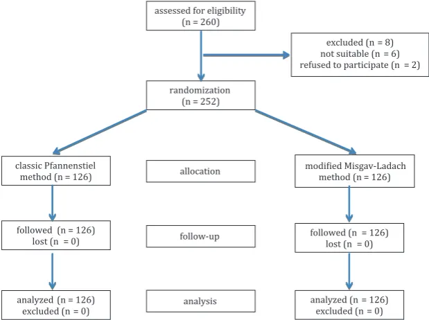
Inclusion criteria were: a gestational age >36 weeks, the first C/S (the women could have delivered vaginally before) and an obstetric indication for C/S. The same 2 surgeons performed all the C/S procedures. Exclusion criteria were: the presence of any additional surgical procedure, such as myomectomy, cystectomy or tubal ligation, placenta previa, placental abruption, preeclampsia, eclampsia, or HELLP syn-drome. A flow diagram showing the selection of the study population is presented in Fig. 1. The patients were random-ized into 2 groups using a computer-generated random num-ber list: PK (n = 126) and MML (n = 126). The organizer informed the surgeons (SG, NS) of the patient’s group assign-ment immediately before the surgery. The nurses who record-ed the VAS scores were blindrecord-ed to the patient’s group. VAS scores were used to assess pain after the operation (on a scale where 0 = no pain and 10 = maximum pain). The anesthesi-ologist decided on the type of anesthesia (general was used in 19% of the patients, spinal or epidural in 81%).
Description of the modified
Misgav-Ladach technique
A Joel-Cohen skin incision was performed with a straight superficial transverse cut in the skin about 3 cm below the line of the spinae iliacae anteriores superiores, and the subcutaneous tissue was opened upwards in the mid-line so as to reach the rectus sheath above the insertion of the pyramidalis muscles.7 The parietal peritoneum was
opened digitally at the upper level of the intermuscular space. The fetus was extracted from a transverse lower uterine segment incision, and the placenta was removed by transabdominal uterine massage combined with light
Fig. 1. The selection of the study population assessed for eligibility
(n = 260)
randomization (n = 252)
excluded (n = 8) not suitable (n = 6) refused to participate (n = 2)
classic Pfannenstiel method (n = 126)
modiied Misgav-Ladach method (n = 126) allocation
analysis follow-up followed (n = 126)
lost (n = 0) followed (n = 126)lost (n = 0)
analyzed (n = 126) excluded (n = 0)
cord traction. Closure of the uterine incision was accom-plished with a 1-layer continuous No. 1 polyglactin 910 suture (Vicryl, Ethicon Inc., Somerville, USA), using ad-ditional hemostatic stitches as required. The visceral and parietal peritoneum and the rectus muscles were left un-sutured. The rectus sheath was closed using a continuous No. 1 polyglactin 910 suture. The subcutaneous tissue was sutured if its depth exceeded 2 cm. The skin was closed with a continuous subcuticular suture.
Description
of the Pfannenstiel-Kerr technique
The skin was opened with a Pfannenstiel incision, and the incision was extended through the subcutaneous tissue until the rectus sheath was exposed; the latter was then opened in the midline.4 Scissors were used to extend the
rectus sheath incision laterally, and to separate it from the pyramidalis and rectus muscles. After lateral extension of the uterine incision with uterine scissors, fetal extraction and removal of the placenta using transabdominal uterine massage combined with light cord traction were performed. Closure of the uterine incision was accomplished with a 1-layer continuous No. 1 polyglactin 910 suture. The vis-ceral and parietal peritoneum were closed with a continuous No. 2/0 polyglactin 910 suture. The rectus sheath was closed with a continuous No. 1 polyglactin 910 suture. The sub-cutaneous tissue was sutured if its depth exceeded 2 cm. The skin was closed with a continuous subcuticular suture.
Primary and secondary measures
of postoperative outcomes
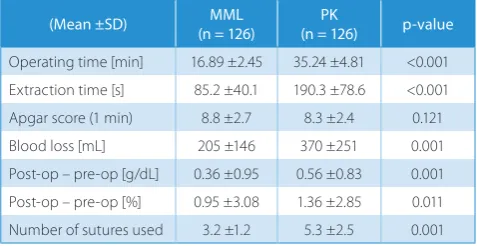
The primary outcome measures were the duration of surgery (between skin incision and skin closure), extrac-tion time (until delivery of the neonate), Apgar score, blood loss, wound complications, and number of sutures used.
Secondary outcome measures were the wound infec-tion, time of bowel restituinfec-tion, VAS scores from 0 to 10 at 6 h and 24 h after the operation, limitations in movement, and analgesic requirements. At 6 weeks after the surgery, patients were evaluated for late complications.
Statistical analysis
Allstatisticalanalyses were performed using RStudio v. 0.98.501 (RStudio Inc., Boston, MA, USA). The Mann-Whitney U test was used to compare the study groups. Probability values <0.05 were considered statistically sig-nificant. For each group, a minimum of 126 subjects was required to have 80% power (α: 0.05) with a 10% difference in Apgar scores.
Results
The patient characteristics are shown in Table 1. There was no difference between the groups in terms of mean maternal age, gestational age, BMI, C/S indication, or the type of anesthesia. There was a significant reduction in to-tal operating time in the MML group (16.9 min) compared with the PK group (35.2 min; p < 0.001), and the mean ex-traction time was significantly shorter in the MML group (p < 0.001). There was no difference in Apgar scores. Pri-mary outcomes are shown in Table 2.
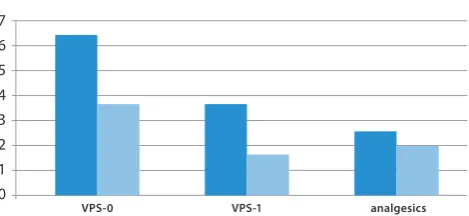
All the patients began a regular diet 6 h after the surgery and were mobilized at 10 h after the surgery. The 6-hour post-op VAS score (VAS0) and the 24-hour score (VAS1) were significantly lower in the MML group (MML: 3.54 VAS0, 1.46 VAS1; PK: 6.36 VAS0, 3.64 VAS1; p < 0.001). Limitation in movement, evaluated 24 h after the opera-tion, was lower in the MML group (Fig. 2), and less anal-gesic was required in the MML group (1.8 doses; Fig. 3). There were no differences in febrile morbidity or the dura-tion of hospitalizadura-tion. At 6 weeks after the operadura-tion, we received no complaints or reports of additional complica-tions related to the surgery.
Discussion
The present study compared the PK and MML meth-ods. In recent studies, shorter operating times have been reported with MML.8–12 Franchi et al. reported similar
op-erating times with both methods, but a shorter extraction
Table 1. Patients’ demographic data
(Mean ±SD) (n = 126)MML (n = 126)PK p-value
Age [years] 31.4 ±4.7 30.2 ±5.4 0.080
BMI [kg/m2] 29.22 ±3.97 30.23 ±5.09 0.251
Gestational age [weeks] 38.82 ±0.6 38.42 ±1.6 0.120 Type of anesthesia n (%)
general regional
22 (17.5) 104 (82.5)
31 (23.6) 95 (76.4)
0.155
Table 2. Operation details
(Mean ±SD) (n = 126)MML (n = 126)PK p-value
Operating time [min] 16.89 ±2.45 35.24 ±4.81 <0.001 Extraction time [s] 85.2 ±40.1 190.3 ±78.6 <0.001
Apgar score (1 min) 8.8 ±2.7 8.3 ±2.4 0.121
Blood loss [mL] 205 ±146 370 ±251 0.001
Post-op – pre-op [g/dL] 0.36 ±0.95 0.56 ±0.83 0.001 Post-op – pre-op [%] 0.95 ±3.08 1.36 ±2.85 0.011
Number of sutures used 3.2 ±1.2 5.3 ±2.5 0.001
MML – modified Misgav-Ladach method; PK – Pfannenstiel-Kerr method.
time with MML.13 We also found a shorter operating time
with MML than with the PK method. A shorter operating time may be particularly important in emergencies as op-posed to elective cases. It is of primary importance that the duration between incision and entrance to the abdomen was shorter, which provides a shorter time for delivering the baby. Although better results for neonatal outcomes would be expected under this condition, in most studies addressing this issue, no difference has been reported for neonatal outcomes between the 2 methods.9,10,13,14 We also
found no difference in Apgar scores between the groups. Another reason for the shorter operating time with MML is that the visceral and parietal peritoneal layers were left unsutured after the closure of the uterus. Suturing the peri-toneal layers is an unnecessary step, because the peritone-um does not heal by the approximation of the wound edges; a new peritoneal layer is formed within 24–48 h. Adhesions are formed as vascular bridges to supply oxygen to ischemic areas of tissue, and necrosis often occurs around peritoneal sutures, providing focal points for adhesions.15–17
More-over, leaving the subcutaneous tissue unsutured does not increase the incidence of wound complications.18,19 In the
present study, we found that significantly fewer sutures were used in the MML group. The reduction in cost achieved in the MML method by using fewer sutures and less anes-thesia is particularly important for developing countries.
Stark and Finkel demonstrated reduced use of antibiotics and less postoperative febrile morbidity with MML.7
How-ever, other studies have found similar results for wound infection in the 2 methods.9,20 A prophylactic antibiotic
was given to all the patients in the present study, and we did not note any infection in either group. As in other recent studies, we found significantly less blood loss in the MML group.21 This was associated with several procedural
dif-ferences: the subcutaneous tissue was not cut, the rectus muscles were stretched instead of being cut and the fascia layer was not opened upwards from the midline.
All the procedures were performed by the same 2 ex-perienced surgeons to avoid variation. A nurse blinded to the patient group recorded the postoperative data (VAS scores, the need for analgesics, scores for movement limita-tion) to prevent bias during the study. Early mobilization is known to reduce the risk of thrombosis, ileus and in-fections. Early restitution of oral intake facilitates physi-cal recovery through rapid replacement of protein loss. The lower analgesic requirements during the early postop-erative period in the MML group are associated with lower tissue trauma due to blunt access to the abdominal cavity, without the blood vessels and nerves of the subcutaneous tissue being incised.19
Higher scores were recorded for VAS0 and VAS1 in the PK group than the MML group in our study. The patients in the MML group reported less postoperative discomfort, indicating that MML is a less traumatic C/S approach. In another study, it was demonstrated that the MML meth-od resulted in better short-term quality of life scores, es-pecially in terms of reduced bodily pain and postoperative complications compared with the PK method.17
Conclusions
We suggest that in the future, with increased experi-ence on the part of surgeons, the minimally invasive MML cesarean section method will be chosen more often than the classic PK method.
References
1. Menacker F, Curtin SC. Trends in cesarean birth and vaginal birth after previous cesarean, 1991–1999. Natl Vital Stat Rep. 2001;49(13):1–16. 2. Thomas J, Paranjothy S; Royal College of Obstetricians and
Gynaeco-logists Clinical Effectiveness Support Unit. The National Sentinel
Cae-sarean Section Audit Report. London: RCOG Press; 2001.
3. Cai WW, Marks JS, Chen CH, Zhuang YX, Morris L, Harris JR. Increased caesarean section rates and emerging patterns of health insurance in Shanghai, China. Am J Public Health. 1998;88:777–780.
4. Pfannenstiel J. On the advantages of a transverse cut of the fascia above the symphysis for gynecological laparotomies, and advice on surgical methods and indications. Samml Klin Vortr Gynakol. 1987; 68:1–22.
5. Kerr JMM. The technic of cesarean section with special reference to the lower uterine segment incision. Am J Obstet Gynecol. 1926;12: 729–734.
6. Joel-Cohen S. Abdominal and Vaginal Hysterectomy: New Techniques
Based on Times and Motion Studies. London: Heinemann; 1972.
7. Stark M, Finkel AR. Comparison between the Joel-Cohen and Pfan-nenstiel incisions in cesarean section. Eur J Obstet Gynecol Reprod Biol. 1994;53:121–122.
8. Song SH, Oh MJ, Kim T, Hur JY, Saw HS, Park YK. Finger-assisted stretching technique for cesarean section. Int J Gynaecol Obstet. 2006;92(3):212–216. 7 6 5 4 3 2 1 0
Fig. 3. Comparison of secondary outcomes (VAS scores and analgesics by number)
VPS-0 VPS-1 analgesics
classic MML 60 50 40 30 20 10 0
Fig. 2. Comparison of primary outcomes
9. Bjorklund K, Kimaro M, Urassa E, Lindmark G. Introduction of the Misgav Ladach caesarean section at an African tertiary centre: A ran-domised control trial. BJOG. 2000;107(2):209–216.
10. Darj E, Nordstrom ML. The Misgav Ladach method for cesarean sec-tion compared to the Pfannenstiel method. Acta Obstet Gynecol Scand. 1999;78(1):37–41.
11. Wallin G, Fall O. Modified Joel-Cohen technique for caesarean deliv-ery. BJOG. 1999;106(3): 221–226.
12. Xavier P, Ayres-De-Campos D, Reynolds A, Guimaraes M, Costa-San-tos C, Patricio B. The modified Misgav-Ladach versus the Pfannen-stiel-Kerr technique for cesarean section: A randomized trial. Acta
Obstet Gynecol Scand. 2005;84(9):878–882.
13. Franchi M, Ghezzi F, Raio L, et al. 2002. Joel-Cohen or Pfannenstiel incision at cesarean delivery: Does it make a difference? Acta Obstet
Gynecol Scand. 2002;81:1040–1046.
14. Naki MM, Api O, Celik H, Kars B, Yasar E, Unal O. Comparative study of Mis-gav-Ladach and Pfannenstiel-Kerr cesarean techniques: A random-ized controlled study. J Matern Fetal Neonatal Med. 2011;24 (2): 239–244. 15. Malvasi A, Tinelli A, Farine D, et al. Effects of visceral peritoneal clo-sure on scar formation at cesarean delivery. Int J Gynaecol Obstet. 2009;105:131–135.
16. Malvasi A, Tinelli A, Guido M, Zizza A, Farine D, Stark M. Should the visceral peritoneum at the bladder flap closed at cesarean sections? A post-partum sonographic and clinical assessment. J Matern Fetal
Neonatal Med. 2010;23: 662–669.
17. Fatusic Z, Hudic I, Sinanovic O, Kapıdzıc M, Hotic N, Music A. Short-term postnatal quality of life in women with previous Misgav Ladach caesarean section compared to Phannenstiel-Dorffler caesarean section method. J Matern Fetal Neonatal Med. 2011;24(9):1138–1142.
18. Federici D, Lacelli B, Muggiasca L, Agarossi A, Cippolla L, Conti M. Cesarean section using the Misgav Ladach method. Int J Gynaecol
Obstet. 1997;57(3):273–279.
19. Kulas T, Habek D, Karsa M, Vukovic M. Modified Misgav Ladach meth-od for cesarean section: Clinical experience. Gynecol Obstet Invest. 2008;65:222–226.
20. Franchi M, Ghezzi F, Balestreri D, et al. A randomized clinical trial of two surgical techniques for cesarean section. Am J Perinatol. 1998; 15:589–594.