Ewa Obłąk
1, Anna Krasowska
2The Influence of Organic Nitrogen Compounds
on Melanoma, Bacterial, and Fungal Cells*
Wpływ organicznych związków azotowych
na komórki czerniaka, bakterii i grzybów
1 Institute of Genetics and Microbiology, University of Wrocław, Poland 2 Faculty of Biotechnology, University of Wrocław, Poland
Abstract
Background. The influence of the quaternary ammonium salt (QAS) N-(dodecyloxycarboxymethyl)-N,N,N- -trimethyl ammonium chloride (IM) and the lysosomotropic aminoester N-N-dimethylaminoethyl dodecanoate hydrochloride (DM-11) on bacterial, fungal, and melanoma cell viability and plasma membrane H+-ATPase activ-ity in Saccharomyces cerevisiae was investigated.
Objectives. The aim was to determine the tested compounds’ activities as potential disinfectants.
Material and Methods. The minimal inhibitory concentration (MIC) was determined in the fungal and bacterial cells (Gram positive and negative strains) on solid YPD and LB medium with different concentrations of the tested drugs (20, 40, 80, 120, 140, 160, 320 µM). Critical micelle concentrations (CMC) were measured at room tempera-ture with a tensiometer using a 24-cm2 Teflon vessel and Wilhelm’s plates. Proton extrusion was determined by pH measurements (computer-linked pH-meter) in distilled water to which a yeast cell suspension (2 mg dry weight), glucose (final concentration 200 mM), either IM or DM-11 (final concentrations of 20, 80 or 140 µM), and KCl (final concentration 100 mM) were successively added.Determination of the ATP concentration in melanoma cells was carried out in 24-well plates at a density of 0.05 × 10⁶ cells per well. ATP determination was performed using the ATP determination kit with luciferase. The MTT (methylthiazole tetrazolium) assay was used to determine the cytotoxicity of the tested agents. This test quantifies the conversion of water-soluble MTT to insoluble purple formazan, which is catalyzed by mitochondrial dehydrogenase of only living cells.
Results. Both tested agents (IM and DM-11) inhibited the growth of Saccharomyces cerevisiae, Candida albicans, Staphylococcus aureus, and Escherichia coli. Moreover, IM influenced the mitochondrial metabolism of the mela-noma cells. The inhibitory activity of the aminoester was only temporary, in contrast to the QAS. Both compounds inhibited proton extrusion by plasma membrane H+-ATPase. This inhibitory effect was concentration dependent. IM strongly reduced the ATP level in melanoma cells.
Conclusions. These results suggest that the modes of action of QASs and aminoesters differ. The QAS influenced the plasma membrane, in contrast to the aminoester, which passed through the plasma membrane and accumu-lated in vacuoles (Adv Clin Exp Med 2010, 19, 1, 65–75).
Key words: quaternary ammonium salt, aminoester, fungi, bacteria, H+-ATPase.
Streszczenie
Wprowadzenie. Badano wpływ czwartorzędowej soli amoniowej chlorku dodecyloksykarboksymetylo-N,N,N- -trimetyloamoniowego (IM) i aminoestru chlorowodorku estru 2-(dimetyloamino)etylowego kwasu laurynowego (DM-11) na przeżywalność komórek czerniaka, bakterii, grzybów oraz aktywność H+-ATPazy błony komórkowej Saccharomyces cerevisiae.
Cel pracy. Określenie wpływu testowanych związków na drobnoustroje i wykorzystanie ich jako potencjalnych dezynfektantów.
Materiał i metody. Minimalne inhibicyjne stężenie (MIC) wyznaczono dla grzybów i bakterii (Gram+ i Gram-), na podłożu stałym YPD lub LB bez związku (badanie kontrolne) i w jego obecności (stosowano IM lub DM-11 w koń-cowych stężeniach 20, 40, 80, 120, 140, 160, 320 µM). Pomiaru krytycznego stężenia micelizacji (CMC)
doko-Adv Clin Exp Med 2010, 19, 1, 65–75 ISSN 1230-025X
OrIGINAL PAPErS
© Copyright by Wroclaw Medical University
The antimicrobial activity of synthetic qua-ternary ammonium salts (QASs) is well known [1–7] and these compounds have been extensively applied as disinfectants and antiseptics. They have several clinical applications such as: preopera-tive disinfection of unbroken skin, application to mucous membranes, disinfection of noncritical surfaces and medical tools [1, 3, 4, 6–11]. They can be also used for hard-surface cleaning and deodor-ization [6].
In addition, QASs are sporostatic, i.e. they inhibit the outgrowth of spores (but not the actual germination processes) [12] and mycobacterio-static [13], although mechanisms of these effects have not been understood, yet.
QASs have also an influence on lipid envel-oped (including human immunodeficiency virus (HIV) and human hepatitis B virus (HBV)) but not nonenveloped viruses [14].
QAS-based products cause loss of human HBV infectivity because of its disintegrationand morphological changes. These compounds are also used as medications [1, 8–12].
It was shown that the application of a wide range of QASs as disinfectants causes the appearance of resistance to these compounds in microorganisms. Thus the plasmid-encoded multidrug resistance gene
qacA from Staphylococcus aureus mediates resistance to ethidium bromide, quaternary ammonium com-pounds, diamidines, and biguanidines [15].
Lysosomotropic aminoesters were a second family of tested compounds [16].
To find compounds which could be poten-tially new disinfectants, a set of chemical agents differing in the number of carbon atoms in the aliphatic tail was synthesized at the Technical University of Wrocław, Poland [17–25] and their biological activities were tested on the yeast
Saccharomyces cerevisiae. The results obtained during several years of research can be summa-rized as follows. The growth inhibitory activity of QASs and lysosomotropic aminoesters, as deter-mined by the minimal inhibitory concentration (MIC), increases with aliphatic chain length up to C10–C14 and then diminishes [17–20]. The sensi-tivity of yeast cells to QASs depends on the cells’ respiratory competence; respiratory-deficient mutants, especially rho– and rhoo, are much more sensitive to QASs than the otherwise isogenic respiratory-proficient original forms [21–23]. The sensitivity of yeast to QASs also depends on its biosynthetic potentials. Mutants with auxotrophy for the biosynthesis of amino acids or nitrogen bases are more sensitive than their prototrophic original strains [21, 22]. Double auxotrophy fur-ther increases the sensitivity.
The last two observations permitted the present authors to advance the hypothesis that QASs inhib-it the transport of low molecular weight nutrients (amino acids and organic bases). This transport is active and requires energy. As the ATP yield in respiratory-deficient mutants metabolizing glucose by the fermentation pathway is lower than in cells with a functioning respiratory chain, the sensitiv-ity of the former to QASs is higher than that of the latter. On the other hand, if QASs inhibit amino-acid uptake and incorporation into proteins, aux-otrophic mutants unable to synthesize the com-pounds from an inorganic nitrogen source under conditions of QAS treatment are condemned to death by amino-acid starvation [23]. This work-ing hypothesis has found experimental support [24, 25]. [14C] leucine uptake is inhibited by QASs in a concentration-dependent fashion. The degree of [14C] leucine uptake inhibition by QASs in yeast cells depends on the general amino-acid permease nano w temperaturze pokojowej na tensometrze w teflonowym naczyniu o rozmiarze 24 cm2, używając bibuły Wilhelma. Aktywność H+-ATPazy błony komórkowej drożdży in vivo określano przez pomiar pH. W tym celu do zawiesiny głodzonych komórek drożdży (2 mg/ml) dodawano kolejno glukozę (200 mM), która indukuje wyrzu-canie protonów przez ATPazę i odpowiednie stężenia badanych związków (IM lub DM-11) oraz 100 mM KCl. Stężenie ATP w komórkach czerniaka mierzono metodą chemiluminescencyjną z wykorzystaniem testu z lucyferazą. Pomiary wykonywano w 24-dołkowych płytkach, gęstość hodowli wynosiła 0,05 × 10⁶ komórek na dołek. Do okreś-lania cytotoksyczności badanych związków użyto testu ze związkiem MTT (methylthiazole tetrazolium). W teście tym następuje konwersja MTT do czerwonego formazanu, która jest katalizowana przez mitochondrialną dehydro-genazę tylko w żywych komórkach (Adv Clin Exp Med 2010, 19, 1, 00–00).
Wyniki. Obydwa testowane związki hamowały wzrost Saccharomyces cerevisiae, Candida albicans, Staphylococcus aureus i Escherichia coli. Związek IM wpływał na mitochondrialny metabolizm w komórkach czerniaka. Inhibicyjne oddziaływanie aminoestru było tylko czasowe w przeciwieństwie do aktywności czwartorzędowej soli amoniowej. Obydwa związki hamowały wyrzucanie protonów przez H+-ATPazę i ten proces był zależny od stężenia związków. Związek IM zmniejszał stężenie ATP w komórkach czerniaka.
Wnioski. Wyniki badań własnych sugerują odmienny mechanizm działania testowanych związków na drobno-ustroje. Czwartorzędowa sól amoniowa oddziaływuje na błonę komórkową w przeciwieństwie do aminoestru, który przenika przez błonę i akumuluje się w wakuolach (Adv Clin Exp Med 2010, 19, 1, 65–75).
(GAP) activity. In yeast cells with derepressed GAP resulting from growth on proline as the nitrogen source, the inhibition is higher than in those with repressed GAP, grown on ammonium salts as the nitrogen source. The results of [14C] leucine uptake inhibition by QASs correlate with MIC [25].
Lysosomotropic compounds at a pH above the pKa value dominate in the unprotonated forms; therefore these drugs cross the plasma membrane at a higher external pH easily and accumulate in acidic cell compartments (vacuoles, lysosomes, and endo-somes). When lysosomotropic drugs exceed the criti-cal micellar concentration, they act as detergents and destroy the vacuoles, causing cellular autolysis [26]. The lysosomotropic compounds caused a change in pH in cells of Saccharomyces cerevisiae [27, 28].
The authors of the present study investigated the influence of a QAS and a lysosomotropic amin-oester on bacterial, fungal, and melanoma cell viabil-ity and on proton extrusion in vivo in view of the postulated role of plasma membrane H+-ATPase of Saccharomyces cerevisiae in generating the trans-membrane potential responsible for nutrient uptake and its role in the regulation of intracellular pH. QASs possess structural similarity to the aminoesters synthesized and studied in the authors’ laborato-ries [19, 20]. Comparative investigations on one of these QASs, N-(dodecyloxycarboxymethyl)-N,N,N- -trimethyl ammonium chloride (IM, Fig. 1a), and the aminoester N-N-dimethylaminoethyl dodecanoate hydrochloride (DM-11, Fig. 1b) were carried out.
Thus the goal was to investigate the antimicro-bial activity of the synthetic compounds (a QAS and an aminoester) in order to explain their mech-anism of action on microorgmech-anisms. This allows one to synthesize new drugs which will be effec-tively active as disinfectants.
Experimental Procedures
Strains
Saccharomyces cerevisiae Σ1278b, a wild-type strain (prototroph), the bacterial strains Staphy
lococcus aureus ATCC 25923 and Escherichia coli
ATCC 25922, and the fungal strain Candida albi cans ATCC 20231 were used in the experiments. The yeast and fungal strains were cultivated on YPD medium (1% Difco yeast extract, 1% Difco bacto peptone, 2% glucose, and 2% Difco bacto agar) at 28°C to the late exponential phase and the bacterial strains were incubated on LB medium (1% Difco yeast extract, 1% Difco Tryptone, 0.5% NaCl, and 2% Difco bacto agar) at 37°C, also to the late exponential phase. The respiratory-deficient
rhoo mutant was obtained by ethidium bromide mutagenesis of the wild strain Σ1278b according to Słonimski [29].
Melanoma
Cell Culture Conditions
The human melanoma A375 cell line (ATCC CrL-1619) was obtained from the American Type Culture Collection. The cells were cul-tured in Dulbecco’s modified Eagle’s medium (DMEM), adjusted to contain 1.5 g/l sodium bicarbonate and 4.5 g/l glucose, and supple-mented with 10% fetal bovine serum (FBS) and antibiotics (10 U/ml penicillin and 10 µg/ml streptomycin) at 37°C in a humidified atmo-sphere of 5% CO2 in air. The cells were routinely passaged by trypsinization.
Chemicals
N-(dodecyloxycarboxymethyl)-N,N,N-trime-thyl ammonium chloride (IM) and N-N-dimethy-laminoethyl dodecanoate hydrochloride (DM-11) were obtained from the laboratory of the Department of Chemistry, Technical University of Wrocław, kindly provided by Prof. S. Witek (Fig. 1a and 1b). The synthesis and properties of the compounds were detailed previously [28, 30, 31].The compounds were dissolved in water and added to YPD agar medium buffered to pH 6 or pH 8 with Sörensen buffer (0.05 M Na2HPO4 × × 12H2O and 0.05 M KH2PO4) to obtain the suit-able final concentrations.
Fig. 1. Chemical structures of the compounds: a) IM(N-(dodecyloxycarboxymethyl)-N,N,N-trimethyl ammonium chloride), b) DM-11 (N-N-dimethylaminoethyl dodecanoate hydrochloride)
Ryc. 1. Struktura chemiczna związków: a) IM (chlorku dodecyloksykarboksymetylo)-N,N,N-trimetyloamoniowego), b) DM-11 (chlorowodorku estru 2-(dimetyloamino)etylowego kwasu laurynowego)
Minimal Inhibitory
Concentration (MIC)
The minimal inhibitory concentration (MIC) was determined in the yeast and fungal cells from the late exponential growth phase in liquid YPD medium. The bacterial cells were cultured on LB medium to the late exponential phase of growth. Then the strains were plated on YPD or LB solidi-fied with 2% agar (Difco) with different concentra-tions of IM or DM-11 (20, 40, 80, 120, 140, 160, 320 µM) or without drug. Triplicate plated sam-ples of 0.1 ml contained 100–200 colony-forming units (c.f.u.). The yeast and fungal colonies were counted after 5 and 9 days of incubation at 28°C and the bacterial colonies after 2 and 5 days of incubation at 37°C and expressed as the minimal inhibitory concentration (MIC).
Yeast Survival Test
The kinetics of the survival of yeast cells treat-ed with IM or DM-11 (both at final concentrations 10 µM) was investigated in liquid YPD medium of pH 6.0 or 8.0 adjusted with Sörensen buffer (0.05 M). Cultures inoculated with 3 × 10³ c.f.u. were incubated at 28°C. Samples (0.1 ml) taken at intervals were plated on YPD medium and the viable count was determined.
Determination of the Critical
Micelle Concentration (CMC)
from Surface Tension
The measurements were performed accord-ing to Devinsky et al. [32] at room tempera-ture in a Nima Technology ST 9000 tensiometer using a 24-cm2 Teflon vessel and Wilhelm’s plates. Solutions of the IM and DM-11 compounds in YPD medium (buffered to pH 6.0 and 8.0 by add-ing 0.05 M Sörensen buffer) and 0.05 M Sörensen buffer (pH 6.0 and 8.0) were added to water at 5-min. intervals to a final concentration of 0.1 μM – 1 mM.
Proton Extrusion Test
Proton extrusion by yeast cells was determined according to [33] and [34, 35] using a computer-linked pH-meter [36]. The yeast cells were cul-tured in YPD medium (1% yeast extract, 1% bacto peptone, and 0.8% glucose; a low concentration of glucose was used to deprive the yeast cells of endogenous glucose) to the late exponential growth phase, collected by centrifugation, washed twice with distilled water, and concentrated to 2 mg dry
weight/ml. In the proton extrusion test, a suspen-sion of yeast cells (2 mg dry weight/ml), glucose (200 mM), compound IM or DM-11 (for final concentrations, see the results described below), and KCl (100 mM) were added successively to the distilled water.
ATP Assay of Yeast Cells
Yeast suspensions of OD560 = 0.1 were incu-bated with 40 and 80 μM DM-11 and IM for 40 minutes at 28°C and then centrifuged (3000 rpm, 5 min.). The supernatant was removed and the sediment was resuspended in 5% TCA to extract ATP. Ten µl of extract was 50-fold diluted in 0.05 M Sörensen buffer (pH 6.0 and 8.0). The ATP extract was used as the substrate for the luciferase assay (ENLITEN® ATP assay; Promega) accord-ing to the manufacturer’s instructions. ATP was extracted from the cells using a 5% trichloroacetic acid (TCA) solution. TCA efficiently releases ATP from microorganisms and cells while inactivating enzymes that might quickly degrade it before mea-surement. Because TCA inhibits the rL/L reaction (luciferase/luciferin), a 50-fold diluted extract was used. Luciferase oxidizes ATP-activated luciferin through a dioxetanone intermediate. It produc-es Pi, AMP, carbon dioxide, and oxyluciferin in an excited state which decays quickly by emit-ting a yellow-green light (max: 560 nm) [37–39]. The results were obtained in rLU/s (relative light units per second) in a MicroLumat LB96P chemi-luminescence plate reader (EG&G Berthold) and the ATP concentration was then read from an ATP standard curve.
Determination of ATP
Concentration
in Melanoma Cells
Cells were cultured for 24 hours in 24-well plates at a density of 0.05 ×10⁶ cells per well in the pres-ence of either IM or DM-11. The ATP concentra-tion was determined using the ATP determinaconcentra-tion kit (ENLITEN® ATP assay; Promega). Briefly, 50,000 and 25,000 cells were collected, washed in PBS, resuspended in 100 µl of distilled water, and boiled for 5 minutes. Ten µl of each sample was added to
90 µl of the reaction solution in the wells of 96-well plates. Plates were analyzed with a MicroLumat LB96P luminometer (EG&G Berthold).
MTT Test
tested agents. This test quantifies the conversion of the water-soluble MTT into insoluble purple formazan, which is catalyzed by mitochondrial dehydrogenase of only living cells. Upon treat-ment of the cells with MTX, which were seeded in 6-well plates, 1350 µl of medium and 150 µl of MTT from a 5 mg/ml MTT stock solution were added and then the cells were incubated for 3 h at 37°C. Then the medium was removed and 1500 µl of acidified isopropanol (rT) was added to the cells to dissolve the formazan crystals and the plates were stirred for 5 min. The mixture was then transferred to 1.5-ml tubes, vortexed, and the optical density was measured at 570 nm, where isopropanol served as a blank. In this experiment, A375 melanoma cells were used.
Results
Although the quaternary ammonium salt IM has a chemical structure similar to that of the aminoester DM-11 (Figs 1a and 1b), the two com-pounds seem to differ in their biological activity. These drugs inhibit the growth of microorgan-isms and their growth-inhibitory activity may be dependent on cell wall structure. Thus, Gram posi-tive bacteria (Staphylococcus aureus) were more sensitive to these compounds than Gram nega-tive (E. coli) (Table 1). Moreover, the quaternary ammonium salt (IM) was more active then the aminoester (DM-11). However, the fungal strain
Candida albicans indicated a similar level of sensi-tivity to both tested compounds (40 µM).
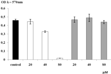
The results the MTT test showed that the A375 melanoma cells were sensitive to the IM compound and their viability was drastically inhibited; however, the aminoester DM-11 had no inhibitory activity (Fig. 2). A decrease in ATP level was observed in the presence of IM in these cells as well (Fig. 3). These two results seem to indicate that IM influences the mitochondrial metabolism of melanoma cells.
Table 1. MIC [µM] of IM and DM-11 tested on Staphylococcus aureus ATCC 25923, Escherichia coli ATCC 25922, and
Candida albicans ATCC 20231 strains
Tabela 1. MIC [µM] testowanych związków IM i DM-11 dla szczepów Staphylococcus aureus ATCC 25923, Escherichia coli
ATCC 25922 i Candida albicans ATCC 20231
Compound
(Związek) MIC [(MIC [µµM] for strains:M] dla szczepów:)
Staphylococcus aureus
ATCC 25923 Escherichia coli ATCC 25922 Candida albicans ATCC 20231
IM 20 80 40
DM-11 40 160 40
Fig. 2. Effect of IM and DM-11 (20, 40, and 80 µM) on
the viability of A 375 melanoma cells, white bars – IM compound, gray bars – DM-11 compound, ±SD (n = 3)
Ryc. 2. Wpływ IM i DM-11 (20, 40 and 80 µM) na
przeżywalność komórek czerniaka A 375, białe słupki – IM, szare słupki – DM-11, średnia ± odchylenie
stan-dardowe (n = 3)
Fig. 3. ATP concentration [nM] in A375 melanoma cells after incubation with IM or DM-11, white bar – IM compound, gray bar – DM-11 compound, ±SD (n = 3)
The compounds inhibited the growth of bak-er’s yeast and their growth-inhibitory activity was pH dependent. At both tested pH levels (6.0 and 8.0), IM was more active than DM-11 in spite of the same number of carbon atoms in the aliphatic chain, and the incubation time had no great influ-ence. The sensitivity of the respiratory-deficient strain rhoo to IM was higher than that of the respiratory-competent strain rho+,in contrast to DM-11, in which the sensitivities of both strains,
rho+ and rhoo, were on the same level after pro-longed incubation (9 days). The differences in the
rho+ and rhoo cells were seen only up to 5 days of incubation and then disappeared (Table 2). This difference in viable count suggests that IM and DM-11 could differ in the character of their bio-logical activity. The QASs, exemplified by IM, have a killing effect, while under the same condi-tions aminoesters, such as DM-11, inhibit growth, especially of the rhoomutant, and the inhibition is only temporary.
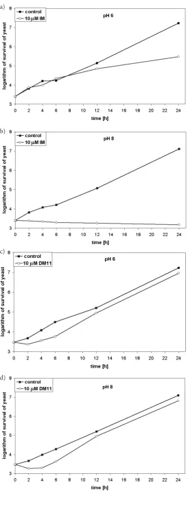
To test this assumption, viable counts in liq-uid cultures of Saccharomyces cerevisiae yeast cells treated with IM or DM-11 were compared. As the sensitivity of the yeast to the compounds in liquid culture is higher than on solid media, compounds at lower concentrations were used. As presented in Figure 4a, yeast cells in pH 6 medium in the pres-ence of IM (10 µM) showed slower growth than the
control (without IM treatment). Their survival was considerably lower than under the control condi-tions. However, at pH 8.0 a cessation of growth of the yeast cells was observed in the presence of IM and their viable count under these conditions (at pH above the pKa of the tested compounds, thus optimal for inhibition) did not change over time (Figure 4b). In contrast, the same culture treated
with DM-11 (10 µM) had only a prolonged growth
lag compared with the untreated control culture at both tested pH levels (pH 6.0 and pH 8.0) (Figs 4c and 4d).
The CMC values should reflect the highest attainable concentrations of the active monomers of the compounds’ molecules. Both tested com-pounds had lower CMCs at pH 8.0 than at pH 6.0. Buffered YPD medium lowered the CMC for IM, in contrast to DM-11, and in Sörensen buffer at pH 6.0 the CMC for IM was 10-times higher than for DM-11. In the case of both tested compounds in buffered YPD (pH 6.0. and pH 8.0), the amount of monomers is similar in all samples; hence the activity of the compounds depends on their struc-ture rather than the concentration of monomers (Table 3).
The results of a previous study by the pres-ent authors indicated that IM inhibited amino-acid uptake by yeast cells with derepressed gen-eral amino-acid permease and the inhibition was concentration dependent [24]. On the other hand, aminoesters, when tested on yeast membrane preparations, showed astrong inhibition of plasma membrane H+-ATPase activity [17, 20, 40, 41].
As glucose-stimulated proton extrusion was proposed as a test of plasma membrane H+-ATPase
in vivo [33, 34], the influence of IM and DM-11 on the efficiency of this process was compared. As shown in Fig. 5, the addition of glucose to a sus-pension of yeast in distilled water led to a rapid decrease in pH and stimulated H+ extrusion. At a lower concentration of IM and DM-11 (20 µM, Figs 5a and 5c), no significant inhibition was observed.
However, the decrease in pH and proton extru-sion by the yeast cells were immediately stopped after adding IM at its minimal inhibitory concen-tration (80 µM). Moreover, an abrupt increase in pH was registered, followed by its slow decrease (Fig. 5b, thick dotted line). The increase in pH was dependent on the reagent concentration and the stimulatory effect of potassium ions is seen. A sim-ilar result was obtained when the influence of the aminoester DM-11 on glucose-stimulated proton extrusion was tested in the presence 140 µM of this compound (Fig. 5d). These changes in pH corre-lated with the acidification rate (thin line). Thus proton extrusion inhibited by IM and DM-11 is concentration dependent. No significant changes in pH were registered in the control experiment (without yeast cells) with both compounds at the concentrations tested.
ATP is essential for plasma membrane H+-ATPase activity. ATP hydrolysis provides the energy needed for the transport of important nutrients and other compounds. The ATP assay Table 2. The influence of the respiratory competence of
the yeast Saccharomyces cerevisiae on sensitivity to IM and DM-11 at pH 6.0 and 8.0
Tabela 2. Wpływ kompetencji oddechowej na wrażliwość
na związki IM i DM-11 drożdży Saccharomycescerevisiae
w pH 6,0 i pH 8,0
Compound
(Związek) MIC [µM]
pH rho+ rhoo
IM 6.0
8.0 4010→∗ 80∗ 10> 5 DM-11 6.0
8.0 14040→40∗ 120→14010→40∗∗ ∗ Growth after 9 days.
Fig. 4. pH-dependent survival of the yeast Saccharomycescerevisiae treated with IM or DM-11 during 24 hours
Ryc. 4. Zależna od pH przeżywalność drożdży Saccharomycescerevisiae trakto-wanych IM i DM-11 w ciągu 24 godz. a)
b)
c)
was used to check if IM and DM-11 influence the ATP level in yeast cells as a consequence of inhibi-tion of plasma membrane H+-ATPase. The results indicate almost the same ATP level in control yeast cells at both pH 6.0 and pH 8.0 (Table 4). The intra-cellular ATP concentration significantly increased after 40 min. of incubation with 80 µM IM at pH
8.0. The effect of DM-11 was lower and seemed relatively constant, regardless of the concentration (Table 4). However, at pH 8.0, both tested
concen-trations of the two compounds caused a greater increase in ATP than at pH 6.0.
Discussion
Quaternary ammonium salts and aminoesters have amphiphilic properties determined by the presence of both polar and non-polar parts in the same molecule. The hydrophilic groups contain Table 3. The critical micelle concentrations (CMCs) for IM and DM-11 ±SD (n = 3)
Tabela 3. Krytyczne stężenie micelizacji (CMC) dla IM i DM-11, średnia ± odchylenie standardowe (n = 3)
Compound
(Związek) CMC [μM] determined in:(CMC [μM] określone w:) Sörensen buffer
(bufor Sörensena) buffered YPD(buforowane YPD)
pH 6.0 pH 8.0 pH 6.0 pH 8.0
IM 5.5 ± 0.5 0.09 ± 0.05 0.87 ± 0.05 0.72 ± 0.05
DM-11 0.53 ± 0.02 0.1 ± 0.01 0.92 ± 0.02 0.77 ± 0.05
Fig. 5. Inhibition of glucose-stimulated proton extrusion by IM or DM-11 in the wild-type strain of Saccharomyces cerevisiae: a) 20 µM IM, b) 80 µM IM, c) 20 µM DM-11, d) 80 µM DM-11; arrows indicate successive additions (to
the indicated concentration) of: Y – yeast cells (2 mg dry weigh), G – glucose (200 mM), IM (20, 80 µM) or DM-11 (20, 80 µM), KCl (100 mM); thick dotted line – pH, thin dotted line – [H+]; a, b and c – glucose-stimulated acidifica-tion rates, IM or DM-11 inhibited and KCl reconstituted, respectively
Ryc. 5. Hamowanie wyrzucania protonów stymulowanego glukozą przez IM lub DM-11 w szczepie Saccharomyces cerevisiae: a) 20 µM IM, b) 80 µM IM, c) 20 µM DM-11, d) 80 µM DM-11; strzałki wskazują dodawanie (w
odpow-iednich stężeniach): Y – komórki drożdży (2 mg suchej masy), G – glukoza (200 mM), IM (20, 80 µM) lub DM-11 (20, 80 µM), KCl (100 mM); gruba linia – pH, cienka linia – [H+]; a, b i c – okresy odpowiednio: zakwaszania stymu-lowanego glukozą, hamowanego przez IM lub DM-11 i odtwarzanego przez KCl
a)
b)
c)
nitrogen, which is actually or potentially posi-tively charged, and hydrophobic tails, represented by the aliphatic chain. These tails enable integra-tion of the compounds into the lipid bilayer of cell membranes, while the positively charged QAS head should protrude, at least at the beginning, from the external surface, repulsed by its pro-tons [42, 43]. This pattern is consistent with the principles governing membrane solubilization by amphiphiles [44]. The membrane integration of the compounds leads to a disorder in membrane function, as manifested by a decrease in amino-acid uptake observed when the permease is dere-pressed in yeast cells grown on proline as a nitro-gen source [20, 25].
The present study showed that the Gram posi-tive bacterium Staphyloccocus aureus was more sensitive to both tested compounds than the Gram negative Escherichia coli due to the possibility of stronger disruption of the Gram-positive cell walls by the compounds after their incorporation. IM was a more toxic drug than DM-11 perhaps because it acts immediately on the cell wall and plasma membrane, while DM-11, as a lysosomotropic compound, penetrates the plasma membrane and concentrates in the cell compartments with the lowest pH (vacuoles). Here, the lysosomotropic compound traps a proton and is transformed into an amphiphilic compound which can be incorpo-rated into the membrane and/or can directly or indirectly interact with the membrane proteins. Both compounds altered Candida albicans at the same level of concentration (Table 1) and also inhibited the growth of the yeast Saccharomyces cerevisiae.
The inhibition of yeast growth depended on the pH of the medium; it was higher at pH 8.0 than at pH 6.0. The mitochondrial respiratory-deficient mutant rhoo was more sensitive to QAS than the isogenic original respiratory-competent rho+ strain, and differences in sensitivity were observed
at both pH 6.0 and pH 8.0. The MIC for the amin-oester DM-11 appears to be absolutely indepen-dent of respiratory competence at both the pH val-ues tested [Table 2]. The inhibitory activity of the aminoester was only temporary, in contrast to the quaternary ammonium salt, which showed a more pronounced killing activity.
The driving force for the transport of many yeast nutrients, especially amino acids, is provided by the electrochemical transmembrane proton gradient generated by H+-translocating ATPase [45]. This plasma membrane enzyme plays a cru-cial role in modulating both intra- and extracel-lular pH and ejects protons outside the membrane against their electrochemical gradients in a pro-cess driven by ATP hydrolysis. The magnitude of the gradient in yeast depends on the presence of other cations, notably K+, which is exchanged for H+ with a 1 : 1 stoichiometry [45].
The investigated aminoesters also blocked glucose-induced proton extrusion by the yeast. However, as their nitrogen-containing head group is charge-free at the beginning, the compound should have access into the cells and protonation will occur either in the cytoplasm or in its com-partments. When considering the proton-extru-sion blocking mechanism, at least three possibili-ties could be envisaged. The very low CMC values, in accordance with higher concentration in the biological activity (MIC, proton extrusion test) of the compounds, are indicative of micelles rather than monomers being active. The compounds incorporated into the phospholipid bilayer would make the plasma membrane permeable to elec-trical charges. The drugs would disturb plasma membrane function and act as inhibitors of proton transport through the plasma membrane. Finally, the compounds could function as inhibitors of ATP’s hydrolyzing activity. In this respect, the activities of both QASs and aminoesters seem to be similar, the duration of their inhibitory effect Table 4. ATP concentration [nM] in S. cerevisiae cells treated with IM and DM-11 ±SD (n = 3)
Tabela 4. Stężenie ATP [nM] w komórkach S. cerevisiae traktowanych IM i DM-11, średnia ± odchylenie standardowe (n = 3)
Compound
(Związek) ATP concentration [nM] in (Stężenie ATP [nM] w S. cerevisiaeS. cerevisiae w obecności badanego związku w zadanym pH) in the presence of the tested compound at the given pH
pH 6.0 pH 8.0
compound concentration
(stężenie związku) compound concentration (stężenie związku) 0 µM
[Control] 40 µM 80 µM [Control]0 µM 40 µM 80 µM IM 3.3 ± 0.1 3.6 ± 0.3 5.2 ± 0.2 2.2 ± 0.7 5.2 ± 0.001 8.8 ± 0.2
being the only difference. The aminoesters are probably less stable and tend to be broken up due to the activity of esterases present in the plasma membrane [46]. The abrupt and drastic increase in pH that immediately follows the addition of IM at the tested concentrations could be easily explained by a disruption of the transmembrane potential resulting from the plasma membrane becoming permeable to protons or electrons.
In contrast to DM-11, IM was cytotoxic to melanoma cells and the viability of the cancer cells was almost completely inhibited at 80 µM
(Fig. 2). The ATP level in the melanoma cells was strong inhibited by 40 µM of IM, while DM-11 decreased ATP level by about 40%. It seems that both compounds, but especially IM, inhibited the synthesis of ATP in the cells. Also rhoo yeast cells were extremely sensitive to the tested com-pounds.
There seems to be a dual mechanism of cell killing by the tested compounds: in simple cells such as yeast, H+-ATPase is blocked, but in human cancer cells such as melanoma, first the ATP level decreases and hence the cells are killed.
References
Xiao Y, Chen J, Fang M, Xing X, Wang H, Wang Y. Li F:
[1] Antibacterial effects of three experimental quaternary
ammonium salt (QAS) monomers on bacteria associated with oral infections. J Oral Sci 2008, 50, 323–327. Thorsteinsson T, Masson M, Kristinsson KG, Hjalmarsdottir MA, Hilmarsson H, Loftsson T:
[2] Soft
antimi-crobial agents: Synthesis and activity of labile environmentally friendly long chain quaternary ammonium com-pounds. J Med 2003, 46, 4173–4181.
Massi L, Guittard F Geribaldi S:
[3] Quaternary bisammonium fluorosurfactants for antimicrobial devices. Prog Colloid Polym Sci 2004, 124, 190–193.
Kourai H, Yabuhara T, Shirai A, Maeda T, Nagamune H:
[4] Syntheses anitimicrobial activities of a series of new
bis-quaternary ammonium compounds. Eur J Med Chem 2006, 41, 437–444. Denyer SP:
[5] Mechanism of action of antibacterial biocides. Int Biodeterior Biodegrad 1995, 36, 227–245. Ohta Y, Kondo Y, Kawada K, Teranaka T, Yoshino N:
[6] Synthesis and antibacterial activity of quaternary
ammo-nium salt-type antibacterial agents with a phosphate group. J Oleo Sci 2008, 57, 8, 445–452. Ohkawa K, Kim H, Lee K:
[7] Biodegradation of Electrospun Poly(e-caprolactone) Non-woven Fabrics by Pure-Cultured Soil Filamentous Fungi. J Polym Env 2004, 12, 211–218.
Lee C:
[8] Structure, conformation and action of neuromuscular blocking drugs. Br J Anaesth 2001, 8, 7755–7769. Koyama K, Shimazu
[9] Y: Benzalkonium chlorides. Drugs and Poisons in Humans 2005, 23, 407–413. Rodrigo G, Rodrigo C, Burschtin O:
[10] A meta-analysis of the effects of ipratropium bromide in adults with acute asthma. Am J Med 1999, 107, 363–370.
Debbash C, de Saint Jean M, Pisella PJ, Warnet J, Baudouin C:
[11] Quaternary ammonium cytotoxicity in a human
conjunctival cell line. J Fr Ophtalmol 1999, 22, 950–958. Russell AD:
[12] Bacterial spores and chemical sporicidal agents. Clin Microbiol rev 1990, 3, 99–119. Russell AD:
[13] Activity of biocides against mycobacteria. J Appl Bacteriol Symp Suppl 1996, 81, 87–101. Resnick L, Varen K, Salahuddin Sz, Tondreau S, Markham PD:
[14] Stability and inactivation of HTLV–III/LAV
under clinical and laboratory environments. JAMA 1986, 255, 1887–1891. Mitchell, BA, Paulsen, IT, Brown, MH, Skurray, RA:
[15] Bioenergetics of the staphylococcal multidrug export
pro-tein QacA: identification of distinct binding sites for monovalent and divalent cations. J Biol Chem 1999, 274, 3541–3548.
DeDuve C, De Barsy T, Poole B, Trout A, Tulkens P, van Hoff F:
[16] Lysosomotropic agents. Biochem Pharmacol
1974, 23, 2495–2519.
Lachowicz TM, Obłąk E, Piatkowski J:
[17] Auxotrophy-stimulated sensitivity to quaternary ammonium salt and its
relation to active transport in yeast. Bul Pol Acad Sci 1992, 40, 173–182.
Lachowicz TM, Witek S, Łuczynski J, Witkowska R, Krasowska A, Kleszczynska H:
[18] Aminoethyl esters of fatty
acids as model lysosomotropic substances. Folia Microbiol 1996, 41, 102–105. Witek S, Goffeau A, Nader J, Łuczynski J, Lachowicz TM, Kuta B, Obłąk E:
[19] Lysosomotropic aminoesters act as
H+-ATPase inhibitors in yeast. Folia Microbiol 1997, 42, 252–254. Obłąk E, Lachowicz TM, Łuczyński J, Witek S:
[20] Lysosomotropic N,N-dimethyl aminoesters and their
quater-nary ammonium salts as plasma membrane and mitochondrial ATPase inhibitors.Cell Mol Biol Lett 2002, 7, 1121–1129.
Obłąk E, Ułaszewski S, Morawiecki A, Witek S, Witkowska R, Majcher K:
[21] Quaternary ammonium salt mutants
in yeast Saccharomyces cerevisiae. Yeast 1989, 5, 273–278. Obłąk E, Witkowska R, Witek S, Lachowicz TM:
[22] respiratory deficiency and sensitivity to a quaternary
ammo-nium salt in yeast Saccharomyces cerevisiae. Genetics of respiratory Enzymes in Yeast. Wrocław University Press, Wrocław 1990, 96–102.
Lachowicz TM, Witkowska R, Obłąk E:
[23] Amino acid auxotrophy increases sensitivity of yeast to a quaternary
ammonium salt IM.Acta Microbiol Pol 1990, 39,157–162. Lachowicz TM, Piatkowski J, Witek S:
Obłąk E, Lachowicz TM, Witek S:
[25] DL-leucine transport in a Saccharomyces cerevisiae mutant resistant to quater-nary ammonium salts. Folia Microbiol 1996, 41, 116–118.
Dubowchik GM, Padilla L, Edinger K, Firestone RA:
[26] reversal of doxorubicin resistance and catalytic
neutraliza-tion of lysosomes by a lipophilic imidazole. Biochim Biophys Acta 1994, 1191, 103–108. Obłąk E, Adamski R, Lachowicz TM:
[27] pH dependent influence of a quaternary ammonium salt and aminoester
on ultrastructure of yeast Saccharomyces cerevisiae. Cell Mol Biol Lett 2003, 8, 105–110. Krasowska A, Chmielewska L, Łuczynski J, Witek S, Sigler K:
[28] The dual mechanism of the antifungal effect of
new lysosomotropic agents on the Saccharomycescerevisiae rXII strain. Cell Mol Biol Lett 2003, 8, 111–120. Słonimski PP, Perrodin G, Croft JH:
[29] Ethidium bromide-induced mutation of yeast mitochondria: complete transformation of cells into respiratory deficient nonchromosomal petites. Biochem Biophys res Comm 1968, 30, 232–239.
Thompson R:
[30] Amphiphilic glycine-based esters as soft antimicrobial agents. PhD Thesis, Goeteborg 1992. Rucka M, Oswiecimska M, Witek S:
[31] New biocides for cooling water treatment. III. Quaternary ammonium salts derivatives of glycine esters. Envir Protec Eng 1983, 9, 25–31.
Devinsky F, Masarova J, Lacko J:
[32] Surface activity and micelle formation of some new bisquaternary ammonium salt. J Colloid Interf Sci 1985, 105, 235–239.
Sigler K, Höfer M:
[33] Mechanisms of acid extrusion in yeast. Biochim BiophysActa 1991, 1071, 375–391. Sigler K, Knotkova A, Kotyk A:
[34] Factors governing substrate-induced generation and extrusion of protons in the yeast Saccharomyces cerevisiae. Biochim Biophys Acta 1981, 643, 572–582.
Kotyk A, Dvorakova M:
[35] Are proton symports in yeast directly linked to H+-ATPase acidification? Biochim Biophys Acta 1992, 1104, 293–298.
Radomski R, Radomska M, Szajnowska K, Wisialski Z:
[36] Microcomputer controlled electrochemical universal
meter. Comput Chem res 1995, 7, 40–46.
Matsuka Y, Neubert JK, Maidment NT, Spigelman I:
[37] Concurrent release of ATP and substance P within guinea
pig trigeminal ganglia invivo. Brlain res 2001, 915, 248–255. Luo L, MacLean DB:
[38] Effects of thyroid hormone on food intake, hypothalamic Na/K ATPase activity and ATP content. Brain res 2003, 973, 233–239.
Hattori N, Sakakibara T, Kajiyama N, Igarashi T, Maeda M, Murakami S:
[39] Enhanced microbial biomass assay
using mutant luciferase resistant to benzalkonium chloride. Anal Biochem 2003, 319, 287–295. Obłąk E, Lachowicz TM, Łuczyński J, Witek S:
[40] The aminoesters as inhibitors of plasma membrane H+-ATPase
in the yeast Saccharomyces cerevisiae. Cell Mol Biol Lett 2004, 9, 755–763. Krasowska A, Chmielewska L, Adamski R, Łuczynski J, Witek S, Sigler K:
[41] The sensitivity of yeast and yeast-like
cells to new lysosomotropic agents. Cell Mol Biol Lett 2004, 9, 675–683. Lichtenberg D, Robson RJ, Dennis EA:
[42] Solubilization of phospholipids by detergents structural and kinetic aspects. Biochim Biophys Acta 1983, 737, 285–304.
Krasowska A, Stasiuk M, Oswiecimska M, Kozubek A, Bien M, Witek S:
[43] Suppression of radical-induced lipid
peroxidation In a model system by alkyl ester of cinnamate quaternary ammonium salts. Z Naturforsch 2001, 56c, 878–885.
Isomaa B, Hagerstrand H, Makela JH:
[44] Effects of amphiphiles on cell membranes and membrane connected events. 11th School on Biophysics of Membrane Transport Poland, Zakopane 1992, 274–289.
Walker GM:
[45] Yeast Physiology and Biotechnology.John Wiley & Sons Ltd Chichester, England 1968. Parkkinen E, Oura E, Soumalainen H:
[46] The esterases of baker’s yeast. I. Activity and localization in the yeast cell. J Inst Brew 1978,84, 5–8.
Address for correspondence:
Ewa Obłąk
Institute of Genetics and Microbiology University of Wrocław
Przybyszewskiego 63/77 51-148 Wrocław Poland
Tel.: +48 71 375 62 24
E-mail: [email protected]
Conflict of interest: None declared

![Fig. 5. Inhibition of glucose-stimulated proton extrusion by IM or DM-11 in the wild-type strain of the indicated concentration) of: Y – yeast cells (2 mg dry weigh), G – glucose (200 mM), IM (20, 80 (20, 80 Saccharomyces cerevisiae: a) 20 µM IM, b) 80 µM IM, c) 20 µM DM-11, d) 80 µM DM-11; arrows indicate successive additions (to µM) or DM-11 µM), KCl (100 mM); thick dotted line – pH, thin dotted line – [H+]; a, b and c – glucose-stimulated acidifica-tion rates, IM or DM-11 inhibited and KCl reconstituted, respectively](https://thumb-us.123doks.com/thumbv2/123dok_us/8771456.1757230/8.595.61.534.386.656/inhibition-stimulated-concentration-saccharomyces-cerevisiae-successive-reconstituted-respectively.webp)
![Table 4. ATP concentration [nM] in S. cerevisiae cells treated with IM and DM-11 ± SD (n = 3)Tabela 4](https://thumb-us.123doks.com/thumbv2/123dok_us/8771456.1757230/9.595.67.530.91.234/table-atp-concentration-cerevisiae-cells-treated-im-tabela.webp)
