Julia K. Bar
1, A, C, D, F, Lesław Zub
2, B, E, G, Anna Lis-Nawara
1, B, D, G,
Leszek Noga
3, C, E, G, Michał Jeleń
1, E, F,
Bogusław Paradowski
4, B, E, F, GExpression and Interactions
Between Cell Adhesion Molecules CD44v6
and E-Cadherin in Human Gliomas*
1 Department of Pathomorphology and Oncological Cytology, Wroclaw Medical University, Poland 2 Department of Neurosurgery, Wroclaw Medical University, Poland
3 Department of Pathophysiology, Wroclaw Medical University, Poland 4 Department of Neurology, Wroclaw Medical University, Poland
A – research concept and design; B – collection and/or assembly of data; C – data analysis and interpretation;
D – writing the article; E – critical revision of the article; F – final approval of article; G – other
Abstract
Background. Gliomas are a heterogenous group of tumors that show the same histological features but differ in their behavior. Gliomas are characterized by biological aggressiveness and extensive infiltrative growth into sur-rounding healthy brain tissue.
Objectives. In this study we estimated CD44v6 and E-cadherin expression and correlation between CD44v6 and E-cadherin in relation to glioma malignancy. We also analyzed simultaneous expression of CD44v6 and E-cadherin in the same tumor sample in order to determine the biological tumor behavior.
Material and Methods. Expression of CD44v6 and E-cadherin was evaluated on ninety-two formalin-fixed paraf-fin-embedded glioma tissue blocks using immunohistochemistry (IHC).
Results. CD44v6 expression was found in 71.6% of gliomas. There was a statistically significant difference between the frequency of positive cases for CD44v6 expression in low (grade I) vs. high (grade IV) as well as in grade I vs. grade II of glioma malignancy (p = 0.001). E-cadherin membrane staining was observed in 28.8% of gliomas. No significant differences were observed between E-cadherin expression and grade of gliomas (p > 0.05). However, re-expression of E-cadherin was found in grade II gliomas. In this group, E-cadherin expression was revealed in 43.3% of the cases. In order to define the relationship between CD44v6 expression and E-cadherin, we analyzed the simultaneous expression of CD44v6 and E-cadherin in the same glioma sample in the whole group and in respect to the degree of glioma malignancy. A positive correlation between studied biomarkers was observed in the analyzed gliomas (p = 0.004) but a simultaneous expression of CD44v6 and E-cadherin revealed no significant differences in respect to glioma malignancy.
Conclusions. Our results showed that the level of E-cadherin might reflect different biological features of gliomas, whereas CD44v6 is associated with tumor cell malignancy. The simultaneous presence of CD44v6 and E-cadherin in a set of low-grade gliomas indicates that both these molecules might strengthen cell migration and may be a hall-mark of glioma invasive growth (Adv Clin Exp Med 2014, 23, 5, 827–834).
Key words: gliomas, CD44v6, E-cadherin, immunohistochemistry.
Adv Clin Exp Med 2014, 23, 5, 827–834 ISSN 1899–5276
ORIGINAL PAPERS
© Copyright by Wroclaw Medical University
Gliomas comprise a heterogenous group of tu-mors that are histologically similar but different in biological behavior [1]. Gliomas show different stag-es of malignancy but the most aggrstag-essive form of brain tumors is glioblastoma [1]. Nevertheless, most low-grade brain tumors display extensive infiltrative
growth into the surrounding normal brain tis-sue [2]. The infiltration pathway of glioma cells into the normal brain parenchyma is a complex process, including detachment of glioma cells from the origi-nal site, adhesion to the ECM (extracellular matrix), remodeling of the ECM, and cell migration [3–5].
Many factors are involved in each step of glioma in-vasion [5]. Up to now, the mechanisms as well as the factors that facilitate glioma cell migration form the primary tumor mass have been unknown [6].
Recent studies focusing on the function and relationship between different proteins involved in progressive growth of human brain tumors re-vealed that surface receptors and adhesion mole-cules might be associated with glioma cell migra-tion [4, 6]. The pattern of glioma cell invasion is related to unique biological features, such as up-regulation of the transmembrane surface receptor and down-regulation of the molecules that regu-late cell-cell adhesion [4, 6].
CD44 is a transmembrane glycoprotein ex-pressed in different types of tumors and normal tis-sues [7]. CD44 exists as multiple isoforms and these isoforms are generated by alternative splicing of up to 20 exons [8]. It has been revealed that CD44 iso-forms containing exon v6 can promote cell mo-tility, inhibit apoptosis and promote tumor pro-gression [9]. CD44v6 are expressed in cancers of different origin and are associated with aggressive tumor behavior [10]. Overexpression of CD44v6 was observed in breast and gastric cancers [11, 12]. In brain tumors, expression of CD44 and its iso-forms was observed [13]. The overexpression of CD44 variant isoforms in gliomas mediates migra-tion and invasion of glioma cells into normal brain parenchyma [13]. Recently, a study showed CD44v6 expression in glioblastomas, whereas CD44v6 was not detected in normal mouse brain or neural pro-genitors [8]. Jijiwa et al. [8] suggest that CD44v6 expression is associated with brain tumor stem cells and CD44v6-targeted therapy may reduce the growth of brain tumor stem cells and non-tu-mor stem cells with different potency. Cadherins are a superfamily of cell surface glycoproteins in-volved in cell-cell adhesion during a variety of bi-ological processes occurring in normal and tumor tissues involving morphogenesis, cell movement, proliferation, and tumor invasion [4, 14]. Cadher-ins mediate cell-cell adhesion and induce cadherin signaling with β-catanin, p120-catenin (p120), and receptor tyrosine kinases (RTK) [14]. Disrup-tion of normal cell-cell adhesion and loss of E-cad-herin play a prominent role in malignant transfor-mation enhancing migration, which occurs during epithelial-to-mesenchymal transition (EMT), lead-ing to invasion and metastasis [14]. Several studies revealed that poorly differentiated and advanced carcinomas commonly have low or undetectable E-cadherin levels [14]. Expression of E-cadherin protein is a rare event in tumor and normal brain tissue [4, 15]. There are studies which showed that E-cadherin expression decreases with tumor grade malignancy when compared to normal brain tissue
and the authors pointed out that epithelial-to-mes-enchymal transition might be involved in glioma progression [16, 17]. Hence, in epithelial tumors down-regulation of E-cadherin leads to a decrease or loss of cell adhesion to other cells as well as de-tachment from the basement membrane, thereby facilitating cells motility [14]. It seems to be inter-esting to study whether E-cadherin is responsible for cells adhesion in gliomas, whether the loss of E-cadherin expression reflects the damage of struc-tural integrity of brain tissue. So far, the balance be-tween cell-cell adhesion molecule members of fami-ly (CAM) and cell motility in primary brain tumors has not been fully analyzed [1, 18]. Moreover, the cooperation between these cell-cell adhesion mol-ecules and their role in glioma progression has not been studied. In this study we estimated CD44v6 and E-cadherin immunohistochemical expression and correlation between CD44v6 and E-cadherin in relation to glioma malignancy. We also analyzed simultaneous expression of both molecules in the same tumor sample in order to determine the bio-logical tumor behavior.
Material and Methods
Tissue Specimens
Immunohistochemical Staining
Immunohistochemical staining (IHC) for the analyzed proteins was performed on paraffin-em-bedded tissue using the Universal Dako LSAB + Peroxidase kit procedure (LSAB+ Kit, HRP, Dako, Copenhagen, Denmark) and 2 primary monoclonal antibodies – anti-CD44v6 and anti-E-cadherin.
Four-micrometer sections forming 1 selected block from each lesion were deparaffinized and boiled in citrate buffer (pH = 6.0) at 700 W in a microwave oven for 3 × 5 min for each anti-body. After microwave treatment, the tissue sec-tions were slowly cooled for 20 min. Endogenous peroxidase reactivity was blocked with 3% H2O2
and nonspecific tissue reactions were blocked with 10% BSA (bovine serum albumin). Tissue specimens were incubated with primary antibod-ies against an epitope encoded by a CD44 exon v6 (clone VFF-7) (Novocastra, United Kingdom) and human E-cadherin (clone 3685) (Novocas-tra) overnight at 4˚C. Following washing with 0.1 M Tris-buffer, pH = 7.4 (TBS), the tissue spec-imens were incubated with secondary biotinylat-ed rabbit anti-mouse IgG antibody (Dako) and with streptavidin-horseradish peroxidase-conju-gated antibody (Dako), both for 15 min at room temperature. After washing with TBS, the antigen-antibody reaction was visualized by DAB (3,3’-Di-aminobenzidine) (Dako) as a chromogen (8 min, room temperature). The sections were counter-stained with hematoxylin and mounted. The in-cubation buffer (TBS) without primary antibody was used as negative control. The internal positive controls were performed according to the manu-facturer’s protocol.
Interpretation
of Immunostaining Results
The preparations were evaluated under a BX-51 Olympus light microscope. The localiza-tions, distribulocaliza-tions, and intensity of immunos-taining were evaluated in the tissue sections. For CD44v6 and E-cadherin, membrane immunos-taining was calculated as the percentage of positive tumor cells in relation to the total number of cells using semiquantative scale as follows: 0 = no stain-ing to 10% of positive cells, 1 = above 10–40% of cells, 2 = 41–60% of cells, 3 = 61–100% of cells.
The intensity of staining was scored as absent (= 0), weak (1+), moderate (2+), and strong (3+). A final score was obtained by multiplying the score for extent and the score for intensity. The immu-nohistochemical analyses were interpreted without prior knowledge of the clinical information.
Statistical Analysis
Correlations between CD44v6, E-cadherin ex-pression, and glioma grade malignancy were sta-tistically studied using the Chi-square test. The relationships between CD44v6 and E-cadherin ex-pression were analyzed by the Chi-square test us-ing STATISTICA 10 PL statSoft program (Cracow, Poland), too. Differences were considered as sig-nificant when p ≤ 0.05.
Results
and grade of gliomas were observed (p > 0.05). However, re-expression of E-cadherin was found in grade II gliomas. In this group, E-cadherin ex-pression was revealed in 43.3% of cases. In or-der to define the relationship between CD44v6 expression and E-cadherin, we analyzed the si-multaneous expression of CD44v6 and E-cad-herin in the same glioma samples in the whole group (n = 92 cases) and in respect to the degree
of glioma malignancy. A positive correlation was observed between studied biomarkers in the an-alyzed gliomas (p = 0.004) (Fig. 3). With respect to the degree of glioma malignancy, simultaneous expression of CD44v6 and E-cadherin did not show significant differences and was found in 3 of 23 (13.0%) gliomas in grade I, 12 of 30 (40.0%) gliomas in grade II, and in 9 of 39 (23.0%) gliomas in grade IV (p > 0.05).
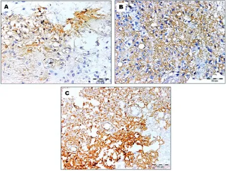
Fig. 1. Immunohistochemical staining for CD44v6 in gliomas: A – CD44v6 expression limited to a small percentage of the tissue of gliomas classified as grade II according to the WHO; B – strong membrane CD44v6 expression in glio-blastoma C – many tumor cells display heterogenous CD44v6 immunostaining (avidin-biotin (ABC) staining ×400).
Table 1. CD44v6 and E-cadherin expression in gliomas
Parameters Immunopositivity
CD44v6 expression E-cadherin expression
Gliomas n positive cases (%) p-value positive cases (%) p-value
grade I 23 8(34.7%)* 6(26.0%)
grade II 30 21(70.0%)* 0.001 13(43.3%) > 0.05
grade IV 39 31(79.4%)** 9(23.0%)
n – number of cases.
Discussion
Despite growing evidence showing the roles of the family of cell adhesion molecules (CAM) in the progressive growth of gliomas, their clinical signif-icance is still unknown. Infiltrative nature of gli-omas is a complex process, and the cell adhesion molecules may play a crucial role in the early stage of brain tumor progression [4, 20, 21]. Holland et al. [3] found that grade II gliomas contain indi-vidual cells with high infiltration capacity over long
distances. The authors [2, 3] suggest that despite complete low-grade tumor resection, these tumors contribute to recurrence but the mechanism of this process is poorly documented. To confirm our hy-pothesis that CD44v6 and E-cadherin are poten-tial biomarkers of aggressive behavior of gliomas, their protein expression and relation between each other, including the degree of glioma malignancy, were evaluated. Our results showed that CD44v6 expression was observed in 71.6% of gliomas and increased with the malignancy grade of gliomas. These findings are consistent with earlier pub-lished data demonstrating that the CD44v6 ex-pression was associated with progression of brain tumors [8]. Our own and other results indicate that CD44v6 might determine aggressive behavior of glioma cells [8, 13]. Heterogeneous pattern of CD44v6 immunostaining in gliomas observed in this paper was also reported by other authors in the case of endometrial carcinoma and osteosarco-ma [22, 23]. In this paper,the differences in the in-tensity and extent of immunostaining for CD44v6 were clearly visible in gliomas presenting different grade of malignancy. In gliomas classified as grade I and II tumors, the immunoreactivity for CD44v6 was observed in tumor core and was limited to 20–30% of brain tumor tissue. By comparison, glioblastomas revealed CD44v6 overexpression in
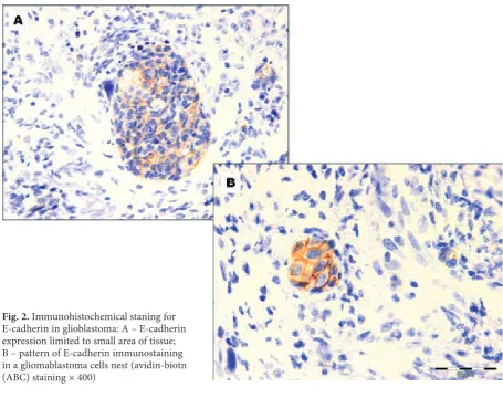
Fig. 2. Immunohistochemicalstaning for E-cadherin in glioblastoma: A – E-cadherin expression limited to small area of tissue; B – pattern of E-cadherin immunostaining in a gliomablastoma cells nest (avidin-biotn (ABC) staining × 400)
a high percentage of tumor tissue (50–80% posi-tive tissue) and CD44v6 expression was higher in invaded cells than in the tumor core. Taking in-to account the role of CD44v6 molecule, our re-sults suggest that tumor cells located at the low-grade tumor margin might possess increased cell motility, which means the spread of cells from the tumor mass could be facilitated [8, 23]. We may also speculate that in this subgroup of low-grade gliomas, the interaction between tumor cells and the environment is important for the initial stage of invasive tumor growth. On the other hand, in glioblastomas, the alteration in adhesion junctions might be important in the formation of tumor structures [8]. This observation is in agreement with the data presented by Kim et al. [6] and Jiji-wa et al. [8], who pointed out the role of CD44v6 in glioma invasion. Based on earlier published da-ta, it is worth underlining that strong CD44v6 ex-pression observed in individual cells of grade II gliomas might induce cell proliferation and migra-tion [20, 23, 24]. On the other hand, this data raises a possibility that CD44v6-positive cells in gliomas with low grade of malignancy might be involved in malignant progression from low-grade astro-cytomas to glioblastomas [20]. We could consid-er such mechanism in our subgroup of CD44v6- -positive gliomas with low malignancy grade. This suggestion might be supported by recent results showing that a knockdown of CD44v6 molecule resulted in suppressed growth of human brain tu-mor stem-like cells in an animal model [8]. Inter-estingly, we found that, irrespective of tumor grade malignancy, in the case of extended CD44v6 ex-pression, individual glioma cells located at the marginal tumor tissue showed strong CD44v6 ex-pression compared to the remaining brain tumor tissue. Jijiwa et al. [8] found that brain tumor stem cells showed a high level of CD44v6 expression. The findings from this paper suggest that cells with strong CD44v6 immunopositivity in glioma spec-imens might reflect cancer stem cell phenotype. However, this observation should be confirmed by identification of cancer stem cell markers [25].
Down-regulation of E-cadherin is considered as one of the main molecular alterations involved in the invasive and aggressive tumor mainte-nance [14, 21, 26]. In gliomas, the role of down- or up-regulation of E-cadherin expression is still con-troversial [4, 16, 26]. Consistent with previous stud-ies which found that E-cadherin expression is a rare event in gliomas [4, 16], in the current study the E-cadherin expression was limited to a small num-ber of analyzed cases and a low percentage of pos-itive brain tumor tissue. The relationship between the decreasing expression of E-cadherin and the increasing grade of malignancy observed by other
migration [21, 26]. This data indirectly indicates that cooperation between CD44v6 and E-cadherin exists, and thereby may promote tumor cells dis-semination [21]. In conclusion, our results clearly showed that the level of E-cadherin might reflect different biological features of gliomas, whereas CD44v6 is associated with tumor cell malignancy.
The simultaneous presence of CD44v6 and E-cad-herin in a set of low-grade gliomas indicates that both these molecules might strengthen cell migra-tion, which may be a hallmark of glioma cell dis-semination. Moreover, further studies are needed to clarify the network of cell-cell adhesive mole-cules in gliomas.
References
[1] Nager M, Bhardwaj D, Canti C, Medina L, Nogues P, Herreros J: β-catenin signalling in glioblastoma multiforme and glioma-initiating cells. Chemother Res Pract 2012, ID 192362, doi:1155/2012/192362.
[2] Wang M, Wang T, Liu Sh, Yoshid D, Teramoto A: The expression of matrix metalloproteinase-2 and -9 in human gliomas of different pathological grades. Brain Tumor Pathol 2003, 20, 65–72.
[3] Holland EC: Glioblastoma multiforme; the terminator. PNAS 2000, 97, 6242–6244.
[4] Lewis-Tuffin LJ, Rodriguez F, Giannini C, Scheithauer B, Necela BM, Sarkaria JN, Anastasiadis PZ: Misreg-ulated E-cadherin expression associated with an aggressive brain tumor phenotype. PLoS ONE 2010, 5, e13665, doi:10.1371/journal.pone.0013665
[5] Ulrich TA, Pardo J, Kumar EM: Mechanical rigidity of the extracellular matrix regulates the structure, motility, and proliferation of glioma cells. Cancer Res 2009, 69, 4167–4174.
[6] Kim Ch-S, Jung Sh, Jung T-Y, Jang W-Y, Sun H-S, Ryu H-H: Characterization of invading glioma cells using molecular analysis of leading-edge tissue. J Korean Neurosurg Soc 2011, 50, 157–165.
[7] Xu Y, Stamenkovic I, Yu Q: CD44 attenuates activation of the hippo signaling pathway and is a prime therapeutic target for glioblastoma. Cancer Res 2010, 70, 2455–2564.
[8] Jijiwa M, Demir H, Gupta S, Leung C, Joshi K, Orozco N, Huang T, Yildiz VO, Shibahara I, Jesus JA, Yong WH, Mischel PS, Fernandez S, Kornblum HI, Nakano I: CD44v6 regulates growth of brain tumor stem cells partially through the AKT-mediated pathway. PLoS ONE 2011, 6, e 24217,doi:10.1371/journal.pone.0024217.
[9] Jung T, Gross W, Zoller M: CD44v6 coordinates tumor matrix-triggered motility and apoptosis resistance. J Biol Chem 2011, 18, 15862–15874.
[10] Hovinga KE, Shimizu F, Wang R, Panagiotakos G, Heijden M: Inhibition of notch signaling in glioblastoma targets cancer stem cell via an endothelial cell intermediate. Stem Cells 2010, 28, 1019–1029.
[11] Brown RL, Reinke I.M, Damerow MS, Perez D, Chodosh LA, Yang I, Cheng C: CD44 splice isoform switching in human and mouse epithelium is essential for epithelial-mesenchymal transition and breast cancer progression. J Clin Inv 2011, 3, 1064–1074.
[12] Lee JL, Wang MJ, Sudhir PR, Chen GD, Chi CW, Chen IY: Osteopontin promotes integrin activation through outside-in and inside-out mechanisms: OPN-CD44V interaction enhances survival in gastrointestinal cancer cells. Cancer Res 2007, 67, 2089–2097.
[13] Dimov I, Tasić-Dimov D, Conić I, Stefanovic V: Glioblastoma multiforme stem cells. Scientific World Journal 2011, 11, 930–958.
[14] Wells A, Yates C, Shepard ChR: E-cadherin as an indicator of mesenchymal to epithelial reverting transitions during the metastatic seeding of disseminated carcinomas. Clin Exp Metastasis 2008, 25, 621–628.
[15] Howng SL, Wu CH, Cheng TS, Sy WD, Lin PC, Wang C, Hong YR: Differential expression of Wnt genes, beta-catenin and E-cadherin in human brain tumors. Cancer Lett 2002, 183, 95–101.
[16] Motta FJN, Valera ET, Lucio-Eterovic AKB, Queiroz RGP, Neder L, Scrideli CA, Machado HR, Carlotti-Junior CG, Marie SKN, Tone LG: Differential expression of E-cadherin gene in human neuroepithelial tumors. Genet Mol Res 2008, 7, 295–304.
[17] Xia M, Xu M, Wang J, Xu Y, Chen X. Ma Y, Su L: Identification of the role of Smad interacting protein 1 (SIP1) in glioma. J Neurooncol 2010, 97, 225–232.
[18] Kaur H, Phillips-Mason PJ, Burden-Gulley SM, Kerstetter-Forgle AE, Basilion JP, Sloan AE, Brady-Kalnay SM:
Cadherin 11, a marker of the mesenchymal phenotype, regulates glioblastoma cell migration and survival in vivo. Mol Cancer Res 2012, 3, 293–304.
[19] Louis DN, Ohgaki H, Wiestler OD, Cavenee WK: WHO classification of tumours of the central nervous system. Lyon, France, International Agency for Research on Cancer (IARC), 2007, 4rd Ed., 14–33.
[20] Nakada M, Kita D, Watanabe T, Hayashi Y, Teng L, Pyko JV, Hamada JI: Aberrant signaling pathways in glioma. Cancers 2011, 3, 3242–3278.
[21] David JM, Rajasekaran AK: Dishonorable Discharge: The oncogenic roles of cleaved E-cadherin fragments. Cancer Res 2012, 72, doi:10.1158/008-5472.CAN-11-3498.
[22] Gun BD, Bahadir B, Bektas S, Barut F, Yurdakan G, Kandemir NO, Ozdamar SO: Clinicopathological signifi-cance of fascin and CD44v6 expression in endometrioid carcinoma. Diag Pathol 2012, 7, 80, http://www.diagnos-ticpathology.org/content/7/1/80.
[23] Deng Z, Niu G, Cai L, Wei R, Zhao X: The prognostic significance of CD44v6, CDH11, and β-catenin expression in patients with osteosarcoma. BioMed Res Int 2013, http://dx.doi.org/10.1155/2013/496193.
[25] Keysar SB, Jimeno A: More than markers: biological significance of cancer stem cell-defining molecules. Mol Cancer Ther 2010, 9, 2450–2457.
[26] Rodriguez FJ, Lewis-Tuffin LJ, Anastasiadis PZ: E-cadherin’s dark side: possible role in tumor progression. Bioch Bioph Acta 2012, 1826, 23–31.
[27] Kim YD, Joo JK, Park YK, Ryu SY, Kim, HS, Noh BK, Lee KH: E-cadherin expression in early gastric carcinoma and correlation with lymph node metastasis. J Surg Oncol 2007, 96, 429–435.
[28] Thiery JP, Seleeman JP: Complex networks orchestrate epithelial-mesenchymal transitions. Nat Mol Cell Biol 2006, 7, 131–142.
Address for correspondence:
Julia K. Bar
Department of Pathomorphology and Oncological Cytology Wroclaw Medical University
Borowska 213 50-556 Wroclaw Poland
Tel.: +48 71 734 39 55
E-mail: [email protected]
Conflict of interest: None declared