Visual stability: perception of stable objects across saccadic eye movements
Thesis by:
Alessio Fracasso
Supervisor:
Professor David Melcher
Center for Mind/Brain Sciences
University of Trento, Italy
02/11/2011
I would like to thank my family, that supported me during these years, my supervisor for mentoring
and giving helpful suggestions and all the PhD students for having great time here in Trento.
Table of Contents
Chapter 1...4
Introduction...4
Eye movements and Visual Stability...5
Saccadic Updating...5
Predictive remapping...10
Relation between saccadic updating and predictive remapping...13
Trans-Saccadic Perception...15
The case of Apparent Motion displays...18
Transformational Apparent Motion...18
Line Motion Illusion...21
Conclusion...23
Chapter 2:...25
Continuous perception of motion and shape across saccadic eye movements...25
Abstract...25
Introduction...26
Experiment 1: Percept of a motion event occurring across the saccade...30
Methods...32
Results ...38
Discussion...41
Experiment 2: Comparing retinotopic versus spatiotopic motion...42
Methods...43
Results...44
Methods...46
Results...50
Experiment 4: Measuring a second-order TAM after-effect...51
Methods...52
Results...55
General Discussion...56
Chapter 3:...61
The role of spatiotemporal distortions in the peri-saccadic unmasking of targets presented in rapid serial presentation...61
Abstract...61
Introduction...62
Experiment 1: Unmasking the Target on RSVP...65
Material and Methods...65
Results...69
Discussion...71
Experiment 2: Spatial displacement of the mask...72
Methods...72
Results...74
Experiment 3: Future versus Backward Remapping...78
Methods...83
Results...84
Discussion...87
General Discussion...87
Chapter 4...94
Remapping of the line motion illusion across eye movements...94
Abstract...94
Introduction...95
Materials and Methods...97
Experiment 1a...99
Experiment 1b...100
Experiment 2...101
Experiment 3...102
Results...103
Discussion...108
General Summary...111
References...115
Chapter 1:
Introduction
The ability of moving freely in the environment gives us the great advantage to directly interact
with it, improving our discriminative abilities. For example, if we were to inspect an object without
the chance to actively moving around it, then we could only rely on the information that we can
extract from a single point of view with respect of the object. We would have restricted access to the
object properties and we would then establish our decisions within those limits. Moving actively
allow us to overcome these limitations and gain access to a more complete set of informations
regarding the object. This would help us decide what to do next, whether or not to interact with an
external object and, in case, providing hints on how to interact. To this extent moving and exploring
the environment augment our discrimination abilities. Moreover, active movements help us to form a
complete sense of space (Trinity-Crapse & Sommer, 2008).
However the remarkable ability to actively move and interact with the environment becomes
adaptive only if the agent is able to distinguish whether a sensorial stimulation is the result of an
external change in the environment (“exafference”, Holtz and Mittelstaedt, 1865) or the effect of its
own movement (“reafference”). Sensory receptors respond irrespective of the source of stimulation,
and this could lead to potentially disadvantageous situations in which active movements of the agent
are confused with changes in the external environment and vice-versa. The nervous system face this
problem with a very general mechanism, keeping track of the movement commands and informing
the sensory system of the incoming movement. This signal is usually referred to as efference copy or
corollary discharge and consists on a copy of the movement command towards relevant sensory
areas for that particular planned movement (Trinity-Crapse & Sommer, 2008).
In this way the system can resolve the exafference / reafference ambiguity taking into account the
input changes, expected only on the basis of the expected movement. Being able to rapidly and
efficiently resolve the ambiguous nature of the sensory input is crucial in order to move efficiently in
the environment. It would be completely useless to be able to run if we could not easily reach the
conclusion that the noisy input to the retina is not due to the world moving around us, but instead the
consequence of our own movement.
In this review I will describe relevant literature about how the visual system takes into account
upcoming movement signals in order to maintain a stable representation of the external world. The
review will be based mainly on saccadic updating, remapping and trans-saccadic memory. Moreover
I will review recent findings on saccadic updating and trans-saccadic perception using apparent
motion displays across eye movements.
Eye movements and Visual Stability
Saccadic Updating
Making eye movements allow us to extract information from the visual field bringing the most
sensitive part of the retina, the fovea, into relevant portions of the field. In normal conditions we
mostly rely on visual inspection to decide how to move in the environment so it is evident that,
especially in the case of eye movements, the nervous system had to deal efficiently with the
exafference / reafference ambiguity to guarantee a fast and efficient processing of the constantly
changing visual input.
Most strikingly, our subjective experience is that we are usually not aware to the changes in the
retinal input due to eye movements, nonetheless eyes are frequently in motion. This subjective
feeling is usually referred to as visual stability.
This subjective insight seems to suggest that we do not rely directly on a retinotopically
organized input, but instead of an eye-position independent representation on the world (Mathot and
Theeuwes, 2011). This would suggest that our conscious visual experience does not depend solely on
our retinotopic input (Harrison & Tong et al, 2009), moreover there exist experimental evidence
which suggest that also the corollary discharge signal that accompany each eye movement does not
have a retinal source based on proprioceptive signals. In a series of elegant experiments (Mays,
Sparks, & Porter, 1987; Sparks & Mays, 1983) it has been shown that when the eyes were passively
set in motion by the experimenter, stimulating the motor neurons right before a saccade,
compensation towards a previously shown saccadic target did not occur, the conclusion was that
proprioception related to the eye-position could not have conveyed sufficient information to set the
corrective saccade needed to reach the target (Wurtz, 2008). A subjective test of the relative
independence of retinal signals to visual stability maintenance has been provided by Descartes, who
pointed out that the world seems to move when the retina was passively displaced tapping on the side
of the eye (Descartes, 1644 in Medendorp 2011).
Recently experimental evidence has been provided suggesting a more central nature of the
corollary discarge generator through a pathway that runs from the superior culliculus (SC) through
the thalamus till the frontal eye fields (FEF), (Sommer & Wurtz, 2006). Inactivation of this pathway
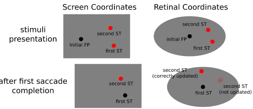
lead to impaired performance on the classical double-step saccade task. In this task two saccade
targets are briefly presented, after targets removal, the participants are instructed to perform two
successive eye-movements to where the targets used to be. Crucially, the nervous system can rely on
the retinotopic trace of the first target to perform the initial movement, but in order to correctly
perform also the second saccade, target position needs to be updated according to the first
eye-movement vector (see figure 1.1). An impairment on the second saccade landing position points
following inactivation of the aforementioned pathway has been taken as evidence for a reliable
source of the corollary discharge accompanying eye-movements, that allowed the system to take into
account motion vectors in the resolving the exafference / reafference ambiguity.
Even though this was the first time that a clear signature of the corollary discharge supporting the
updating process was shown, it has to be noted that the deficit on the updating task was limited to a
19%. This observation seems to suggest not only the existence of alternative pathways for the
updating process, as could be suggested by in a cognitive degeneracy approach (Friston & Price,
7
2003), since the same function can be performed by a variety of different pathways in the brain. The
clear advantage would be to have a system extremely resilient to damage, which could recover
quickly and efficiently. Moreover we cannot exclude that other processes might be involved in
maintaining visual stability and updating across saccades, possibly with different sources of the
upcoming movement signal other than SC (Berbam & Wurtz 2008 and Prevosto et al 2009).
For example there exists experimental evidence for a direct involvement of parietal areas in
saccadic updating in humans. Morris and colleagues (Morris et al, 2007) reported that stimulation of
intra parietal sulcus (IPSp), distorts eye trajectories towards the contralateral hemifield on the second
saccade of the classical double-step saccade task, while leaving unaltered end point and trajectory of
the first one. This results suggest that IPSp is crucial for the ongoing updating mechanism of eye
movements in humans. Evidence for the existence of a saccadic updating mechanism in humans has
been reported also using fMRI (Medendorp et al, 2003; Merriam et al, 2003).
This is not the only existing proposed explanation tor the updating of sensory input with
eye-movement information, another relevant proposal comes from theoretical neuroscience. In a
remarkable attempt to model sensorimotor transformation through implementation of basis functions,
it has been shown that an updating behavior to compensate for eye movements emerges as a natural
property on the hidden layer of a a three-levels neural network. The aim of the network was to
integrate eye position and eye-centered position (retinal location of a stimuli) on a single head
centered representation (Deneve et al 2001). Notably, the authors took advantage of the neurally
plausibile approach of population coding (Georgopulos et al, 1982), moreover this proposal did not
assume the existence of any peculiar signal associated wit the eye movement, but only considers
fixed eye position on the various fixations, as successive snapshots.
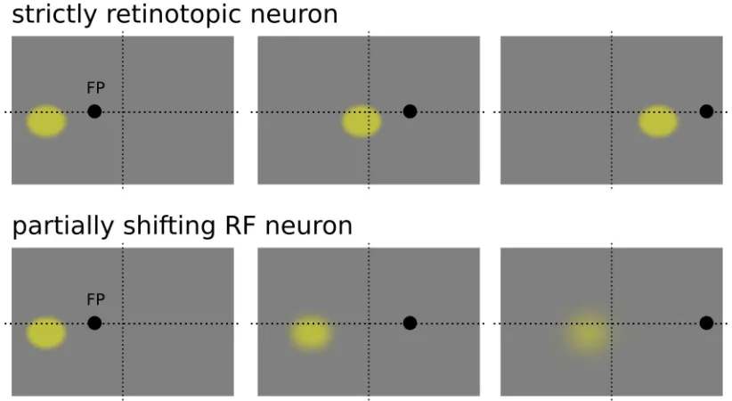
Nodes in the hidden units of the network showed a clear partially shifting behavior, meaning that
receptive fields were not purely eye-centered since the preferential position in eye-centered
coordinates changed consistently with eye position. Interestingly, this gain modulated shift was in the
opposite direction of the eye displacement, as one would expect for a saccade compensatory
mechanism. This kind of shifting behavior is consistent with what has been observed experimentally
(Duhamel, 1997 Nature, the analysis was performed on a set of neurons on the ventral intraparietal
area of the monkey, see figure 2.1). It's interesting to note that this view does not assume that the
system does not update for reafferent sensorial information in order to compensate for shifts due to
the eye movement, instead it states that the mechanism that takes into account sensory reafference is
built-in in the architecture of the system itself, embedded as an emerging property.
Figure 2.1: strictly retinotopic receptive field (upper panels) and partially shifting receptive field (downward panels) behaviour, in the former the receptive field changes its position according to the eye position, keeping its response unaltererd for a different position in space, in the latter eye position modulates the gain of the neuron response, with the neuron being sensitive to the same position in space
Predictive remapping
One additional mechanism that has been show to intervene whenever a monkey performs an eye
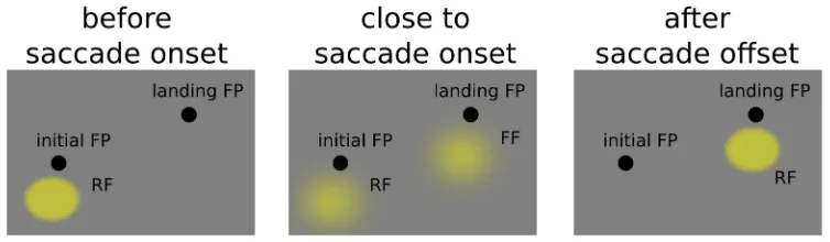
movement is predictive remapping. Neurons in FEF and Lateral Intraparietal Cortex (LIP) have
shifting receptive fields that predicts the response to the visual stimulus that will fall on the receptive
field after the completion of the saccade (Duhamel 1992). The peculiar behavior of the response of
these neurons is essentially different from those described above since their response is predictive of
what will fall on the receptive field after the eye movement, it anticipates the movement itself. Not
only a stimulus briefly shown right before the onset of a saccade elicit activity on the receptive field
that will be stimulated only after the saccade but, the closer we get to the eye movement onset, the
larger the response of the “future” receptive field (FF) will be with respect to the “current” receptive
(RF) field (Kusunoky & Goldberg 2003, see figure 3.1). This peculiar process has been shown to
have a dynamic nature that evolves during time along the perisaccadic interval, gradually decreasing
the response on the current RF and increasing in the FF.
Figure 3.1: predictive behaviour of an LIP neuron, during the perisaccadic interval the neuron becomes sensitive to a spatial location that will be brought into the receptive field only after saccade completion. The neuron becomes gradually less sensitive to the current location and shifts its sensitivity towards the future location in space.
This neurophysiological evidence is tightly coupled with the well known behavioural
phenomenon of perisaccadic mislocalization. When a stimuli is briefly presented around the
perisaccadic interval participants reports its location as mislocalized along the direction of the eye
movement. Such perisaccadic mislocalization has been studied and replicated various times (Honda
1989; Matin et al. 1970; Burr et al, 1997; De Pisapia et al, 2010). One possible explanation is that, in
the highly spatially inaccurate moments around the onset of the eye movement, the brain extracts the
location of stimulation as the average between the response of the current and the future receptive
field associated with the stimuli, the first one being aligned with current fixation, whereas the second
being shifted along the direction of the eye movement, averaging the two lead to gross
mislocalizations of target stimuli along the direction of the eye movement (Kusunoky & Goldberg
2003).
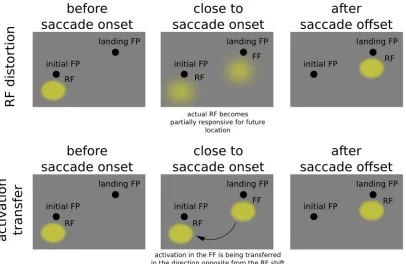
It is important to note that neurophysiological data suggests that the receptive field associated
with a particular location in space-time is actually distorted by the upcoming eye-movement,
meaning that a particular neuron, right around the onset of the eye movement, becomes sensitive to
stimuli outside the region of space that would elicit a response at stable fixation. Specifically this
region is displaced along the direction of the intended eye movement.
Figure 4.1: upper panels, neuron showing a predictive remapping behaviour, during the perisccadic interval it becomes sensitive for location that will be brought into the receptive filed only after saccade completion. Lower panels, alternative account based on an transfer of activation between the future towards the current receptive field
This form of remapping is not compatible with a simple mechanism that takes into account
eye-movement vectors and just compensate saccade-induced shifts. In fact receptive fields distortions are
in the same direction of the saccadic movement, not in the opposite.
Another proposal is that predictive remapping reflects an activation transfer right before the
initiation of the saccade starting from the future receptive field towards the current one (Cavanagh et
al 2010; Hunt & Cavanagh, 2011; Mathot & Theuvees, 2011, see figure 4.1), also this second
proposal can be ruled out considering the nature of the perisaccadic distortion. This idea is intriguing
because the transfer of activation would follow a direction opposite to the saccadic vector, then
compatible with a compensation mechanism for the upcoming eye movement, reconciling the
predictive remapping evidence with the saccadic updating data. But the supposed transfer of
activation that should take place from the future receptive field towards the current receptive field
cannot take place in practice, since current and the future RF are in fact the same receptive field. The
difference in activation found along the perisaccadic interval for different spatial locations
(Kusunoky & Goldberg, 2003) is the result of a different sensitivity of the same receptive field for
two different locations, the current and the future (that are the location covered by the receptive field
before before the eye movement and after eye movement completion, respectively). What changes
during the perisaccadic interval is RF orientation in space-time of the visual receptive field, (Hall &
Colby, 2011, Burr & Morrone, 2011), not an activation transfer among the same receptive field, how
this dynamic change in space-time orientation might help to maintain visual stabilty is not clear yet,
but various attempts has been made arguing that this distortion could lead to a form of local and
transient spatiotopy across single fixations, an area in which extends temporally and spatially around
the onset of the eye-movement that let information on the previous fixation to be integrated with
upcoming information on the next planned fixation (Burr & Morrone, 2011).
Relation between saccadic updating and predictive remapping
Attempts has been made to investigate the relation between the two aforementioned mechanisms
closely related to visual stability. As stated above it has been shown that SC inactivation leads to
impaired performance on the dual-step saccade task, a task that requires efficient spatial updating
across saccades to be performed correctly. Superior culliculus inactivation is thought to disrupt the
neural pathway mediating the corollary discharge signal that informs the nervous system about the
upcoming eye-movement. (Wurtz & Sommer, 2006). Moreover, it has been shown that visual
neurons in the parietal cortex shift their activity in space right before the onset of the eye movement.
Specifically the sensitivity of these neurons shift to a position that will be occupied by the receptive
field only after the saccade, with this location being referred as future receptive field (Duhamel et al,
1992).
An open question is whether corollary discharge signal mediating spatial updating influences
directly the shifting behaviour of the receptive fields shown in visual, parietal and frontal cortex
(Hall & Colby, 2011) for perisaccadic presented stimuli. This empirical question has been
investigated by Sommer & Wurtz (2006) by selectively impairing the middle dorsal (MD) thalamus
(a crucial node in the corollary discharge pathway) and measuring the behavior of the FEF shifting
receptive fields before and after inactivation. Results showed that the magnitude of the shifting
receptive field on the tested neurons (as measured by future field activity) was severely reduced after
inactivation. Notably, MD inactivation did not changed significantly activity on the current receptive
field of the FEF area tested, moreover it did not modify monkeys ability to perform the actual
eye-movement. The main conclusion of this study is that there is a direct link between the SC corollary
discharge generator and the shifting behavior shown for retinotopic FEF neurons.
One question however remains open. It has been shown with neurophysiology and neuroimaging
studies that neurons in visual, parietal (LIP) and frontal cortices shown a peculiar predictive
behaviour during the perisaccadic interval, being responsive to locations that will be brought into
view only after eye movement completion. On the other hand, experimental data (VIP neurons,
Duhamel et al, 1997) and computational approaches to sensorimotor transformation showed how
multisensory neurons (neurons integrating information between different sensory inputs, for example
coding eye position and stimuli in retinal coordinated, eye-preferred positions) presented a gain field
modulated partially shifting behavior. Crucially this dual “shifting” behaviour from a purely retinal
based coordinate system (eye-centered reference frame) is in the opposite direction: along the same
vector of the eye-movement on the perisaccadic remapping case, on the opposite direction for the
partially shifting, gain modulated RF (see figures 2.1 and 3.1). How this evidence can be reconciled
under the same theoretical framework is still a matter of debate. It could be that the two mechanism
jointly act to update sensorimotor information separately, or they could be related to the corollary
discharge signal generating in the superior colliculus (Wurtz & Sommer, 2006). As perisaccadic
remapping signal seems to be directly related to this updating signal, also the gain modulated RF
could be modulated by the upcoming signal, even though this second system is embedded in the
architecture of the system. One way to tackle this issue would be to study the temporal dynamics of
gain modulated RF in active vision, and test the eventual influence of CD on this dynamic.
Trans-Saccadic Perception
Other than egocentric cues providing information about saccade metrics and the expected
reafference signal deriving from those movements (as the corollary discharge), the brain can also
take advantage of allocentric cues, that is to derive object location by its relative position with
respect of other objects in the world, independently of the observer's gaze.
A possible mechanism to maintain space constancy would be by matching images from
successive fixations ignoring the attributes of the saccade vector (Deubel et al, 1996). However, the
larger the saccade amplitude, the less the retinal overlap between subsequent images will be, so a
mechanism based only this principle would be useless in the case of large retinal displacements,
paradoxically the case where a process allowing space constancy would be more of use.
An alternative strategy would be the use egocentric cues (as those discussed in the previous
paragraphs), combining the informations regarding the upcoming eye-movement with the positions
of the relevant objects in the visual field, in a convenient frame of reference (that could be assumed
to be the head, Pouget et al, 2008; Sommer and Wurtz, 2008). For example behavioural studies
seems to suggest that stimuli location can be correctly updated across saccadic eye movements just
on the basis of egocentric cues, as the characteristics of the upcoming saccades, in an experimental
settings that aimed to remove as much as possible allocentric indexes (Prime et al, 2006).
Using the paradigm of saccadic suppression of intrasaccadic displacement (SSID, Bridgeman et
al, 1975; Deubel et al, 1996) it has been shown that the nervous system uses a combination of both
egocentric and allocentric cues to maintain a stable representation of the world (Nimeier et al, 2003).
The basic SSID paradigm is the following. A target is displayed at a certain spatial location well
before a saccade occurs. As soon as the eyes started to move and a saccade is detected the target is
shifted towards another spatial location, usually in the same or opposite direction of the
eye-movement, not in the orthogonally to saccadic vector. The task of the observer could be to report
whether the change in location has been perceived or to report the perceived direction of
displacement, if any, or to guess otherwise. The principal finding is that the displacement threshold is
much higher in the saccadic condition than in a fixation condition. Up to one-third the size of the
saccade (Bridgeman et al. 1975).
The influence of allocentric cues on this kind of task has been established (Deubel, 2004) as well
as the active involvement of egocentric cues (Nimeier et al, 2003) showing how displacements
thresholds scales with saccadic amplitude and post-saccadic eye position scatter ratios (post-saccadic
eye position standard deviation. which is assumed to reflect the uncertainty of eye position towards
saccadic target following the eye movement).
The proposed model of trans-saccadic integration shows that the brain uses eye-position signals
in interpreting post-saccadic retinal information (egocentric cues), only when the retinal information
provide a reliable measure for interpreting the new post-saccadic information inflow. The different
weights associated with allocentric and egocentric cues in combination is a function of the reliability
of these different sources of information, giving the overall percept of trans-saccadic visual stability.
This observation reflects the quantitative counterpart of a crucial aspect of spatial constancy, that is
the assumption of visual stability (Mathot and Theewues 2011). Our visual system exploits the fact
that that world remains stable at least for the small duration of the eye-movement.
Even though this model is extremely intriguing theoretically and can predicted new empirical
findings that found subsequent experimental confirmation its building blocks are not cognitive
plausible. Its implementation takes the form of a Bayesian integration of information that changes
the outcome of the predicted response as a function of the reliability of the input signals. Similar
remarkable attempts has been made to build model to describe multisensory integration (Ernst and
Banks, 2002; Alais and Burr, 2006) and positional information during the perisaccadic interval
(Binda et al, 2008).
These models behave remarkably well in predicting participant behavior but it's not clear how the
neural system could perform the computations to obtain these response. However, there has been
attempts to implement a Bayesian explanatory framework in a biologically plausible network, taking
advantage of a hierarchical scheme (Friston, 2002).
The main aim of the network was to inferring the causes of sensory input and learning the
relationship between input and cause (sensory input) using the same unifying principle, that is to
minimize the error between the observed inputs and the expected input based on a generative model
aimed to infer which sensory stimulation caused that particular input. This idea is similar to what
Irwine Rock stated about perception as an inferential process (Rock, 1997), but expressed using a
mathematical formalism. The model has been successfully applied to model and predict
electroencephalography phenomena (as the mismatch negativity wave and P300) as well as
psychophysical results as priming. These attempts show how a powerful Bayesian explanatory
framework can be implemented in a biologically plausible fashion, which further support that
trans-saccadic perception may rely on a accurate weighting between allocentric and egocentric cues
available in different conditions.
The case of Apparent Motion displays
Transformational Apparent Motion
Recent behavioral evidence seems to support and extend previous data on trans-saccadic
perception suggesting that not only allocentric or egocentric cues are taken into account in order to
keep vision stable across eye movements. Also features of relevant stimuli are taken into
consideration and are likely to be updated across eye-movements (Fracasso et al, 2010).
Apparent motion phenomenon provide a series of interesting tests for trans-saccadic percetion.
One interesting perceptual property of apparent motion is that our visual system fills in the entire
motion path, rather than seeing two discrete events (Kolers, 1972; Morgan, 1976). A dot flashed in
two different locations, given the right timing parameters, is seen to move through the entire
trajectory between point A and point B. This property of apparent motion makes it the perfect test of
the hypothesis that perception bridges the saccade. In fact, Rock and Ebenholtz (1962) had already
reported a version of trans-saccadic apparent motion many years ago. In their experiment observers
were asked to synchronize left/right eye movements with two alternating flashing lights visible
through two vertical slits. In this way the illuminated vertical lines would be presented at the fovea
after each eye movement. Various kinds of apparent motion exists, but one type of apparent motion is
of particularly interest for trans-saccadic perception, namely transformational apparent motion
(TAM, Tse & Caplovitz, 2006).
This particular type of apparent motion has been studied recently (Tse, 1998), it occurs when
two spatially overlapping shapes are presented discretely in time, this results on a percept of a single
object that transform smoothly and illusorily from the first shape into the second as if the sequence
were animated, as shown in figure 5.1.
Crucially, figural parsing plays an essential role in determining the perceived direction of TAM
(Tse et al., 1998; Tse and Logothetis, 2002). Figural parsing involves a comparison of contour and
surface relationships among successive scenes. Based of contour and surface relationships between
successive images in the sequence, the visual system appears to infer which figures at time 2 are
derived from which figures at time 1. If a given figure has a different shape at different times, a
continuous deformation between those shapes is constructed and perceived. It's likely that the new
figure is inferred to be a change in the shape of an already existing figure, as if the two images were
merged into a single spatio-temporal object. The processing of shape information must accompany
the motion processing that subserves the percept of TAM. The perceived motion depends on how
figures at time 1 have been matched to figures at time 2. The mechanism involved into TAM
perception is two-fold: the first step is to identify candidates at both instants, and the second is to
match them. Usually these steps are referred to parsing and matching steps.
These steps are of particular interest also in trans-saccadic perception since it is still a matter of
debate whether object features are maintained across eye movements. The allocentric/egocentric sets
of indexes that might help keeping a stable updating are focused on the maintenance of object
location irrespective of large retinal shifts due to eye-movements. However it is not clear if also
19
feature characteristics are in some ways stored and updated as object locations seems to be
( Cavanagh et al, 2010; Melcher, 2010).
Interestingly, when participants were asked to perform and eye movement between the two
images of the TAM sequence, they report perceiving a clear and vivid motion between subsequent
snapshots (see figure 6.1), that did not differ significantly from a condition in which the stimuli in
the sequence were aligned both in spatial and retinal coordinates (subjects were required to maintain
stable fixation while presented with the sequence).
When retinal coordinates of stimulation were matched across the saccade motion reports were
consistently lower than when stimulation was matched in spatial coordinates, see figure 6.1 for
details.
20
These results suggests that the parsing / matching mechanisms involved in TAM perception
continues across the eye movements even though the stimuli are not aligned in retinal coordinates.
Moreover object location does not seem to be the only characteristics maintained across the
eye-movement, also shape features seems to be updated to help keeping vision stable.
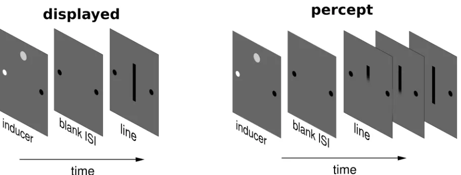
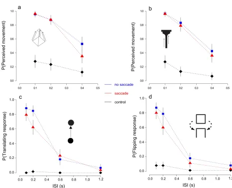
Line Motion Illusion
The line motion illusion (LMI) provides an interesting test for spatial updating across saccades
and the role egocentric indexes on maintaining visual stability. This illusion occurs when a static line,
shown shortly after an inducer (flashed stimulus) appears to radiate away from the location where the
inducer had been presented (figure 7.1).
Available data suggests that this illusion occurs independently of attention (Fuller and Carrasco,
2006). Together with the classical method of subjective reports of motion perception this particular
apparent motion illusion can be measured using the motion cancellation method, giving an objective
measure of its strength, less prone to subjective bias than simple subjective reports. To investigate the
21
spatiotopic LMI, strength of the illusion was measured, as a function of the inducer’s contrast, for
trials in which the eye was stationary or in which a horizontal saccade was made during the ISI
between the inducer and the presentation of the line. We varied the timing of the stimuli with respect
to the saccade to test whether the LMI effect was influenced by saccade timing, as would be
predicted by an active remapping explanation for spatiotopic motion perception.
Either with subjective reports and the motion cancellation method results highlighted a cost in
performing a saccade between the subjective snapshot of the apparent motion display. Moreover,
subjective reports of motion were linked to the metric of the performed saccade, namely faster
saccades lead to larger proportion of motion reports than slower eye movements. This latter effect
was related to the distracter effect of the flash on the requested eye-movement, a form of a well
known phenomena, the remote distractor effect (RDE, Bompnas and Sumner, 2009). When the flash
was presented outside the RDE time window around the requested saccade signal, subjective reports
of LMI across saccades increased consistently.
In trans-saccadic memory literature usually no cost of a single saccade is observed, with virtually
indistinguishable capacity measures at fixation and when asked to perform an eye movement while
presented with the subsequent frames (Prime et al, 2006).
It is important to note that the task adopted to measure visual memory across saccades usually
employed a comparison of the subsequent snapshots presented across the eye movement (though see
Bays and Husain, 2008).
The case of line motion illusion in particular and apparent motion in general provide a different
experimental setup in which the frames need not only to be compared but actually combined in order to obtain the motion percept. In our opinion the lack of any saccade cost in visual memory task
as opposed to the drop in LMI strenght across eye movements described above can be accounted by
this crucial difference in the underlying task, posing a difference between trans-saccadic memory and
trans-saccadic perception.
Other recent studies of trans-saccadic perception using apparent motion displays aimed to study
how performing an eye-movement lead to an illusory perception of the apparent motion display
(Szinte and Cavanagh, 2011), given the updating error associated with each eye movement. This case
is interesting since the perception of motion was so compelling that authors did not measured the
strength of the illusion across saccades but focused directly on the estimation of the updating error.
This provides another case of trans-saccadic perception across eye movements, irrespective large
stimuli shift in retinal coordinates.
Conclusion
A number of phenomena contribute to reach visual stability across eye movement. The corollary
discharge signal that accompany each eye-movement appear inform the sensory areas of the
incoming reafference input signals due by the agent movement and not by actual changes in the
external environment, this signal seems to be directly involved in the updating of information across
eye movement, as tested with the well known double-step saccade task (Wurtz and Sommer, 2006).
The phenomenon of shifting receptive fields seems to provide a mechanism that anticipate the
outcome of the incoming eye movement, with retinotopic neurons being sensitive to information
presented outside the spatial range of their receptive field during the perisaccadic interval. Most
importantly these two neruphysiological phenomena seems to be strinctly related as recent evidence
seems to suggest (Wurtz and Sommer, 2006), with corollary discharge signal directly modulating the
amount of neurons showing shifting receptive field behavior across eye-movements in the frontal eye
fields.
Not only internal neural signals representing the metrics of the upcoming eye movement are
taken into account to solve the matching problem across saccades. Allocentric cues also plays a role
since a reasonable assumption of the system is that the world does not change during the short time
of a saccade. Moreover it has been shown that egocentric and allocentric cues are optimally
combined, giving the overall percept of trans-saccadic visual stability (Nemeier et al, 2003).
Recent behavioral evidence seems to show how information between subsequent fixations can be
combined to give a single percept, as in the case of transformational apparent motion and line motion
illusion, even if retinal coordinates of stimulation are shifted in the direction opposite to the eye
movement. With these kind of paradigm also the cost of a single saccade can be shown. The nature
of this task seems to suggest that experimental setups where an active integration between fixations
is needed are well suited to study the behavioural indexes underlying mechanisms of visual stability.
Chapter 2:
Continuous perception of motion and shape across saccadic eye
movements
Published as:
Continuous perception of motion and shape across saccadic eye movements
Fracasso A, Caramazza A, Melcher D
Journal of Vision, (2010)
.
Abstract
Although our naïve experience of visual perception is that it is smooth and coherent, the actual
input from the retina involves brief and discrete fixations separated by saccadic eye movements.
This raises the question of whether our impression of stable and continuous vision is merely an
illusion. To test this, we examined whether motion perception can “bridge” a saccade in a
two-frame apparent motion display in which the two two-frames were separated by a saccade. We found
that transformational apparent motion, in which an object is seen to change shape and even move in
three dimensions during the motion trajectory, continues across saccades. Moreover, participants
preferred an interpretation of motion in spatial, rather than retinal, coordinates. The strength of the
to give rise to a motion-from-shape after-effect, even when the motion was defined by a
second-order shape cue (“phantom transformational apparent motion”). These findings suggest that motion
and shape information are integrated across saccades into a single, coherent percept of a moving
object.
Keywords: saccades, visual stability, transformational apparent motion, motion after effect
Introduction
The fact that we typically make several saccadic eye movements every second means that the
position of objects on the retina is constantly changing. Thus, one of the fundamental questions of
vision science is how we keep track of the location of objects across saccades (for review, see: Bays
& Husain, 2007; Melcher & Colby, 2008; Wurtz, 2008). But a perhaps more basic question is how
our naïve perception of a smooth and continuous visual flow is built out of a series of relatively
brief visual snapshots that are separated by abrupt jumps, like in a poorly filmed home movie. This
problem is made even more clear by the fact that the new input to the eyes in each fixation must
travel through the visual system before it reaches awareness, necessitating around 120 – 200 ms
(Genetti, Khateb, Heinzer, Michel, & Pegna, 2009; Liu, Agam, Madsen, & Kreiman, 2009; Thorpe,
Fize, & Marlot, 1996), and that visual input is partially suppressed while a saccade is performed
(Burr, Morrone, & Ross, 1994). The issue of achieving stable perception based on discrete and
discontinuous input is particularly troublesome in the case of visual motion. While the brain is
extremely efficient in integrating motion cues over time and space over a period of seconds (Burr &
Santoro, 2001; Neri, Morrone, & Burr, 1998), motion detectors are typically assumed to operate in
position the same object is integrated across saccades (Melcher & Morrone, 2003), then this
impressive ability to integrate motion over time would be essentially useless.
There are essentially three main ideas about how visual stability is maintained (for review, see
Melcher & Colby, 2008). The first is that our impression of smooth perception is essentially an
illusion (Dennett, 1992). Failures to detect changes in the position of an object across a saccade
(Bridgeman, Hendry, & Stark, 1975), for example, argue against detailed information being
maintained across saccades. In the case of motion perception, this theory would predict that motion
processing begins anew with each fixation, since any matching of object location across the saccade
would be based solely on memory (Irwin, 1991).
A second idea is that our impression of visual stability comes from cross-saccadic priming, in
which our post-saccadic perception is influenced by what was previously seen. A clear example
comes from studies of reading, in which information about the word to the right of fixation (the
“parafoveal preview”) primes us to quickly read the word after the saccade (Rayner, 1998, 2009).
Similar results, in which post-saccadic perception is influenced by what was seen before the
saccade, have been reported for color perception (Wittenberg, Bremmer, & Wachtler, 2008), time
perception (Burr, Tozzi, & Morrone, 2007), motion perception (Melcher & Morrone, 2003), object
recognition (Van Eccelpoel, Germeys, De Graef, & Verfaillie, 2008) and face perception (Melcher,
2005; van Boxtel, Alais, & van Ee, 2008). Such cross-saccadic priming might contribute to the
subjective impression that the world is stable, since the post-saccadic stimulus would be processed
quickly and efficiently (Khayat, Spekreijse, & Roelfsema, 2004a, 2004b). However, this theory still
maintains the idea that perception is essentially discrete and tied to individual fixations.
The third, and most radical, proposal is that conscious perception fuses information from before
and after the saccade into a single, coherent percept. This idea agrees with the common, naïve
could simply be wrong. In fact, early attempts to demonstrate the “fusion” of dot patterns across a
saccade were without success (Bridgeman & Mayer, 1983; Irwin, Yantis, & Jonides, 1983).
Likewise, the finding that changing the case of all letters in a word (McConkie & Zola, 1979) had
little effect on reading behavior suggests that abstract codes are used in integrating information
across saccades in reading.
More recently, however, the idea of trans-saccadic perception has been revived based on two
types of evidence. The first is the discovery of dynamic receptive fields (RFs) which change their
sensitivity around the time of eye movements (Duhamel, Colby, & Goldberg, 1992; Melcher &
Colby, 2008; Wurtz, 2008). This “re-mapping” involves both prediction (before the saccade, the
neuron responds to a stimulus in its future RF) and a type of memory trace updating mechanism in
which a neuron continues to respond, after the saccade, to the stimulus in its old RF. One important
implication of these findings is that the neural activity bridges the saccade, rather than showing
discrete and discontinuous firing patterns. The second type of evidence comes from changes in
visual perception, such as peri-saccadic mislocalization, which have been reported around the time
of saccades (Matin & Pearce, 1965; Ross, Morrone, Goldberg, & Burr, 2001). These findings
suggest that the brain anticipates the saccade and uses this information to update spatial information
and match it across saccades. However, most experiments have looked at localization of briefly
flashed stimuli in laboratory settings. It is less clear how everyday perception, in which objects
rarely appear and disappear during saccades, would be influenced by dynamic receptive fields.
We directly tested the predictions of this third, trans-saccadic perception hypothesis by studying
apparent motion. One interesting perceptual property of apparent motion is that our visual system
“fills in” the entire motion path, rather than seeing two discrete events (Kolers, 1972; Morgan,
1976). A dot flashed in two different locations, given the right timing parameters, is seen to move
it the perfect test of the hypothesis that perception bridges the saccade. In fact, Rock and Ebenholtz
(1962) had already reported a version of trans-saccadic apparent motion many years ago. In their
experiment observers were asked to synchronize left/right eye movements with two alternating
flashing lights visible through two vertical slits. In this way the illuminated vertical lines would be
presented at the fovea after each eye movement. The displacement of the vertical line directly
followed the size and direction of the saccade. Although the retinal position of the flash was
constant, participants reported seeing motion in external (in their terms, “phenomenal”) space.
In line with this observation it has been shown that participants are able to detect changes in the
position of a moving object across a saccade (Gysen, De Graef, & Verfaillie, 2002). In contrast to
the Rock and Ebenholtz studies, recently replicated by Szinte and Cavanagh (2009), studies of
change detection for moving objects measured the ability to notice changes in the expected position
of the stimulus rather than to perceive smooth trans-saccadic motion. Thus, we adapted the Rock
and Ebenholtz technique to study the perception of a coherent motion sequence across saccades.
In a new set of experiments, we built upon the Rock and Ebenholtz finding in four ways. First,
we added motion orthogonal to the direction of the saccade in order to disentangle motion caused
by the saccade from motion of the stimulus. This also resulted in the two stimuli being shown in
different visual hemi-fields (and thus to different cerebral hemispheres), providing a greater
challenge for mechanisms of trans-saccadic perception. Second, we varied the temporal delay
between the two flashes in order to provide a more fine-tuned measure of motion perception. Third,
and most importantly, we used “transformational apparent motion” (TAM), in which two differently
shaped stimuli are perceived, when shown in an apparent motion display, as smoothly changing
shape over time (Tse, Cavanagh & Nakayama, 1998; Tse, 2006; Tse & Caplovitz, 2006; Tse &
estimate of the reliability of TAM perception judgments and used this stimulus to measure a
transformational apparent motion after-effect.
The overall aim of these four experiments was to test whether motion can carry shape
information—in addition to spatial location—across the saccade. If perception essentially begins
anew with each fixation, then there should be little or no impression of smooth motion across
saccades in the TAM condition. The ability to integrate the two stimuli into continuous shape-based
motion, however, would provide strong evidence that pre- and post-saccadic information are
combined into a single, trans-saccadic perceptual event.
Experiment 1: Percept of a motion event occurring across the
saccade
The aim of the first experiment was to measure the smooth and continuous perception of motion
across a saccadic eye movement. Following the example of Tse and Logothetis (2002), we
presented two stimuli, which differed in location or shape, separated by a blank delay of varying
duration (Figure 1.2). We expected that the perception of smooth apparent motion would decrease
for longer blank delays (ISI) between the first and second stimuli. In addition, we investigated the
influence of saccades on transformational apparent motion. While Tse and Logothetis (2002) had
matched both the retinal and spatial location of the two stimuli, we investigated whether spatially
matching the external location of stimuli across the saccade, despite a change in retinal coordinates,
was sufficient to support the perception of object-based motion.
Varying the blank delay between the two stimulus frames was also important in order to allow
sufficient time to make a saccade during this blank period between the first and second frames. We
100 ms of the presentation of the first frame was spent preparing for the saccade (trials in which
saccade onset was less than 100 ms were excluded from analysis). Then there was a blank delay,
with no stimulus, during which participants moved their eyes to the new fixation position. As a
result of the saccade, the two different stimuli were always shown in opposite visual hemifields.
The minimum blank delay duration was chosen to be 100 ms for three main reasons. First, this
allowed enough time for subjects to make a saccade on the majority of trials, even for the shortest
blank delay duration (saccade onset less than 200 ms). Second, this brief ISI gave a strong
impression of motion without a saccade. Finally, a blank delay of at least 100 ms was necessary to
avoid the suppression of trans-saccadic displacement of the stimulus (Deubel, Bridgeman, &
Schneider, 2004).
Methods
Observers
Six observers participated in the experiment (two authors and four participants naïve to the aims
of the experiment). Informed consent was obtained for all participants and all subjects reported
normal or corrected-to-normal vision.
Stimuli and Apparatus
Stimuli were presented on a PC using Matlab software and presented on a gamma corrected
Iiyama CRT 1900 monitor running at 85 hz (resolution: 1280 x 1024, short persistence phosphors).
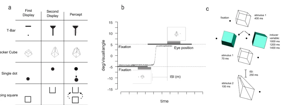
Figure 1.2a shows the four different types of stimuli used during the experiment; each configuration
consisted of 2 frames in which the shape or vertical position of the stimulus was changed. Both
shapes were modified from studies by Tse and colleagues (Tse, Cavanagh & Nakayama, 1998; Tse,
2006; Tse & Caplovitz, 2006; Tse & Logothetis, 2002). The T-bar stimulus subtended 8.8 x 4.5
deg/visual angle whereas the short and expanded Necker cube subtended 2.9 x 3.3 and 2.9 x 4.4
deg/visual angle, respectively. For these stimuli, background was set to white (CIE coordinates: x =
0.28; y = 0.30; luminance: 80 cd/m2), and the stimuli were black (CIE: x = 0.35; y = 0.37;
luminance: 0.25 cd/m2). The fixation point consisted of a red (CIE: x = 0.56; y = 0.33; luminance:
70 cd/m2) circle that subtended 0.4 degrees of visual angle.
The other two types of stimuli used in this experiment were a single black disk (1.4 deg/visual
angle diameter) that could shift its vertical position from the first to the second frame by 4
motion sequence whose first frame consisted of a black wireframe rectangle (2 deg/visual angle
side) and the second frame consisted of the same square, shifted vertically by 2 deg/visual angle,
with one side missing (Figure 1.2a). The typical percept for this sequence is that of a square that
flips in the third dimension until it reach its final position depicted on frame 2, as described by Rock
(1997). For this second group of stimuli (disk and flipping square), the screen background was set
to gray (CIE coordinates: x = 0.28; y = 0.31; luminance: 8.8 cd/m2).
For the control experiment, the stimuli were presented on a gray background. The Necker cube
subtended 3.1 and 6 deg/visual angle for the contracted and the expanded version, respectively.
Color used were CIE: x = 0.20; y = 0.32; luminance: 58.6 cd/m2, CIE: x = 0.27; y = 0.31;
luminance: 4.8 cd/m2 and CIE: x = 0.26; y = 0.34; luminance: 9.8 cd/m2. The fixation point
consisted of a black circle that subtended 0.4 degrees of visual angle presented either to the left or
the right of the stimuli (4.5 deg/visual angle).
Observers sat in a dimply lit room and viewed the screen binocularly at a distance of 57 cm,
with their heads stabilized by a chin rest. Right eye position was monitored using an EyeLink 1000
Desktop Mount (SR Research, Ontario, Canada) sampling at 500Hz. Eye position was recorded for
each trial and saved for offline analysis.
Procedure
Prior to the experiment, subjects were presented with practice trials showing examples of both
smooth apparent motion (ISI between the two stimuli of ~105 ms) and non-motion (ISI of ~1200
seconds). During training, participants were shown the “no saccade” condition, which involved
maintaining gaze on the fixation point throughout the trial. In the main experiment, all three
separate, interleaved blocks in randomized order. The four different stimulus types were divided
into two blocks, with two different types of stimuli presented in random order within each block.
One type of blocks contained the T-bar and Necker cube stimuli, while the other blocks contained
the moving disk and flipping cube apparent motion stimuli.
Each trial began with the participant looking at the fixation point and then pressing a button
when ready. For the first group of stimuli (the T-Bar and the Necker cube), the first stimulus was
presented for 400 ms, followed by a variable blank that could vary between 105 ms (9 frames), 210
ms (18 frames) or 400 ms (34 frames) and then the second stimulus display for a further 400 ms. In
the “no saccade” condition, participants viewed a fixation point to the left or right of the display and
stimuli were shown at the center of the screen.
In the “saccade” condition (Figure 1.2b), the fixation point was displaced during the trial to the
other side of the screen, requiring a 10 degree saccade. This saccade cue occurred ~300 ms into the
trial (25 frames), when the first stimulus was still visible, leaving participants ~210ms (18 frames)
to ~500ms (43 frames) to move their eyes, depending on the condition.
Trials in which participants executed the saccade before the first stimulus disappeared (saccadic
latency < 105 ms), as well as trials in which saccades were too short (amplitude < 8.5 degrees) or
too slow (such that the saccade was not started by the onset of the second motion frame were
excluded from further analysis (mean saccade latency was 184 ms, mean saccade amplitude was
9.8˚ visual angle). Please note that in the saccade condition, the spatial coordinates of the two
stimuli on the screen were matched but they were always shown in different retinal coordinates. In
total, 27% of trials on the saccade condition were excluded based on eye movements for these
blocks of trials (30% for ISI = 105ms, 23% for ISI = 210ms and 28% for ISI = 400ms, see Figure
In the other blocks (with the moving disk and flipping square apparent motion stimuli), the
procedure was identical except that four different ISI’s were tested (105, 210, 600 and 1200 ms) and
subjects were requested to perform a 14 deg/visual angle saccade. Again, trials in which participants
executed the saccade before the first stimulus disappeared (saccadic latency < 105 ms), as well as
trials in which saccades were too short (amplitude < 12.5 degrees) or too slow, such that participants
failed to make a saccade by the time of the second motion frame, were excluded from further
analysis (mean saccade latency was 182ms, mean saccade amplitude was 13.4 deg/visual angle). In
total, 22% of trials on the saccade condition were excluded based on eye movements (50% for ISI =
105ms, 26% for ISI = 210ms, 19% for ISI = 600ms and 12% for ISI = 1200ms, see Figure 2.2).
The third condition provided a control for the saccade condition by replicating the same retinal
stimulation but without the intervening saccade. The participant maintained fixation at the center of
the screen. The first stimulus was shown on one side of the screen (distance of 5 degrees) while the
second stimulus was shown on the opposite side of the screen. This condition tested the possible
role of large receptive fields (radius of 10 degrees or more) which might integrate the two motion
stimuli despite different retinal locations. Such large-range spatial pooling might have been a
potential confound for the saccade condition, thus necessitating this control condition. In each of the
three conditions, observers were presented with a total of 20 trials for each ISI level for the T-bar
and the Necker cube (120 trials overall) and 10 trials for each ISI level for the disk and the flipping
square stimuli (80 stimuli overall)
The motion stimuli were oriented along the vertical axis of the screen in the “saccade” and “no
saccade” conditions (Figure 1.2). After each trial, subjects were requested to report whether they
perceived motion in the vertical axis or, instead, perceived the appearance/disappearance of the
stimuli. Subjects gave their responses by pressing a button on the keypad. For the T-Bar and the
Necker cube trials, subjects could report one of the following three choices: no motion, moving up
or moving down. These choices were included because it has been shown that the T-bar stimuli has
a preferential direction of perceived motion that goes from the single point to the bar itself (Tse &
Caplovitz, 2006), whereas the direction of the perceived motion for the Necker cube depends on the
subjective interpretation of the first display orientation (Tse & Logothetis, 2002).
For the blocks of trials showing the black disk and flipping square apparent motion sequences,
subjects could report one of the following three choices: no motion, translating or flipping, Thus, it
was possible to explicitly test whether subjects perceived a shape change between the two frames in
In the case of the Necker cube, the direction of perceived shape change (forward or backward in
depth) depended on the initial interpretation of the ambiguous shape. To directly test the perceived
shape transformation, we ran an additional control condition (Figure 1.2c) which biased the
perceived orientation of the Necker cube by presenting a solid cube (Tse & Logothetis, 2002).
Participants were presented with 2 different conditions (“saccade” and “no saccade”), in interleaved
blocks (48 trials for each block, 96 trials for each condition). Each trial began with the participant
looking at the fixation point and then pressing a button when ready. The first stimulus of the Necker
cube TAM (stimulus 1) sequence was presented for 400 ms (see Figure 1.2), followed by a solid
biasing shape (inducer) remaining on the screen for a variable time (1000 ms, 1200 ms or 1400 ms),
in order to prevent participants from anticipating the saccade. In the “saccade” condition stimulus 1
was shown again for 6 flips (~70 ms) together with a 9 deg/visual angle displacement of the fixation
point on the other side of the screen. Participants were asked to perform a saccade towards the
displaced fixation point. After an ISI of 250 ms, stimulus 2 of the transformational apparent motion
sequence was presented for ~100 ms (9 frames). Thus participants had approximately 320 ms to
shift their gaze to the new fixation position. Subjects were requested to report the direction of the
perceived motion (“forward” or “backward” in depth, see Figure 1.2) or to report that no motion
was perceived. The “no saccade” condition was identical except that the fixation point remained on
the initial position. To ensure that subjects correctly performed the eye movement and maintained
fixation as requested we implemented a gaze contingent display that checked eye position online. If
a saccade was not performed correctly (the saccade did not occur during the 250 ms blank ISI
between stimulus 1 and stimulus 2 or did not land inside an area of 3 deg / visual angle around the
target fixation point) or if participants did not maintain fixation as requested in “no saccade” trials,
then the trial was repeated at the end of the block. During offline analysis, an additional 12% of the
Since the procedure for the Necker cube and the T-Bar, the black disk and the flipping square,
and the control condition (with the Necker cube) were all run in separate blocks with different
parameters, data was analyzed separately for each of these three types of trials.
Results
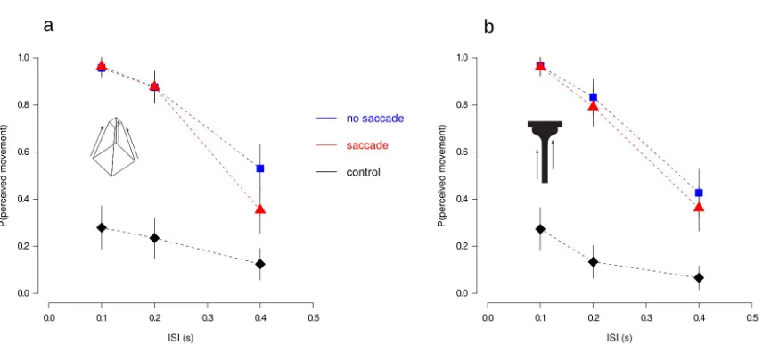
In the “no saccade” condition, the proportion of trials in which subjects reported coherent
motion decreased as a function of the delay (ISI) duration between the two stimuli (Figure 3.2). In
sharp contrast, participants did not report seeing coherent vertical motion in the control condition
(Figure 3.2, diamonds). The main finding was that performance in the saccade condition (Figure
3.2, triangles) was similar to that found in no-saccade trials (Figure 3.2, squares) indicating that
transformational apparent motion occurred across saccadic eye movements, in non-retinal
coordinates.
For the Necker cur and the T-Bar stimuli we performed a repeated measures analysis of variance
(ANOVA) on subject proportion of perceived movement (“up” and “down” responses were pooled
together, given the bistability of the Necker cube). We also report post hoc comparisons reaching
significance after Bonferroni correction. A 3 (ISI) x 3 (viewing condition) x 2 (stimuli) repeated
measures ANOVA showed a main effect of ISI, F(2,10)=18 . 366, p<. 001, η2=. 79 , a main
effect of viewing condition, F(2,10)=18 . 386 p<. 001, η2=. 78 , and a significant ISI x viewing
condition interaction F(4,20)=8 .249, p<. 001, η2=.63 . Stimulus type did not influence the
proportion of perceived movement,F(1,5)=0 . 435, p>. 05, ns . Bonferroni-corrected
comparisons failed to reveal differences between the no-saccade and the saccade condition for ISI’s
Similar results were found with the moving disk and flipping square apparent motion stimuli. To
look specifically at the change in shape in the flipping square trials, only “flipping” responses were
examined (no motion and translation responses were pooled together). Again, there were main
effects of ISI and viewing condition ( F(3,15)=33 . 137, p<.001, η2=. 86 and
F(2,10)=23 . 108, p<.001, η2=. 82 , respectively) and a significant interaction between the two,
F(6,30)=11 . 998, p<.001, η2=. 70 . Also in this case Bonferroni corrected comparisons did not
The results for the control condition with the “biased” Necker cube followed the same trend
(Figure 4.2). The reported direction of motion was effectively biased by the inducer in both the
saccade (t(6)=3. 566, p<. 001 ) and no-saccade (t(6)=4 . 290, p<. 001 ) trials. There was no
difference between and saccade and no-saccade conditions (t(6) < 1, n.s.).
Discussion
The main finding of the first experiment was that TAM and apparent motion perception
continues across saccades. Participants reported a compelling percept of object transformation in
both the no-saccade and the tran-saccadic motion condition. This finding suggests that the smooth
perception of motion in non-retinal coordinates reported with location-defined apparent motion
(Rock & Ebenholtz, 1962; Cavanagh & Szinte, 2009; Szinte and Cavanagh, 2009) occurs also with
a more complex, shape-defined TAM.
Previous studies have reported that the visual system performs poorly in detecting intrasaccadic
displacements of stimuli (Bridgeman et al., 1975). At a first sight our results might seem to be in
conflict with these reports, but there are considerable differences between our paradigm and the
classic saccadic suppression of displacement (SSD) paradigm. First of all, in the SSD paradigm the
saccadic target is displaced while in our method the stimuli were presented in the centre of the
screen, not as the saccade target. Secondly in our case the displacement of the stimulus was
considerably larger, around 4 deg/visual angle, than the usual displacement of ~1 deg/visual angle
adopted in the classical paradigm. Thirdly, and perhaps most importantly, we included a blank delay
between the vertical displacement so that it did not occur surreptitiously during the saccade (and
during saccadic suppression). It has been shown (Deubel et al., 1996) that blanking the target
considerably lowers the threshold for detection of saccadic displacements of target stimuli. Thus, it
is perhaps not surprising that observers in our experiments could easily detect the shift between
successive frames of the apparent motion sequence. What was more striking was the finding that
subjects perceived coherent, vertical motion at the center of the screen even though the two stimuli
were presented in two completely different retinal positions.
Experiment 2: Comparing retinotopic versus spatiotopic motion
In the first experiment, subjects were able to integrate two stimuli in different retinal positions
into a coherent motion perception in spatiotopic coordinates. However, the spatiotopic percept was
by far the simplest interpretation of the display. Thus, it is not clear whether the spatiotopic
apparent motion display could be possible. To test the preference for spatial or retinal coordinates in
trans-saccadic motion, we included two different post-saccadic stimuli, one at the retinal location
and one in the spatially-matched location on the screen.
Methods
Observers
Six observers participated in this experiment, two authors and four naïve observers, two of
whom participated also on the first experiment, All observers had normal or corrected-to-normal
vision. Informed consent was obtained for all participants.
Stimuli and Apparatus
Each stimuli comprised two frames: the first display consisted of a single bar (1 X 5 deg visual
angle) with two possible orientations (90° or 45°), while in the second display two bars were
presented, one in the same spatiotopic position of the screen and the other one in the same
retinotopic coordinates after the saccade (see Figure 5.2a). The orientation of the bars in the second
display depended on the bar orientation in the first display. If the bar in display one was at 90°, the
orientations of the bars in the second display were 45° and 135°, while if the bar in display one was
at 45°, the orientations were 0° and 90°. Background was set to white (CIE coordinates: x = 0.28; y
= 0.30; luminance: 80 cd/m2) stimuli color was set to black (CIE: x = 0.35; y = 0.37; luminance:
0.25 cd/m2). The fixation point was a red (CIE: x = 0.56; y = 0.33; luminance: 70 cd/m2) circle that