Morphological characterization of
Cryptosporidium parvum
life-cycle stages in an
in vitro
model system
H. BOROWSKI1, R. C. A. THOMPSON1*, T. ARMSTRONG1andP. L. CLODE2
1WHO Collaborating Centre for the Molecular Epidemiology of Parasitic Infections,Veterinary and Biomedical Sciences,
Murdoch University,South Street,Murdoch,WA 6150,Australia
2Centre for Microscopy,Characterisation and Analysis,The University of Western Australia,35 Stirling Hwy,Crawley,
WA 6009,Australia
(Received 30 January 2009; revised 25 May, 15 June and 19 June 2009; accepted 22 June 2009; first published online 20 August 2009)
S U M M A R Y
Cryptosporidium parvumis a zoonotic protozoan parasite that mainly affects the ileum of humans and livestock, with the potential to cause severe enteric disease. We describe the complete life cycle ofC. parvumin anin vitrosystem. Infected cultures of the human ileocecal epithelial cell line (HCT-8) were observed over time using electron microscopy. Additional data are presented on the morphology, development and behavioural characteristics of the different life-cycle stages as well as determining their time of occurrence after inoculation. Numerous stages ofC. parvumand their behaviour have been visualized and morphologically characterized for the first time using scanning electron microscopy. Further, parasite-host interactions and the effect ofC. parvumon host cells were also visualized. An improved understanding of the parasite’s biology, proliferation and interactions with host cells will aid in the development of treatments for the disease.
Key words : Cryptosporidium parvum, morphology, host cell interaction, phylogenetic affinity, gregarines, electron microscopy.
I N T R O D U C T I O N
Cryptosporidium is a protozoan enteric parasite of humans and other vertebrates (Fayer et al. 1997). Numerous Cryptosporidium species have been de-scribed (Smithet al. 2005) most of which are specific to their vertebrate host. The speciesC. parvumis of medical and economic relevance as it affects both humans and cattle with its primary site of infection being the gastrointestinal tract. It affects the epi-thelial lining of the ileum, resulting in self-limiting diarrhoea in immunocompetent individuals or in life-threatening diarrhoeal diseases in immunocom-promised individuals.
Belonging to the phylum of apicomplexan para-sites, Cryptosporidium shares common life-cycle features and morphological characteristics with other members of this phylum (Tetleyet al. 1998). Initially,Cryptosporidiumwas categorized as a cocci-dian parasite (Levine, 1988). However, more recent studies show that Cryptosporidium lacks key mor-phological characteristics of coccidians and is insen-sitive to anti-coccidial agents (O’Donoghue, 1995 ; Fayer et al. 1997 ; Carreno et al. 1999). Further
phylogenomic analysis has since revealed that Cryptosporidiumis most closely related to gregarines (Barta and Thompson, 2006).Cryptosporidiumshares many features in common with gregarines, including an extracytoplasmic location and connection to the host cell via a myzocytosis-like feeding mechanism (Barta and Thompson, 2006). The primary differ-ence between these two groups is that Crypto-sporidiuminduces the host cell to overlay it with the host cell apical membrane (Barta and Thompson, 2006 ; Butaevaet al. 2006). The parasite appears on the surface of cells, residing in a parasitophorous vacuole (PV) between the cytoplasmic membrane and the apical membrane (Huanget al. 2004).
Critically, the mechanisms of Cryptosporidium pathogenesis are not fully understood, but both parasite stimuli as well as host immune responses are thought to play critical roles (Barta and Thompson, 2006). The life cycle and the mechanisms of infection by Cryptosporidium have recently been reviewed in detail by Smithet al. (2005) and Borowskiet al. (2008). Importantly, previous studies by Hijjawi et al. (2001, 2004) described the life cycle ofC. par-vum in vitro, using light microscopy while more recent studies by Valigurovaet al. (2008) described the morphology of various life-cycle stages of 2 dif-ferentCryptosporidiumspecies from mice and toads in vivousing electron microscopy.
The aim of this study was to expand on this earlier work and to gain a better understanding of the biology and relationship with host cells of the * Corresponding author : WHO Collaborating Centre
for the Molecular Epidemiology of Parasitic Infections, Veterinary and Biomedical Sciences, Murdoch University, South Street, Murdoch, WA 6150, Australia. Tel :+(08) 9360 2466. Fax :+(08) 9360 6285. E-mail : a.thompson@ murdoch.edu.au
economically and medically important species, C. parvum. In this study the human ileocecal epi-thelial cell line HCT-8 was used as anin vitromodel to monitor the developmental process ofC. parvum and to study the effects upon target cells. Crypto-sporidiumwas observed to proliferate in our culture system for 5 days. Hence, infected cells were moni-tored for this period, with data obtained using scanning (SEM) and transmission (TEM) electron microscopy. From this, a more complete life cycle ofC. parvumhas been visualized and the behaviour and morphological characteristics of numerous life-cycle stages described for the first time with the aid of SEM.
M A T E R I A L S A N D M E T H O D S
Cell culture
The C. parvum cattle isolate used during this study was originally obtained from the Institute of Parasitology, University of Zurich. Oocysts were subsequently passaged through, and purified from, infected ARC/Swiss mice as described by Meloni and Thompson (1996). For routine passaging, HCT-8 cells were cultured in RPMI medium with 2 g Lx1sodium bicarbonate, 0.3g Lx1L-glutamine, 3.574 g Lx1HEPES buffer (15 mmol Lx1) and 10 % fetal calf serum (FCS) at pH 7.4, 37xC with 5 % CO2.
Pre-treatment of oocysts
C. parvum oocysts were bleached with 200ml of household bleach in 10 ml of water for 30 min at room temperature (RT). Sterilized oocysts were inoculated into excystation medium (0.5 % trypsin, pH of 2.5) for 30 min at 37xC. Excysted oocysts were resuspended in maintenance medium consisting of the RPMI medium described above plus 3 g Lx1 sodium bicarbonate, 0.2 g Lx1 bovine salt, 1 g Lx1 glucose, 250mg Lx1 folic acid, 1 mg Lx1 4 amino benzoic acid, 500mg Lx1 calcium pentothenate, 8.75 mg Lx1ascorbic acid and 1 % FCS.
Cell line infection and cell-free culture
Twenty-four h prior to an infection of cells with C. parvum, HCT-8 cells were plated onto thermonox cover slips in 24-well plates. Each cell was then infected with C. parvum pre-treated oocysts (15 000 per cm2) in 1 ml of maintenance medium and maintained at 37xC with 5 % CO2. Infected cultures were sampled and processed for microscopy at 6 h, 7 h, 24 h, 48 h, 72 h, 96 h and 120 h post-inoculation.
Sample preparation for electron microscopy
Cover-slips with adherent cells were fixed in 2.5 % glutaraldehyde in 1rPBS. Additionally, to
investigate extracellular C. parvum stages present within the supernatant of infected cells, medium from cells was aspirated and added to an equal volume of 5 % glutaraldehyde in 2rPBS. Cellular material within this supernatant was subsequently attached to poly-L-lysine coated glass cover-slips for SEM investigation by applying several drops of concentrated supernatant and incubating for 20 min.
All samples were post-fixed in 1 % OsO4in PBS, and then dehydrated in a graded series of ethanols using a Pelco Biowave Microwave Processor. Samples destined for SEM were then critical-point dried, mounted on stubs with carbon tabs, and coated with 3 nm platinum for high-resolution imaging. Samples destined for TEM were infil-trated and embedded in Spurr’s Resin. Thermonox cover-slips were removed under liquid nitrogen, and samples re-embedded. Sections, approximately 100 nm thick, were cut on a diamond knife and mounted on copper grids.
Imaging
SEM images were acquired at 3 kV using the in lens secondary electron detector, on a Zeiss 1555VP field emission SEM. TEM sections were viewed unstained at 120 kV using a JEOL 2100 TEM. Images were digitally acquired with a Gatan SC1000 ORIUS digital camera.
R E S U L T S A N D D I S C U S S I O N
All recognizedC.parvumlife-cycle stages (Table 1) were observed on the surface of epithelial cells or in the supernatant. Small trophozoites (<1mm) were observed as early as 6 h post-inoculation with well-distinguishable meronts I and free merozoites type I being observed after 24 h. This implies that oocyst excystation and sporozoite invasion must have occurred immediately post-inoculation. Consistent with this, previous studies by Forney et al. (1999), observed sporozoites invading host cells as early as 5 min post-inoculation at the point of sporozoite emergence from the oocyst. Subsequent infection increased over the next 2–3 days as a result of ongoing oocyst excystation, and the ability of C. parvum to replicate asexually. After 2 days post-infection, only inoculated oocysts, sporozoites, trophozoites, meronts I and merozoites type I were observed in culture. After 3 days, meronts II as well as mero-zoites type II were seen, while gametocytes were not observed until 4 days post-inoculation.
This timeline of Cryptosporidium development correlates with previous light microscopic data from in vitro cultures of C. parvum (Hijjawi et al. 2001, 2002, 2004), as well as electron microscopy studies of Cryptosporidium sp. ‘ toad ’ and C. muris from experimentally infected toads and mice respectively
(Valigurova et al. 2008). This consistency with an in vivosystem confirms the validity of our in vitro model system ofC. parvuminfection.
In addition to the widely accepted life-cycle stages, we observed stages that are not commonly reported or known to date (Table 1). Such stages include trophozoites that formed within parent stages with-out host cell invasion, and extracellular accumu-lations of trophozoites.
Oocyst excystation and sporozoite host cell invasion Following inoculation, intact C. parvum oocysts were only ever found in the supernatant and these were ovular, 5mmr7mm in size and possessed a smooth surface (Fig. 1A). This correlates with pre-vious findings of Hijjawiet al. (2001). On the surface of these oocysts a cleft was visible and presumably it is along this cleft that the oocyst opens to release sporozoites during the excystation process. Excysted oocysts were observed adhered to cells after 24 h but not before. It is known that oocysts possess surface lectins that are thought to hinder their adherence to host tissuein vivo until the target tissue is reached where they then mediate attachment to host cells (Kuznar and Elimelech, 2006). In the present study, oocysts were directly applied to target cells but did not attach immediately. Perhaps without passage through the gastrointestinal tract, oocysts do not
begin to express the surface lectins needed for host cell adherence until after extended exposure to host cells or simply until a certain progression of time after excystation stimuli are initiated. The outer membrane of excysted oocysts appeared rough (Fig. 1B). This is probably due to membrane per-foration during the excystation processes.
Sporozoites observed in this study measured about 5mmr0.5mm and showed well-defined apical ends (Fig. 1B–D). The hatching sporozoite shown in Fig. 1B as well as the sporozoite on the surface of the host cell in Fig. 1D, both showed an enlarged posterior region and a well-defined apical region, which appeared thin and elongated. Sporozoite apical organelle discharge is known to mediate host cell contact and according to previous findings already occurs during excystation (Snelling et al. 2007). As the sporozoites shown in Fig. 1B and D appear to be making host cell contact, apical or-ganelle discharge of molecules, which are discharged in the presence of host cells, might account for the occurrence of this thin and elongated apical region. In contrast, sporozoites isolated from supernatant did not display this apical elongation and their shape remained largely regular along their entire length (Fig. 1C). Sporozoites were only observed up to 48 h post-inoculation. As sporozoites are known to invade host cells within 5 min after excystation (Forney et al. 1999) it can be assumed that after Table 1. Distinguishing characteristics ofCryptosporidiumlife-cycle stages
Time
(h) Stage Size Morphological Feature Fig.
0 Oocyst 5r7mm Ovular, smooth surface with cleft for
sporozoite release
1A
>24 Excysted oocyst 5r7mm Perforated surface 1B
>3 Sporozoite 5r0.5mm Rough surface, pointed apical region (elongated when in proximity to host cells), rounded posterior region
1B–D
>6 Early trophozoite <1mm Smooth surface formed by the host cell apical membrane, hood like shape
1E
>24 Trophozoite 2.5mm Epicellular, smooth surface, electron dense band, feeder-organelle, PV, cytoplasmic granulation, hood like shape
2A–G
>24 Trophozoite clusters
— Merging of apical membranes
engulfing individual parasites
2D–G
>24 Meronts I 1.5mm Epicellular, smooth surface 5A,B
>24 Merozoites Type I
0.4r1mm Rod like shape, pointed apical region, rough surface
5E–H
>72 Meronts II 3.5mm Epicellular, smooth thick membrane 6A
>72 Merozoites Type II
0.5–1mm Round, rough surface 6A–C
>96 Microgamont 2r2mm Extracellular, densely packed with microgametes
8A–C
>96 Microgamete 0.1mm Spherical, rough surface 8A–C
>96 Macrogamont 4r5mm Extracellular, ovular, rough surface 7A–C
48 Extracellular
trophozoite
2mm Forms within parent stage, rough surface 9A
96 Extracellular
meront
<2mm Round, extracellular
accumulation of trophozoites
48 h all oocysts have excysted and that no new sporozoites are produced. Invasion of host cells by sporozoites is shown in Fig. 1E. Invading sporozoites eventually become completely encapsulated by the host cell apical membrane, which initially appeared as a hood-like structure at this early invasion stage (Fig. 1E). A similar process has also been described in thein vivotoad system by Valigurovaet al. (2008). As part of the infection process, sporozoites trans-form into the trophozoite stage and undergo mer-ogony, which leads to trophozoite growth. This initial trophozoite stage measured<1mm in diam-eter (Fig. 1E) and was observed as early as 6 h post-inoculation, showing that host cell infection
followed by parasite development occurs rapidly after parasite-host tissue contact.
Trophozoites on the host cell surface 48 h post-inoculation
During the invasion process,C. parvumsporozoites always remain epicellular and appear to transform into the trophozoite stage extracytosolic. Thus, stages that result from host cell invasion show a similar cellular location as gregarines, with the only differ-ence being that Cryptosporidium becomes encapsu-lated by a host cell apical membrane (Fig. 2A–G ; Barta and Thompson, 2006). Trophozoites shown in Fig. 1. Oocyst excystation and sporozoite host cell invasion. (A) Intact oocyst from supernatant, 3 h post-inoculation. (B) Oocyst excystationin vitro, 48 h post inoculation. (C) Free sporozoite isolated from supernatant 3 h
post-inoculation. (D) Free sporozoite on host cell, 7 h post-inoculation. Arrows indicate the apical regions of sporozoites. (E) Encapsulated by the host cell apical membrane, invading sporozoites transform into the trophozoite stage epicellularly, at 6 h and 24 h respectively. Scale bars : (A) 4mm ; (B) 2mm ; (C,D,E) 1mm.
Fig. 2A–G may have developed from either spor-ozoites or from merspor-ozoites type I after host cell in-vasion. Trophozoites varied in size depending upon their developmental stage. Early trophozoites ob-served from 6 h post-inoculation onwards, were f1mm in size (Fig. 1E), whereas well-developed trophozoites observed after 24 h post-inoculation, were up to 2.5mm (Fig. 2B). These mature tropho-zoites appeared attached to the surface of cells, but were separated from the host cell via an electron-dense band (Fig. 2C). The formation of a feeder or-ganelle structure is hypothesized for all C. parvum stages and is shown here in Fig. 2A. Previous mor-phological studies by Huanget al. (2004) have already described this extracytosolic location of the parasite and its feeder organelle attachment. Our data show sporozoites becoming engulfed by the host cell apical membrane. This phenomenon was proposed (Perkins et al. 1999 ; Elliott and Clark, 2000 ; Polloket al. 2003 ; Chenet al. 2004a,b, 2005 ; Hashimet al. 2006) and recently confirmed by Valigurovaet al. (2008). It has been suggested that the parasite induces the host cell to encapsulate itself with the host cell apical mem-brane (Borowskiet al. 2008) to escape the hosts de-fence mechanisms.
zoites (Fig. 6A–C). Chenet al. (2000) reported that zoite stages express surface lectins that have adhesive properties. It is possible therefore, that these lectins also facilitate the attachment of infective zoites to each other. Taken together, these data suggest that excysted sporozoites and merozoites can be adherent to each other during host cell attachment and in-vasion of cells (Fig. 6C). This close proximity of invading stages results in the merging of membranes between individuals, forming clusters of rapidly growing trophozoites.
The effect on host cell microvilli
Protozoan parasites possess gliding motility. Gliding motility of sporozoites has been observed in previous studies (Barneset al. 1998 ; Riggset al. 1999 ; Wetzel et al. 2005) leading to the suggestion that gliding along the host cell surface is a prerequisite to host cell invasion.C. parvumgliding trails along host cell surfaces were observed in this study (Fig. 3A). These gliding trails were evident as trails of elon-gated microvilli between an excysted oocyst and newly formed trophozoites ; extending up to 15mm. AsC. parvumis known to utilize microvilli material
to establish itself in its niche (Bonnin et al. 1995), it can be suggested that gliding sporozoites use microvilli material for their gliding motion and thus cause this abnormality seen in gliding trails. As gliding trails were not observed in all cases of oocyst excystation, it appears that not all sporozoites glide along the host membrane surface until they are ready to invade a cell at a particular area. Some sporozoites are thought to invade directly at their origin of ex-cystation (Fig. 2F), exhibiting gliding movements in a small area only, whereas other sporozoites might even travel through the culture medium (Fig. 1C) before establishing host cell contact and invading cells some distance from excystation.
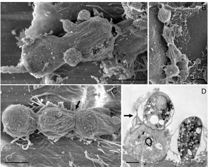
Microvillus abnormality has not only been ob-served in gliding trails, but also around developing trophozoites (Fig. 3B and C). Abnormally abun-dant microvilli were frequently found to surround trophozoites (Fig. 3B) and in some cases were significantly elongated (Fig. 3C). As the expression of microvilli around trophozoite clusters was found to be higher than in non-infected areas, C. parvum might even induce the production of microvilli to satisfy its need for microvillar components. Micro-villi of infected cells were also seen to appear to Fig. 3. The parasite’s effect on host cell microvilli. (A) Gliding trail composed of elongated microvilli between
an excysted oocyst and trophozoites 3 days post-inoculation. (B,C) Abnormal microvilli clusters surround trophozoites 48 h post-inoculation. Scale bars : 2mm.
become incorporated into the membrane engulfing the parasites and to aid in securing the parasite to the host cell surface (Fig. 2B and G). This phenom-enon has not been described before, but microvillar material has been identified in the membrane com-ponents engulfing the parasite using molecular tech-niques (Bonninet al. 1995).
Binary fission and syzygy in theC. parvumlife cycle Additional C. parvum stages were also found that were characteristically different from those described above. Figure 4A and B raise the possibility that C. parvum may undergo binary fission, i.e. the splitting of a parent cell into 2 daughter cells, other than during the course of merogony and shizogony. In Fig. 4A, two microgamonts that seem to be in the process of binary fission, are engulfed by a single membrane that may have initially encapsulated the parent trophozoite on the host cell surface. The finding of 2 parasites that appear to be dividing from 1 parent and seem to be the same life-cycle stage (Fig. 4A), demonstrates that C. parvum possibly undergoes asexual reproduction other than in the course of merogony. As explained below, asC. par-vum progresses through its life cycle, it appears to
become more extracellular. This is evident in stages that appear to have little or no attachment to host cells (Fig. 4A–C). All of these stages are likely to be microgamonts, one of which is clearly visible in Fig. 4A. Microgamonts occur later in the life cycle once sexual reproduction has been initiated. Thus, these stages show a more extracellular location. Our observations raise the question whether C. parvum stages with more epicellular location are still attached to and encapsulated by the host cell apical mem-brane, or have broken contact with host cells, but still retain the surrounding outer host cell membrane.
Our data also indicate that C. parvum might employ syzygy (Fig. 4C). Syzygy is defined as the association of gamonts (pre-gametes) end-to-end or in lateral pairing prior to the formation of gametes that may be employed to ensure genetic diversity and has been described in gregarines (Landers, 2001 ; Lacombe et al. 2002 ; Barta and Thompson, 2006 ; Toso and Omoto, 2007). Thus, it is reasonable that syzygy may occur in closely relatedCryptosporidium species. Connecting discs between adjacent parasites are visible (Fig. 4C and D) and these appear to be the same as the basal discs already described in Cryptosporidium by Valigurova et al. (2008). The C. parvum stages involved in syzygy shown in
C
Fig. 4C and D are probably microgamonts. As sy-zygy was first observed when sexual life-cycle stages occurred in culture, it can be suggested that pre-dominantly gamont stages (here microgamonts) employ syzygy. This correlates with findings on gregarines and further supports the affinity of Cryptosporidiumwith these apicomplexans (Landers, 2001 ; Toso and Omoto, 2007).
Meronts and merozoites
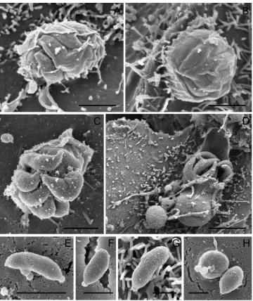
Trophozoites that result from sporozoite host cell invasion develop into meronts I. Meronts I with visible internal merozoites type I, were observed as early as 24 h post-inoculation (Fig. 5A and B). Like trophozoites, meronts I were found to be attached to host cells and engulfed by the host cell
apical membrane (Fig. 5A and B). Developing mer-onts measured approximately 1.5mm in diameter (Fig. 5A and B), whereas mature meronts at the stage of merozoite release were larger, measuring 2.5mm (Fig. 5C and D). From this, it appears that tropho-zoites developing into meronts I undergo merogony and begin to form internal merozoites, before they have reached their full size. Merozoites seemed to be aligned in a parallel orientation within intact meronts I (Fig. 5A and B) which is consistent with observations by Hijjawiet al. (2001). Once excysted, the merozoites type I in a meront numbered 6 or 8, which also correlates with previous findings by Hijjawi et al. (2001). Excysting merozoites type I showed a rod-like shape with a pointed apical region. In contrast to excysting oocysts, the membranes of meronts I appear not to become perforated to Fig. 5. Meronts I and merozoites type I. (A,B) Developing meronts I with internal merozoites type I. (C,D)
Mature meronts I at the stage of merozoite I excystation. (E,F,G) Free merozoites type I showing well-defined apical regions (arrow) at 6 h of culture. (H) Merozoite type I host cell invasion 6 h post-inoculation. Scale bars : (A,B,C,E,F) 1mm ; (D) 2mm ; (G,H) 0.5mm.
facilitate infective zoite release. During merozoite release, the membranes engulfing the parasite still appeared smooth (Fig. 5C and D) and seemed to either open up (Fig. 5 D) or rupture (Fig. 5C). When merozoites type I are released from the meront a residual body is left behind (Fig. 5C). Similar ob-servations have been made on the Cryptosporidium sp. ‘ toad ’ model (Valigurovaet al. 2008).
Free merozoites type I were seen in cell culture from 24 h post-inoculation onwards (Fig. 5E–H). Merozoites, which previously were probably in-correctly referred to as microgametes (Thompson et al. 2005) type I were 0.4mmr1mm, with both a rounded and a pointed end (Fig. 5E–H). The pointed end appeared similar to that of sporozoites, which houses the apical organelles for host cell invasion (Tetley et al. 1998). These free merozoites must be adhered to the host cells in some manner (Fig. 5E–5H), otherwise they would have been removed during sample preparation. Surface lectins which have been detected on infective zoite stages
(Chenet al. 2000) might mediate this attachment to host tissue. Merozoites appear initially to adhere to host cells along their full body length, but become stouter as cellular invasion occurs (Fig. 5H). Thus it can be hypothesized that after initial host cell attachment involving surface lectins, a re-orientation of merozoite organelles occurs, bringing the apical complex into host cell contact to initiate cellular invasion.
Once merozoites type I are present, the parasite employs 2 ways to replicate in its host which both occur concurrently : (i) it progresses via asexual re-production by the formation of meronts I and (ii) it progresses via sexual reproduction by formation of meronts II which then further develop into micro-and macrogamonts (Borowskiet al. 2008).
meronts I (Fig. 6A). Merozoites type II were round and measured between 0.5mm and 1mm in diameter (Fig. 6A–C). Similar to meronts I, the membrane engulfing the merozoites appeared smooth and not perforated for their release (Fig. 6A) suggesting the host cell origin of this membrane.
Similar to the adhesion of sporozoites to each other via surface lectins, merozoites type I and type II can also adhere to each other and co-invade cells (Fig. 6B and C).
Microgamonts and macrogamonts
Merozoites type II that are released in culture, invade cells to transform into either micro- or macrogamonts. Four days post-inoculation micro-and macrogamonts were observed for the first time (Figs 7 and 8). Both gamont stages appeared to have less contact with host cells than trophozoites or meronts. This supports our suggestion that the fur-ther the parasite progresses in its life cycle, the more extracellular it appears to become. Macrogamonts were ovular and measured about 4mmr5mm (Fig. 7A). Feeder organelle attachment of a macro-gamont is visible in Fig. 7A. The surface of this macrogamont appeared rough, suggesting that it has
already broken host cell contact leaving the host cell-derived membrane that once surrounded it behind. Such empty ‘ attachment-zones ’ often show a ring-like structure (Fig. 7C), presumably where feeding organelles were attached. These findings correlate with observations by Valigurova et al. (2008) who described a similar extracellular location of macrogamonts, their detachment from host cells, the granular structure of their feeder organelles, as well as detachment zones in vivo, in their Crypto-sporidiumsp. ‘ toad ’ model.
Microgamonts were rounder and smaller than macrogamonts, measuring about 2mmr2mm (Fig. 8A–C). They contained a large number of microgametes (Fig. 8A and C), which are released to fertilize macrogamonts. This observation is not consistent with findings by Hijjawiet al. (2001), who identified only 16 microgametes in 1 microgamont with light microscopy. This difference is likely to be due to the difficulty resolving such structures with light microscopy. Microgametes measure about 0.1mm in diameter and are spherical. Hijjawi et al. (2001) believed that they were non-flagellated which is confirmed by our SEM study. The microgamonts shown in Fig. 8A seem to be surrounded by pre-sumably host cell-derived membrane. Fig. 8C shows Fig. 7. Macrogamonts and their attachment zones. (A) Macrogamont with feeder organelle (arrow) 4 days
post-inoculation. (B) Developing macrogamont with feeder organelle (arrow). (C)Cryptosporidium parvumattachment zones 5 days post-inoculation. Scale bars : (A,C) 2mm ; (B) 1mm.
a feeder organelle attachment of the same origin as that of the macrogamont in Fig. 7A. Approximately half of all microgamonts seen in culture possessed a stalk-like structure that either appeared as if the stalk had broken (Fig. 8D) or seemed to attach the parasite to the host cell (Fig. 8C). Again, a similar finding has also been reported by Valigurovaet al. (2008). This stalk on microgamonts might result from the parasite being released from the attachment zones described above in a similar manner to macro-gamonts. The microgamonts that possess a stalk (Fig. 8C and D) appeared to have less host cell con-tact compared to those which did not show such a structure (Fig. 8A and B). Additionally, their mem-brane appeared perforated (Fig. 8C) in contrast to microgamonts without this stalk.
Like trophozoites, microgamonts were also ob-served to occur in clusters of 2 or more. The ac-cumulation of microgamonts (Fig. 8D) probably resulted from microgamonts that had released from their attachment zone and adhered to each other, whereas the 2 adjacent microgamonts in Fig. 8B are more likely to be a result of merozoite type II host cell invasion in close proximity.
Development ofC. parvumin host cell culture without invasion of host cells
The present study is consistent with the intra-cellular life cycle ofC. parvumdescribed by Hijjawi et al. (2001). The stages described above all resulted from host cell invasion, yet, complete cell-free de-velopment of C. parvumhas also been documented at the light microscope level (Hijjawi et al. 2004) in vitro. From our own observations, we hypothesize that extracellular life-cycle stages might be part of the C. parvum life cycle, or co-exist resembling rudimentary stages of an ancestral life cycle. Our data show that C. parvum can develop extracellularly in the presence of host cells in anin vitromodel, without invasion. We have observed stages ofC. parvumthat have been described before at the light microscopic level but are not as yet considered as part of the life cycle.
Life-cycle stages of C. parvum that developed extracellularly showed a rough surface like that of previously described free parasite stages (Fig. 5E–H and Fig. 6A–C), and they did not appear to be engulfed by the host cell apical membrane.
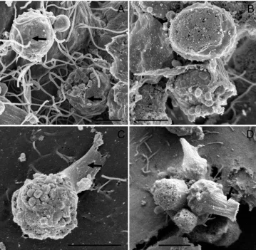
After 48 h inoculation, we observed what we be-lieve to be trophozoite development directly within an oocyst (Fig. 9A). The membrane of the oocyst releasing the trophozoites appeared rough and per-forated, a characteristic already observed during the release of sporozoites (Fig. 1B). Similar to these so-called intracellular trophozoites, trophozoites that formed directly within an oocyst measured ap-proximately 2mm. As these stages were observed as early as 48 h post-inoculation (Fig. 9A) it can be hypothesized that the trophozoites developed within an inoculated oocyst, and were eventually released into culture.
From our observations it can be suggested that each sporozoite and each merozoite is capable of
undergoing merogony and transforming into a trophozoite stage, without invasion of host cells (Fig. 9A). This suggestion is supported by our findings of a proposed extracellular meront 4 days post-inoculation (Fig. 9B). The meront measured approximately 8mm in diameter and was round in shape. The zoite stages observed within the meront, measured up to 2mm and appeared to be densely packed. It is likely that this extracellular meront has formed through the clumping of infective zoite stages in culture, of which each single one has under-gone merogony to form a trophozoite. In support of this, accumulations of merozoites and/or tropho-zoites, as well as the formations of meronts in cell-free culture, have already been described (Hijjawi et al. 2004).
Critically, how extracellular trophozoites and meronts progress in their life cycle without cellular invasion is still not understood. Each extracellular trophozoite might progress in a manner typical of an intracellular trophozoite, or each single tropho-zoite might transform into the next life-cycle stage. The observation that C. parvum also develops ex-tracellularly, despite the presence of host cells, fur-ther supports the proposal that Cryptosporidium has a close affinity with gregarines (Carreno et al. 1999 ; Barta and Thompson, 2006 ; Valigurovaet al. 2007).
C O N C L U S I O N S
For the first time, our study reveals the morphology of each stage in the currently accepted life cycle of C. parvum. The development of life-cycle stages described here extends previous reports that were based on light microscopic findings (Hijjawi et al. 2001, 2002, 2004). Data from our in vitro model system are further supported by similar morpho-logical observations fromin vivostudies onC. muris and Cryptosporidium sp. ‘ toad ’ (Valigurova et al. 2008). Surprisingly, the speciesC. parvumobserved in our study shows more similarities to the species Cryptosporidium sp. ‘ toad ’ than to C. muris (Valigurovaet al. 2008).
Apart from revealing the morphology of accepted life-cycle stages in C. parvum our study describes previously unreported features of C. parvum life-cycle stages, which include their morphological structure and interaction with host cells. Further, extracellular stages of C. parvum are reported and characterized for the first time in an in vitromodel of cultured host cells at an electron microscopic level.
It is not clear whether extracellular stages of C. parvumoccur in vivo, and whether they are part of the parasite’s life cycle or represent rudimentary stages of an ancestral life cycle. Future studies are needed to identify the existence of these stagesin vivo and clarify their nature.
Fig. 9. Development ofCryptosporidium parvumin host cell culture without the invasion of host cells. (A) Trophozoite development within an inoculated oocyst after 2 days. (B) Possible extracellular meront after 4 days of culture. Scale bars : (A) 2mm ; (B) 1mm.
R E F E R E N C E S
Barnes, D. A., Bonnin, A., Huang, J. X., Gousset, L., Wu, J., Gut, J., Doyle, P., Dubremetz, J. F., Ward, H. and Petersen, C.(1998). A novel multi-domain mucin-like glycoprotein ofCryptosporidium parvum mediates invasion.Molecular and Biochemical Parasitology96, 93–110.
Barta, J. R. and Thompson, R. C. A.(2006). What is Cryptosporidium? Reappraising its biology and phylogenetic affinities.Trends in Parasitology22, 463–468.
Bonnin, A., Gut, J., Dubremetz, J. F., Nelson, R. G. and Camerlynck, P.(1995). Monoclonal antibodies identify a subset of dense granules inCryptosporidium parvumzoites and gamonts.The Journal of
Eukaryotic Microbiology42, 395–401.
Borowski, H., Clode, P. L. and Thompson, R. C. A.
(2008). Active invasion and/or encapsulation ?
A reappraisal of host-cell parasitism byCryptosporidium. Trends in Parasitology24, 509–516.
Butaeva, F., Paskerova, G. and Entzeroth, R.(2006). Ditrypanocystis sp. (Apicomplexa, Gregarinia, Selenidiidae) : the mode of survival in the gut of Enchytraeus albidus(Annelida, Oligochaeta,
Enchytraeidae) is close to that of the coccidian genus Cryptosporidium.Tsitologiia48, 695–704.
Carreno, R. A., Martin, D. S. and Barta, J. R.(1999). Cryptosporidiumis more closely related to the gregarines than to coccidia as shown by phylogenetic analysis of apicomplexan parasites inferred using small-subunit ribosomal RNA gene sequences.Parasitology Research
85, 899–904.
Chen, X. M., Huang, B. Q., Splinter, P. L., Orth, J. D., Billadeau, D. D., McNiven, M. A. and
LaRusso, N. F.(2004a). Cdc42 and the actin-related protein/neural Wiskott-Aldrich syndrome protein network mediate cellular invasion byCryptosporidium parvum.Infection and Immunity72, 3011–3021.
Chen, X. M. and LaRusso, N. F.(2000). Mechanisms of attachment and internalization ofCryptosporidium parvumto biliary and intestinal epithelial cells. Gastroenterology118, 368–379.
Chen, X. M., O’Hara, S. P., Huang, B. Q., Splinter, P. L., Nelson, J. B. and LaRusso, N. F.(2005). Localized glucose and water influx facilitates Cryptosporidium parvumcellular invasion by means of modulation of host-cell membrane protrusion. Proceedings of the National Academy of Sciences, USA
102, 6338–6343.
Chen, X. M., Splinter, P. L., Tietz, P. S., Huang, B. Q., Billadeau, D. D. and LaRusso, N. F.(2004b). Phosphatidylinositol 3-kinase and frabin mediate Cryptosporidium parvumcellular invasion via activation of Cdc42.The Journal of Biological Chemistry279, 31671–31678.
Elliott, D. A. and Clark, D. P.(2000).Cryptosporidium parvuminduces host cell actin accumulation at the host-parasite interface.Infection and Immunity68, 2315–2322.
Fayer, R., Speer, C. A. and Dubey, J. P.(1997). The general biology ofCryptosporidium. InCryptosporidium and Cryptosporidiosis(ed. Fayer, R.), pp. 1–41. CRC Press LLC, New York, USA.
Forney, J. R., DeWald, D. B., Yang, S., Speer, C. A. and Healey, M. C.(1999). A role for host
phosphoinositide 3-kinase and cytoskeletal remodeling duringCryptosporidium parvuminfection.Infection and Immunity67, 844–852.
Hashim, A., Mulcahy, G., Bourke, B. and Clyne, M.
(2006). Interaction ofCryptosporidium hominis andCryptosporidium parvumwith primary human and bovine intestinal cells.Infection and Immunity74, 99–107.
Hijjawi, N. S., Meloni, B. P., Morgan, U. M. and Thompson, R. C. A.(2001). Complete development and long-term maintenance ofCryptosporidium parvumhuman and cattle genotypes in cell culture.International Journal for Parasitology31, 1048–1055.
Hijjawi, N. S., Meloni, B. P., Ryan, U. M., Olson, M. E. and Thompson, R. C. A.(2002). Successful in vitro cultivation ofCryptosporidium andersoni: evidence for the existence of novel extracellular stages in the life cycle and implications for the classification of Cryptosporidium.International Journal for Parasitology
32, 1719–1726.
Hijjawi, N. S., Meloni, B. P., Ng’anzo, M., Ryan, U. M., Olson, M. E., Cox, P. T., Monis, P. T. and Thompson, R. C. A.(2004). Complete
development ofCryptosporidium parvumin host cell-free culture.International Journal for Parasitology34, 769–777.
Huang, B. Q., Chen, X. M. and LaRusso, N. F.(2004). Cryptosporidium parvumattachment to and
internalization by human biliary epithelia in vitro : a morphologic study.The Journal of Parasitology90, 212–221.
Kuznar, Z. A. and Elimelech, M.(2006). Cryptosporidiumoocyst surface macromolecules significantly hinder oocyst attachment.Environmental Science and Technology40, 1837–1842.
Lacombe, D., Jakowska, S. and Silva, E.(2002). GregarineCephaloidophora communis mawrodiadi, 1908 in the barnacle Euraphia rhyzophorae, Oliveira, 1940 from Brazil.Memo´rias do Instituto Oswaldo Cruz
97, 1057–1061.
Landers, S. C.(2001).Pterospora floridiensis, a new species of acephaline gregarine (Apicomplexa) from the maldanid polychaete Axiothella mucosa in St. Andrew Bay, Florida.Systematic Parasitology48, 55–59.
Levine, N. D.(1988)The Protozoan Phylum Apicomplexa, Vols 1 and 2. CRC, Boca Raton, FL, USA.
Meloni, B. P. and Thompson, R. C. A.(1996). Simplified methods for obtaining purified
oocysts from mice and for growingCryptosporidium parvumin vitro.The Journal of Parasitology82, 757–762.
O’Donoghue, P. J.(1995). Cryptosporidium and cryptosporidiosis in man and animals.International Journal for Parasitology25, 139–195.
Pollok, R. C., McDonald, V., Kelly, P. and Farthing, M. J.(2003). The role ofCryptosporidium
parvum-derived phospholipase in intestinal epithelial cell invasion.Parasitology Research90, 181–186.
Riggs, M. W., McNeil, M. R., Perryman, L. E., Stone, A. L., Scherman, M. S. and O’Connor, R. M.
(1999).Cryptosporidium parvumsporozoite pellicle antigen recognized by a neutralizing monoclonal antibody is a beta-mannosylated glycolipid.Infection and Immunity67, 1317–1322.
Smith, H. V., Nichols, R. A. and Grimason, A. M.
(2005).Cryptosporidiumexcystation and invasion : getting to the guts of the matter.Trends in Parasitology
21, 133–142.
Snelling, W. J., Lin, Q., Moore, J. E., Millar, B. C., Tosini, F., Pozio, E., Dooley, J. S. and Lowery, C. J.
(2007). Proteomics analysis and protein expression during sporozoite excystation ofCryptosporidium parvum(Coccidia, Apicomplexa).Molecular and Cellular Proteomics : MCP6, 346–355.
Tetley, L., Brown, S. M., McDonald, V. and Coombs, G. H.(1998). Ultrastructural analysis of the sporozoite ofCryptosporidium parvum.Microbiology
144, 3249–3255.
Thompson, R. C. A., Olson, M. E., Zhu, G., Enomoto, S., Abrahamsen, M. S. and Hijjawi, N. S.(2005). Cryptosporidiumand cryptosporidiosis.Advances in Parasitology59, 77–158.
Toso, M. A. and Omoto, C. K.(2007). Ultrastructure of theGregarina niphandrodesnucleus through stages from unassociated trophozoites to gamonts in syzygy and the syzygy junction.The Journal of Parasitology93, 479–484.
Valigurova, A., Hofmannova, L., Koudela, B. and Vavra, J.(2007). An ultrastructural comparison of the attachment sites betweenGregarina steiniand
Cryptosporidium muris.The Journal of Eukaryotic Microbiology54, 495–510.
Valigurova, A., Jirku, M., Koudela, B., Gelnar, M., Modry, D. and Slapeta, J.(2008). Cryptosporidia : epicellular parasites embraced by the host cell membrane.International Journal for Parasitology38, 913–922.
Wetzel, D. M., Schmidt, J., Kuhlenschmidt, M. S., Dubey, J. P. and Sibley, L. D.(2005). Gliding motility leads to active cellular invasion byCryptosporidium parvumsporozoites.Infection and Immunity73, 5379–5387.