BioMedCentral
BMC Physiology
Open Access
Research article
Decrease of PECAM-1-gene-expression induced by
proinflammatory cytokines IFN-
γ
and IFN-
α
is reversed by TGF-
β
in
sinusoidal endothelial cells and hepatic mononuclear phagocytes
Katrin Neubauer, Alexander Lindhorst, Kyrylo Tron, Giuliano Ramadori and
Bernhard Saile*
Address: University of Göttingen, Department of Internal Medicine, Section of Gastroenterology and Endocrinology, Göttingen, Germany
Email: Katrin Neubauer - [email protected]; Alexander Lindhorst - [email protected]; Kyrylo Tron - [email protected]; Giuliano Ramadori - [email protected]; Bernhard Saile* - [email protected]
* Corresponding author
Abstract
Background and aim: The mechanisms of transmigration of inflammatory cells through the sinusoids are still poorly understood. This study aims to identify in vitro conditions (cytokine treatment) which may allow a better understanding of the changes in PECAM (platelet endothelial cell adhesion molecule)-1-gene-expression observed in vivo.
Methods and results: In this study we show by immunohistochemistry, that there is an accumulation of ICAM-1 (intercellular cell adhesion molecule-1) and ED1 positive cells in necrotic areas of livers of CCl4-treated rats, whereas there are few PECAM-1 positive cells observable. After the administration of CCl4, we could detect an early rise of levels of IFN-γ followed by an enhanced TGF-β protein level. As shown by Northern blot analysis and surface protein expression analysed by flow cytometry, IFN-γ-treatment decreased PECAM-1-gene-expression in isolated SECs (sinusoidal endothelial cells) and mononuclear phagocytes (MNPs) in parallel with an increase in ICAM-1-gene-expression in a dose and time dependent manner. In contrast, TGF-β-treatment increased PECAM-1-expression. Additional administration of IFN-γ to CCl4-treated rats and observations in IFN-γ-/- mice confirmed the effect of IFN-γ on PECAM-1 and ICAM-1-expression observed in vitro and increased the number of ED1-expressing cells 12 h after administration of the toxin.
Conclusion: The early decrease of PECAM-1-expression and the parallel increase of ICAM-1-expression following CCl4-treatment is induced by elevated levels of IFN-γ in livers and may facilitate adhesion and transmigration of inflammatory cells. The up-regulation of PECAM-1-expression in SECs and MNPs after TGF-β-treatment suggests the involvement of PECAM-1 during the recovery after liver damage.
Background
Inflammatory cells leave the circulation at sites of local
that are classified as rolling, activation, tight adhesion, transmigration and passage across the basement
mem-Published: 8 May 2008
BMC Physiology 2008, 8:9 doi:10.1186/1472-6793-8-9
Received: 13 November 2007 Accepted: 8 May 2008
This article is available from: http://www.biomedcentral.com/1472-6793/8/9
© 2008 Neubauer et al; licensee BioMed Central Ltd.
tory processes in the liver are thought to differ from those in other organs. It has been suggested that SECs of the nor-mal liver – in contrast to capillary endothelial cells – only express a restricted set of adhesion molecules [7-9].
PECAM-1 is known to be expressed on the surface of endothelial cells, circulating platelets, monocytes, granu-locytes and some subsets of T-lymphocytes. PECAM-1 has been proposed as one of the main players in transend-othelial migration of neutrophils, monocytes, and natural killer cells in both in vivo and in vitro models, since by the use of antibodies directed against PECAM-1, transmigra-tion and inflammatransmigra-tion could be significantly reduced [10-12]. However, the pathomechanisms involving PECAM-1 are still a matter of debate, since in a PECAM-1-knock-out-mouse model, transmigration of inflammatory cells and inflammatory damage were almost unchanged [13].
In our previous studies, we have demonstrated that PECAM-1 is expressed in SECs and MNPs of the rat liver. Furthermore, we have shown that the PECAM-1 expres-sion in SECs and MNPs is decreased after CCl4-treatment in vivo before inflammatory phagocytes accumulate in the pericentral area of rat livers and following IFN-α -treat-ment of cultured SECs and MNPs in vitro. In contrast, ICAM-1-gene expression is upregulated early after CCl4 administration, reaching a maximum after 24 h. Thereaf-ter, ICAM-1 is decreasingly expressed in SECs, reaching the control level of the value at 0 h after 48–72 h. In MNPs ICAM-1 is maximally expressed 6 h after CCl4 administra-tion and thereafter expression declines to control levels 72 h after CCl4 administration. [14,15].
Since we have seen that there is a down-regulation of PECAM-1 in the rat liver after the administration of CCl4, in this study we aimed to identify further cytokines that are responsible for the down-regulation of PECAM-1 on SECs and on transmigrating mononuclear phagocytes (MNPs), but also to identify cytokines that allow the reversal of this effect on both cell types.
A pivotal role in pathomechanisms of liver injury has been assigned to IFN-γ next to IFN-α [16] and it has been demonstrated that IFN-γ can induce IFN-α expression in MNPs [17]. TGF-β is a known pleiotropic cytokine with profibrogenic and anti-inflammatory capacities [18]. In this study we measured the concentration of IFN-γ and TGF-β in the rat liver at different time points after CCl4 -administration paying particular attention to the early time points. Furthermore we tested the modulation of PECAM-1 and ICAM-1-expression in SECs and MNPs fol-lowing IFN-γ-treatment in vitro and in vivo. We also stud-ied the effect of TGF-β on PECAM-1 expression in isolated SECs and MNPs. Whereas IFN-γ reduces PECAM-1 expres-sion in parallel with the increase of ICAM-1 expresexpres-sion,
TGF-β up-regulates both molecules and is also able to counteract the down-regulating effect of IFN-γ on PECAM-1 expression in vitro.
These data strongly suggest an early down-regulation of PECAM-1 in parallel to an ICAM-1 up-regulation as a pos-sible important mechanism for the adhesion and transmi-gration of inflammatory cells. However, further in vitro and in vivo studies are required to confirm this hypothe-sis. The TGF-β induced PECAM-1 up-regulation may be of importance during the recovery phase.
Results
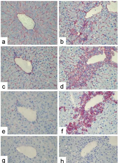
PECAM-1-immunoreactivity in livers from normal rats and from rats treated with CCl4: Comparison to ED1-, and ICAM-1-immunoreactivity
Immunostaining of sections from a normal rat liver using a monoclonal antibody against rat PECAM-1 revealed positivity along the sinusoids in a similar pattern to ICAM-1 (Figure 1), suggesting PECAM-1 positivity of SECs. Staining of sequential sections by antibodies against PECAM-1 and against markers of MNPs (ED1) suggests that the positivity along the sinusoids of the normal rat liver is also partly due to MNPs (Figure 1).
To analyze PECAM-1 immunoreactivity under conditions of inflammation, we used a model of CCl4-induced liver damage, which is characterized by the accumulation of inflammatory cells (mostly MNPs) starting 12 h after the administration of CCl4 and peaking after 48 h, when peri-central necrotic areas can be seen. The possibility that some PECAM-1 positive cells are myofibroblasts (MFB), activated HSCs (hepatic stellate cells), or liver myofibrob-lasts deriving from cells different from quiescent HSCs can be excluded since isolation of these cell types at different time points after CCl4 administration and primary cul-tures of HSC or liver myofibroblasts from normal rat liv-ers never showed any PECAM-1-gene expression (data not shown).
At 48 h after a single dose of CCl4 there were significant numbers of ICAM-1 and ED1 positive cells within the necrotic pericentral areas (Figure 1). However, the ICAM-1- and EDICAM-1-immunoreactive cells of the necrotic area were PECAM-1 negative (Figure 1).
Expression of IFN-γ, IFN-α and TGF-β in CCl4-treated rat livers
During recent years, an important role for IFN-γ and
BMC Physiology 2008, 8:9 http://www.biomedcentral.com/1472-6793/8/9
Indirect immunodetection of PECAM-1, ICAM-1, and ED1 in sections of normal rat or acutely damaged livers (48 h after a sin-gle CCl4-administration)
Figure 1
Indirect immunodetection of PECAM-1, ICAM-1, and ED1 in sections of normal rat or acutely damaged livers (48 h after a single CCl4-administration). Sections of normal rat liver tissue (a, c, e) or acutely damaged rat liver tissue (b,
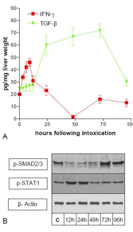
the livers of untreated controls (time point 0 h), there was an early steep increment in IFN-γ expression with a maxi-mum 9 h after CCl4 administration, reaching a level of 46 pg per mg of liver wet weight. At 24 h after CCl4 adminis-tration the IFN-γ concentration peaked at the level of the control livers. At 96 h after CCl4 administration the IFN-γ expression returned to the level seen in the livers of untreated rats. (Figure 2). TGF-β expression only slightly increased during the first 12 h after CCl4-administration. However it increased significantly thereafter with a maxi-mum after 72 h, reaching a level of 67 pg per mg of liver wet weight and returned to the level seen in the livers of untreated rats after 96 h (Figure 2).
Since measurement of cytokine levels in over the whole liver gave no information on possible local variations in concentration we performed Western Blot analysis of p-STAT1 and p-SMAD2/3 in SECs isolated at different time points after CCl4 administration. p-STAT1 was found to be increased at 12 h and 24 h after CCl4-administration, indi-cating that a receptor-mediated IFN signal transduction lasts longer than suspected from the cytokin levels of the total liver. In order to investigate at which time points after CCl4-administration TGF-β induces intracellular sig-naling in SECs we performed Western Blot analysis of p-SMAD2/3. Whereas TGF-β expression is increased between 24 h and 72 h after CCl4-administration, p-SMAD2/3 is decreased in SECs isolated 24 h–48 h after CCl4-administration. Thereafter p-SMAD2/3 is found to be increased.
IFN-γ decreases PECAM-1 transcript level in liver SECs and MNPs
We analysed the modulation of the PECAM-1 specific transcript level in cultured SECs and MNPs following treatment with IFN-γ (Figure 3). SECs at day one after iso-lation were cultured in the absence or presence of IFN-γ under serum reduced conditions (0.3% FCS). Testing dif-ferent concentrations of IFN-γ (100, 1000, 10000 U/ml) indicated that the strongest effects of IFN-γ on the PECAM-1 transcript level were found at a concentration of 10000 U/ml. As shown in Figure 3A, IFN-γ causes a decrease in the PECAM-1 specific transcript level. In con-trast the ICAM-1 mRNA level in SECs was increased by IFN-γ-treatment. Furthermore, different stimulation peri-ods (4 h, 8 h, 12 h, and 24 h of incubation with IFN-γ) were compared indicating maximal effects on PECAM specific transcripts after 8 to 12 h of stimulation. It is note-worthy, that an increase of ICAM-1 and a parallel decrease of PECAM-1 could already be observed after 4 h of treat-ment (Figure 3B) suggesting that the regulation of the two genes may take place at the same time.
Since we were interested in the modulation of PECAM-1 by IFN-γ not only on SECs but also on the transmigrating
MNPs, we also tested MNPs. In MNPs from normal rat liv-ers at day 1 after isolation cultured in the absence or pres-ence of IFN-γ (100, 1000, 10000 U/ml) a maximal decrease of PECAM-1 specific transcripts was revealed using 10000 U/ml of IFN-γ for 12 h. At the same time the
(A) Expression of TNF-α, IFN-γ, and TNF-β during acute liver damage (CCl4-model)
Figure 2
(A) Expression of TNF-α, IFN-γ, and TNF-β during acute liver damage (CCl4-model). Livers taken at differ-ent time points after a single administration of CCl4 were homogenized and used with a human-TNF-α and human-TNF-β EIA-kit or rat-IFN-γ ELISA-kit. Cytokine concentra-tion is indicated as pg per mg of liver wet weight. Error bars indicate standard deviation of the mean value of four experi-ments from four different animals. (B) Western Blot analysis of p-SMAD2/3 and p-STAT-1 in SECs during acute liver dam-age (CCl4-model). SECs from untreated control rats (c) and different time points after CCl4 administration were isolated.
BMC Physiology 2008, 8:9 http://www.biomedcentral.com/1472-6793/8/9
(A) IFN-γ decreases PECAM-1 transcript level and elevates the ICAM-1 transcript level in liver SECs or MNPs
Figure 3
(A) IFN-γ decreases PECAM-1 transcript level and elevates the ICAM-1 transcript level in liver SECs or MNPs.
ICAM-1 mRNA level was increased in MNPs following IFN-γ-treatment (Figure 3A).
TGF-β-treatment increases PECAM-1 transcript level in cultured SECs and MNPs from normal rat livers
Since we had noted the TGF-β levels increasing during the recovery phase of CCl4 induced liver injury, we wanted to analyze whether TGF-β could have reverse effects on PECAM-1-expression compared to IFN-γ. When SECs or MNPs were cultured in the presence of TGF-β for different time periods (4 h, 8 h, 12 h, and 24 h) a maximal increase of PECAM-1 specific transcript level was reached after 12 h to 24 h of stimulation. In addition, cells were treated with TGF-β in different concentrations (1, 3 and 10 ng/ ml) for 12 hours under serum reduced conditions (0.3%
FCS). While no effect of TGF-β on the PECAM-1 mRNA-level was detectable at a concentration of 1 ng/ml, treat-ment with TGF-β at a concentration of 3 or 10 ng/ml resulted in an increased PECAM-1 specific transcript level in SECs and in MNPs (Figure 4). The strongest effects were revealed using 10 ng/ml of TGF-β. An increase of ICAM-1 specific transcript levels was observed in SECs following the treatment with TGF-β. There was no significant increase in ICAM-1 specific transcript level in MNPs.
In order to investigate whether TGF-β is capable of revers-ing the effect of IFN-γ on PECAM-1 and ICAM-1 expres-sion on SECs or MNPs or whether both cytokines simply have inverse but independent effects we additionally cul-tured SECs or MNPs with IFN-γ and TGF-β. Northern Blot
TNF-β increases the PECAM-1 transcript level and ICAM-1 transcript level in liver SECs and MNPs
Figure 4
BMC Physiology 2008, 8:9 http://www.biomedcentral.com/1472-6793/8/9
TNF-β reverses the effect of IFN-γ on PECAM-1 transcript level but has no additional effect on the ICAM-1 transcript level in liver SECs and MNPs
Figure 5
TNF-β reverses the effect of IFN-γ on PECAM-1 transcript level but has no additional effect on the ICAM-1 transcript level in liver SECs and MNPs. Northern blot analysis of total RNA extracted from primary cultures of SECs and MNPs at day 1 after isolation either untreated (c) or treated with IFN-γ 10000 (104)U/ml (I) or IFN-γ together with
TNF-β (10 ng/ml) (I+T) for 12 h. Five µg of total RNA was separated on agarose gel, blotted and hybridised with a 32P-dCTP-labeled cDNA probe specific for rat PECAM-1. The same membranes were hybridised with a cDNA specific for rat ICAM-1 or for β- and γ-actin. This figure shows results of five experiments from five different isolations. The lower panel shows a statistical eval-uation of densitometric scans. Error bars indicate standard deviation of the mean value after normalization on β,γ-actin expres-sion. P-value was tested to show significant differences of control cells compared to cells treated with IFN-γ or IFN-γ +TNF-β. * indicates p < 0.05.
analysis revealed that coadministration of IFN-γ and
TGF-β inhibits the PECAM-1-downregulating effect of IFN-γ on both cell types. On the other hand coadministration of both cytokines had no additional effect on upregulation of ICAM-1 (Figure 5).
Surface expression of PECAM-1 and ICAM-1 on SECs and MNPs following IFN-α-, IFN-γ – or TGF-β-treatment
To demonstrate that IFN-γ – or TGF-β treatment also influ-ences PECAM-1 and ICAM-1 surface expression, we fur-ther investigated the PECAM-1-gene-expression in SECs
and MNPs following treatment with IFN-γ or TGF-β by flow cytometry.
SECs. On MNPs only an insignificant increase in ICAM-1 expression was observed.
Constant PECAM-1 specific transcript level in CCl4 -treated IFN-γ-/- mice livers and reduced increase of the
ICAM-1 specific transcript level
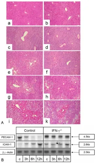
For a functional proof that IFN-γ leads to an early decrease of PECAM-1 and to an early increase of ICAM-1 specific transcripts in vivo we used an IFN-γ-/- mice knockout model (B6.129S7-Ifngtm1Ts) [22]. In control mice (female C57BL mice), similar CCl4-administration levels to those previously used in rats leads to an early decrease of PECAM-1 specific transcripts and to an increase of 1 specific transcripts. Whereas an increase of ICAM-1 specific transcripts can also be observed in IFN-γ-/- mice livers after CCl4-administration, no significant decrease of PECAM-1 specific transcripts was evident (Figure 7A). A histological comparison after inducing acute liver damage by CCl4 showed that the degree of damage in the IFNγ -/-mice was less than in the control -/-mice. Furthermore the damaged pericentral areas were smaller, less fatty and the occurrence of necrotic areas, which were also smaller in the liver sections from IFNγ-/- mice, was observed later (48 h vs. 24 h after CCl4 administration). Even 72 h after CCl4 administration the hepatocytes are more vital (Fig. 7B).
Treatment of rats with IFN-γ enhances the decrease of PECAM-1-gene-expression in CCl4-treated rat livers in
parallel to an enhanced increase of the ICAM-1 gene-expression
To further analyse whether the effects of IFN-γ observed in vitro in the previous experiments can be reproduced in vivo, rats were treated either with CCl4 or with IFN-γ and CCl4 in parallel. Measurement of the IFN-γ concentration in liver homogenates by means of ELISA revealed an increase of 5–10% in the IFN-γ concentration in liver tis-sue following the intraperitoneal treatment with 50000 U IFN-γ. Since, from the in vitro and in vivo experiments we expected the effects of IFN-γ within the first 10 h after treatment, livers were analyzed (3 h, 6 h, 12 h after treat-ment) for changes in PECAM-1 and ICAM-1-gene-expres-sion by Northern blot analysis (Figure 8).
When rats were treated with IFN-γ and CCl4 the increase of ICAM-1 transcript level and the decrease of PECAM-1 transcript level was enhanced compared to rats treated with CCl4 alone. This difference between rats treated with CCl4 and IFN-γ to rats treated with CCl4 alone was only detectable up to 6 h following the treatment.
Treatment of rats with IFN-γ enhances the accumulation of ED1-expressing cells in CCl4-treated rat livers
Since differences in adhesion molecule expression follow-ing the additional treatment with IFN-γ could be observed up to 6 h after treatment, sections of rat livers were ana-lysed by indirect immunostaining with a monoclonal antibody against the ED1 antigen at 3 h, 6 h, and 12 h after the treatment to analyze whether the amount of recruited inflammatory cells was influenced. The staining of control livers revealed the previously observed pattern of non-parenchymal cells situated around the vessels. At 12 h after CCl4-treatment the number of ED1-positive cells around the vessels and along the sinusoids increased. Interestingly, when rats were treated with the combina-tion of IFN-γ and CCl4 the number of ED1 positive cells at 12 h was significantly higher than in the livers of animals treated with CCl4 alone (Figure 9).
Discussion
Since we have seen that there is a down-regulation of PECAM-1 in rat livers after the administration of CCl4, in this study we aimed to identify cytokines that were responsible for the down-regulation of PECAM-1 on SECs and on transmigrating MNPs, but we also wanted to iden-tify cytokines that allow the reversal of this effect on both cell types. In this study, we show that in CCl4-treated rat livers there is an early rise of levels of IFN-γ and – during the recovery phase – an increase of TGF-β-protein level. Similar observations for TGF-β levels after induced liver damage have also been reported by others [23,24]. How-ever especially measurements of the early time points are missing in the literature. This might be the reason for reports of a decrease of IFN-γ after inducing liver damage
Surface expression of PECAM-1 and ICAM-1 in SECs and MNPs under the treatment of TNF-β or IFN-γ Surface expression of PECAM-1 and ICAM-1 in SECs
Figure 6
BMC Physiology 2008, 8:9 http://www.biomedcentral.com/1472-6793/8/9
(A) HE staining of liver sections of C57BL/6 mice (a, c, e, g, i) and B6
Figure 7
[25]. In vitro studies indicated that IFN-γ-treatment induced a decrease of PECAM-1-gene-expression in paral-lel with an increase in ICAM-1-gene-expression. Further-more, TGF-β-treatment resulted in an increase of PECAM-1-expression in SECs and MNPs. TGF-β – also inhibited the effect of IFN-γ treatment. However, the temporal course of the cytokine expression in vivo after administra-tion of the damaging noxae does not perfectly match the temporal course of changes in the expression of PECAM-1 or ICAM-1. This has two major causes. Firstly, in vivo,
many intercellular contacts and additional cytokines have an impact on PECAM-1 and ICAM-1 expression on SECs and MNPs e.g. IFN-α [15] which shows a similar expres-sion profile compared to IFN-γ. Secondly, local differ-ences in the concentration of the cytokines can lead to increased levels of the cytokines around SECs or MNPs when levels in the total liver are already decreased. The observation that IFN-γ or TGF-β signalling as measured by p-STAT1 or p-SMAD2/3 is delayed and can be observed when the levels of IFN-γ or TGF-β are already decreasing supports this thesis.
The in vitro effect of IFN-γ could be confirmed in vivo, since the administration of IFN-γ (0,4 µg per mg liver wet weight, which is comparable to the concentration used for the in vitro treatment (0.01 to 0.1 µg/ml of IFN-γ)), together with CCl4 further decreased the PECAM-1 tran-script level and also further increased the number of ED1-positive cells in the liver at 12 h after the administration of CCl4 compared to the single CCl4 administration to the animals. In contrast, in IFN-γ-/- mice the lack of IFN-γ may be responsible for the missing early decrease of PECAM-1 and the reduced increase of ICAM-1.
To study the involvement of cell adhesion molecules dur-ing inflammation and tissue damage in vivo several
Treatment of rats with IFN-γ enhances the number of ED1 positive cells
Figure 9
Treatment of rats with IFN-γ enhances the number of ED1 positive cells. Animals were treated with CCl4 or with CCl4 and IFN-γ and livers were taken for analysis 3 h (3 h), 6 h (6 h) or 12 h (12 h) after treatment. Sections were stained by indirect immunohistochemistry using a mono-clonal antibody against ED1. Positive cells were counted in 10 purviews (250 × magnification). Error bars indicate stand-ard deviation of the mean value of 10 counted purviews. P-value was tested to show significance differences of rats 12 h after treatment with CCl4 compared to treatment with CCl4 and IFN-γ. * indicates p < 0.05.
Treatment of rats with IFN-γ enhances the decrease of the PECAM-1 specific transcript level in CCl4-treated rat livers in parallel to an enhanced increase of the ICAM-1 specific tran-script level
Figure 8
BMC Physiology 2008, 8:9 http://www.biomedcentral.com/1472-6793/8/9
approaches have been used. It has been difficult to block inflammation using reagents directed against single mol-ecules. Several studies, however, revealed that the transmi-gration of monocytes and of neutrophils could be inhibited using antibodies against PECAM-1 in vitro [12], and in vivo, as the administration of antibodies against PECAM-1 or the administration of the soluble domain 1 of PECAM-1 blocked the accumulation of leukocytes in different models of inflammation [10,11,26,27]. In PECAM-1-knock-out-mice, however, only a slowing of inflammatory cells transmigrating through the basement membrane was observed. This slowing had no effect on the total number of transmigrated cells [13].
In vivo data showing a reduced PECAM-1 expression in inflammatory cells have been interpreted as if PECAM-1 down-regulation were a consequence of the transmigra-tion [28]. Our results demonstrate that in vitro and in vivo treatment with IFN-γ down-regulates the expression of PECAM-1 in liver cells at an early time point. The in vitro studies reveal a decreased PECAM-1-expression on SECs and on MNPs. These data support the hypothesis that PECAM-1 has to be down-regulated to facilitate transmi-gration. A confirmation of this hypothesis could be seen in a model of abdominal aortic transplantation. The transmigration of MNPs was unexpectedly enhanced when aortic allografts from PECAM-1 knock-out-mice were transplanted compared to transplanted allografts from wild type mice [29]. The authors suggested that this phenomenon might be due to the fact that PECAM-1 is concentrated at interendothelial junctions and is thereby important for the structural stability of the endothelial barrier. In PECAM-1 deficient grafts this endothelial sta-bility could be reduced during the inflammatory episode after transplantation making it easier for macrophages to transmigrate through the endothelium.
Another relevant finding of the in vivo experiments is the increase of TGF-β concentration in the liver during the recovery phase after CCl4-administration, when IFN-γ – and IFN-α concentration have already been decreased. TGF-β is a pleiotropic cytokine with profibrogenic and anti-inflammatory capacities [18]. The data presented in this study show that in cultured SECs and MNPs, TGF-β increases PECAM-1-expression. Furthermore, TGF-β inhibited the effect of IFN-γ on the PECAM-1 transcript level. Although an up-regulation of PECAM-1-gene-expression by TGF-β-treatment has been observed using a human macrophage cell line (U937 cells) [30], this is the first report showing similar effects on endothelial cells and on tissue macrophages. These data indicate a different modulation of PECAM-1 by IFN-γ and TGF-β in SECs and MNPs. Under normal conditions SECs are an efficient bar-rier against corpusculate matter. Early production of
IFN-induce an increase of ICAM-1 expression, but also a decreased PECAM-1 expression, which is essential for the tight adhesion and for the transmigration of inflamma-tory cells into the parenchyma. During liver repair cytokines such as TGF-β may be effective in reestablishing the endothelial barrier.
Whereas IFN-γ has previously been shown to exert antifi-brotic effects in man and mice by abrogating TGF-β signal-ling [31,32], inducing an acute liver damage in IFN-γ knockout mice revealed that the degree of damage was reduced and delayed when compared to control mice. This suggests that, least in the early time points after administration of a damaging noxae, IFN-γ is involved in an increase of liver damage without antagonizing TGF-β. From these data, blockage of IFN-γ signalling in the early time points after liver damage, thereby inhibiting down-regulation of PECAM, could be a valuable tool in the treat-ment of patients during the early phases of liver damage.
Methods
AnimalsMale Wistar rats (8 weeks old) (Charles River, Sulzberg, Germany), female C57BL/6 mice and B6.129S7-Ifngtm1Ts (IFN-γ-deficient) mice (The Jackson Labora-tory, Bar Harbor, ME) received humane care and were kept according to the institution's and the National Insti-tutes' of Health guidelines.
Reagents
Human recombinant IFN-γ was purchased from R&D sys-tems (Wiesbaden, FRG). Human TGF-β and IFN-α were purchased from Sigma (Deistendorfen, FRG). The mono-clonal antibody against rat PECAM-1 was purchased from Natutec (Frankfurt, FRG). The monoclonal antibodies against rat ICAM-1 was from Genzyme (Cambridge, MA, USA), against pSTAT1, pSMAD2/3 and β-actin were from Santa Cruz Biotechnology (Heidelberg, Germany) and against ED1 was from Biermann (Biermann, Wiesbaden, Germany).
cDNA probes
Rat and mouse PECAM-1 and ICAM-1 specific cDNAs were generated as described earlier [14,15]. Northern blot results were normalised to an oligonucleotide probe spe-cific for 28S rRNA [33] or with a chicken α-actin cDNA exhibiting cross reactivity with β- and γ-actin, which was a gift from A. Schwartz [34]. Furthermore, a PCR-generated cDNA directed against rat IFN-α was used mapping posi-tions 140–509 of the published sequence [35].
Inducing liver damage
described [36-38]. Control animals were treated with maize-oil only. Six animals in each group were sacrificed 3 h, 6 h, 9 h, 12 h, 24 h, 48 h, 72 h, and 96 h after a single high dose CCl4 administration. Furthermore, 16 animals were treated with IFN-γ (50000 IU/100 g body weight which is about 0.4 µg per mg of rat liver, assuming that about 80% of the intraperitoneally injected IFN-γ reaches the liver) in addition to CCl4-treatment. Animals were sac-rificed 3 h, 6 h, 12 h, and 24 h after the treatment, since we expected IFN-γ induced modulation of gene expres-sion to take place at early time points after the administra-tion. For inducing liver damage in mice a carbon-tetrachloride/maize oil solution (50% v/v) was adminis-trated intraperitoneally with a concentration of 1 ml CCl4/kg.
Immunohistology
APAAP-immunostaining was performed as described [39]. Negative controls were performed by replacing the primary antibody by murine IgGs. ED1 positive cells were counted in 10 purviews (250 × magnification) of immu-nostainings of two independent series of experiments.
Measurement of IFN-γ, IFN-α and TGF-β in liver tissue
Liver specimens (40 mg) were homogenized and used with a human-IFN-α and-TGF-β EIA-kit (Chemicon, Tam-emcula, USA) or rat-IFN-γ ELISA-kit (Genzyme Diagnos-tics, Cambridge, Massachusetts, USA). Measurements were carried out according to the manufacturers' proto-cols.
Cell isolation and culture conditions
MNPs from normal rat livers were isolated according to the method of De Leeuw et al. [40] as described previously [41]. SECs from normal rat livers were obtained according to Knook et al. [42] with modifications as described [14,15,36]. MNPs were cultured in M-199 supplemented with 10% FCS. SECs were cultured on collagen-coated tis-sue culture plates in endothelial cell medium (Promo cell, Heidelberg, FRG) supplemented with epidermal growth factor (10-11 mg/ml), basic fibroblast growth factor (10-9 mg/ml), 0.15% insulin and 2% FCS. The medium was replaced every day. Purity of SECs was more than 91% as tested by incorporation of dioctadecyl tetramethyl indo-carbocyanine perchlorate acetylated low density lipopro-tein and electron microscopy of SECs as described in [14,15,43]. Freshly isolated MNPs from normal livers were more than 99% viable and more than 98% pure (as tested by ED1/ED2 staining). On day 1 of culture, SECs and MNPs were washed three times with Gey's balanced salt solution and were incubated in serum reduced (0.3% FCS) culture medium with increasing concentrations of IFN-α (10, 100, 1000, U/ml), IFN-γ (10, 100, 1000, 10000 U/ml which is 0.01 to 0.1 µg/ml), or TGF-β (1, 3, and 10 ng/ml).
Flow cytometric quantification of PECAM-1 and ICAM-1 expression in isolated liver cells
For quantification of PECAM-1 and ICAM-1 immunoflu-orescence after indirect immunostaining, we used flow cytometry of MNPs and SECs on day one after isolation (Epics ML, Coulter, Kerfeld, Germany). Negative controls were performed using murine IgGs instead of the primary antibody.
Northern Blot analysis of total RNA
Total RNA was isolated according to Chirgwin [44] and was separated by agarose gel electrophoresis, transferred onto nylon membranes and hybridised with specific 32P dCTP or 32P dATP labeled cDNA probes as described [43]. cDNA probes were labelled by Random priming®. The oligo recognizing the 28S rRNA was end labelled using 32P dATP. Hybridization was carried out over 2 h at 68°C using the Quick Hyb® solution. Post hybridization – washes were performed twice for 15 minutes at room tem-perature and twice for 5 – 15 minutes at 50°C in 2 × SSC containing 0.1% SDS. Nylon filters were exposed to X-ray films at -80° and finally densitometric scans were per-formed.
Western Blot Analysis
Cells were lysed in hot Laemmli buffer (95°C) and proc-essed by sodium dodecyl sulphate polyacrylamide gel electrophoresis (SDS-PAGE; 9% polyacrylamide) under reducing conditions [45]. The protein content of the cell lysates was calculated by the Coomassie Protein Assay (Pierce, Rockford, IL) and 30 µg protein per lane were applied. Proteins were transferred onto Hybond-ECL nitrocellulose membranes according to [46]. Immunode-tection was performed according to the ECLWestern blot-ting protocol. Antibodies against p-STAT1 and p-SMAD2/ 3 were used at 2.5 µg/ml solutions, β-actin at 1:1000 solu-tion, and peroxidase-labeled anti-mouse and anti-rabbit immunoglobulins were each used at a 1:1000 dilution.
Statistical analysis
Densitometric analysis of the blots was performed using the program Scion Image Beta 4.0.2 (NIH, Washington, USA). Statistical evaluation was done by means of the Wil-coxon test.
Abbreviations
BMC Physiology 2008, 8:9 http://www.biomedcentral.com/1472-6793/8/9
Authors' contributions
KN participated in the design and coordinated the study, performed and supervised the rat and mouse liver damage and drafted the manuscript. AL removed the tissues, iso-lated the cells and carried out the immunohistochemistry and FACS analysis. KT performed the molecular studies. GR participated in the design of the study and critically reviewed the manuscript. BS participated in the design and coordination of the study, performed the immunob-lotting and ELISAs, interpreted the results, helped to draft the manuscript and critically reviewed and revised its final version. All authors have read and approved the manu-script.
Acknowledgements
The authors are indebted to A. Herbst for excellent technical assistance. This work was supported by the DFG SFB 402 TP C6 and D3
References
1. Bianchi E, Bender JR, Blasi F, Pardi R: Through and beyond the wall: late steps in leukocyte transendothelial migration. Immunol Today 1997, 18:586-91.
2. Carlos TM, Harlan JM: Leukocyte-endothelial cell adhesion molecules. Blood 1994, 84:2068-2075.
3. Dunon D, Piali L, Imhof BA: To stick or not to stick: The new leu-kocyte homing paradigm. Current Opinion in cell Biology 1996,
8:714-723.
4. Madri JA, Graesser D: Cell migration in the immune system: the evolving interrelated roles of adhesion molecules and proteinases. Dev Immunol 2000, 7:103-16.
5. Sallusto F, Lanzavecchia A: Understanding dendritic cell and T-lymphocyte traffic through the analysis of chemokine recep-tor expression. Immunol Rev 2000, 177:134-40.
6. Springer TA: Traffic signals for lymphocyte recirculation and leukocyte emigration: the multistep paradigm. Cell 1994,
76:301-314.
7. Couvelard A, Scoazec JY, Feldmann G: Expression of cell-cell and cell-matrix adhesion proteins by sinusoidal endothelial cells in the normal and cirrhotic human liver. Am J Pathol 1993,
143:738-52.
8. Scoazec J-Y, Feldmann G: In situ immunotyping study of endothelial cells of the human hepatic sinusoid: result and functional implication. Hepatology 1991, 14:789-797.
9. Scoazec JY, Feldmann G: The cell adhesion molecules of hepatic sinusoidal cells. J Hepatol 1994, 20:296-300.
10. Bogen S, Pak J, Garifallou M, Deng X, Muller WA: Monoclonal anti-body to murine PECAM-1 (CD31) blocks acute inflamma-tion in vivo. J Exp Med 1994, 179:1059-1064.
11. Liao F, Ali J, Greene T, Muller WA: Soluble domain 1 of platelet-endothelial cell adhesion molecule (PECAM) is sufficient to block transendothelial migration in vitro and in vivo. J Exp Med 1997, 185:1349-57.
12. Muller WA, Weigl SA, Deng X, Phillips DM: PECAM-1 is required for transendothelial migration of leukocytes. J Exp Med 1993,
178:449-60.
13. Duncan GS, Andrew DP, Takimoto H, Kaufman SA, Yoshido H, Spell-berg J, de la Pompa JL, Elia A, Wakeham A, Karan-Tami B, Muller WA, Senaldi G, Zukowski MM, Mak TW: Genetic evidence for func-tional redundancy of Platelet endothelial cell adhesion mol-ecule-1 (PECAM-1): CD31-deficient mice reveal PECAM-1 dependent and PECAM-1 independent functions. J Immunol
1999, 162:3022-3030.
14. Neubauer K, Wilfling T, Ritzel A, Ramadori G: PECAM-1 expres-sion in sinusoidal endothelial cells of the liver. J Hepatol 2000,
32:921-32.
15. Neubauer K, Ritzel A, Saile B, Ramadori G: Decrease of platelet-endothelial cell adhesion molecule 1-gene-expression in inflammatory cells and in endothelial cells in the rat liver
fol-16. Koerber K, Sass G, Kiemer AK, Vollmar AM, Tiegs G: In vivo regu-lation of inducible no synthase in immune-mediated liver injury in mice. Hepatology 2002, 36:1061-9.
17. Taylor JL, Grossberg SE: The effects of interferon-alpha on the production and action of other cytokines. Semin Oncol 1998,
25:23-9.
18. Nakao A: Is TGF-beta1 the key to suppression of human asthma? Trends Immunol 2001, 22:115-8.
19. Saile B, Ramadori G: Inflammation, damage repair and liver fibrosis--role of cytokines and different cell types. Z Gastroen-terol 2007, 45:77-86.
20. Tiegs G: Cellular and cytokine-mediated mechanisms of inflammation and its modulation in immune-mediated liver injury. Z Gastroenterol 2007, 45:63-70.
21. Pinzani M, Rombouts K: Liver fibrosis: from the bench to clinical targets. Dig Liver Dis 2004, 36:231-242.
22. Dalton DK, Pitts-Meek S, Keshav S, Figari IS, Bradley A, Stewart TA:
Multiple defects of immune cell function in mice with dis-rupted interferon-gamma genes. Science 1993, 259:1739-1742. 23. Czaja MJ, Flanders KC, Biempica L, Klein C, Zern MA, Weiner FR:
Expression of tumor necrosis factor-alpha and transforming growth factor-beta 1 in acute liver injury. Growth Factors 1989,
1:219-226.
24. Grasl-Kraupp B, Rossmanith W, Ruttkay-Nedecky B, Mullauer L, Kammerer B, Bursch W, Schulte-Hermann R: Levels of transform-ing growth factor beta and transformtransform-ing growth factor beta receptors in rat liver during growth, regression by apoptosis and neoplasia. Hepatology 1998, 28:717-726.
25. Daniluk J, Chibowski D, Szuster-Ciesielska A, Kandefer-Szerszen M:
Effect of carbon tetrachloride intoxication and ethanol ingestion on interferon production in mice. Arch Immunol Ther Exp (Warsz) 1994, 42:325-330.
26. Murohara T, Delyani JA, Albelda SM, Lefer AM: Blockade of plate-let endothelial cell adhesion molecule-1 protects against myocardial ischemia and reperfusion injury in cats. J Immunol
1996, 156:3550-3557.
27. Vaporciyan AA, Delisser HM, Yan H, Thom SR, Jones ML, Ward PA, Abelda SM: Involvement of platelet endothelial cell adhesion molecule-1 in neutrophil recruitment in vivo. Science 1993,
262:1580-1582.
28. Christofidou-Solomidou M, Nakada MT, Williams J, Muller WA, Delisser HM: Neutrophil platelet endothelial cell adhesion molecule-1 participates in neutrophil recruitment at inflam-matory sites and is down-regulated after leukocyte extrava-sation. J Immunol 1997, 158:4872-8.
29. Ensminger SM, Spriewald BM, Steger U, Morris PJ, Mak TW, Wood KJ: Platelet-endothelial cell adhesion molecule-1 (CD31) expression on donor endothelial cells attenuates the devel-opment of transplant arteriosclerosis. Transplantation 2002,
74:1267-73.
30. Lastres P, Almendro N, Bellon T, Lopez-Guerrero JA, Eritja R, Bern-abeu C: Functional regulation of platelet/endothelial cell adhesion molecule-1 by TGF-beta 1 in promonocytic U-937 cells. J Immunol 1994, 153:4206-18.
31. Weng HL, Wang BE, Jia JD, Wu WF, Xian JZ, Mertens PR, Cai WM, Dooley S: Effect of interferon-gamma on hepatic fibrosis in chronic hepatitis B virus infection: a randomized controlled study. Clin Gastroenterol Hepatol 2005, 3:819-828.
32. Weng H, Mertens PR, Gressner AM, Dooley S: IFN-gamma abro-gates profibrogenic TGF-beta signaling in liver by targeting expression of inhibitory and receptor Smads. J Hepatol 2007,
46:295-303.
33. Barbu V, Dautry F: Northern blot normalization with a 28S RNA oligonucleotide probe. Nucl Acid Res 1989, 17:7115-7119. 34. Schwartz RJ, Haron JA, Rothblum KM, Dugaiczyk A: Regulation of
muscle differentiation: Cloning of sequences from alpha-actin messenger ribonucleic acid. Biochemistry 1980,
19:5883-5890.
35. Estler HC, Grewe M, Gaussling R, Pavlovic M, Decker K: Rat tumor necrosis factor-alpha. Transcription in rat Kupffer cells and in vitro posttranslational processing based on a PCR-derived cDNA. Biol Chem Hoppe Seyler 1992, 373:271-81.
Publish with BioMed Central and every scientist can read your work free of charge "BioMed Central will be the most significant development for disseminating the results of biomedical researc h in our lifetime."
Sir Paul Nurse, Cancer Research UK
Your research papers will be:
available free of charge to the entire biomedical community
peer reviewed and published immediately upon acceptance
cited in PubMed and archived on PubMed Central
yours — you keep the copyright
Submit your manuscript here:
http://www.biomedcentral.com/info/publishing_adv.asp
BioMedcentral 37. Neubauer K, Eichhorst ST, Wilfling T, Buchenau M, Xia L, Ramadori
G: Sinusoidal ICAM-1 upregulation precedes the accumula-tion of LFA-1 positive cells and tissue necrosis in a model of CCl4-induced acute rat liver injury. Lab Invest 1998, 78:185-194.
38. Yokoi Y, Namihase T, Kuroda H, Komatsu I, Miyazaki A, Watanabe S, Usui K: Immunocytical detection of desmin in fat-storing cells (Ito-cells). Hepatology 1984, 4:709-714.
39. Herbst H, Heinrichs O, Schuppan D, Milani S, Stein H: Temporal and spatial patterns of transin/stromylesin RNA expression following toxic injury in rat liver. Virchows Arch B 1991,
60:295-300.
40. De Leeuw AM, Mc Carthy SB, Geerts A, Knook DL: Purified rat liver fat-storing cells in culture divide and contain collagen. Hepatology 1984, 4:392-403.
41. Armbrust T, Ramadori G: Functional characterization of two different Kupffer cell populations in normal rat liver. J Hepatol
1996, 25:518-528.
42. Knook DL, Blansjaar N, Sleyster EC: Isolation and characteriza-tion of Kupffer and endothelial cells from the rat liver. Exp Cell Res 1977, 109:317-329.
43. Neubauer K, Knittel T, Armbrust T, Ramadori T: Accumulation of fibrinogen during short term and long term rat liver injury. Gastroenterology 1995, 108:1124-1135.
44. Chirgwin JM, Przybyla AB, Mac Donald RJ, Rutter WJ: Isolation of biochemically active ribonucleic acid from sources enriched in ribonuclease. Biochemistry 1979, 18:5294-5300.
45. Laemmli U: Cleavage of structural protein during the assemby of the head of bacteriophage T4. Nature 1970, 227:680-682. 46. Towbin H, Staehelin T, Gordon J: Electrophoresis transfer of
proteins from polyacrylamide gels to nitrocellulose sheets: procedure and some applications. Proc Natl Acad Sci USA 1979,