R E S E A R C H
Open Access
Saturated long-chain fatty acid-producing
bacteria contribute to enhanced colonic
motility in rats
Ling Zhao
1†, Yufen Huang
2†, Lin Lu
1†, Wei Yang
1, Tao Huang
1, Zesi Lin
3, Chengyuan Lin
1,4, Hiuyee Kwan
1,
Hoi Leong Xavier Wong
1, Yang Chen
5, Silong Sun
2, Xuefeng Xie
2, Xiaodong Fang
2,5, Huanming Yang
6, Jian Wang
6,
Lixin Zhu
7*and Zhaoxiang Bian
1*Abstract
Background:The gut microbiota is closely associated with gastrointestinal (GI) motility disorder, but the mechanism(s) by which bacteria interact with and affect host GI motility remains unclear. In this study, through using metabolomic and metagenomic analyses, an animal model of neonatal maternal separation (NMS)
characterized by accelerated colonic motility and gut dysbiosis was used to investigate the mechanism underlying microbiota-driven motility dysfunction.
Results:An excess of intracolonic saturated long-chain fatty acids (SLCFAs) was associated with enhanced bowel motility in NMS rats. Heptadecanoic acid (C17:0) and stearic acid (C18:0), as the most abundant odd- and even-numbered carbon SLCFAs in the colon lumen, can promote rat colonic muscle contraction and increase stool frequency. Increase of SLCFAs was positively correlated with elevated abundances ofPrevotella,Lactobacillus, and Alistipes. Functional annotation found that the level of bacterial LCFA biosynthesis was highly enriched in NMS group. Essential synthetic genesFabswere largely identified from the generaPrevotella,Lactobacillus, andAlistipes. Pseudo germ-free (GF) rats receiving fecal microbiota from NMS donors exhibited increased defecation frequency and upregulated bacterial production of intracolonic SLCFAs. Modulation of gut dysbiosis by neomycin effectively attenuated GI motility and reduced bacterial SLCFA generation in the colon lumen of NMS rats.
Conclusions:These findings reveal a previously unknown relationship between gut bacteria, intracolonic SLCFAs, and host GI motility, suggesting the importance of SLCFA-producing bacteria in GI motility disorders. Further exploration of this relationship could lead to a precise medication targeting the gut microbiota for treating GI motility disorders.
Keywords:Gastrointestinal motility disorder, Gut microbiota, Neonatal maternal separation, Saturated long-chain fatty acids
Background
Disordered gastrointestinal (GI) motility is one major symptom frequently presented in patients suffering with functional GI disorders (FGIDs) and other bowel
diseases [1,2]. Chronic or recurrent episodes of dysregu-lated GI motility severely impact patients’ quality of life [3]. The underlying mechanisms of disordered GI motil-ity are multifaceted. Previous studies have revealed con-tributions of immune activation, ionic channels and neurohumoral dysregulation to GI motor dysfunction [4–6]. However, current etiological understanding of dis-ordered GI motility is incomplete, which limits the de-velopment of personalized and precisely effective medicine for GI motility disorders.
* Correspondence:[email protected];[email protected]
†Ling Zhao, Yufen Huang and Lin Lu contributed equally to this work. 7Digestive Diseases and Nutrition Center, Department of Pediatrics, The State
University of New York at Buffalo, 3435 Main Street, 422BRB, Buffalo, NY 14214, USA
1Chinese Medicine Clinical Study Center, Jockey Club School of Chinese
Medicine, Hong Kong Baptist University, Kowloon Tong, Hong Kong SAR, China
Full list of author information is available at the end of the article
The gut microbiota is thought to be an important fac-tor associated with disordered GI motility [7]. For ex-ample, patients with irritable bowel syndrome (IBS), especially for diarrhea-predominant IBS (IBS-D), showed a lower diversity and a higher instability of gut microbial community [8,9]. Altered composition of the gut micro-biota was also found in children and adults with chronic functional constipation [10–12]. Regulation of the gut microbiota by probiotics can improve bowel movement frequency in up to 70% of functionally constipated pa-tients [13]. Transplantation of fecal microbiota from IBS-D suffers significantly accelerated colonic transit in germ-free (GF) mice [14]. GF mice colonized with fecal microbiota from patients with slow transit constipation exhibit lower fecal frequency, delayed GI transit time, and weaker spontaneous contractions of colonic smooth muscle [15]. These results point directly to a conclusion that the gut microbiota serves a pivotal role in GI motil-ity disorder. But the mechanism by which microbiota interact with and affect host GI motility remains unclear.
It has been proposed that the effect of the gut micro-biota on host GI motility partly derives from release of bacterial end-products of fermentation or molecules de-rived from host and gut microbiota co-metabolism [16,
17]. One study found that microbial products, such as li-popolysaccharides (LPS), can regulate GI motility through activating toll-like receptor 4 (TLR4) signaling that in-creases survival of nitrergic neurons [18]. Short-chain fatty acids and lactic acids, produced from bacterial fermenta-tion of dietary fiber and resistant starch, can regulate in-testinal motor action, probably through regulating peptide YY secretion and cholinergic neurons [19]. In another study, GF mice displayed accelerated GI motility after colonization with spore-forming microbes, the metabolites of which could stimulate enteric serotonin release [20]. Therefore, gut microbiota-derived substances appear to be the link between gut microbiota and host GI motility.
A rodent model of neonatal maternal separation (NMS) is a well-established model characterized by long-term colonic dysfunction [21]. Our previous study found that colon tissue isolated from adult NMS rats showed a higher level of muscle amplitude [22]. Such enhanced colonic motility is involved in upregulation of L-type calcium channels in colonic smooth muscle cells [22] and increased serotonin production from colonic enterochromaffin cells (ECs) [23]. Meanwhile, gut mi-crobial disturbance is also reported in both mice and rats subjected to NMS [24, 25], but the association of changed gut microbiota with colonic dysmotility in an NMS model is yet to be investigated.
In this study with an NMS rat model, metabolic profil-ing of feces and luminal contents of the GI tract of ex-perimental animals was preformed to identify featured microbiota-derived metabolites. A group of saturated
long-chain fatty acids (SLCFAs) was remarkably in-creased in the colon lumen and feces of NMS rats. After confirming their stimulatory effects on colon motility, function-based metagenomic analysis was done and found hyperactive synthesis of SLCFAs by the gut microbiota of NMS rats. The relationship between SLCFA-producing bacteria and host colon motility were further clarified by transplanting fecal microbiota from NMS donors into pseudo germ-free (GF) rats and modulating gut micro-biota in NMS rats by neomycin intervention. Based on these experiments, we aimed to clarify the linkage be-tween gut microbiota and host colonic motility phenotype and to determine which bacteria are the primary agents manipulating host GI colonic dysmotility.
Results
Excess intracolonic SLCFAs are associated with GI motility in NMS model
an excess of intracolonic SLCFAs is associated with en-hanced GI motility in NMS rats.
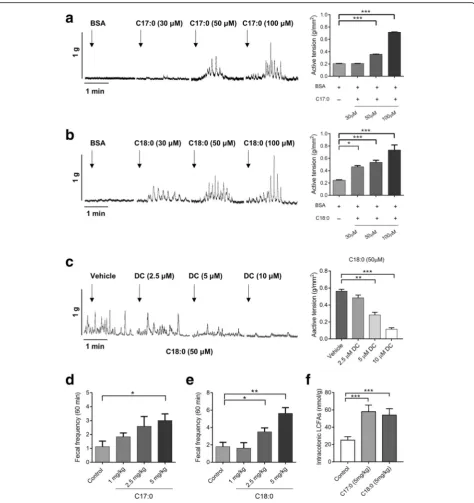
SLCFAs stimulate rat colonic contraction and defecation Lipid perfusion has been shown to enhance colon motil-ity in normal subjects and patients with IBS-D [26]; however, the effects of SLCFAs on colonic motility have not been reported. To determine these effects, we stud-ied C17:0 and C18:0, the most abundant even- and odd-numbered carbon SLCFAs in the colon lumen of NMS rats, using an organ bath system as described in our previous study [27]. Compared with baseline (fatty acid-free BSA), C17:0 (50 and 100 μM) and C18:0 (30, 50, and 100 μM) dose-dependently enhanced contrac-tion amplitudes of colonic circular muscles (Fig. 3a, b). Acetylcholine treatment was used as the positive control (Additional file 1: Figure S2A). C2:0, another saturated FA found to be increased level in fecal VOCs of NMS rats, was tested as a negative control (Additional file 1: Figure S2B). It is well known that GPR40 and GPR120, expressed in the endocrine cells of the colon, can be
activated by free LCFAs [28–30]. To test which free long-chain fatty acid receptors are involved in such SLCFA-induced muscle contraction, isolated colonic segments were separately treated with different doses of DC260126 (GPR40 antagonist) and AH7614 (GPR120 antagonist) prior to introduction of C18:0. We found that DC260126 in doses of 5 and 10 μM can effectively suppress C18-induced colonic contraction, but AH7614 showed no effects (Fig. 3c and Additional file 1: Figure S2B). Furthermore, this effect of SLCFAs on colonic mo-tility was confirmed in normal rats in vivo through orally administrating SLCFAs at dosages of 1, 2.5, and 5 mg/ kg. Both C17:0 (5 mg/kg) and C18:0 (2.5 and 5 mg/kg) notably increased rat defecation frequency (Fig. 3d, e). The cumulative amount of intracolonic SLCFAs was sig-nificantly increased in rats given 5 mg/kg of C17 or C18 (Fig. 3f ), and this amount is close to the total SLCFA level in the colonic contents from NMS rats. These find-ings revealed that SLCFAs can accelerate rat colonic mo-tility, and such action is possibly involved in GPR40 signaling.
Changed fecal microbiome is associated with SLCFA generation in NMS rats
Generally, intraluminal SLCFAs are mainly derived from diets or synthesized by host and gut microbiota. A previous study screened six genes (Fasn, Scd1, Srebp1, CD36, Fabp2, and Cav1) related to lipogenesis and lip-olysis based on RT-PCR array and found no difference in their levels between adult NMS and control rats [31]. These evidences indicate that NMS rats have nor-mal levels of host fatty acid synthesis, digestion, and
absorption. Another piece of evidence has revealed that odd-numbered carbon SLCFA C15:0 and C17:0 can be produced by bacteria only [32]. Hence, we hypothesized that increase of SLCFAs is associated with the gut microbiota.
Gut dysbiosis in NMS rodents has been previously re-ported based on 16s ribosomal RNA sequencing [24,
25]. However, such approach cannot be used to obtain information related to metabolic function. To investigate whether gut dysbiosis results in abnormal SLCFAs, and
what the key manipulators are, we performed metage-nomic sequencing analysis of fecal samples collected on postnatal day (PD) 56 from NMS and control rats (n= 8/group). In total, 83.01% of the high-quality sequencing reads (6.6 GB per sample on average) were used to
generate 2.7 million contigs without ambiguous bases (minimum length of 500 bp), which allowed on average 74.75% of the reads in each sample to be mapped (Add-itional file 2: Table S2). Metagenomic results showed that microbial richness and α-diversity was reduced in
NMS rats at gene level (Additional file 1: Figure S3A). Microbial β-diversity was distinct between both groups based on either principal coordinate analysis (PCoA) analysis (Fig. 4a) or Bray-Curtis dissimilarity (Add-itional file 1: Figure S3B). Taxonomy of the microbes was profiled at phylum, genus, and species levels (Add-itional file2: Tables S3–S5). At the phylum level, Bacter-oidetes, Firmicutes, Proteobacteria, Deferribacteres, and Actinobacteriadominated the fecal microbial communi-ties of both groups, but without statistical differences (Additional file 1: Figure S3C). Taxonomic profiles of both groups were compared at genus level (Add-itional file 1: Figure S3D) and revealed that relative abundances ofBacteroides,Blautia, andParabacteroides were slightly decreased, while abundances of Prevotella, Lactobacillus, Alistipes, and Ruminiclostridumwere sig-nificantly increased in fecal microbiota of NMS rats (Fig. 4b). At species level, 168 species were significantly altered, and a majority of them (90 species) were found significantly correlated with at least one of the fecal SLCFAs (Additional file 2: Table S6). More specifically, multiple species from the genera Prevotella, Lactobacil-lus, and Alistipes were positively correlated with fecal C17:0 or/and C14:0 (Spearman’s correlation, adjustedp < 0.05; Fig.4c and Additional file2: Table S6). Moreover, pathway analysis of gene and genome (KEGG) orthologs (KO) found that the levels of bacterial fatty acid biosyn-thesis and elongation were highly enriched in the NMS model group, whereas fatty acid degradation level ap-peared no difference in NMS rats in comparison to con-trols (Fig. 4d). Ten genes mapped in fatty acid synthesis were identified in both groups. Of these, five genes, en-coding to synthetases and elongases (FabD, FabF, FabG, FabZ, and FabI) responsible for bacterial LCFA synthesis and elongation [33, 34], showed elevated relative abun-dances in the NMS group (Fig. 4e). Moreover, abun-dances of genes TesA, YciA, and FadL, encoding to thioesterase and long-chain fatty acid transporter that takes charge of termination of LCFA elongation and LCFA transport across bacterial cytoplasmic membranes [35, 36], were also elevated in the NMS group (Fig. 4e). Notably, the abundant elongatingFabgenes that are in-dispensable for LCFA generation were expressed in the genera Prevotella, Lactobacillus, and Alistipes (Add-itional file 2: Table S7), suggesting that they participate SLCFA formation. In addition, five Fad genes encoding
to oxidase enzymes were mapped in fatty acid
β-oxidation, one important pathway for LCFA degrad-ation. The level ofFadAwas significantly reduced in the NMS group while other genes showed no difference (Fig. 4f ). Taken together, metagenomic results suggest hyperactive bacterial SLCFA synthesis in NMS rats, with contributions from the genera Prevotella, Lactobacillus, andAlistipes.
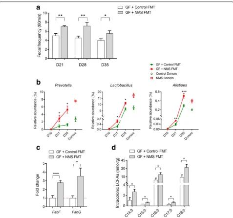
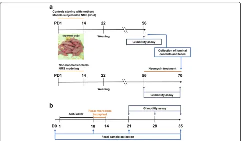
Fecal microbiota from NMS donors enhances stool frequency and intracolonic SLCFAs in pseudo GF rats To investigate whether gut microbiota of NMS rats pro-motes SLCFA production in the colon lumen and en-hances bowel motility, fecal microbiota prepared from NMS or NH donors were transplanted to pseudo GF rats (n= 6/group). The experimental process is showed in Fig.1b. The pseudo GF model was induced by provid-ing rats with water spiked with antibiotic cocktail (ABX), according to a published method [37]. During GF model establishment, rats from different cages consumed simi-lar volumes of ABX water (Additional file 1: Figure S4A), indicating there would be no difference in bacteri-cidal action for GF modeling. Fecal microbiota extracted from donors were prepared as PBS suspensions, and or-ally administrated to pseudo GF rats for 5 consecutive days. One week after fecal microbial transplant (FMT), accumulation of fecal output within 60 min was found to be significantly increased in GF rats colonized with microbiota of NMS donors (NMS FMT) relative to con-trols that has been colonized with microbiota of control rats (Control FMT); the change was maintained over the following 2 weeks (Fig. 5a). In addition, the baseline of defecation number was higher in GF recipients than conventional rats, which possibly relates to watery or shapeless stools presumably caused by continuous infu-sion of ABX water. A similar fecal phenotype has been reported in other antibiotic-induced GF studies [38,39].
Microbial DNA extracts from fecal samples of GF rats on day 0 (baseline), day 10 (after ABX treatment), day 21 (1 week after transplant), and day 35 (3 weeks after transplant) were subjected to 16s ribosomal RNA se-quencing analysis to determine the dynamic change of fecal enterotypes (Additional file2: Table S8). Compared with the baseline, dramatic loss of microbial richness and DNA integrity observed in ABX-treated rats indi-cates that the antibiotic-induced pseudo GF model was well-developed (Additional file 1: Figure S4A-C). The ecological community of feces collected from colonized GF rats was then compared with that of the donors from diversity and taxonomic perspectives. PCoA analysis showed a certain similarity of bacterial β-diversities be-tween recipients and donors (Additional file 1: Figure S4D). Specifically, changes of 49 genera from NMS do-nors, accounting for 50% of identified bacteria, also ap-peared in GF rats with NMS FMT (Additional file 2: Table S9).
increased after FMT, and such bacterial profile of GF recipients on day 35 was closer to that of donors (Fig. 5b). Quantitative polymerase chain reaction (qPCR) analysis found that relative levels of two indis-pensable LCFA-synthesized genesFabFandFabGwere increased in the colon lumens of GF rats receiving fecal microbiota of NMS donors on day 35 (Fig.5c). Consist-ently, the levels of individual SLCFAs (from C14:0 to C18:0) were obviously raised in the colon lumen (Fig.5d). These results indicate that featured enterotype
of NMS rats could result in elevation of stool frequency and SLCFA generation.
Microbial modulation effectively reduces GI motility and intracolonic SLCFA production in NMS rats treated with neomycin
To further confirm the relationship between
SLCFA-producing bacteria and bowel motility, the anti-biotic drug neomycin, previously reported as efficient in the modulation of gut dysbiosis and improvement of
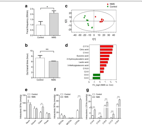
bowel symptoms in patients with IBS [40, 41], was chosen to orally administrate to NMS rats (150 mg/kg), twice daily, from postnatal days (PD) 56 to 70 (Fig. 1a). Control groups were treated with PBS. Stool frequency was monitored every 3 days during drug intervention. As shown in Fig.6a, model rats with oral gavage of PBS presented greater stool pellet output within 60 min. En-hanced defecation number was effectively attenuated and ultimately returned to normal after 14 days of neo-mycin treatment. The gut transit time became prolonged in neomycin-treated NMS rats, but was shorter than control rats (Fig.6b). Genus-specific PCR analysis found that levels of Prevotella, Lactobacillus, and Alistipes were downregulated in the colon lumens of the neomycin-treated group (Fig. 6c). Also, the levels of SLCFA synthetic genes FabF and FabG were reduced (Fig. 6d). In line with changes of SLCFA-producing bac-teria and genes, intracolonic levels of individual SLCFAs were notably attenuated after intervention (Fig.6e).
Discussion
In this study, we observed an altered gut microbiota in NMS rats characterized by increased capacity for gener-ation of SLCFAs, which were proved to enhance rat colonic contraction ex vivo and stool frequency in vivo. Further, with microbiota intervention studies includ-ing FMT and antibiotic treatment, we found that SLCFA-producing bacteria contribute to the acceler-ation of colonic motility in rats.
SLCFAs have been reported to regulate GI motility in humans, most of studies concerned their effects on the upper GI tract. A study of 10 healthy men showed that upper intestinal infusion of oils enriched with C18:0 sig-nificantly reduced gastric motility [42]. The similar in-hibitory impact of SLCFAs on the upper gut motility has also been reported in patients with FGIDs [43]. LCFA-induced slower upper GI motility influences food intake and energy metabolism, demonstrating the im-portance of SLCFAs in maintenance of body weight as well as in obesity progression [44]. However, we found no difference in the body weight and luminal SLCFAs in the small intestine between NMS rats and controls. A previous study found that increase of defecation was not accompanied by a concomitant change in food consump-tion in NMS model [45]. Thus, the upper GI motility is possibly not affected by SLCFAs in the NMS model.
The effects of LCFAs on the lower GI tract, i.e., the colon, is another story. Some studies have reported that lipids can increase colon motility in normal subjects and patients with IBS-D [26, 43]. Infusion of unsaturated LCFAs have been shown to accelerate colon transit in human [46, 47]. But the effects of SLCFAs specifically on the colon motor function have not been reported. This study is the first to provide direct evidence for the
promotional action of SLCFAs on colon motility. Com-pared with SLCFAs, short-chain fatty acids (C3:0 and C4:0) show low potency for stimulating the colonic motor [48]. Also, we found that C2:0, present increased level in fecal VOCs of NMS rats, cannot induce muscle contraction of the isolated colon. This suggests that LCFAs have distinct and specific effects on the colon. Furthermore, elevated muscle contraction induced by C18:0 can be significantly inhibited by a GRP40 antagon-ist but not a GRP120 antagonantagon-ist, indicating that GPR40 is specifically involved in the SLCFAs-stimulated colon motility. Briscoe et al. have reported that fatty acids with carbon chain lengths greater than six are able to activate GPR40, thereby giving rise to elevation of calcium secre-tion and release [49]. Such GPR40-dependent Ca2+
rise can be blocked by inhibition of L-type calcium channels or opening of the K(ATP) channel [50]. These evidences suggest that LCFAs stimulate colon motility possibly through upregulation of GPR40-dependent Ca2+ influx. We intend to explore this possibility in further study.
Generally, luminal SLCFAs are derived from either lip-olysis of dietary fats or host/bacterial fatty acid synthesis [43,51, 52]. A previous study reports normal expression of enzymes for host lipolysis, transport and lipogenesis at the mRNA level in NMS model rats [31]. Moreover, SLCFAs with odd-numbered carbon atoms, such as C15:0 and C17:0, can be produced by bacteria only [32]. If this is true then, elevation of individual SLCFAs from C14 to C18 in the colon lumens of NMS model should be the results of gut bacterial actions. In our study, the combination of metabolomic and metagenomic results points to a significant correlation between changes of dominant bacteria and levels of excretive SLCFAs. KO analysis of fecal metagenome showed obvious elevation in the level of fatty acid synthesis and elongation. These findings also support the hypothesis that gut bacteria en-gage in formation of intracolonic SLCFAs.
level of bacterial SLCFA synthesis is characteristic of the NMS-associated enterotype.
The genera Prevotella, Lactobacillus and Alistipes ex-pressing essentialFabgenes showed elevated abundances in NMS rats and GF rats receiving fecal microbiota of NMS donors, and were significantly reduced in NMS rats after neomycin treatment. Probably, these three genera are important manipulators for intracolonic SLCFA dis-ruption. Interestingly, these SLCFA-producing bacteria have been linked with host GI motility both in clinical and laboratory studies. For example, abundances of Lactobacil-lus,PrevotellaandAlistipes spp.are significantly decreased in patients with constipation [10–12]. Conversely, increase of the genusLactobacillushas been reported in patients of IBS-D [58]. Higher abundances of Prevotella spp. are found in children with chronic diarrhea [59]. The genera Prevotella, Lactobacillus or Alistipes showed increased abundance in chronic stress-induced rodents character-ized by GI dysmotility [24,25].Lactobacillusstrains accel-erate intestinal transit in GF rats and enhance ileal contraction in guinea-pig [60,61]. These lines of evidence consistently support a linkage between SLCFA-producing bacteria and host colonic motility; however, further studies are needed to identify the precise roles of specific species or strains in modulating host colonic motility.
By profiling fecal VOCs, we noticed that acidic sub-stances, including SLCFAs and fermented products (e.g., lactate and acetate) were significantly elevated in NMS group, corresponding to the raised acidity in stools. Meanwhile, increase of fecal TCA intermediates citrate and succinate in NMS rats suggest a higher metabolic level of citrate cycle. It is supported by fecal metage-nomic results that a significant enrichment of citrate cycle module in NMS group relative controls (different genes mapping in TCA cycle: 398 genes for control and 486 genes for NMS group, adjust p< 0.05). Of them, succinate can stimulate water secretion from intestinal segments [62], indicating that increased fecal consistency of NMS rats can be attributed to altered microbial me-tabolites. In contrast, fecal butyrate and valerate showed reduced levels in the NMS group. Previous study has found that butyrate is beneficial to maintenance of the intestinal epithelial barrier through facilitating tight junction assembly [63]. Soderholm et al. revealed that NMS enhances vulnerability of the colonic mucosal bar-rier to stress [64]. Probably, deficiency of bacterial butyr-ate production is one cause of the weak colonic barrier of the NMS model. This evidence indicates the import-ance of bacterial metabolites in controlling both stool characteristics and colonic barrier function.
Conclusion
This study determined that NMS rats have excessive levels of intracolonic SLCFAs, and these SLCFAs are
specifically related to accelerated colonic movement and increased fecal output. Such elevated SLCFAs is contrib-uted by hyperactive synthetic action of gut bacteria, spe-cifically by the genera Prevotella, Lactobacillus and Alistipes. Fecal microbial transplantation and antibiotic modulation revealed a causal relationship between SLCFA-producing bacteria and host colonic motility. These findings clarify the stimulatory effects of SLCFAs on the colon motility and provide novel insight in gut microbiota-driven GI dysmotility, which may lead to a therapeutic intervention targeting specific gut micro-biota for treating GI motility disorders.
Methods
Animals
All Sprague-Dawley (SD) rats used for this study were obtained from the Laboratory Animal Services Centre of The Chinese University of Hong Kong. Rats were main-tained on a 12-h light/dark cycle with free access to food and water under specific pathogen-free (SPF) condition. Materials including cages, diets, water, and litters were sterilized, and stool frequency was tested in a clean hood with a UV lamp for pseudo GF rats. All animal experi-ments have followed the Animals Ordinance, Depart-ment of Health, Hong Kong SAR, China.
Neonatal maternal separation modeling and colonic motility assessment
Referring to our previous study [65], the procedure of the NMS model was performed as shown in the timeline (Fig. 1a). Briefly, four pregnant SD rats were housed in-dividually in cages. Randomly, newborn pups from two mothers were assigned to NMS group and pups from another two dams were assigned to non-handled control group. NMS pups were separated from their dams dur-ing the period of postnatal days (PD) 2 to 14 for 3 h daily whereas control pups remained with their mothers constantly. All pups were weaned on PD 22, and only male pups (n= 8/group) with similar body weight was used for further experiments. GI motility was individu-ally evaluated in rats 8 weeks later. Fecal pellets were collected to measure pH level and water contents, and were used for analyses of metagenomics and metabolo-mics. The detail methods for assessment of motor func-tion and stool characteristics were described in Additional file1. The luminal contents of ileum (10 cm proximal to the cecum), cecum, and proximal colon (5 cm distal to the cecum) were collected after CO2
days (from PD 56 to 70). Meanwhile, other NMS or con-trol rats were orally gavaged with PBS.
Pseudo germ-free modeling and fecal microbial transfer The experimental procedure is shown in Fig. 1b. The antibiotic cocktail (ABX), containing ampicillin (1 g/L), neomycin (1 g/L), and metronidazole (0.5 g/L) were pre-pared in drinking water as previously described [37]. The ABX-contained water was ad libitum supplied for 12 normal SD rats (n= 6/group) to establish the pseudo GF model. No difference in the record of daily and total water consumption (Additional file1: Fig. S4A) indicates that rats in different cages ingested similar volumes of ABX. To ensure elimination of the gut microbiome, fecal pellets were frequently collected during experiment for real-time monitoring of dynamic changes of gut micro-biota. The very low levels of fecal total DNA quality and bacterial diversity (Additional file1: Fig. S4B and C) in-dicate that the pseudo GF model was successfully established after 10 days of ABX-water intervention. Fur-thermore, donors’ feces from either NMS or control group (n= 8/group) were individually pooled and com-pletely homogenized in pre-reduced PBS at 1 ml per pel-let referring to a published method [20]. One milliliter of the settled suspension was daily administered to pseudo GF rats for five consecutive days. The stool fre-quency was weekly determined in colonized GF rats. Fecal samples were collected on Day 0, 10, 21 and 35 for further microbial analysis, and colonic contents were collected on Day 35 for assessment of SLCFA producing genes and SLCFA amounts.
Preparation of saturated long-chain fatty acids for colonic motility assessments
To evaluate the effects of SLCFAs on host colonic motil-ity, dominant odd- and even-numbered carbon SLCFAs C17:0 and C18:0 were chosen to individually test their influences on the muscle contraction of rat isolated colon segments ex vivo and rat stool frequency in vivo.
C17:0 and C18:0 (Cat#: 506-12-7 and 57-11-4,
Sigma-Aldrich, St. Louis, MO USA) were solubilized with fatty acid free-bovine serum albumin (BSA, Cat#: 9048-46-8; Sigma-Aldrich, USA) to generate a series of concentrations (10, 30, 50 and 100μM) for organ bath system-based colonic contraction test ex vivo [66]. The concentration range were calculated in accordance with SLCFA levels found in the colon contents of NMS rats with 1.1 g/cm3 of density of the colonic contents reported in a previous study [67]. To test which long-chain fatty acid receptors mediate SLCFA-stimulated colonic contrac-tion, selective GPR40 antagonist DC260126 (DC, Cat#: 346692-04-4, Tocris Bioscience, UK) and GPR120 antag-onist AH7614 (AH, Cat#: 6326-06-3, Tocris Bioscience, UK) was prepared at working dosages of 2.5, 5, and 10μM
according to previous studies [68,69]. Acetylcholine (Ach, Cat#: 60–31-1, Sigma, St. Louis, MO USA) and C2:0 (Cat#: 127-09-3, Sigma, St. Louis, MO USA) were applied as positive and negative control, respectively. The experi-mental procedure was described in Additional file1. Fur-thermore, both SLCFAs were prepared as different dosages (1, 2.5 and 5 mg/kg, dissolved in 5% ethanol) for in vivo measurement of stool frequency in normal SD rats (n= 8/group). Each rat orally treated with either 1 mL of SLCFAs or vehicle solution was located in an individual cage for defecation frequency assessment. Fecal pellets were collected for determination of intracolonic SLCFA levels.
Total DNA isolation and metagenomic sequencing Total bacterial DNA was isolated and purified from fecal pellets (precisely weighted 200 mg) using a stool DNA Isolation Kit (Qiagen, Valencia, CA). All samples were sequenced based on the Illumina Hiseq 4000 platform (paired-end; insert size, 350 bp; read length, 150 bp). After removal of adaptor and low-quality reads, the remaining reads were filtered to eliminate the host DNA genome based on the genome reference ofRattus norve-gicus by SOAPalign v2.21 [70]. Finally, 105.81 Gb high-quality pair-end reads for the 16 rats samples (n= 8/group) was acquired with an average of 6.61 Gb per sample in groups of NMS and control (Additional file2: Table S2).
Construction of the gene catalog
The reads were assembled into contigs for all samples using the assembly software SOAPdenovo v2.0455. Aver-age 75.67% of the total reads were used to generate 1.6 million contigs without ambiguous bases (minimum length of 500 bp). ORFs were predicted from the assem-bled contigs using the MetaGeneMark v3.26 programme [71]. The 6,116,823 ORFs longer than 100 bp covered 89.67% of the total length of the contigs and about half (50.51%) of the ORFs appeared complete. All ORFs were clustered by CD-hit v4.6.4 to construct a non-redundant gene catalog using a stringent criterion of 95% identity at the nucleotide level over 90% of the length of the shorter ORFs [72]. The final non-redundant gene set contained 1,462,418 ORFs with an average length of 728 bp.
Diversity analysis, taxonomic assignment and functional characterization
BLAST (v 2.2.26, default parameter) with at least 80% overlap of query. Taxonomic identification was per-formed as 65% identity for phylum, 85% identity for genus, 95% identity for species [74, 75]. The taxonomic abundance was calculated based on gene abundance, and protein sequences of the predicted genes were searched using National Center for Biotechnology Infor-mation BLASTP against the KEGG gene database (v79). Each protein was assigned to the KEGG group by the highest scoring annotated hits. Significance in the rela-tive abundance of genes, KOs, phylum, genera, and spe-cies between both groups were compared by two-tailed Wilcoxon rank-sum test (Additional file 2: Tables S3-S7). The relationship between the abundance of each species and SLCFAs contents was assessed by the Spear-man’s correlation [76]. Enrichment in NMS or control group was then determined according to the higher rank-sum.
Determination of bacterial DNA quality and 16s ribosomal RNA amplicon sequencing
DNA extracts was prepared from feces (precisely weighted 200 mg) of donors and colonized GF rats. The DNA yield and quality were determined spectrophoto-metrically by the NanoDrop™ ND-2000 (Thermo Fisher Scientific Ltd., Waltham, MA, USA). The DNA integrity was determined through 1% agarose gel (w/v). Further-more, the extracted DNA (30 ng) was amplified with universal primers (515F and 806R) to obtain the V4 re-gions of the 16S rRNA gene. The PCR products were purified and sequenced on the Illumina Hiseq 2500 plat-form. After raw data filtering and merging [77, 78], the numbers of tag and operational taxonomic unit (OTU) were generated from 50 samples (Additional file2: Table S8). Bacterial diversity and taxonomy was obtained from OTU table by using QIIME software package [79].
PCR analysis of genus-specific bacterial 16s rDNA and SLCFA synthetic genes
Genus-specific primers for Prevotella, Lactobacillus and Alistipes were obtained from published studies [32, 80]. Bacterial DNA extracts (50 ng) of colonic contents were amplified with pairs of primers and Power SYBR GREEN Master Mix (Applied Biosystems, Foster city, CA, USA) based on an ABI StepOne Plus Sequence Detection Sys-tem (Applied BiosysSys-tems, Foster city, CA, USA). The cycling conditions were as follows: 95 °C for 10 min, followed by 40 cycles of 95 °C for 30 s, 52 °C for 30 s,
and 72 °C for 1 min. Moreover, such SYBR
GREEN-based PCR detection also applied to testify ex-pression of LCFA synthetic FabFand FabG genes from colonic contents. The primers for FabF and FabG were designed as previously described [57]. The expression of
each genus and gene was normalized to the level of 16S rRNA.
Metabolite extraction and SLCFA quantification
Fecal samples or luminal contents (100 mg) were com-pletely homogenized with five-fold volume of ice-cold distilled water. After high-speed centrifugation (13,000 rpm for 15 min at 4 °C), water extractions were transferred to a new 2-mL tube. Subsequently, a five-fold volume (500 μL) of methanol was added into the pellet sample. The mixture was completely homoge-nized and centrifuged again. Methanol extractions were combined with the previous water extractions. Strongly vortex and centrifugation again, the resulting superna-tants were obtained for derivatization processing. Mean-while, a quality control (QC) sample pooling all rat samples was prepared using the same protocol. For quantification of luminal SFAs in the small and large in-testines, external calibration solution of SFA standards from C2:0 to C18:0 were prepared at a series of concen-trations (from 1 to 20,000 ng/ml). Isotope labeled C13-myristic acid was used as internal standards.
Abso-lute quantities of individual SFAs were normalized to the sample weight.
GC/MS-based metabolomic analysis and data processing Fecal metabolites were derivatized by BSTFA with 1% TMCS based on a previously method [81]. A gas chroma-tography coupled with a mass spectrum (GCMS-QP2010 systems, Shimaduzu Co., Tokyo, Japan) was applied for fecal metabolome analysis. Fecal derivatives were sepa-rated by a DB-5 MS fused-silica capillary column (30 m × 250μm i.d.; Agilent J&W Scientific, Folsom, CA), chem-ically bonded with a 5% phenyl-95% methylpolysiloxane cross-linked stationary phase (0.25 μm film thickness). The detail sample preparation and analytical parameters of instrumental conditions were showed in Add-itional file 1. The metabolic signals were deconvoluted, aligned and normalized to final data matrix through using R-scipt with xcms package (R version 3.4.2). The processed matrix was introduced into the software SIMCA-P (Version 11.0, Umetrics, Umea, Sweden) for principal component analysis (PCA) and partial least squares-discriminant analysis (PLS-DA). Metabolic fea-tures were selected by combination of the variable im-portance in the projection (VIP) threshold (VIP > 1.0) and the Student’s t-test (p< 0.05). Metabolites were identified by the national institute of standards and technology (NIST) library (over 95% matched similarity).
Statistical analysis
Wilcoxon rank-sum test. For determination of metabo-lites, genes, and colonic motor in animal studies, Mann-Whitney test was used for statistics between two groups, and one-way ANOVA was used for comparison among more than two groups. These statistical analyses were performed in GraphPad Prism 6 (GraphPad soft-ware lnc., CA, USA). Statistical significance is defined as p< 0.05.
Additional files
Additional file 1Figure S1.Fecal characteristics and body weight in NMS rats and controls.Figure S2.Muscle amplitudes of rat isolated colonic segments with different treatment.Figure S3.Phylogenetic profiles of fecal microbiomes of NMS and control rats.Figure S4.Fecal microbial community in pseudo GF rats at duration of FMT experiment.
Table S1.Fecal metabolites with significant difference between NMS rats and controls. (DOCX 784 kb)
Additional file 2Table S2.Data production, quality control, assembly result and gene prediction resulted from fecal metagenomic sequencing analysis.Table S3.Taxonomic profiles of fecal microbiota in NMS and control groups at phylum level.Table S4.Taxonomic profiles of fecal microbiota in NMS and control groups at genus level.Table S5.
Taxonomic profiles of fecal microbiota in NMS and control groups at species level.Table S6.Correlation between species and saturated long-chain fatty acids determined by fecal metagenomic and metabolomic analyses.Table S7.Identified KOs involved in fatty acid synthesis and degradation.Table S8.Data production and quality control of fecal samples from 16s rRNA amplicon sequencing analysis.Table S9.Taxonomic changes at genus level between colonized GF rats and donors. (XLSX 73 kb)
Abbreviations
ABX:Antibiotic cocktails; Acaca: Acetyl-CoA carboxylase; C14:0: Tetradecanoic acid; C15:0: Pentadecanoic acid; C16:0: Palmitic acid; C17:0: Heptadecanoic acid; C18:0: Octadecanoic acid; C2:0: Acetic acid; C4:0: Butyric acid; C5:0: Valeric acid; FabD: Malonyl CoA-acyl carrier protein; FabF: 3-Oxoacyl-[acyl-carrier-protein] synthase II; FabG: 3-Oxoacyl-[acyl-carrier protein] reductase; FabI: Enoyl-[acyl-carrier protein] reductase I; FabZ: Hydroxyacyl-[acyl-carrier-protein] dehydratase; FadA: Acetyl-CoA acyltransferase; FadB: 3-Hydroxybutyryl-CoA dehydrogenase; FadD: Long-chain acyl-CoA synthetase; FadE: Acyl-CoA dehydrogenase; FadH: 2,4-Dienoyl-CoA reductase; FadL: Long-chain fatty acid transport protein; Fas: Fatty acid synthase (bacteria type); NMS: Neonatal maternal separation; PCoA: Principal coordinate analysis; SLCFAs: Saturated long-chain fatty acids; TesA: Acyl-CoA thioesterase I; VIP: Variable importance in the projection; VOCs: Volatile organic compounds; YciA: Acyl-CoA 12 thioesterase
Acknowledgments
We thank the colleagues of Shenzhen Key Laboratory of Human Commensal Microorganisms and Health Research and Shenzhen Engineering Laboratory of Detection and Intervention of Human Intestinal Microbiome for their assistance in microbial sequencing and analysis. We also thank Dr. Martha Dahlen (CA, USA) for her critical appraisal for this manuscript.
Funding
This project was supported by grants from Faculty Research Grant of Hong Kong Baptist University (FRG2/15-16/001 and FRG2/16-17/003), the Research Grants Council of Hong Kong Collaborative Research Fund (C2012-15G), and Guangdong-Hong Kong Technology Cooperation Funding Scheme (2016A050503039).
Availability of data and materials
Fecal metagenomic sequencing reads are deposited in the National Center for Biotechnology Information (NCBI) database with accession code PRJNA419985. Other data that support the findings of this study are available from the corresponding author upon reasonable request.
Authors’contributions
ZXB, XDF, LXZ, and LZ conceived the idea and designed this study. LZ and LL performed metabolomics, in vivo and ex vivo experiments using animals. YF and YC contributed to metagenomic and 16s rRNA sequencing analysis. WY and ZSL conducted bacterial DNA extraction and relevant PCR analysis. LZ, TH, CYL, and HLW prepared manuscript. HMY, JW, and LXZ provided guidance for both experimental design and manuscript preparation. All authors read and approved the final manuscript.
Ethics approval
The use and care of all animals were performed in accordance with the Animals Ordinance, and experimental operators obtained licenses issued by the government of the Hong Kong Special Administrative Region Department of Health, Hong Kong SAR, China.
Competing interests
The authors declare that they have no competing interests.
Publisher’s Note
Springer Nature remains neutral with regard to jurisdictional claims in published maps and institutional affiliations.
Author details
1
Chinese Medicine Clinical Study Center, Jockey Club School of Chinese Medicine, Hong Kong Baptist University, Kowloon Tong, Hong Kong SAR, China.2BGI Genomics, BGI-Shenzhen, Shenzhen, China.3Preparatory Office of Shenzhen-Melbourne Institute of Life Sciences and Bioengineering, Guangzhou University of Chinese Medicine, Guangzhou, China.4YMU-HKBU Joint Laboratory of Traditional Natural Medicine, Yunnan Minzu University, Kunming, China.5The Second Affiliated Hospital of Guangzhou University of Chinese Medicine, Guangzhou, China.6BGI-Shenzhen, Shenzhen, China. 7
Digestive Diseases and Nutrition Center, Department of Pediatrics, The State University of New York at Buffalo, 3435 Main Street, 422BRB, Buffalo, NY 14214, USA.
Received: 13 December 2017 Accepted: 1 June 2018
References
1. Corazziari E. Definition and epidemiology of functional gastrointestinal disorders. Best Pract Res Clin Gastroenterol. 2004;18:613–31. 2. Bassotti G, Antonelli E, Villanacci V, Salemme M, Coppola M, Annese V.
Gastrointestinal motility disorders in inflammatory bowel diseases. World J Gastroenterol. 2014;20:37–44.
3. Lacy BE, Weiser K. Gastrointestinal motility disorders: an update. Dig Dis. 2006;24:228–42.
4. Ohama T, Hori M, Ozaki H. Mechanism of abnormal intestinal motility in inflammatory bowel disease: how smooth muscle contraction is reduced? J Smooth Muscle Res. 2007;43:43–54.
5. Beyder A, Farrugia G. Targeting ion channels for the treatment of gastrointestinal motility disorders. Therap Adv Gastroenterol. 2012;5:5–21. 6. Hansen MB. Neurohumoral control of gastrointestinal motility. Physiol Res.
2003;52:1–30.
7. Quigley EM. Microflora modulation of motility. J Neurogastroenterol Motil. 2011;17:140–7.
8. Matto J, Maunuksela L, Kajander K, Palva A, Korpela R, Kassinen A, Saarela M. Composition and temporal stability of gastrointestinal microbiota in irritable bowel syndrome–a longitudinal study in IBS and control subjects. FEMS Immunol Med Microbiol. 2005;43:213–22.
9. Pozuelo M, Panda S, Santiago A, Mendez S, Accarino A, Santos J, Guarner F, Azpiroz F, Manichanh C. Reduction of butyrate- and methane-producing microorganisms in patients with irritable bowel syndrome. Sci Rep. 2015;5:12693.
10. Khalif IL, Quigley EM, Konovitch EA, Maximova ID. Alterations in the colonic flora and intestinal permeability and evidence of immune activation in chronic constipation. Dig Liver Dis. 2005;37:838–49.
12. de Meij TG, de Groot EF, Eck A, Budding AE, Kneepkens CM, Benninga MA, van Bodegraven AA, Savelkoul PH. Characterization of microbiota in children with chronic functional constipation. PLoS One. 2016;11:e0164731. 13. Kim SE, Choi SC, Park KS, Park MI, Shin JE, Lee TH, Jung KW, Koo HS, Myung SJ. Constipation research group of Korean Society of N, motility: change of fecal Flora and Effectiveness of the short-term VSL#3 probiotic treatment in patients with functional constipation. J Neurogastroenterol Motil. 2015;21:111–20. 14. De Palma G, Lynch MD, Lu J, Dang VT, Deng Y, Jury J, Umeh G,
Miranda PM, Pigrau Pastor M, Sidani S, et al. Transplantation of fecal microbiota from patients with irritable bowel syndrome alters gut function and behavior in recipient mice. Sci Transl Med. 2017;9https:// doi.org/10.1126/scitranslmed.aaf6397.
15. Ge X, Zhao W, Ding C, Tian H, Xu L, Wang H, Ni L, Jiang J, Gong J, Zhu W, et al. Potential role of fecal microbiota from patients with slow transit constipation in the regulation of gastrointestinal motility. Sci Rep. 2017;7:441.
16. Barbara G, Stanghellini V, Brandi G, Cremon C, Di Nardo G, De Giorgio R, Corinaldesi R. Interactions between commensal bacteria and gut sensorimotor function in health and disease. Am J Gastroenterol. 2005;100:2560–8. 17. Triantafyllou K, Chang C, Pimentel M. Methanogens, methane and gastrointestinal motility. J Neurogastroenterol Motil. 2014;20:31–40. 18. Anitha M, Vijay-Kumar M, Sitaraman SV, Gewirtz AT, Srinivasan S. Gut
microbial products regulate murine gastrointestinal motility via toll-like receptor 4 signaling. Gastroenterology. 2012;143:1006–1016 e1004. 19. Cherbut C. Motor effects of short-chain fatty acids and lactate in the
gastrointestinal tract. Proc Nutr Soc. 2003;62:95–9.
20. Yano JM, Yu K, Donaldson GP, Shastri GG, Ann P, Ma L, Nagler CR, Ismagilov RF, Mazmanian SK, Hsiao EY. Indigenous bacteria from the gut microbiota regulate host serotonin biosynthesis. Cell. 2015;161:264–76.
21. Bulbul M, Babygirija R, Cerjak D, Yoshimoto S, Ludwig K, Takahashi T. Impaired adaptation of gastrointestinal motility following chronic stress in maternally separated rats. Am J Physiol Gastrointest Liver Physiol. 2012;302:G702–11. 22. Zhang M, Leung FP, Huang Y, Bian ZX. Increased colonic motility in a rat
model of irritable bowel syndrome is associated with up-regulation of L-type calcium channels in colonic smooth muscle cells. Neurogastroenterol Motil. 2010;22:e162–70.
23. Bian ZX, Zhang M, Han QB, Xu HX, Sung JJ. Analgesic effects of JCM-16021 on neonatal maternal separation-induced visceral pain in rats. World J Gastroenterol. 2010;16:837–45.
24. De Palma G, Blennerhassett P, Lu J, Deng Y, Park AJ, Green W, Denou E, Silva MA, Santacruz A, Sanz Y, et al. Microbiota and host determinants of behavioural phenotype in maternally separated mice. Nat Commun. 2015;6:7735.
25. Pusceddu MM, El Aidy S, Crispie F, O'Sullivan O, Cotter P, Stanton C, Kelly P, Cryan JF, Dinan TG. N-3 polyunsaturated fatty acids (PUFAs) reverse the impact of early-life stress on the gut microbiota. PLoS One. 2015;10:e0139721.
26. Deiteren A, Camilleri M, Burton D, McKinzie S, Rao A, Zinsmeister AR. Effect of meal ingestion on ileocolonic and colonic transit in health and irritable bowel syndrome. Dig Dis Sci. 2010;55:384–91.
27. Lin CY, Zhang M, Huang T, Yang LL, Fu HB, Zhao L, Zhong LL, Mu HX, Shi XK, Leung CF, et al. Spexin enhances bowel movement through activating L-type voltage-dependent Calcium Channel via Galanin receptor 2 in mice. Sci Rep. 2015;5:12095.
28. Edfalk S, Steneberg P, Edlund H. Gpr40 is expressed in enteroendocrine cells and mediates free fatty acid stimulation of incretin secretion. Diabetes. 2008;57:2280–7.
29. Paulsen SJ, Larsen LK, Hansen G, Chelur S, Larsen PJ, Vrang N. Expression of the fatty acid receptor GPR120 in the gut of diet-induced-obese rats and its role in GLP-1 secretion. PLoS One. 2014;9:e88227.
30. Ichimura A, Hirasawa A, Hara T, Tsujimoto G. Free fatty acid receptors act as nutrient sensors to regulate energy homeostasis. Prostaglandins Other Lipid Mediat. 2009;89:82–8.
31. Paternain L, Martisova E, Milagro FI, Ramirez MJ, Martinez JA, Campion J. Postnatal maternal separation modifies the response to an obesogenic diet in adulthood in rats. Dis Model Mech. 2012;5:691–7.
32. Kumagai H, Maisawa S, Tanaka M, Takahashi M, Takasago Y, Nishijima A, Watanabe S. Intestinal microbiota and secretory immunoglobulin a in feces of exclusively breast-fed infants with blood-streaked stools. Microbiol Immunol. 2012;56:657–63.
33. Qiu X, Choudhry AE, Janson CA, Grooms M, Daines RA, Lonsdale JT, Khandekar SS. Crystal structure and substrate specificity of the
beta-ketoacyl-acyl carrier protein synthase III (FabH) from Staphylococcus aureus. Protein Sci. 2005;14:2087–94.
34. Cronan JE, Thomas J. Bacterial fatty acid synthesis and its relationships with polyketide synthetic pathways. Methods Enzymol. 2009;459:395–433. 35. Schweizer E, Hofmann J. Microbial type I fatty acid synthases (FAS): major
players in a network of cellular FAS systems. Microbiol Mol Biol Rev. 2004;68: 501–17. table of contents
36. Dellomonaco C, Clomburg JM, Miller EN, Gonzalez R. Engineered reversal of the beta-oxidation cycle for the synthesis of fuels and chemicals. Nature. 2011;476:355–9.
37. Ijssennagger N, Belzer C, Hooiveld GJ, Dekker J, van Mil SWC, Muller M, Kleerebezem M, van der Meer R. Gut microbiota facilitates dietary heme-induced epithelial hyperproliferation by opening the mucus barrier in colon. Proc Natl Acad Sci U S A. 2015;112:10038–43.
38. Nicklas W, Keubler L, Bleich A. Maintaining and monitoring the defined microbiota status of Gnotobiotic rodents. ILAR J. 2015;56:241–9. 39. Ge X, Ding C, Zhao W, Xu L, Tian H, Gong J, Zhu M, Li J, Li N.
Antibiotics-induced depletion of mice microbiota induces changes in host serotonin biosynthesis and intestinal motility. J Transl Med. 2017;15:13.
40. Basseri RJ, Weitsman S, Barlow GM, Pimentel M. Antibiotics for the treatment of irritable bowel syndrome. Gastroenterol Hepatol (N Y). 2011;7:455–93. 41. Ahmad OF, Akbar A. Microbiome, antibiotics and irritable bowel syndrome.
Br Med Bull. 2016;120:91–9.
42. French SJ, Conlon CA, Mutuma ST, Arnold M, Read NW, Meijer G, Francis J. The effects of intestinal infusion of long-chain fatty acids on food intake in humans. Gastroenterology. 2000;119:943–8.
43. Feinle-Bisset C, Azpiroz F. Dietary lipids and functional gastrointestinal disorders. Am J Gastroenterol. 2013;108:737–47.
44. Matzinger D, Degen L, Drewe J, Meuli J, Duebendorfer R, Ruckstuhl N, D'Amato M, Rovati L, Beglinger C. The role of long chain fatty acids in regulating food intake and cholecystokinin release in humans. Gut. 2000;46:688–93. 45. Coutinho SV, Plotsky PM, Sablad M, Miller JC, Zhou H, Bayati AI, McRoberts
JA, Mayer EA. Neonatal maternal separation alters stress-induced responses to viscerosomatic nociceptive stimuli in rat. Am J Physiol Gastrointest Liver Physiol. 2002;282:G307–16.
46. Spiller RC, Brown ML, Phillips SF. Decreased fluid tolerance, accelerated transit, and abnormal motility of the human colon induced by oleic acid. Gastroenterology. 1986;91:100–7.
47. Kamath PS, Phillips SF, O'Connor MK, Brown ML, Zinsmeister AR. Colonic capacitance and transit in man: modulation by luminal contents and drugs. Gut. 1990;31:443–9.
48. Hurst NR, Kendig DM, Murthy KS, Grider JR. The short chain fatty acids, butyrate and propionate, have differential effects on the motility of the Guinea pig colon. Neurogastroenterol Motil. 2014;26:1586–96.
49. Briscoe CP, Tadayyon M, Andrews JL, Benson WG, Chambers JK, Eilert MM, Ellis C, Elshourbagy NA, Goetz AS, Minnick DT, et al. The orphan G protein-coupled receptor GPR40 is activated by medium and long chain fatty acids. J Biol Chem. 2003;278:11303–11.
50. Shapiro H, Shachar S, Sekler I, Hershfinkel M, Walker MD. Role of GPR40 in fatty acid action on the beta cell line INS-1E. Biochem Biophys Res Commun. 2005;335:97–104.
51. Niot I, Poirier H, Tran TT, Besnard P. Intestinal absorption of long-chain fatty acids: evidence and uncertainties. Prog Lipid Res. 2009;48:101–15. 52. Smith S, Witkowski A, Joshi AK. Structural and functional organization of the
animal fatty acid synthase. Prog Lipid Res. 2003;42:289–317. 53. Parsons JB, Rock CO. Bacterial lipids: metabolism and membrane
homeostasis. Prog Lipid Res. 2013;52:249–76.
54. Heath RJ, Rock CO. Enoyl-acyl carrier protein reductase (fabI) plays a determinant role in completing cycles of fatty acid elongation in Escherichia coli. J Biol Chem. 1995;270:26538–42.
55. Jeon E, Lee S, Lee S, Han SO, Yoon YJ, Lee J. Improved production of long-chain fatty acid in Escherichia coli by an engineering elongation cycle during fatty acid synthesis (FAS) through genetic manipulation. J Microbiol Biotechnol. 2012;22:990–9.
56. Garwin JL, Klages AL, Cronan JE Jr. Beta-ketoacyl-acyl carrier protein synthase II of Escherichia coli. Evidence for function in the thermal regulation of fatty acid synthesis. J Biol Chem. 1980;255:3263–5. 57. Chen P, Torralba M, Tan J, Embree M, Zengler K, Starkel P, van Pijkeren JP,
58. Carroll IM, Chang YH, Park J, Sartor RB, Ringel Y. Luminal and mucosal-associated intestinal microbiota in patients with diarrhea-predominant irritable bowel syndrome. Gut Pathog. 2010;2:19.
59. Gilchrist CA, Petri SE, Schneider BN, Reichman DJ, Jiang N, Begum S, Watanabe K, Jansen CS, Elliott KP, Burgess SL, et al. Role of the gut microbiota of children in diarrhea due to the protozoan parasite Entamoeba histolytica. J Infect Dis. 2016;213:1579–85.
60. Husebye E, Hellstrom PM, Sundler F, Chen J, Midtvedt T. Influence of microbial species on small intestinal myoelectric activity and transit in germ-free rats. Am J Physiol Gastrointest Liver Physiol. 2001;280:G368–80. 61. Massi M, Ioan P, Budriesi R, Chiarini A, Vitali B, Lammers KM, Gionchetti P,
Campieri M, Lembo A, Brigidi P. Effects of probiotic bacteria on
gastrointestinal motility in Guinea-pig isolated tissue. World J Gastroenterol. 2006;12:5987–94.
62. Inagaki A, Ichikawa H, Sakata T. Inhibitory effect of succinic acid on epithelial cell proliferation of colonic mucosa in rats. J Nutr Sci Vitaminol (Tokyo). 2007;53:377–9.
63. Peng L, Li ZR, Green RS, Holzman IR, Lin J. Butyrate enhances the intestinal barrier by facilitating tight junction assembly via activation of AMP-activated protein kinase in Caco-2 cell monolayers. J Nutr. 2009;139:1619–25. 64. Soderholm JD, Yates DA, Gareau MG, Yang PC, MacQueen G, Perdue MH.
Neonatal maternal separation predisposes adult rats to colonic barrier dysfunction in response to mild stress. Am J Physiol Gastrointest Liver Physiol. 2002;283:G1257–63.
65. Chung EK, Zhang XJ, Xu HX, Sung JJ, Bian ZX. Visceral hyperalgesia induced by neonatal maternal separation is associated with nerve growth factor-mediated central neuronal plasticity in rat spinal cord. Neuroscience. 2007;149:685–95.
66. Huang S, Rutkowsky JM, Snodgrass RG, Ono-Moore KD, Schneider DA, Newman JW, Adams SH, Hwang DH. Saturated fatty acids activate TLR-mediated proinflammatory signaling pathways. J Lipid Res. 2012;53:2002–13. 67. Lupton JR, Ferrell RG. Using density rather than mass to express the
concentration of gastrointestinal tract constituents. J Nutr. 1986;116:164–8. 68. Hu H, He LY, Gong Z, Li N, Lu YN, Zhai QW, Liu H, Jiang HL, Zhu WL, Wang
HY. A novel class of antagonists for the FFAs receptor GPR40. Biochem Biophys Res Commun. 2009;390:557–63.
69. Houthuijzen JM, Oosterom I, Hudson BD, Hirasawa A, Daenen LGM, McLean CM, Hansen SVF, van Jaarsveld MTM, Peeper DS, Jafari Sadatmand S, et al. Fatty acid 16:4(n-3) stimulates a GPR120-induced signaling cascade in splenic macrophages to promote chemotherapy resistance. FASEB J. 2017; 31:2195–209.
70. Li R, Yu C, Li Y, Lam TW, Yiu SM, Kristiansen K, Wang J. SOAP2: an improved ultrafast tool for short read alignment. Bioinformatics. 2009;25:1966–7. 71. Zhu W, Lomsadze A, Borodovsky M: Ab initio gene identification in
metagenomic sequences. Nucleic Acids Res 2010, 38:e132.
72. Li W, Godzik A. Cd-hit: a fast program for clustering and comparing large sets of protein or nucleotide sequences. Bioinformatics. 2006;22:1658–9. 73. Liu R, Hong J, Xu X, Feng Q, Zhang D, Gu Y, Shi J, Zhao S, Liu W, Wang X,
et al. Gut microbiome and serum metabolome alterations in obesity and after weight-loss intervention. Nat Med. 2017;23:859–68.
74. Qin J, Li R, Raes J, Arumugam M, Burgdorf KS, Manichanh C, Nielsen T, Pons N, Levenez F, Yamada T, et al. A human gut microbial gene catalogue established by metagenomic sequencing. Nature. 2010;464:59–65. 75. Feng Q, Liang S, Jia H, Stadlmayr A, Tang L, Lan Z, Zhang D, Xia H, Xu X, Jie
Z, et al. Gut microbiome development along the colorectal adenoma-carcinoma sequence. Nat Commun. 2015;6:6528.
76. Karlsson FH, Tremaroli V, Nookaew I, Bergstrom G, Behre CJ, Fagerberg B, Nielsen J, Backhed F. Gut metagenome in European women with normal, impaired and diabetic glucose control. Nature. 2013;498:99–103. 77. Magoc T, Salzberg SL. FLASH: fast length adjustment of short reads to
improve genome assemblies. Bioinformatics. 2011;27:2957–63.
78. Edgar RC. UPARSE: highly accurate OTU sequences from microbial amplicon reads. Nat Methods. 2013;10:996–8.
79. Caporaso JG, Kuczynski J, Stombaugh J, Bittinger K, Bushman FD, Costello EK, Fierer N, Pena AG, Goodrich JK, Gordon JI, et al. QIIME allows analysis of high-throughput community sequencing data. Nat Methods. 2010;7:335–6. 80. Qiu XY, Li X, Wu Z, Zhang F, Wang N, Wu N, Yang X, Liu YL. Fungal-bacterial
interactions in mice with dextran sulfate sodium (DSS)-induced acute and chronic colitis. RSC Adv. 2016;6:65995–6006.
81. Gao X, Pujos-Guillot E, Sebedio JL. Development of a quantitative metabolomic approach to study clinical human fecal water metabolome
![Fig. 4 Alteration of fecal microbiome is closely associated with SLCFA generation in NMS rats (carboxylase; Fas, fatty acid synthase (bacteria type); FabD, Malonyl CoA-acyl carrier protein; FabF, 3-oxoacyl-[acyl-carrier-protein] synthase II; FabG,3-oxoacyl](https://thumb-us.123doks.com/thumbv2/123dok_us/9069840.1899434/7.595.60.538.87.593/alteration-microbiome-associated-generation-carboxylase-synthase-malonyl-synthase.webp)