21
Preliminary Evaluation of Learning Performance of the Simplest Bovine
Trans-rectal Palpation Phantom for Training Veterinary Students
Zolhavarieh, S. M.1*; Sadeghi-nasab, A.1; Ghanbari, S.2; Mirshokraei, P.3; Ruhi athar, M.4
1- Department of Clinical Sciences, Faculty of Veterinary Science, Bu-Ali Sina University, Hamedan, Iran.
2- Department of Educational Sciences, Faculty of Humanities, Bu-Ali Sina University, Hamedan, Iran.
3- Department of Clinical Sciences, School of Veterinary Medicine, Ferdowsi University of Mashhad, Mashhad,
Iran.
4- Department of Foreign languages, Farhangian University. Hamedan, Iran.
*
Correspondence:Zolhavarieh, S. M.
Email: [email protected] , [email protected]
Abstract
Traditionally, animal exploitation in veterinary education in disciplines such as obstetrics is common worldwide. In
addition, it is clear that veterinary schools are expected to provide sufficient opportunity for developing students’ necessary
skill in bovine trans-rectal palpation by graduation. However, the veterinary medical education should be refined and
animal exploitation in education be superseded by using non-harmful alternatives. Thus, a phantom was developed as a
potential alternative to overcome the present educational problem in Bu-Ali Sina University.The phantom was composed of
a fiberglass rear-half of the cow that was designed with detailed inside structures of pelvis and abdominal cavity of cow.
Two opening on the cranial and dorsal aspects of the phantom were made to provide proper guidance while an instructor
followed a student’s hand movement. By installing a slaughterhouse-derived cow reproductive tract inside the pelvis and a
real rectum above it, an approximately real environment was provided. To find out how it can be useful in the training of
students on cow trans-rectal examination, 31 veterinary theriogenologists and 110 fifth-year veterinary students were asked
to fill out a developed attitude questionnaire anonymously. The results of instructors' and students' responses to the
questionnaire indicated that the Phantom provided approximately a realistic physical environment and a comfortable and
satisfying approach for teaching/learning cow trans-rectal examination. Accordingly, we can embed the phantom as a
supplementary teaching tool in the bovine reproduction course of veterinary medical curriculum.
Keywords: Cow, Bovine trans-rectal palpation, Education, Phantom, Slaughterhouse-derived reproductive tract
Introduction
Contemporary dairy and beef farming focus on
combining high production with acceptable fertility.
Therefore, the early diagnosis of reproductive
disorders and early detection of pregnancy are critical
points for improving a herd’s production status. Thus,
22 IJRHR (2016), 1(1): 21-30
considered central to bovine reproduction
examination in veterinary medicine practice (Bossaert
et al. 2009). Accordingly, knowing the trans-rectal
examination is an indispensable skill for veterinarians.
In the traditional didactic program, teaching of
trans-rectal palpation to veterinary students is carried
out in live cows. In addition to this fact that it is an
extremely expensive training method in any
department of theriogenology and that the
ever-growing financial burden affects the budget of
veterinary medical schools, there are several reports
about risk of abortion or animal death due to rectal
palpation, particularly when it is carried out by novice
students (Bossaert et al. 2009). If the financial
problems are accepted even in part, animal
exploitation in education should be superseded by
using non-harmful alternatives because it is strongly
against the animal rights.
To that end, some alternative educational
methods have been considered to compensate
insufficient traditional training in veterinary medical
curriculum. Educational simulation has emerged at
the forefront of technologies and processes when it
comes to the education and training of veterinary
medical students (Salazar, 2002).
Many studies have indicated that the use of
simulators, compared to the traditional hands-on
methods, provides comparable results (Akpan, 2001;
Bernardo, 2003; Scherzer et al. 2010). Other
researchers, nevertheless, argue against these
alternatives and claim that 3D virtual reality
modalities, Haptic technology, computer simulations
and high quality videos are not comparable with real
touch of tissues and organs (Akpan, 2001;Aziz et al.
2002). They claim that throughout the training,
students should be able to know how to use their
hands and they should develop their touch-based
skills (Older, 2004). This is due to the fact that this
sense is a very important means of apprehending
substantial reality and that the most tangible evidence
of the actual existence in our environment is
touch-based (Aziz et al. 2002; Older, 2004; Wolkomir,
2000).
Although developing such skills in the students
of veterinary medicine is necessary, there are few
animal simulators designed specifically for using in
veterinary education and it may incite the further
development of such technologies (Scalese and
Issenberg, 2005).
Development of cow trans-rectal palpation
simulators has long been considered by researchers.
For example, Bovine Rectal Palpation Simulator (at
the University of Glasgow, UK, 2003) and Real-time
Visio-Haptic Deformable Bovine Rectal Palpation
Simulator (at the Universiti Teknologi PETRONAS,
Malaysia, 2010) have been developed as the teaching
tools to supplement the existing training methods
(Ahmad and Sulaiman, 2010; Baillie et al. 2003).
These simulators involving a phantom haptic device
have focused on basic fertility examination and
diagnosis pregnancy skills, such as touching a virtual
cervix, uterus and ovaries (Ahmad and Sulaiman,
2010; Baillie et al. 2005). Recently, the Breed’n Betsy
simulation model, consisting of an artificial cow’s
pelvis surrounded by a metal frame, in which an
artificial vulva and anal sphincter are installed, has
been developed (Bossaert et al. 2009).
It is clear that they cannot provide the whole
predictable conditions in a real reproductive system
23
complete procedure (Kustritz et al. 2009).
Furthermore, simulators are very expensive and
cannot provide all anatomical variations (Aziz et al.
2002).
As a remedy, a Bovine Trans-rectal Palpation
Phantom has been developed in Bu-Ali Sina
University as the simplest and the most affordable
supplementary teaching tool for training the manual
bovine trans-rectal examination. Although the use of
models in veterinary education was demonstrated as a
method accordance with animal welfare
considerations (Knight, 2008), it follows that the
study aimed to determine the preliminary validation
of this phantom as a proper supplementary teaching
tool to embed in the bovine reproduction course of
veterinary medical curriculum.
Materials and methods
Materials
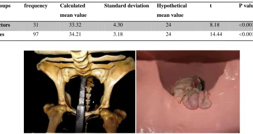
The Bovine Trans-rectal Palpation Phantom was
composed of a fiberglass rear-half of the cow that was
designed with detailed inside structures of pelvis and
abdominal cavity and was based on the anatomy of a
dual-purpose Iranian-Holstein mixed breed (Fig. 1).
Also, at the caudal part of its body, two openings
were built as anus and vulva. In addition, three hooks
were installed to secure a slaughterhouse-derived
rectum in its anatomic location above the vagina. For
installing a slaughterhouse-derived reproductive tract
of cow, inside the pelvis and ventral to the rectum, a
rigid pipe was attached proximal to the opening of
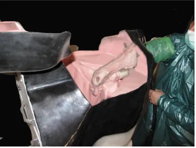
vulva (Fig. 2). Cranial and dorsal parts of phantom
were open to provide proper guidance while an
instructor followed a student’s hand movement inside
the pelvis (Fig. 3).
Purpose
The validation strategy for this phantom was
aimed towards answering three basic research
questions according to users’ opinions:
1: Can practice with the Bovine Trans-rectal
Palpation Phantom simulate performance in actual
environment for teaching/learning purpose?
2: Is the learning practice made comfortable by
using this phantom?
3: Is there any learning satisfaction when the
phantom is used for teaching the manual trans-rectal
palpation?
Data Collection Procedure
The descriptive-survey involved fifth-year
veterinary students and veterinary theriogenologists.
During the first stage of the study, students were
theoretically taught by using just traditional materials.
In the following stage, the Bovine Trans-rectal
Palpation Phantom was embedded in the lecture
materials. In the third stage, students performed
trans-rectal palpation under close supervision on-farm
situation. Finally, based on Cochran formula, 31
instructors (theriogenologists) and 110 fifth-year
veterinary students were selected from the population
by employing simple random sampling. All
instructors and students were asked to fill out
questionnaires developed and distributed among them
24 IJRHR (2016), 1(1): 21-30
statements that assessed the reality (10 questions),
comfortable learning (6 questions) and learning
satisfaction (8 questions). The modal response was in
the form of a Likert scale, consisting of five
categories and ranging from "Very Much" (score 5) to
"Very Little" (score 1) for all of the statements.
Data Analysis
Content validity of questionnaires was confirmed
by 16 experts of veterinary theriogenology and
educational measurement. The results of Cronbach
alpha (0.86) indicated high reliability of the
questionnaires. The results of Kolmogorov-Smirnov
test showed that the distribution of the data was
normal. Since the research adopted interval scale and
the data was normal, the researcher employed
one-sample t-test. To calculate the assumed mean for each
variable, the number of the questions was multiplied
by the mean of total responses ranging from 5 (very
much) to 1 (very little). Accordingly, the assumed
means calculated were 30, 30 and 24 for feeling
reality, comfortable learning and learning satisfaction
respectively. The calculated mean was equal to the
mean of the total responses provided by the
participants filling out the questionnaire. The data was
submitted to SPSS (version 18) in order to calculate
the t-test.
Results
The male teachers (n=31/31) and 97 students
(43/97 men and 54/97 women) responded to the
questionnaire. The findings showed that there were no
significant differences between men and women
concerning all responses (p<0.001).
According to Table 1, the values of “t” at the level of α=0.001 are more than “critical t values”
indicating significant differences between calculated
mean values and hypothetical mean values in both
groups of instructors and trainees. Because the
calculated mean values (in both groups) are more than
the hypothetical mean value (=30), we can conclude
that the phantom can create approximately realistic
physical environment. Furthermore, Table 2 shows
that the values of “t” at the level of α=0.001 are more than “critical t values” that indicate significant
differences between calculated mean values and
hypothetical mean values in both groups. Because the
calculated mean values (based on instructors’ and trainees’ opinions) are more than the hypothetical
mean value (=30), it can be concluded that learning of
the cow trans-rectal examination using this device is
very comfortable. Moreover, Table 3 reveals the
learning satisfaction on the basis of users’ opinions, because the values of “t” at the level of α=0.001 are more than “critical t values” indicating significant
differences between calculated mean values and
hypothetical mean values in both groups. Because the
calculated mean values (based on instructors’ and trainees’ opinions) are more than the hypothetical
mean value (=24), it can be concluded that there is a
25 Table 1. T-values for both groups’ feeling of reality
Groups frequency Calculated
mean value
Standard deviation Hypothetical
mean value
t P value
Instructors 31 37.16 9.88 30 4.03 <0.001
Trainees 97 34.21 7.30 30 5.67 <0.001
Table 2. T-values for both groups’ feeling of comfort
Groups frequenc
y
Calculated
mean value
Standard deviation Hypothetical
mean value
t P value
Instructor
s
31 22.35 3.14 30 7.73 <0.001
Trainees 97 21.97 3.15 30 12.39 <0.001
Table 3. T-values for both groups’ learning satisfaction
Groups frequency Calculated
mean value
Standard deviation Hypothetical
mean value
t P value
Instructors 31 33.32 4.30 24 8.18 <0.001
Trainees 97 34.21 3.18 24 14.44 <0.001
Figure 1: The Bovine Trans-Rectal Palpation Phantom has been designed with detailed inside structures of pelvis
26 IJRHR (2016), 1(1): 21-30
Figure 2: For installing a slaughterhouse-derived reproductive tract of cow, inside the pelvis and ventral to the
rectum, a rigid pipe was attached proximal to the opening of vulva.
Figure 3: Cranial and dorsal parts of phantom are open to provide proper guidance while instructor follows the student’s
hand movement inside the pelvis.
Discussion
The manual trans-rectal palpation is a
common veterinary practice applied to
reproduction programs management because
accessibility of reproductive structure from
rectum is an easy, cost-effective, accurate
and efficient method. Thus, it should be
acquired by veterinary students, especially if
they expect a professional career in bovine
reproduction (Simões, 2012). A traditional
method for the purpose of training students
involves using live animals in
slaughterhouses (Lopes and Rocha, 2006). It
is a traumatic and stressful situation for
animals; however, in the slaughterhouses,
minimized stress for animals should be
provided just before they are dead. Some
partial solutions for the student training
purposes were studied, like the use of the 3D
animations (Scherzer et al. 2010) or
computer assisted learning without living
cows. In response to the need for finding the
ways of supplementing existing methods for
training veterinary students to perform
bovine trans-rectal palpation with animal
welfare considerations, a Bovine Trans-rectal
Palpation Phantom has been developed as a
27 Probably, using the Bovine Trans-rectal
Palpation Phantom, like other simulators, can
provide a standardized experience for all
trainees without showing unpredictable
behaviors such as fatigue.
Moreover, it can be readily available at
any time and place without the limitation in
the number of examinations allowed. Due to
the fact that the simulation represents a safe
environment and there are no bad
consequences when the trainees make
mistakes, they can learn from their mistakes
and correct them in simulation environment.
Another issue that is closely related to safety
is animal welfare considerations that may
raise some ethical questions about the
exploitation of a real patient as an
educational resource while the simulator does
not have the same ethical and welfare
limitations.
Although cadaver is mostly applicable in
the teaching of anatomy, in the traditional
bovine reproductive teaching, live cows are
used (Aziz et al. 2002; Lopes and Rocha,
2006), because the understanding of anatomy
of live organs and anatomical variations are
very important components (Zucconi et al.
2002). However, most of simulators cannot
create environment just like real-life (Aziz et
al. 2002; Older, 2004). For instance, the
users' comments (trainees and teachers) of
the Bovine Rectal Palpation Simulator at the
University of Glasgow indicated that the
subjects have not felt a full reality of the
rectal environment because of the absence of
feces and peristaltic contractions. Another
limitation is related to trainees' single finger
contact with haptic device because detailed
examination of ovaries and membrane slip
requires the use of all fingers (Baillie et al.
2005). Thus, these limitations reduce the
fidelity of this simulator.
To compensate this deficiency, the
Bovine Trans-rectal Palpation Phantom was
equipped with a slaughterhouse-derived real
bovine reproductive organ. Accordingly,
there is no limitation to palpate the whole of
reproductive tract by all fingers that provide
high-fidelity three-dimensional interaction
for examiner. The results of Table 1 show
that instructors and trainees felt a realistic
environment when they used the phantom,
although there were some comments
claiming that it was better if there was a
broad ligament and free border of broad
ligament could be found. Furthermore, while
instructors can provide the students with the
sessions for any states of reproduction cycle
and gestation status, the Bovine Rectal
Palpation Simulator at the University of
Glasgow and the Real-time Visio-Haptic
Bovine Rectal Palpation Simulator at the
Universiti Teknologi PETRONAS can only
conduct basic skills in the trans-rectal
palpation independently of the complete
28 IJRHR (2016), 1(1): 21-30
pregnancy and some anatomical variations
(Ahmad and Sulaiman, 2010; Baillie et al.
2003).
According to the users' comments
(trainees and teachers) of the phantom in the
present research and other studies, the cow
trans-rectal palpation is difficult to learn and
perform by novice veterinary students and
requires repetitive practice to get more
experience in the identification of structures
palpated (Baillie et al. 2005; Penny, 2002).
Thus, during the teaching trans-rectal
palpation with a real cow, the teacher should
provide the students guidance as they
perform the procedure (Simões, 2012).
However, providing effective guidance can
be difficult because the students' hand
movement is not visible and they cannot
describe the palpated structures carefully;
therefore, the teaching method can be both
limited and variable by using live cow
(Baillie et al. 2005). According to the
instructors and students, the teacher is able to
provide more effective guidance and
feedback on their performance during the
training by this device via following the
student’s hand movement. It was confirmed on users’ opinions.
Although some comments of students
and instructors who have used the Bovine
Trans-rectal Palpation Phantom stressed the
appropriacy of the device for learning trans-
rectal palpation (Table 3), they mentioned
that it was not sufficient by itself. This
limitation is not unique to our study but true
for other devices in this field too (Bossaert et
al. 2009). We should consider the potentiality
of this device to supplement the existing
teaching methods.
Since this device will enable students to
make more effective use of animals as a
learning resource, bovine trans-rectal
palpation course should be comprised of
several stages (Knight, 2007). At first,
students learn the procedure theoretically.
Then, they progress to simulated procedure
by the Bovine Trans-rectal Palpation
Phantom. Finally, students perform
trans-rectal palpation under close supervision
on-farm situation for practical concerns.
According to the comments written by the
instructors and students, this phantom
illustrated some additional benefits including
time and cost savings, increased student
confidence and the saving of substantial
numbers of animal lives who might be
injured throughout veterinary courses
(Simões, 2012).
It is expected to embed the
supplementary teaching tools in veterinary
curriculum and emphasize the use of these
devices to develop student’s understands of
animal welfare science and animal welfare
issues, not only during their education but
29 References
Ahmad, I. and Sulaiman, S. (2010). Evaluation
of Real-Time Visio-Haptic Deformable
Bovine Rectal Palpation Simulatorn; jv
wdtd. Paper presented at International
Symposium in the Information
Technology (ITSim).
ieeexplore.ieee.org.
Akpan, J. P. (2001). Issues associated with
inserting computer simulations into
biology instruction: A review of the
literature. Electronic Journal of Science
Education, 5(3).
Aziz, M. A., Mckenzie, J. C., Wilson, J. S.,
Cowie, R. J., Ayeni, S. A. and Dunn, B.
K. (2002). The human cadaver in the
age of biomedical informatics. The
Anatomical Record, 269, 1, 20-32.
Baillie, S., Crossan, A., Brewster, S., Mellor, D.
and Reid, S. (2005). Validation of a
bovine rectal palpation simulator for
training veterinary students. Studies in
Health Technology and Informatics,
111, 33-36.
Baillie, S., Crossan, A., Reid, S. and Brewster,
S. (2003). Preliminary development and
evaluation of a bovine rectal palpation
simulator for training veterinary
students. Cattle Practice, 11, 2, 101-106.
Baillie, S., Mellor, D. J., Brewster, S. A. and
Reid, S. W. J. (2005). Integrating a
bovine rectal palpation simulator into an
undergraduate veterinary curriculum.
Journal of Veterinary Medical
Education, 32, 1, 79-85.
Bernardo, T. M. (2003). New technology
imperatives in medical education.
Journal of Veterinary Medical
Education, 30. 4.
Bossaert, P., Leterme, L., Caluwaerts, T., Cools,
S., Hostens, M., Kolkman, I. (2009).
Teaching transrectal palpation of the
internal genital organs in cattle. Journal
of Veterinary Medical Education, 36, 4,
451-460.
Knight, A. (2007). Humane teaching methods in
veterinary education. Veterinary
Review, 126, 16-21.
Knight, A. (2008). Humane Teaching Methods Demonstrate Efficacy in Veterinary Education. Responsibilities–The 4th R, 119.
Kustritz, R., Ghenowet, PJ and Tibary, A.
(2009). Efficacy of training in
theriogenology as determined by a
survey of veterinarians. Journal of
American Veterinary Medical
Association, 229, 514-521.
Lopes, G. and Rocha, A. (2006). Teaching
bovine rectal palpation with live cows in
the slaughterhouse: Is it worthwhile?
Reproduction in Domestic Animals, 41,
6, 510-513.
Older, J. (2004). Anatomy: A must for teaching
the next generation. The Surgeon, 2, 2,
30 IJRHR (2016), 1(1): 21-30
Penny, C. (2002). Education-A University View.
Cattle Practice, 10, 4, 255-256.
Salazar, I. (2002). Coming changes in veterinary
anatomy: what is or should be expected?
Journal of Veterinary Medical
Education, 29, 3, 126-130.
Scalese, R. J., and Issenberg, S. B. (2005).
Effective use of simulations for the
teaching and acquisition of veterinary
professional and clinical skills. Journal
of Veterinary Medical Education, 32, 4,
461.
Scherzer, J., Buchanan, M. F., Moore, J. N., and
White, S. L. (2010). Teaching veterinary
obstetrics using three-dimensional
animation technology. Journal of
Veterinary Medical Education, 37, 3,
299-303.
Simões, J. (2012). Manual transrectal palpations
performed by veterinary. REDVET -
Revista electrónica de Veterinaria, 13, 3,
181-188.
Wolkomir, R. (2000). Charting the terrain of
touch. Smithsonian, 31(3), 38.
Zucconi, W. B., Guelfguat, M., and Solounias,
N. (2002). Approach to the educational
opportunities provided by variant
anatomy, illustrated by discussion of a
duplicated inferior vena cava. Clinical