[ 77 ]
III.
Some Notes on Soil Protozoa.B y C. H. Martin, M .A ., and K. R Le w in, B .A ., Rothamsted Experim ental
Station.
Communicated by Horace T. Brow n, F .R .S.
(Received December 5, 1913,—Read February 5, 1914.)
[Plates 5 a n d 6.]
Contents.
I. Introduction ...
II. Methods of Preparation and C u l t u r e ... ... . . III. Cucumber bed— Vahlkampfia soli, n. sp., Amoeba cucumis, n. sp IY. Seedling bed—Amoeba gobanniensis, n. sp...
Y. Conclusions. . ... ... VI. Literature List . ...
VII. Description of P l a t e s ...
Introduction.
I t is rather a curious fact th a t it has been left to agricultural chemists to bring into prominence the im portant p art th a t free-living Protozoa may play in the soil. A t the present time it seems to us th a t the prevalent idea as regards the distribution of free-living protozoa is th a t they can exist in the trophic state only in definite accumulations of fresh w ater (e.g., pools, rivers, lakes) or in the sea. I t seems also to be generally held th a t after the drying up of small pools the cysts of protozoa are transported by currents of air, and th a t it is to these wind-borne cysts th a t the prevalence of protozoa in cultures of ordinary soils may be mostly attributed. I t is very difficult to trace the historical origin of this view, but it is evident th a t to the early workers on protozoa the idea of this limitation of their active life to water was unthought of.
I t is unnecessary to refer here to the works of very early writers, by whom the spread of disease was sometimes attrib u ted to invisible forms of life present in the air. I t would seem, however, from the following quotation th at Ehrenberg held a
far wider view as regards the distribution of protozoa than th a t which is fashionable a t present. In his great work ‘ Die Infusionsthierchen als Yollkommene Organismen/ p. 496, he s a y s :—
Jeder der bekannten lebenden Korper besitzt eine Organisations-Feuchtigkeit. So lange er diese, den ihn besturmenden physikalischen N aturkraften entgegen- kampfend, in seinen Hauptorganen erhalt, so lange ist er lebend ; sobald sie durch
( 3 1 5 . ) Published separately, May 30,1914. PAGE
78 MESSRS. C. H. MARTIN AND K. R. L EW IN : SOME NOTES ON SOIL PROTOZOA.
Hitze Frost oder eigene Schwache verloren geht, oder durch und durch erstarrt, erfolgt der Tod, der auch auf manche andere Weise eintreten kann. Diese Organisa- tions-Feuchtigkeit nehmen Kaferlarven im dlirrsten Holze, Mottenlarven im diirrsten Pelze, Infusorien und Mooswurzeln, Samen, dergl., im diirrsten Sande aus dem Dunste der Atmosphare in sich auf, bleiben fleischig und feucht und nassen sogar ihre Umgebung. Lebende Dammerde bleibt feucht.”
I t is important to note that at the time this work was written the cysts of Protozoa were still unknown, and th a t therefore, for Ehrenberg, life meant trophic life, and not latent life in the cyst.
By later workers this wide view of the distribution of protozoa seems to have become gradually more limited. For example, Dujardin in his account of the protozoa in the ‘ Histoire Naturelle des Zoophytes/ 1841, p. 169, states :—
“ Certains Infusoires vivent, non pas simplement dans les eaux, mais dans les sites habituellement humectes, comme les touffes de mousses, et surtout les couches minces d’oscillaires, sur la terre ou sur les murs hum ides; pour les trouver, il suffit d’agiter et de presser dans un vase d’eau successivement plusieurs touffes de mousse prise au pied des arbres, dans les lieux frais, ou au bord des ruisseaux ; ou bien de placer dans une soucoupe, avec un peu d’eau, la pellicule enlevee k la surface du sol couvert d’oscillaires.”
From this quotation it is evident th a t Dujardin regarded the existence of the Protozoa in their trophic state in some saturated soils as certain, but even in the case of saturated soils later writers seem to have believed th at the forms met under these conditions were mostly present as cysts. I t is to Stein th at we owe the discovery th at nearly all free-living protozoa can encyst, and it is probably to him also th at we owe the present limitation of our view "as to the distribution of protozoa. On p. 20 of his work ‘ Der Organismus der Flagellaten’ (1878) he states :—
“ Die encystirten Infusionsthiere erwachen, wenn sie friiher oder spater unter Wasser gesetzt werden, zu neuem Leben und durchbrechen meist schon nach wenigen Stunden ihre sich erweichende und aufquellende Cyste. Sie konnen aber auch in trockenem Zustande, gleich Pflanzensamen und Pflanzensporen, durch die Winde auf weite Entfernungen hin fortgefuhrt und auf den verschiedensten Gegenstanden, z. B. auf dem ausgebreiteten Heu der Wiesen, auf den Moosiiberziigen alter Gemauer, zwischen der rissigen Rinde alter Baume oder im Sande der Dacher abgesetzt werden. Uebergiesst man dann solche Gegenstande mit Wasser, so liefern die daran haftenden Cysten ebenfalls schon nach wenigen Stunden die freie bewegliche Infusorienform.”
This view seems to have appealed to Butschli since in his work on Protozoa (I Abtheilung, Sarcodina und Sporozoa, 1880-82) he states on p. 162 :—
MESSRS. C. H. MARTIN AND K. R. LEW IN: SOME NOTES ON SOIL PROTOZOA. 7 9
“ Dass es sich in diesen Fallen meist um Formen handelt, die durch W inde im encystirten oder zum Theil vielleicht auch nicht encystirten Zustand gewissermaassen verschlagen wurden, diirfte keinem Zweifel unterliegen.
“ In dieselbe Kategorie dlirfen wir vielleicht auch die von Cienkow sky auf
Pferdemist beobachtete Diplophrys stercoreum und das unter ahnlichen Verhaltnissen getroffene Platoum stercoreum, sowie den jedenfalls zur gleichen G attu n g gehorigen,
von Gabriel in feuchter, m it thierischen Excrementen durchsetzter Erde gefundenen sogen. Troglodytes rechnen, deren nachste Verwandte ja das siisse W asser bewohnen.”
Finally it is to this view of the distribution of protozoa th a t we probably owe a recent interesting paper by E. M. Puschkarew.* Puschkarew left sterilised plates exposed to the air and examined the cultures th a t he so obtained. O f the 13 forms he obtained in this way all can be found in soil, b ut it seems fairly clear th a t he believed th a t nearly all these forms in his cultures owed their origin to cysts from dried pools on the banks of the river^which had been transported by the wind, cp. p. 331.
“ H erbst und W inter 1910/11 waren nun zum Teil sehr regnerisch und also ungiinstig fur meine Arbeit. N un kam aber das Friihjahr 1911 m it relativ wenig Regen und endlich der Sommer m it seiner grossen H itz e ; viele Siimpfe waren ganz ausgetrocknet; das Wasser in Fliissen und Seen stand kolossal niedrig ; der meistens mit Algen bewachsene Fluss- und Seeboden war zum Teil von W asser b e fre it; die ausgetrockneten Algen waren m it Protozoencysten bedeckt und die Luftstromungen konnten letztere, zusammen m it feinen Schlammteilchen, vom Boden in die Hohe reissen und zu entfernten O rten transportieren.”
Of recent years a large amount of our knowledge of th a t difficult group the Amoebae has been obtained from animals cultivated from soils (Nagler and Glaser), but it is interesting to note th a t little if any attention has been paid to the biology of the forms thus obtained, in their natural s t a t e ; and we believe th a t to most biologists the idea th a t ordinary soils contain a large number of protozoa in an active condition is a new one. The view th a t the presence of such protozoa in excessive numbers may be the cause of soil sickness was, we believe, first put forward by Bussell and
Hutchinson in a paper “ On the Effect of P artial Sterilisation of Soil on the Production of Plant-Food.”t
In working with these soil-protozoa, so long as one was confined to culture methods it was obviously impossible to decide how far any given culture of protozoa gave a true picture of the life in a soil, and how far the organisms in the culture were simply derived from cysts. There were, of course, certain indications, as one of us has pointed out in a previous paper, th a t certain faunas were connected with certain soils,
* “ Ueber die Verbreitung der Susswasserprotozoen durch die Luft,” ‘Archiv fiir Protistenkunde/ Band 28.
80 MESSES. C. H. MARTIN AND K. R. LEW IN: SOME NOTES ON SOIL PROTOZOA.
but even then one was left absolutely in the dark as to what degree of saturation of water was necessary in these soils before the animals found in the cultures could lead an active life in the soil.
By a method which one of us has described in a recent letter to ‘ N ature/ No. 2 2 6 6 ,
vol. 91, 1913, and which is given in detail below, it is possible to obtain results which, though minimal, are definite as to the state of the fauna in any given soil at any moment. Up to the present the only soils we have examined thoroughly by this method are (1) a cucumber bed, and (2) a seedling bed obtained from the layer of soil underneath the tu rf in a neighbouring field and free from any admixture of manure.
In this paper we propose giving some account of the organisms found in an active state in these two soils by this method, together with some further details on the life-cycles of these forms in cultures. In a future paper we hope to return to this question of the existence of active protozoa in the soil in greater detail.
We should like to take this opportunity of thanking Dr. Bussell for the great interest which he has taken in this work. W e wish also to thank Prof. Minchin for his great kindness in allowing us to write up our results in his department at the Lister Institute, and Miss Bhodes for the trouble she has taken over the drawings.
Methods of Preparation and Culture.
The only method by which, so far as we are aware, the presence of active protozoa in the soils can be satisfactorily demonstrated is one which one of us has recently
described in a letter to * N ature/ For this purpose a small quantity of the soil to be examined is added to about an equal quantity of a saturated aqueous solution *of
picric acid ; the mixture is then stirred very thoroughly, so th at the protozoa which are situated on the liquid films between the soil particles are freed. The mixture is then allowed to stand for some time (12—24 hours), and it will be found that the scum which rises to the surface contains a proportion of the bacteria and protozoa of the soil. Cover-slip preparations can then be made by floating cover-slips upon the surface of the mixture. For the purpose of this work the cover-slips which one of us described in a paper on “ A Note on the Protozoa from Sick Soils, with some Account of the Life-Cycle of a Flagellate Monad,”* are very convenient. The cover-slip smears thus obtained can be put in corrosivef or simply washed out in 70-per-cent, alcohol and then stained and mounted in the ordinary way. I f the organisms in the soil are scarce it will be found an advantage, for purposes of searching to over-stain in eosin.
There can be little doubt th at this method is not an ideal one, and that it only gives minimal results, still in suitable soils quite good results can be obtained, for example, in the cucumber soil, the fauna of which is described below, up to 150 Amoebae have been obtained on a single film. I t is to be hoped, however,
* ‘ Roy. Soc. Proc./ B, vol. 85.
MESSES. 0 . H. MARTIN AND K. R. LEW IN: SOME NOTES ON SOIL PROTOZOA. 81
th a t in the near future some better method will be found for dealing with this problem, more particularly in poor clay soils, though a t present we must confess it is very hard to discover one.
I t would we feel be prem ature a t present to attem pt a formal list of the culture media on which soil protozoa flourish. In all cases of cultures of soil protozoa, so far as we are aware, up to the present, as Vahlkam pf clearly insisted in his paper on the biology, etc., of Amoeba Umax* the protozoa feed upon the bacteria of the culture, and hence almost any culture media on which soil bacteria flourish will probably support a large number of protozoa.
Therefore in those cases in which the expression “ pure animal c u ltu re ” is used we only wish to indicate th a t the culture contained only one form of protozoon, though of course it contained large numbers of bacteria. I t may of course be possible in the future to obtain cultures of some saprozoic protozoa free from bacteria, and in certain cases we have found indications th a t certain Amoebae show a distinct preference for certain culture media, though here, again, this effect may be a secondary one due to the encouragement of a certain type of bacteria.
Up to the present we have mainly used solid media for our cultures, as we find th at they are far more convenient for isolating any given form. W e used two types of culture media, one an ordinary agar made up of 1000 c.c. meat extract and 15 grm. of agar ; but we have found a culture medium of Friedberger and Keiter described in Kolle and Wasserm ann’s ‘ Handbuch der pathogenen Mikro-. organismen/ vol. 1, gives very good results for most soil protozoa; it consists of a horse-dung agar made up of three lumps of horse dung and 500 c.c. of water, this m ixture is boiled for one and a half hours, then filtered through cloth, and finally about 8 grm. of agar is added. In many cases where it is used to get a very strong growth of protozoa it is advisable to add a small amount of water or dilute albumen to the culture plates to about a depth of 2 mm. This addition of water seems to obviate the vacuolated appearance which some workers have noted as characteristic of culture Amoebae on plates.
The stock cultures are made up by adding a little soil directly to the plates. I f these stock cultures are examined from time to time it will be found th a t in any given culture there is a more or less definite succession of animal forms. By selecting the time and method of culture it will probably be found possible to get pure animal cultures of any of these forms.
In this connection it may be interesting to note the results obtained from two cultures which would seem to indicate th a t the protozoa in the soil are to a great extent feeders on bacteria, and th a t the number of bacteria in the mud from ordinary fresh water pools does not seem to offer equal scope for true feeders on bacteria. Two cultures were made from the cucumber soil on manure agar to which, in place of water, Molisch’s solution was added. In these cases the algae got the upper hand
* * Arch. Protistenk.,’ vol. 5.
of the bacteria on the culture, and possibly as a result of this the culture from a protozoon point of view was a very poor one. The counter-experiment was made
with some of the cultures of Amoeba proteus, which were placed with some mud on manure-agar plates, and here the bacteria did not develop to anything like the
extent to which they would have done from an ordinary soil culture, and none of the ordinary soil protozoa were found.
§2 MESSRS. 0. H. MARTIN A N l) It. R. L E W lN : SOME NOTES ON SOIL PROTOZOA.
Cucumber Bed.
In July, 1913, through the kindness of a grower in the neighbourhood of Rothamsted we had an opportunity of examining a sick cucumber border. In order to give a fair picture of the very special conditions prevailing in these cucumber beds we have thought it well to give the history of this particular border bed from its origin. I t was made up in February, 1912, of one part by volume of light pasture soil, one part of heavy pasture soil, and two parts by volume of horse manure. During the season (April to June 15) the beds were either mulched and given in alternate weeks a small dose of chemical food or were given a dressing of soil, cow- dung, and soot. After June 15 the same treatm ent was applied with longer intervals. Towards the end of the year two-thirds of the bed was removed and replaced by horse manure. On December 27, 1912, the bed was steam sterilised (20 minutes on a steam grid) and then treated as in the previous year. The water content on the date on which we first examined the bed (August 1, 1913) was in the top layer 62 per cent, by weight, and in the bottom layer 55 per cent. On October 7, 1913, the water content was 57 per cent, in a sample from the bottom. Probably the water content does not vary much in these beds. At first sight this water content may seem high to those who have had no experience of estimations of soil moisture, but the soil a t the time of examination was only damp to the touch and there was no sign of excessive water.
Preparations were made of the fresh soil by the picric acid method described above, and the following organisms were discovered in the trophic Condition on the films so prepared :—
1. Euglypha, ? sp. (Plate 5, fig. 1). 2. Trinema, ? sp.
3. Vahlhampjia soli, n. sp. (Plate 5, fig. 10). 4. Amoeba cucumis, n. sp. (Plate 6, fig. 20).
5. Flagellate, ? gen. (Plate 5, fig. 3). 6. Chilodon sp. (Plate 5, fig. 2). 7. Ciliate, ? gen.
8. Ciliate, ?gen. •
Of these the thecamcebae were probably present in this soil in considerable numbers, more than is indicated by the number of forms obtained on the films.
This is probably due to the fact th at under the most favourable circumstances it is rather difficult to get good preparations of these thecamoebse from a numerical point of view by the cover-slip method.
MESSRS. C. H. MARTIN AND K. R. LEW IN: SOME NOTES ON SOIL PROTOZOA. 8 3
specimens in an active trophic condition, with large numbers of ingested bacteria, were counted on a single film. These were probably the dominant protozoa in this soil during the time it was under examination.
The flagellates were very rare on these films, and this was rath er curious, as in cultures from this soil they were very numerous (Prowazekia and Copromonas, etc.). I t would seem th a t this result can be explained by one of the two following hypotheses:—
(a) The films gave a true picture of the life in this soil, and flagellate forms were not present in large numbers in the trophic condition, possibly because the water- content of the soil was not large enough to allow them sufficient liberty for long free p a th s ; or
( b The flagellates were present in an active condition, b u t the films did not give a ) true picture, because the long flagella m ight have become entangled in the soil particles, and thus have prevented the flagellates being carried to the surface-film.
A t present wre are inclined to accept the former of these views, because in preparations made in the above manner from other soils we have found fair numbers of flagellates in the trophic phase.
The ciliates in these preparations are also very rare, and here it seems probable th a t their scarcity on the films represents fairly the state of affairs in this soil. I t is possible th a t in absolutely saturated soils ciliates may play an active part as a bacterial check, but it is difficult to believe th a t they can exercise an important
rdle in a sick soil-bed like the one under examination. On three occasions ciliates were found in fresh soil-smears : one appeared to be a Chilodon (Plate 5, fig. 2), and the two other forms have not been identified with any certainty.
Of these forms we only propose to deal a t length with the two Amoebae, since not only were they probably the most im portant checks on the bacteria in this soil a t the time of examination, but they also gave the most interesting results on the cultures.
Vahlkampjia soli (n. sp. ). To judge from the evidence of the fresh films, this m ust have been the dominant form in the cucumber bed during the month of August. Although this animal agrees in many important points with the Amoeba Umax of Vahlkampf’s classical paper, yet there are certain small points of difference which seem to justify the formation of a new species for the reception of these forms.
In life Vahlkampjia soli is a very active form, showing the characteristic movements of an amoeba of the Umax group. In progression a single, large pseudopodium, composed almost entirely of ectoplasm, is usually thrown out, and the granular entoplasm seems to stream into it. Very often the posterior end shows a characteristic tufted appearance, and it is interesting to note th a t this appearance is often found in animals on fresh smears (Plate 5, fig. 10), showing th a t these forms were actively moving through the soil a t the time they were fixed by the addition of
8 4 MESSRS. C. H. MARTIN AND K. R. L E W IN : SOME NOTES ON SOIL PROTOZOA.
picric acid. A contractile vacuole is present, and is usually discharged a t the posterior end. The period of active life on these culture-plates a t 18° C. seems to be about 3-4 days. In life certain highly retractile granules are fairly frequent in the endoplasm, especially of young forms. In fresh smears prepared from this cucumber soil by the picric acid method, the trophic stages of this animal were very abundant during the m onth of A ugust. The forms thus found were usually very active, and contained large numbers of ingested bacteria (Plate 5, fig. 10). On some smears indications of division were found (P late 5, fig. 4), thus clearly showing th a t
Vahlkampjia soli flourished in this soil a t this date. VahlJcampJia soli was found to do fairly well on m anure-agar cultures with water, and we also tried plates made up
of a straw decoction, which had been recommended by Va h lk a m pf. In stained forms from young cultures the nucleus is a spherical body, with a large, darkly- staining karyosome of rath er elliptical form. The membrane is a fairly definite structure, and ranged upon it there appear to be a number of masses staining w ith eosin. These forms from young cultures corresponded in every way with the forms found in the fresh films, and it would seem th a t this appearance of the nucleus is correlated w ith the intense reproductive activity characteristic of this stage. In older cultures the karyosome is rounder, smaller, and d e n se r; some of the stages of division appear to resemble very closely stages figured by Glaser in the division of
Amoeba tachypodia, and probably some of th e stages figured by Aragao for Amoeba
diplomitotica.
I t is highly probable, however, th a t there are a very large number of amoebae for which this statem ent will be found true.
A s regards the behaviour o f th e cytoplasm during division, it is perhaps worth noting th a t the rounding up which Glaser has stated to be alw ays characteristic of dividing amoebae is not seen in th is form, and th a t th e stage o f nuclear division does not seem to bear an absolutely fixed relation to the progress o f the cytoplasmic division.
The nuclear division of this amoeba belongs to th a t very difficult type, for which
MESSRS. C. H. MARTIN AND K. R. LEW IN: SOME NOTES ON SOIL PROTOZOA. 85
one side into a mass of irregular granules (Plate 5, fig. 11). This stage seems to correspond in some respects with th a t figured by Glaser for Amoeba , but in this case he is inclined to refer the origin of these granules to extra-karyosomic chromatin. The dumb-bell-shaped karyosome is now broken through (Plate 5, figs. 12 and 13), and the two polar masses gradually pass to the opposite extremities of the dividing animal (Plate 5, fig. 14). The connecting bar, which seems to consist of a mixture of the chromatin granules mentioned above and an achromatic mass, becomes gradually thinner a t its middle, and, as by this process chromatin granules are brought much closer together, it becomes a t the same time much more con spicuous (Plate 5, fig. 15). D uring this stage, for some unknown reason, the chromatin becomes massed in three volumes, viz. the two polar masses, and the median portion of the connecting bar, so th a t the characteristic appearance shown in Plate 5, fig. 15, is originated by two polar masses, an intervening clear space, and a darkly-stained central bar. The daughter nuclei now become separated through the snapping of this connecting bar, and in the reconstruction of the nuclei it would appear th a t the wedge-shaped mass derived from the connecting bar loses its chromatin in the form of irregular granules, which fuse with the karyosome (Plate 5, fig. 16). I t will be obvious from this account th at, though we differ with Glaser
over some slight points of detail, yet in this form also we can find no trace of the existence of a centriole.
In life the cysts of these amoebse are very characteristic objects on the culture- plates. They have an outer gelatinous coat, to which foreign objects become attached, and an inner resistant wall of a yellowish colour. The cysts are frequently grouped together in a culture, and this feature appears to be somewhat important, as upon it some of the earlier workers, Zo p f, place similar forms in the group of Mycetozoa. In his account of Copromyxa protea in ‘ Die Pilzthiere oder Schleimpilze,’ Zo p f states on p. 133 :—
“ Da auf trocknen M istculturen die Amoeben gewohnlich nicht alle neben, sondern zum Theil liber einander kriechen, so entstehen die eingangs erwahnten Sporen- haufchen ( So r t) (fig. 31, I, II).”
The m ature cysts are extraordinarily difficult to stain. I t seems, however, from early stages of encystation, such as is shown in Plate 6, fig. 19, th a t there is a single central nucleus, and we have no reason to suppose th at the cyst is anything but a protective cyst. Under ordinary cultural conditions this form emerges from
the cysts as a typical Umax-amoeba ; in the process of excystation the cyst swells considerably and the amoeba can be seen moving round actively in the cyst for some
time before it emerges through a small opening in the cyst wall.
86 MESSES. C. H. MARTIN AND K. R. LEWIN: SOME NOTES ON SOIL PROTOZOA.
it was found that, if a plate of manure-agar were heavily infected with the cysts and covered to a depth of about 2 mm. with tap-water containing 0*25 per cent. NaCl and 0'05 per cent. MgS04, after about 16-20 hours’ incubation at 23° C., flagellate forms of this amoeba appear sporadically. On three occasions an example of the flagellate was continuously watched under a 2'5 mm. water-immersion objective, and was seen to become amoeboid, lose its flagella by absorption, and turn into a typical specimen of the amoeba. Permanent preparations of this transition have also been obtained (Plate 6, fig. 18). The flagellate stage (Plate 5, fig. 17) is generally ovoid, with an oblique anterior end, but some specimens are markedly piriform, the pointed end being anterior. The two flagella arise anteriorly and are each about equal in length to the body. The nucleus is situated near the insertion of the flagella, and in stained preparations a fibril connecting the nuclear membrane with the common root of the flagella can be detected.
A contractile vacuole is present, occurring laterally in the posterior third of the body. I t arises as a group of small vacuoles, which coalesce to one large one -that bulges out the body-wall and eventually bursts like a bubble. Its period is two to three minutes.
The cytoplasm is fairly homogeneous, and a t the oblique anterior end there seegns to be a slight differentiation recalling ectoplasm. 'The flagellate is flexible, but shows no tendency to put out pseudopodia.
Movements.—These are quite rapid, translocation being effected by a steady shouldering movement, during which the animal rotates on its long axis. The flagella are hardly to be made out unless the flagellate be momentarily a t rest. They appear to whip right back to the sides of the body.
Feeding.—Bacteria are ingested a t the oblique anterior surface, where a sort of ectoplasmic differentiation is seen. They pass by the nucleus and are finally digested in the median and posterior parts of the body.
Transition to Amoeba.—-The flagellate remains in one place, and the outlines of its body appear slightly wavy. Then it becomes roughly spherical and protrudes pseudopodia in all directions. Almost at once the spherical form is lost and a typical amoeboid appearance is attained. The flagella lose their anterior position and become lateral, or often terminal, but for about seven minutes emerge always in the neighbourhood of the nucleus, to which they are attached. After this time the connection presumably breaks, and the position of the nucleus is no index to th at of the flagella, which continually tend towards the hind end a t any moment. Shortly after attaining independence, the flagella are active only in a minor degree, and soon become motionless. About 15 minutes (hanging drop preparation; less under cover slip) after the first signs of change, the flagella are absorbed by a pseudopodium which pushes out close to their origin, thrusts them aside, and flows along them and engulfs them.
MESSRS. C. H. MARTIN A N D K. R. L E W lN : SOME NOTES ON SOIL PROTOZOA. 8 7
flagellate was seen to round up, protrude pseudopodia, and take on typical amoeboid form, and after 2^ minutes gradually to recover the definite flagellate shape. After this it was watched continuously for 25 minutes, and then showed no signs of transition. In this instance of th e flagellate’s attaining the amoeboid form, the flagella did not lose their connection with the nucleus.
Te x t-f ig. 1.— Transformation of Flagellated into Amoeboid Stage of soli.
1, Flagellate stage; 2, start of transformation; 3, 1 minute after start; 4, 2 minutes; 5, 3 minutes; 6, 7 minutes; 7, 9 minutes; 8, 12 minutes; 9, 15 minutes; 10, 17 minutes.
The significance of these flagellate stages is a t present unknown, but whether their appearance forms grounds for removing the ^max-amoebae from the group of true Amoebae and placing them amongst the Proteomyxa is a question th at future work m ust decide.
Amoeba cucumis (n. sp.).
cultures three to four days old, and cysts were not frequent until the eighth or ninth day. Active forms associated with cysts have, in fact, been met with in cultures over a month old, though these forms were then of rather small size. In young cultures the typical appearance seems to be an almost spherical form with rather fine radiating pseudopodia composed almost entirely of ectoplasm. Frequently, however, forms are met with in which a kind of wave of ectoplasm is thrown out. As far as can be seen there is no definite contractile vacuole. This form is not nearly
such a typical devourer of bacteria as Vah , since it not infrequently ingests small flagellates, cysts, etc.
The cytoplasm has a tendency to take up stains rather deeply. The nucleus is almost central, it consists in young cultures of a large, darkly staining karyosome separated by a considerable space from a distinct membrane, and in this space small particles, composed apparently of chromatin, lie, their presence being more noticeable in forms from older cultures. The division of this form seems to show many points of similarity to th at described by Glaser for Amoeba
In the early stages of division the karyosome becomes much swollen and the cytoplasm of the body becomes very definitely rounded up (Plate 6, fig. 22). The karyosome then becomes broken up into a number of small chromatin masses, and these become arranged in a plate which may extend over half ol the diameter of the animal (Plate 6, fig. 23). The chromatin-granules are a t first present in a number of rows, but later they become arranged in a double row, thus giving rise to two distinct equatorial plates (Plate 6, fig. 24). These two plates, which lie in a differentiated tract in which we can find no trace of a definite fibril structure, become gradually shifted apart and a t the same time more concentrated (Plate 6, fig. 26). A t about this stage the first signs of division are seen in the cytoplasmic body. The chromatin-granules now become lumped into masses which coalesce to form a new karyosome. An odd feature of this division is the prominence of a thin thread connecting the daughter-individuals (Plate 6, fig. 28), which seems absolutely identical with th at figured by Glaser for Amoeba lamellipodia. In many cases encystation seemed to be preceded by an association of two individuals correlated with some rather complicated nuclear changes; to these, however, we hope to return on a future occasion. The ripe cyst in life is a thin-walled, colourless structure (Plate 6, fig. 29). The nucleus lies rather eccentrically, and the cytoplasm is at certain stages crowded with darkly staining masses which appear to be extruded by chromatin granules.
In the process of excystation it appears th at the cyst-wall is dissolved. For example, an excysting form was picked up a t 3.30; a t this time the cyst-wall was quite definite and the nucleus rather elongate. Vacuoles now made their appearance at the periphery of the cytoplasm and under the cyst-wall, and by 3.58 the wall was completely dissolved and the typical membranous pseudopodia had made their appearance.
MESSRS. C. H. MARTIN AND K. R. LEW IN: SOME NOTES ON SOIL PROTOZOA. 89
Seedling Bed.
In March of this year one of us had an opportunity of examining, through the kindness of Mr. Pitt of Abergavenny, the soil of a frame bed in which seedling
cauliflowers were being grown. The bed was made this year by taking the first spit of soil after the tu rf had been removed from a neighbouring field and mixing it with sand and leaf mould ; no manure was added, and the frame was not heated, so th at the conditions were as different from those in the preceding soil as could be imagined.
The active fauna found by means of the picric acid method was not as rich in the number of individuals but far richer in the number of species than the cucumber soil. This case seems to present an interesting analogy from a faunistic point of view to results obtained on the grass plots a t Bothamsted, where, as is well known, the untreated plot still gives a large number of species, whereas in some of the plots which have received heavy manure of one kind for many years the number of species has been cut down to four or five.
The same phenomenon seems to be shown in rich infusions, in which as a rule one or other protozoon gets an upper hand, whereas in ordinary fresh-water pools the fauna, though far richer from the point of view of the number of species to be met with, is far poorer in the actual numbers of individuals. In fresh preparations of this soil, of which only a few were carefully examined, the following were some of the protozoa met with in a trophic condition :—
1. Euglypha, ? sp. (Plate 5, fig. 5). 2. Chlamydophrys, ? sp. (Plate 5, fig. 6). 3. Amoeba gobannicnsis, n. sp. (Plate 5,
fig. 7).
4. Amoeba, ? sp. (Plate 5, fig. 8).
5. Amoeba, ? sp. (P late 5, fig. 9). 6. Amoeba, ? sp.
7. Flagellate amoeba.
8. Bodo caudatus (Plate 0, figs. 35, 36, and 37).
On cultures from this soil Amoeba diploidea, a monad and sp. ? and a small ciliate were also m et with. I t is probable th a t a more careful search would have revealed Amoeba diploidea in a trophic condition on fresh films of this soil.
In this paper we only propose to note some of the forms met with in this soil with the exception of an amoeba, Amoeba n. sp., which seems of especial interest since it is evidently so closely allied to the Amoeba cucumis found in the cucumber soil.
The Chlamydophrys found in this soil seems to be rather an interesting form as it differs in respect of its size and behaviour of the pseudopodia from the Chlamydophrys
which one of us cultured out of a soil two years ago and has kept under culture ever since in the hope of being able to confirm Schaudinn’s account ol the life-cycle of this form. Further details of this form will probably be published in a future paper.
DO MESSRS. C. H. MARTIN A ND K. R. L E W IN : SOME NOTES ON SOIL PROTOZOA.
An amoeboid form which was rath er interesting from the readiness with which it assumed flagellate condition was m et with in this soil. I t seemed to divide in the amoeboid condition.
A relatively large amoeba was also m et with in fresh films and in cultures from this soil.
A Bodo which we are inclined to identify with the , Bodo caudatus of Dujardin,
was met with both on the fresh smears and in the cultures. As will be remembered, this species is characterised by a peculiar method of multiple division, and it is, as far as we are aware, the only flagellate of a Bodo type for which this method of division has been described (Plate 6, figs. 35, 36, and 37).
From the point of view of nomenclature it is interesting to observe th a t this
Bodo has a typical kinetonucleus, and this would seem to emphasise a point already brought out by Alexeieff as to the very unsatisfactory state of the classification of
the genera Bodo and Prowazekia. Some stages of the division of this form are shown.
The behaviour of the nucleus seems rath er complicated and will be dealt with more fully by one of us in a future paper in the ‘ Zoologischer Anzeiger.’
Amoeba gobanniensn. sp.
This amoeba was m et with on the fresh films of the seedling soil and on cultures made from it on m anure-agar and water.
Amoeba gobanniensis is again extraordinarily like the Amoeba lamellipodia
described by Glaser (16) and the previously described Amoeba but here again there are certain distinctive features which seem to show the necessity for the formation of a new species, in order to avoid the danger of including two different forms under the same specific name.
In life this amoeba is a very sluggish form, and is . readily recognised by the extraordinary development of its ectoplasm. The characteristic prickle pseudopodia
of Amoeba cucumis (cf.Plate 6, fig. 21) are absent in this form, in which the resting form is characterised by a large plate-like ectoplasmic pseudopodium surrounding the
animal. In movement a long axial pseudopodium from the entoplasmic core seems to be thrown out by the animal.
The nucleus in the stained form consists of rather a small karyosome, between which and the membrane lie a number of irregular granules.
The behaviour of this animal in division recalls in many points the division of
MESSRS. C. H. MARTIN AND K. R. LEW IN: SOME NOTES ON SOIL PROTOZOA. 91
characterised by the behaviour of the ectoplasm. Secondly, the band of chromatin granules in the early stage of division is not nearly so broad as in the corresponding
stage of Amoeba cucumis, and in the later stages the spindle seems always slightly smaller. Thirdly, there is a more definite indication of fibrous structure in the
spindle of Amoeba gobanniensis (Plate 6, fig. 32).
The stages leading up to encystation seem to show similar nuclear complications to those th a t have been noticed in Amoeba , and the cyst seems to be very similar.
Conclusions.
I t seems generally agreed th a t further examination of the Amoebae will necessitate the splitting up of the genus Amoeba into a number of genera. The first and much needed step in this reform has already been taken by Chatton (13), by the
formation of the genus Vahlkampjia for the group of Umax amoebae. W hether it would not be better to p u t this genus and all the other amoebae which show a
flagellate stage in their life-cycle apart from the gamete stage into the group Proteomyxa seems to us an open question. There can be no doubt th a t in this genus
Vahlkampjia a number of quite definite species are included which can probably be best separated by minute differences in the behaviour of the nucleus during division. I t will probably be found necessary in the same way to form another genus for the
lameUipodia group of amoebae, which would again have to be broken up into a number of species in a similar manner.
The main purpose of this introductory paper has not, however, been the study of these amoebae from a specific point of view, so much as the proof which we hope to have brought of the existence of a relatively frequent trophic Protozoan fauna in certain soils and the rough indication of some possible methods of dealing with this fauna. How far this fauna under certain conditions exercises a deleterious influence on plant growth is rather a question for the agriculturist than the zoologist.
92 MESSRS. C. H. MARTIN AND K. R. L E W IN : SOME NOTES ON SOIL PROTOZOA.
LITERATURE.
1. Alexeieff, “ La Division nucleaire et l’Enkystement chez quelques Amibes, I—III,” ‘ Compt. Rend. Soc. Biol./ vol. 70.
2. Idem,“ Sur le Stade Flagella dans, revolution des Amibes limax,” ‘ Compt. Rend. Soc. Biol./ vol. 72 (1912).
3. Idem “ Sur les Caracteres Cytologiques et la Systematique des Amibes du,
groupe Limax ( . Nceglerianov. gen. et H artm annia nov. gen.) et des Amibes Parasites des Vert^bres ( nov. gen.),” ‘ Bull. Soc.
Zool. France/ vol. 37 (1912).
4. Idem “ Quelques Remarques Complementaires sur la Systematique des Amibes, du groupe Limax,” ‘ Bull. Soc. Zool. France/ vol. 37 (1912).
5. Idem “ Systematisation de la Mitose dite ‘ p rim itive/” ‘ Arch. Protistenk./, vol. 29 (1913).
6. AragAo, “ Amoeba diplomitotica” ‘ Mem. do Inst. Oswaldo Cruz/ vol. 1 (1909)* 7. Behla, ‘ Die Amoben, insbesondere vom parasitaren und kulturellen
Stand-punkte/ Berlin, 1898.
8. Beyerinck, M. W., “ Kulturversuche mit Amoeben auf festen Substraten,” ‘ Centralb. f. Bakt. u. Parasit., Jen a/ vol. 19 (1896), and vol. 21 (1898). 9. Butschli, “ Protozoa,” ‘ Bronn’s Thierreich/
10. Brodsky, “ Divisionand Encystment of (Davy),” ‘Biol. Zeitsch./ vol. L (1910).
11. Chatton, “ Sur quelques genres d’Amibes libres et parasites,” ‘ Bull. Soc. Zool. France, vol. 37 (1912).
12. Idem “ La Structure du Noyau et la Mitose chez les Amoebiens,” ‘Archives de, Zool. Expbr./ vol. 5 (1910)
13. Chatton and Lalung- Bonnaire, “ Une Amibe Lim ax (Vahlkampfia, N. G.) dans l’intestin humain,” ‘ Bull. Soc. Path. E xot./ vol. 5.
14. Dangeard, “ Etudes sur le developpement des Organismes inferieurs,” ‘ Le Botaniste/ vol. 11 (1910).
15. Frosch, P., “ Zur Frage der Reinzuchtung der Amceben,” ‘ Centralb. f. Bakt. u. Parasit./ vol. 21.
16. Glaser, “ Uber die Teilung einiger Amoben,” ‘ Arch. Protistenk./ vol. 25 (1912).
17. Idem “ Kernteilung Encystierung und Reifung von , Amoeba m ir a ” ‘ Arch. Protistenk./ vol. 27 (19,12).
18. Hartmann and Chagas, “ Uber die Kernteilung von Amoeba hyalina (Davy), ‘ Mem. Inst. Oswaldo Cruz./ vol. 2 (1910).
20. Nagler, “ Entwicklungsgeschichtliche Studien liber Amceben,” ‘ Arch.
P rotistenk./ vol. 15 (1909).
21. Idem,“ Studien liber Protozoen aus einem Almtumpel— Amoeba H artm .anni”
‘ Arch. P rotistenk./ vol. 22 (1911).
22. Idem “ Die Kern- und Centriolteilung bei Amoben,” ‘ Arch. Protistenk./, vol. 26 (1912).
23. Puschkarew, “ U ber die V erbreitung der Siisswasser-protozoen durcb die L uft,” ‘ Arch. P rotistenk./ vol. 28 (1913).
24. Vahlkampf, E., “ Biologie und Entwieklungsgechichte von Amoeba Umax,”
‘ Arch. P rotistenk./ vol. 5, p. 167 (1905).
25. Walker, “ Die Technik der Arnoebenzuchtung/, ‘ Centralb. flir B akter./ vol. 50 (1911).
26. Wasielewski and Hirschfeld, “ Untersuchungen li. K ulturam oben,” ‘ Abh. Heidelberg. Akad. Wiss. M ath.-naturw. K l.’ (1910).
27. Wherry, “ Studies on the Biology of an Amoeba of the Limax group,” ‘ Arch. P rotistenk./ vol. 31 (1913).
28. Whitmore, “ Studien liber Kulturam oben aus Manila,” ‘ Arch. Protistenk.,’ vol. 23 (1911).
29. Zopf, “ Die Pilztiere oder Schleimpilze,” ‘ Encyclopadie der N aturw iss./ Breslau, 1885.
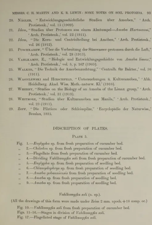
D ESC R IPTIO N OF PLATES.
Plate 5.
Fig. 1.— Eugl ypha sp. from fresh preparation of cucumber bed. ,, 2.— Chilodon sp. from fresh preparation of cucumber bed.
,, 3.— Flagellate from fresh preparation of cucumber bed.
,, 4.— Dividing Vahlkampjia soli from fresh preparation of cucumber bed. ,, 5.—Euglypha sp. from fresh preparation of seedling bed.
,, 6.—Chlamydophrys sp. from fresh preparation of seedling bed. ,, 7.—Amoeba gobanniensis from fresh preparation of seedling bed. ,, 8.—Amoeba sp. from fresh preparation of seedling bed.
,, 9.—Amoeba sp. from fresh preparation of seedling bed.
MESSRS. C. H. MARTIN AND K. R. L E W IN : SOME NOTES ON SOIL PROTOZOA. 9 3
Vahlkampjia soli (n. sp.).
(All the drawings of this form were made under Zeiss 2 mm. apoch. -J-18 comp, oc.) lig . 10.— Vahlkampjia soli from fresh preparation of cucumber bed.
Figs. 11-16.—Stages in division of Vahlkampjia soli.
9 4 MESSES. C. H. MARTIN A N D K. R. LEW IN : SOME NOTES ON SOIL PROTOZOA
Plate 6.
Vahlkampjia (n. sp.). Fig. 18.—Transition to amoeboid stage (whole length of flagella not shown).
„ 19.— Cyst of Vahlkam pjia soli.
Amoeba cucumis (n. sp.). (Zeiss 2 mm. apoch.+ 12 comp, oc.)
Fig. 20.—Amoeba cucumis from fresh preparation of cucumber bed. „ 21.— Amoeba cucumis from young culture.
Figs. 22-28.-—Stages in division of Amoeba cucumis.
Fig. 29.— Cyst of Amoeba cucumis.
Amoeba gobanniensis (n. sp.). (Zeiss 2 mm. apoch. + 12 comp, oc.) Fig. 30.—Amoeba gobanniensis from culture.
Figs. 31-34.—Stages in division of Amoeba gobanniensis.
Bodo caudatus.
(Zeiss 1'5 mm. apoch.+ 18 comp, oc.) Fig. 35.—Bodo caudatus.
„ 36.— Multiple division of Bodo caudatus.
4 $ . ' M m ,
w
12
jSaJS
MSB
JL ■J B lijM L j
,
® | J
#»
9
r
j
;/
4 ;
OF 8
£21
c 11 IX
§ § i | p ©
H
%
i y r
«
*
-B p H R S r
14
.-V--[^■flSlu
f
^il^StSSj^BT.
13'^•ST ...TSH*/
■ £ i j
m
i
®
p
- \
CP^#&P-■0 ' ^v<V/j M
w w
‘ '*v'» " -•' • •' '.v V ';i: '
-S ii.
I7
• B
r J % « w
H
j K ^
ibiSs
< . . . iw - ; , : •' / ?•-•'? k ^ p . y
M JV
19
o
2 0
'*WWr
• l
21
' '*ww
o o
i-J (mi
: * v
j k»3 « i v ly*
; v > v * - .v* 1 ’NmWIWIIIIhi
ri®. #s
NS
l i N V J sH : t* K 5 f l T » i
M B ■* i, : J2 t m J ^ s f^
? i ,# m /:
Jill
v \
I 7
iKjyfHMIm i .V1 1 1
w _ ... .
I^MSf
^ ^ 3s;
*t
7
# % iKfll
\
i *
*If»
«. # V
V
II
W
m
di: 'M < W :
>*•*
tiS S f^ d
w
'f9Bm M
3
27
/ A ♦
/ •
• • , "i& ?# 31»
* * 2 a
! Jj
/.«
W 4 'i l m i
%*.
1
#•31
♦ :IZ W 2.9K )
33# 9 0
Cl
34■ R \
( # 0 t ] 35
■7 r \ %JA \ J r * rvi
36
37