Correlations of Biomechanical Characteristics with Ball Speed in Penalty Corner Push-In
Studying the Effect of the Concentration and Sintering Temperature on the Structural and
Optical Properties of Zinc Ferrite (ZnFe
2O
4) Preparing by Sol-Gel
Abbas S. Khamas1, Sabah I. Abbas2 & Hashim A. Alyusr3
1,2,3
Department of Physics, College of Science, University of Wasit, Iraq.
Received 10th August 2017, Accepted 19th September 2017
Abstract
Zinc ferrite nanoparticles have been synthesized by using the sol-gel method from Fe(NO3)3.9H2O and
Zn(NO3)2.6H2O by adopting molecular concentrations (0.2 M and 0.4 M) .The samples were sintered at two temperatures
(600 °C , 800°C) for two hours. The spinal phase of zinc ferrite structure and crystallite size was examined by XRD spectrum, pattern show that nanoparticles structure exhibit mixed phase of 𝛼-Fe2O3 and ZnFe2O4. On the optical properties
for all the films were studied by recording the transmittance and absorbance spectra in the range of (200-1000) nm. The results showed that the energy band gap for allowed direct electronic transition varies from (2.63 to 2.97)eV at sintering temperature 600 °C and concentration of 0.2 M,(2.32 to 2.70)eV at sintering tempeature 800 °C and concentration of 0.4 M.
Keywords: Zinc Ferrite, Nanoparticle , Sol-Gel Methods.
© Copy Right, IJRRAS, 2017. All Rights Reserved.
Introduction
Nano size zinc ferrite has been the particular subject of study because of its unusual structural, electrical, optical and magnetic properties, which differ from their bulk counterpart. Zinc ferrite is particularly also important because of the strong tetrahedral site preference of zinc ions in zinc ferrite. Zinc ferrite nanoparticles have extremely important in the gas sensor applications [1]. Many methods have been used to prepare magnetic ferrite nanomaterials such as sol-gel methods [2], co-precipitation[3], mechanochemical synthesis [4], hydrothermal/ solvolysis thermal [5,6], spray pyrolysis [7]. The sol–gel methods have been used to prepare of different mixed metal oxides, nanomaterials, nanoscale, nonporous oxides [8]. The sol gel processes have given numerous advantages such as best mixing of the raw materials and excellent homogeneity, ultrafine and reproducible zinc ferrites with small size distribution. The homogeneous microstructure of zinc ferrite indicating the ability to control the electric-magnetic properties and heat treatments temperature which decrease the impurities generated during the preparation and variation in the composition [2]. At many works before this results shows the effect of increasing and decreasing of concentrations at all compounds that used. Some researches show the increasing of grain size by decreasing zinc concentrations [9]. And in another
Correspondence Abbas S.Khamas.
E-mail: [email protected] Ph. +9197898 53995
hand surface structural can be changed with zinc concentrations [10]. Annealing temperature affected directly at grain size and lattice constant affect by concentrations, by increasing temperature grain size increasing too [11]. At this we will see the effect of increasing concentrations for all using materials and discussion results in any case.
Experimental Procedure
ZnFe2O4 nanoparticles were prepared by sol-gel
methods. The mixture consist of three solution, (0.2M) of iron nitrate Fe(NO3)3.9H2O, (0.2M) of zinc nitrate
Zn(NO3)2.6H2O, were used as a precursor solution and
were gelatin by adding 100 ml of citric acid (C6H8 O7)
solution with concentration (0.2M). The next concentrations is (0.4M) of iron nitrate, (0.4M) of zinc nitrate also 100 ml of citric acid. The control on pH of the solution was fined at (7) by using many drops of ammonia and the solution was heated on the hot plate at 60 Cº for 30 min.. The temperature of solution increased to 80Cº for (8) hours, the solution was turned into gel. The gel material dried by Leave it several hours. Every gel compounds sintering at (600 Cº and 800 Cº) for two hours, to prepare two samples can by studied before grinding process. The structure and the average grain size of the spinal zinc ferrites nanoparticles were measured by X-Ray diffractometer (6000-Shimadzu) by using CuKa (α) radiation source with a wavelength, λ=1.54060 Å. Optical properties studied by using (UV-300 Nano) .
International
Journal of Recent Research and Applied Studies
Abbas et al. 2017 ISSN: 2349 – 4891
Results and Discussion Structural properties XRD tests
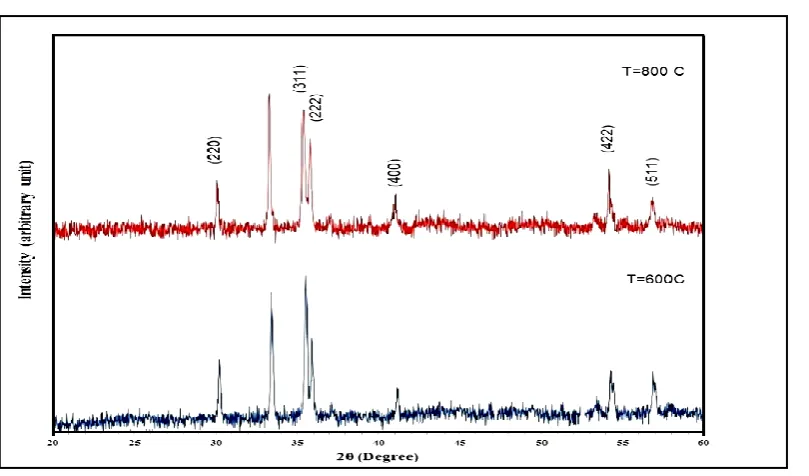
Results of X-Ray diffraction shows increasing of crystalline grain size with increasing of sintering temperature , also the samples that have low concentrations more larger grain size then samples have
high concentrations, and that returned to zinc concentrations level [10]. Results will illustrate in a fig.(1) for samples have concentration 0.2 M and fig.(2)
for samples have concentration 0.4 M .
Figure I
XRD test for 0.2 M at two different sintering temperature
Figure II
XRD test for 0.4 M at two different sintering temperature
The tables can be more clear for explain the changed in structural because in above figures it too
Table 1
Structural calculation of 0.2 M samples
Annealing temperature
Cº
2θ (Deg.) FWHM (Deg.) dhkl Exp.(Å) G.S (nm) hkl phase
600 Cº
29.9 0.191363 2.982942 43 (220) ZnFe2O4
33.2 0.138568 2.699141 60 (104) 𝛼-Fe2O3
35.2 0.207136 2.544568 40 (311) ZnFe2O4
35.5 0.217136 2.525347 38 (222) ZnFe2O4
43.0 0.246651 2.103812 35 (400) ZnFe2O4
49.5 0.277136 1.839396 32 (313) ZnFe2O4
54.1 0.277136 1.692849 32 (422) ZnFe2O4
56.6 0.461894 1.624062 20 (511) ZnFe2O4
800 Cº
29.9 0.28 2.982942 29.67205559 (220) ZnFe2O4
33.2 0.23 2.691855 35.9000404 (104) 𝛼-Fe2O3
35.2 0.32 2.541343 25.78350033 (311) ZnFe2O4
35.5 0.42 2.522173 20.06933049 (222) ZnFe2O4
42.8 0.51 2.108132 16.79729245 (400) ZnFe2O4
49.5 0.46 1.839396 18.94021243 (313) ZnFe2O4
54.1 0.32 1.693831 27.58845165 (422) ZnFe2O4
56.6 0.46 1.622848 19.5409925 (511) ZnFe2O4
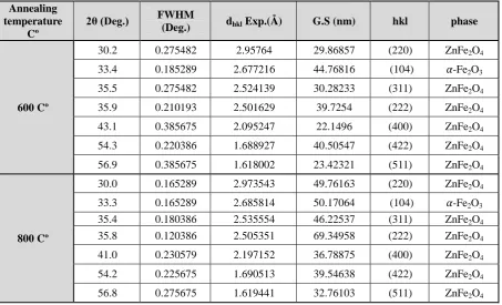
Table 2
Structural calculation of 0.4 M samples
Annealing temperature
Cº
2θ (Deg.) FWHM
(Deg.) dhkl Exp.(Å) G.S (nm) hkl phase
600 Cº
30.2 0.275482 2.95764 29.86857 (220) ZnFe2O4
33.4 0.185289 2.677216 44.76816 (104) 𝛼-Fe2O3
35.5 0.275482 2.524139 30.28233 (311) ZnFe2O4
35.9 0.210193 2.501629 39.7254 (222) ZnFe2O4
43.1 0.385675 2.095247 22.1496 (400) ZnFe2O4
54.3 0.220386 1.688927 40.50547 (422) ZnFe2O4
56.9 0.385675 1.618002 23.42321 (511) ZnFe2O4
800 Cº
30.0 0.165289 2.973543 49.76163 (220) ZnFe2O4
33.3 0.165289 2.685814 50.17064 (104) 𝛼-Fe2O3
35.4 0.180386 2.535554 46.22537 (311) ZnFe2O4
35.8 0.120386 2.505351 69.34958 (222) ZnFe2O4
41.0 0.230579 2.197152 36.78875 (400) ZnFe2O4
54.2 0.225675 1.690513 39.54638 (422) ZnFe2O4
Abbas et al. 2017 ISSN: 2349 – 4891
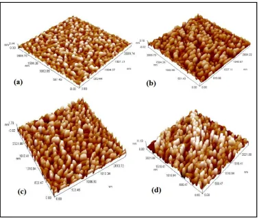
AFM tests
The results tests of atomic force microscopy for zinc ferrite powder a small grain size for samples consisted of high zinc concentration, and larger in low concentrations. The next fig.(3) will illustrate 3D image of zinc ferrite compounds. And the table (3) shows all grain size values for all samples to be clearly
explanation. The direction of grain's growth is upper towards and the grain had more crystallinity by increasing of sintering temperature. By increasing concentrations cracks disappear and grooves will be more tightly and almost cant seen. The values distributed from (70.35 nm) to (118.99 nm).
Figure III
AFM (a) & (b) 0.2 M at 600 Cº (c) & (d) 0.4 M at 800 Cº.
Table 3
AFM values according to concentrations and Annealing temperature
concentrations (M)
annealing temperature (Cº)
Avg. Diameter (nm)
Root mean square (nm)
Roughness Avg.
(nm) size (nm)
0.2 600 88.84 0.678 0.585 2507-2568 800 98.67 0.509 0.509 2507-2512
0.4 600 70.35 0.413 0.341 2517-2527 800 82.82 2.82 2.39 2527
Optical properties
Optical properties was measured by (UV-3000 Nano device) . Transmittance shows a little drop of samples that have (0.4 M) specially at 600 Cº sintering treatments , at 800 Cº in both samples didn't shows any clearly changes . fig.(4) will shows transmittance of both sintering degree ( 600 Cº and 800 Cº) . Absorbance
Figure IV
Transmittance (a) concentration of (0.2 M) (b) concentration of (0.4 M)
Figure V
Absorbance (a) concentration of (0.2 M) (b) Concentration of (0.4 M)
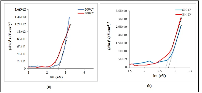
As mentioned above the height of zinc concentrations and large grain size as a results of that, sintering temperature didn't affect clearly of absorbance [9] .And for that if zinc concentrations are high the solution of zinc ferrite if deposit as a thin films will get a homogeneous surface films [12]. Energy gap of zinc
ferrite decreasing by increasing sintering temperature and increasing with increasing of concentrations, and that because of large grain size and large of crystalline size. The next fig.(7) illustrate that and table (4) shows the values of energy gap.
Table 4
Energy gap values
Concentrations M
Sintering at 600 Cº
Sintering at 800 Cº
0.2 M 2.63 2.32
Abbas et al. 2017 ISSN: 2349 – 4891
Figure VII
Energy gap (a) concentration 0.2 M (b) Concentration 0.4 M
Conclusions
ZnFe2O4 nanoparticles were successfully
prepared by sol-gel methods by adopting molecular concentrations (0.2 M and 0.4 M).The powder XRD analysis revealed presence of the mixed phases of ZnFe2O4 and 𝛼-Fe2O3 in the synthesized nanomaterials.
The ZnFe2O4 belongs to face centered regular spinel
cubic structure and the crystallization increases with increasing of sintering temperature. AFM figures shows that the concentration (0.2M) has an homogeneous distribution of nanoparticle than of (0.4 M).The band gap energy of ZnFe2O4 nanoparticles was obtained to be
(2.63 e V for 0.2 M , 2.79e V for 0.4 M) at 600Cº and be (2.32 e V for 0.2 M , 2.70 e V for 0.4 M) at 800Cº. The refractive index of the films increased with increase of annealing temperature.
References
1. Sivakumar, Manickam, et al. "Fabrication of zinc ferrite nanocrystals by sonochemical emulsification and evaporation: observation of magnetization and its relaxation at low temperature." The Journal of Physical Chemistry B 110.31 (2006): 15234-15243. 2. Zahi, S. "Synthesis, permeability and microstructure
of the optimal nickel-zinc ferrites by sol-gel route." Journal of Electromagnetic Analysis and Applications 2.01 (2010): 56.
3. Hariani, Poedji Loekitowati, Muhammad Faizal, and Dedi Setiabudidaya. "Synthesis and properties of Fe3O4 nanoparticles by co-precipitation method to removal procion dye." International Journal of Environmental Science and Development 4.3 (2013): 336.
4. Yang, Huaming, et al. "Synthesis of ZnFe 2 O 4 nanocrystallites by mechanochemical reaction." Journal of Physics and Chemistry of Solids 65.7 (2004): 1329-1332.
5. Sinthiya, M. Maria Angelin, et al. "Synthesis of Zinc Ferrite (ZnFe2O4) Nanoparticles with
Different Capping Agents." Int J Chem Tech Re s 7 (2014): 2144-2149.
6. Xu, Huayun, et al. "A comparative study of nanoparticles and nanospheres ZnFe2O4 as anode material for lithium ion batteries." Int J Electrochem Sci 7.9 (2012): 7976-83.
7. Takayama, Akio, Masayuki Okuya, and Shoji Kaneko. "Spray pyrolysis deposition of NiZn ferrite thin films." Solid State Ionics 172.1 (2004): 257-260.
8. Gatelytė, Aurelija, et al. "Sol-gel synthesis and characterization of selected transition metal nano-ferrites." Materials Science 17.3 (2011): 302-307. 9. El-Saaey, Ahmed Saied Faheim, et al. "Effect
Investigation of Zn Substitution on the characterization of Cobalt Ferrite Nano Particles Prepared Co-precipitation method."
10. Jiang, Nan-Nan, et al. "Influence of zinc concentration on structure, complex permittivity and permeability of Ni–Zn ferrites at high frequency." Journal of Magnetism and Magnetic Materials 401 (2016): 370-377.
11. Singhal, Sonal, et al. "Effect of Zn substitution on the magnetic properties of cobalt ferrite nano particles prepared via sol-gel route." Journal of Electromagnetic Analysis and Applications 2.06 (2010): 376.