O R I G I N A L R E S E A R C H
Open Access
Gene expression signature in mouse
thyroid tissue after
131
I and
211
At exposure
Nils Rudqvist
1*, Johan Spetz
1, Emil Schüler
1, Britta Langen
1, Toshima Z. Parris
2, Khalil Helou
2and Eva Forssell-Aronsson
1Abstract
Background:131I and211At are used in nuclear medicine and accumulate in the thyroid gland and may impact normal thyroid function. The aim of this study was to determine transcriptional profile variations, assess the impact on cellular activity, and identify genes with biomarker properties in thyroid tissue after131I and211At administration in mice.
Methods:To further investigate thyroid tissue transcriptional responses to131I and211At administration, we generated a new transcriptional dataset that includes re-evaluated raw intensity values from our previous131I and211At studies. Differential transcriptional profiles were identified by comparing treated and mock-treated samples using Nexus Expression 3.0 software. Further data analysis was performed using R/Bioconductor and IPA.
Results:A total of 1144 genes were regulated. Hierarchical clustering subdivided the groups into two clusters containing the lowest and highest absorbed dose levels, respectively, and revealed similar transcriptional regulation patterns for many kallikrein-related genes. Twenty-seven of the 1144 genes were recurrently regulated after131I and211At exposure and divided into six clusters. Several signalling pathways were affected, including calcium, integrin-linked kinase, and thyroid cancer signalling, and the peroxisomal proliferator-activated receptor network.
Conclusions:Substantial changes in transcriptional regulation were shown in131I and211At-treated samples, and 27 genes were identified as potential biomarkers for131I and211At exposure. Clustering revealed distinct differences between transcriptional profiles of both similar and different exposures, demonstrating the necessity for better understanding of radiation-induced effects on cellular activity. Additionally, ionizing radiation-induced changes in kallikrein gene expression and identified canonical pathways should be further assessed.
Keywords:Radiation biology; Microarray; Radiation biomarkers; Radionuclide therapy; Transcriptomics; Radiogenomics
Background
Medical applications for radionuclides are rapidly devel-oping. Theβparticle-emitting131I is frequently included in therapy regimens of various thyroid disorders due to selective uptake of the isotope in thyroid tissue and is also administered bound to tumour-seeking agents for therapeutic and diagnostic purposes [1–4]. Theα particle-emitting 211At is a suitable therapeutic radionuclide due to, e.g., a nearly optimal therapeutic linear energy transfer value of emittedαparticles of 98.8 keV/μm [5].211
At-la-belled tumour-seeking pharmaceuticals have been utilized both in humans and in animals [6–8]. Similar to unbound 131
I, selective uptake of unbound211At also occurs in thy-roid tissue [9–11], and administration of free 131I and 211
At or 131I- and 211At-labelled radiopharmaceuticals have been shown to result in thyroid irradiation [6, 12]. Additionally, nuclear accidents often involve an atmos-pheric release of131I, as was the case in connection with the Chernobyl accident, which resulted in an increased in-cidence of thyroid cancer in children [13, 14].
Despite the risk of exposing thyroid tissue to 131I and 211
At, the understanding of radiation-induced effects is far from complete and molecular biomarkers of absorbed dose or radiation-induced effects on thyroid tissue are yet to be identified. Biomarkers are useful to indicate achieved therapeutic effects or estimate risk exposure and evaluate * Correspondence:[email protected]
1
Department of Radiation Physics, Institute of Clinical Sciences, Sahlgrenska Cancer Center, Sahlgrenska Academy, University of Gothenburg, SE-413 45 Gothenburg, Sweden
Full list of author information is available at the end of the article
© 2015 Rudqvist et al.Open AccessThis article is distributed under the terms of the Creative Commons Attribution 4.0 International License (http://creativecommons.org/licenses/by/4.0/), which permits unrestricted use, distribution, and reproduction in any medium, provided you give appropriate credit to the original author(s) and the source, provide a link to the Creative Commons license, and indicate if changes were made.
the quality and severity of side effects. RNA microarray analysis is a semi-quantitative method to identify changes in genome-wide transcriptional patterns between two or more samples. The result is a transcriptional profile, i.e. a snapshot of the radiation-induced cellular activity at the mRNA level. This can be used to determine the impact of radiation on biological functions and canonical pathways, to predict upstream regulation of target molecules, and for biomarker discovery without the risk of bias in focus-ing on a specific set of signallfocus-ing pathways only.
Few investigations on global gene expression effects ofα and βparticle irradiation have been performed in normal (thyroid) tissue in vivo. There are, however, a few in vitro studies on global gene expression in fibroblasts and cancer cells afterαparticle exposure [15, 16]. Additionally, effects on gene expression for a set of pre-defined genes after α particle irradiation have been measured both in cancer cells in vitro and in vivo in xenografted tumours [17–19]. Previously, we have published results showing substantial differences between transcriptional profiles in thyroid tissue in vivo after different131I or211At exposures, varying absorbed dose, dose rate and time after administration [20–22]. We then identified potential biomarkers for each type of exposure separately and concluded that biological response to radiation is complex and that it is difficult to predict or extrapolate radiation-induced effects for other exposure parameters.
The aim of this work was to re-evaluate the previously obtained transcriptional response in thyroid after admin-istration of 131I− and free 211At in mice from different exposure conditions to gain a better understanding of variations in transcriptional regulation on absorbed dose, dose rate, time after administration and radiation quality.
Methods
Study design
In this study, we further investigate the thyroid transcrip-tional response to 131I and 211At exposure by using nor-malized intensity values from three different experiments where separate analyses of each experiment have been published elsewhere [20–22]. All normalized intensity values (normalization was performed according to ex-periment) from these three experiments were imported together into Nexus Expression 3.0 (BioDiscovery; El Segundo, CA) for filtering, linear modelling and deter-mination of differentially expressed genes.
No new tissue sampling and analysis was thus performed in this study, but a reanalysis of all these data together in a new analysis by Nexus Expression. Data presented in the present work are therefore novel and have not been previ-ously published.
A brief summary of the methods used in the three ori-ginal studies is as follows: a total of 44 female Balb/c nude mice (CAnN.Cg-Foxn1nu/Crl, Charles River Laboratories
International, Inc., Salzfeld, Germany) (n= 2–3 per group) were i.v. injected with various amounts of131I or211At in the tail vein and killed at 1, 6, 24 or 168 h after adminis-tration, or mock-treated (Table 1). Thyroid, kidney, liver, lung and spleen tissue samples were collected and imme-diately snap-frozen in liquid nitrogen and stored at−80 °C until RNA extraction. The present study contains data on transcriptional changes in thyroid glands. The transcrip-tional response in the kidneys, livers, lungs and spleens has been published elsewhere [23–25].
The methods for determination of absorbed doses have been reported previously with separate analysis of each experiment [20–22]. In short, the mean absorbed dose was calculated according to conventional Medical Internal Radiation Dose (MIRD) formalism. We used previously published data on relative activity concentra-tion of 131I and 211At in thyroid in mice [26], an absorbed fraction of 0.742 and 1 for radiation emitted from 131I and 211At, respectively [27], and a standard mouse thyroid mass of 3 mg.
Gene expression analysis
Genome-wide transcriptional analysis using RNA microarray of thyroid tissue has been described else-where [20, 21]. Briefly, RNA samples were analysed using MouseRef-8 Whole-Genome Expression Beadchips (Illu-mina; San Diego, CA, USA). Nexus Expression 3.0 (BioDis-covery; El Segundo, CA) was used to identify statistically significant differentially expressed transcripts (≥1.5-fold change) with a Benjamini-Hochberg adjusted p value cut-off of 0.01 between irradiated and control tissues. RNA microarray data from irradiated samples were compared
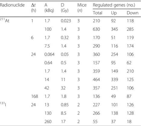
Table 1Number of regulated genes in thyroid tissue in mice 1–168 h after administration of131I and211At
Radionuclide Δt (h)
A (kBq)
D (Gy)
Mice (n)
Regulated genes (no.)
Total Up Down
211At 1 1.7 0.023 3 210 92 118
100 1.4 3 630 345 285
6 1.7 0.32 3 170 51 119
7.5 1.4 3 290 116 174
24 0.064 0.05 3 360 254 106
0.64 0.5 3 157 95 62
1.7 1.4 3 359 149 210
14 11 3 464 339 125
42 32 3 357 251 106
168 1.7 1.8 3 136 49 87
131I 24 13 0.85 2 227 101 126
130 8.5 2 266 138 128
260 17 2 55 37 18
Absorbed dose was calculated using MIRD formalism
Abbreviations:Δtexposure time,Ainjected activity,Dabsorbed dose
with RNA microarray data from control animals from each separate experiment to ensure that differential expression of genes reflects radiation-induced changes and not varia-tions between different control groups. There was one dif-ference in study design between the present and previous studies. In the present work, data from tissue samples from animals administered 0.064 and 0.64 kBq211At and killed 24 h after administration were compared with data from tissue samples from control animals killed the same day. However, in the previous paper with analysis of the tran-scriptional response at 24 h after211At administration, ani-mals administered 0.064 and 0.64 kBq were compared with controls killed earlier (killed simultaneously as animals injected with 1.7, 14 and 42 kBq, also at 24 h after ad-ministration but on another day) [20]. In the present paper, the term “regulated transcripts/genes” is used synonymous to “statistically significant differentially expressed transcripts/genes”.
Hierarchical clustering of regulated transcripts according to their transcriptional regulation profile was performed using the hclust function (stats package, version 3.1.1) with the complete linkage algorithm and Lance-Williams dis-similarity update formula in the R statistical computing en-vironment (version 0.97.551, http://www.r-project.org) [28]. Heat maps were produced using the heatmap.2 function (gplots package, version 2.14.2).
Upstream regulation, diseases and functions and canon-ical pathway analyses were generated using the Ingenuity Pathway Analysis tool (IPA, Ingenuity® Systems, www.in-genuity.com; Redwood City, CA) with Fisher’s exact test (pvalue <0.05).
Gene expression data discussed in this publication have been deposited at the NCBI’s Gene Expression Omnibus (GEO accession numbers: GSE32306 [20], GSE54594 [21] and GSE66089 [22]).
Results
Regulated genes
In the present study, 1144 genes (1164 transcripts) were regulated. The number of regulated genes in each group varied between 55 (17 Gy, 24 h, 131I) and 630 (1.4 Gy, 1 h 211At) (Table 1). Hierarchical clustering subdivided the exposure groups into two larger clusters: one smaller branch containing groups with higher absorbed dose levels (1.4–32 Gy from 211At and 8.5 Gy from 131I at 24 h) and a larger branch with the remaining groups (Fig. 1).
At both 1 h and 6 h after 211At administration, a higher absorbed dose/dose rate resulted in a higher number of regulated genes (Table 1). In contrast, at 24 h after 211At administration, the number of regulated genes varied non-monotonously with absorbed dose. A slight increase in the number of genes regulated after 131
I exposure was seen between 0.85 and 8.5 Gy and a
decrease between 8.5 and 17 Gy. Transcriptional profiles at 1, 6 and 168 h clustered together, with the exception of 1.4 Gy at 1 h (Fig. 1). At 24 h, the transcriptional pro-files for 0.05 and 0.5 Gy clustered together while the profiles for 1.4, 11 and 32 Gy clustered together with highest similarity between 11 and 32 Gy. Furthermore, upregulation of 110 genes was shared between 0.05 and 1.4 Gy at 24 and 1 h after 211At administration, respect-ively. For 131I exposure, the transcriptional profiles for 0.85 and 17 Gy clustered together, whereas the response after 8.5 Gy was more similar to that after 1.4, 11 and 32 Gy at 24 h following 211At administration. In the groups exposed to 1.4 Gy, the number of regulated genes decreased from 630 to 290 between 1 and 6 h, but increased to 359 at 24 h (Table 1). Additionally, the tran-scriptional profiles of these groups showed little similar-ity in the cluster analysis (Fig. 1). Lastly, an equal amount of injected activity (1.7 kBq, 211At) resulted in 210, 170, 359 and 136 regulated genes at 0.023, 0.32, 1.4 and 1.8 Gy after 1, 6, 24 and 168 h, respectively (Table 1). The transcriptional profiles of these groups were similar and clustered together with the exception of 1.4 Gy at 24 h which was clearly different (Fig. 1).
Kallikrein 1 and kallikrein 1-related peptidases
Hierarchical clustering of all regulated transcripts revealed similar transcriptional regulation profiles for 13 kallikrein genes belonging to the peptidase S1 family (Klk1,Klk1b1, Klk1b4, Klk1b5, Klk1b8, Klk1b9, Klk1b11, Klk1b16, Klk1b21, Klk1b22, Klk1b24, Klk1b26 and Klk1b27 (A in Figs. 1 and 2). All 13 genes were upregulated at 11 and 32 Gy 24 h after 211At administration. For an absorbed dose of 1.4 Gy after211At exposure, 9/13 kallikrein genes were upregulated at 24 h but downregulated at 1 h (three additional genes were downregulated at 1 h), while at 6 h, 1 and 4 genes were down- and upregulated, respectively. Furthermore, 4/13 genes were upregulated at 1.8 Gy 168 h after injection of 211At. At 24 h after 131I administration, 4/13 genes were upregulated after 17 Gy, but not regu-lated at the lower absorbed dose levels.
Recurrently regulated genes after131I and211At exposure
Twenty-seven of the 1144 regulated genes were regu-lated in≥9 of the 13 groups (Fig. 3). Hierarchical cluster-ing divided the 27 genes into six groups: 1) Atp2a1, Ckm,Eno3,Pvalb,Tnnc2,Tnni2,Tnnt3; 2)Coq10b,Ctgf, Dbp, Per1; 3) Mfsd2; 4) Ltf; 5) Ccl8, Ly6g6d, S100a8, S100a9 and 6) Aoc3, Ccl9, Clec2d, Cpa3, Dpt, Fstl1, Lum,Mup2,Ogn,Scara3. In cluster 1, genes were down-regulated at 1 h following 211At administration. This changed at 6 h where 0.32 and 1.4 Gy showed down-and upregulation, respectively. At 24 h, the regulated genes were upregulated and, notably, the fold change increased with absorbed dose. At 24 h after131I administration, all of
these genes were upregulated with high fold changes (21–180) after 8.5 Gy, while two genes were upregu-lated to low extent after 17 Gy. Genes in cluster 2 were upregulated after211At administration while131I
administration resulted in both up- and downregula-tion. The Mfsd2 gene, the only gene in cluster 3, was up- and downregulated after 211At and 131I adminis-tration, respectively. TheLtf gene, sole gene in cluster Fig. 1Results from hierarchical clustering analyses of genes and exposure groups after131I and211At exposure. Genes were hierarchically clustered according to log2-ratio values using gplots package in R/Bioconductor. Thecolumnsrepresent genes androwsrepresent exposure type. Cluster containing kallikrein genes is denotedA
Fig. 2Regulation of 13 kallikrein genes belonging to the peptidase S1 family after131I and211At exposure. Values indicate fold change of differential gene expression.Greenandred coloursindicate up- and downregulation of genes, respectively. Higher saturation of colours indicates higher fold change
Fig. 3Regulation of 27 recurrently regulated genes after131I and211At exposure. Values indicate fold change of differential gene expression. Greenandred coloursindicate up- and downregulation of genes, respectively. Higher saturation of colours indicates higher fold change. The relationships between genes and thyroid function, ionizing radiation, and thyroid cancer and biological function of genes have been assessed using literature reports
4 with almost opposite regulation toMfsd2, was down-regulated after 211At administration with the exception of a 22-fold upregulation after 0.5 Gy at 24 h, and up-regulated after 131I administration. Cluster 5 was simi-lar to cluster 4, and 211At administration resulted in downregulation with few exceptions of upregulation at 24 h and 131I administration generally resulted in up-regulation. Genes in cluster 6 were generally downregu-lated with the exception of upregulation at, e.g., 1.4–32 and 17 Gy 24 h after 211At and 131I administration, respectively.
Canonical pathway analysis: calcium, integrin-linked kinase and thyroid cancer signalling
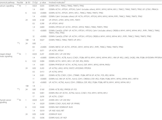
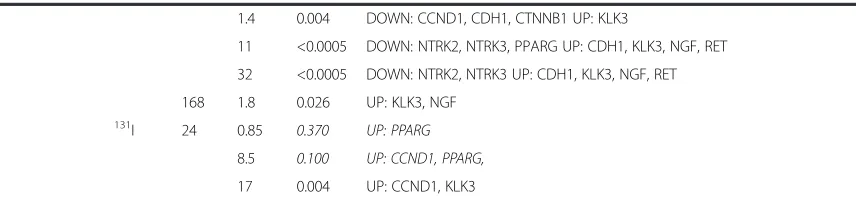
In the present study, calcium, integrin-linked kinase and thyroid cancer signalling were the top three canonical pathways generated using the IPA comparison analysis tool (Table 2). An impact on calcium signalling was sta-tistically significant in all groups except at the two low-est and the highlow-est absorbed dose levels 24 h after211At and 131I administrations, respectively. Integrin-linked kinase signalling was statistically significant in all groups except at low absorbed dose levels at 1 and 24 h, and at the lowest and highest absorbed dose levels 24 h after 211
At and131I administration, respectively. Genes associ-ated with calcium and integrin-linked kinase signalling were generally downregulated early and at low absorbed doses and upregulated at later time points and at higher absorbed doses. Thyroid cancer signalling was statisti-cally significant in all groups except at 0.023 and 0.05 Gy, 1 and 24 h following211At administration, re-spectively, and at 0.85 and 8.5 Gy 24 h after131I admin-istration (Table 2). Additionally, the number of genes involved in the different canonical pathways at each time point generally increased with absorbed dose (with exceptions between 11 and 32 Gy).
Diseases and function analysis: thyroid cancer and disturbed thyroid function
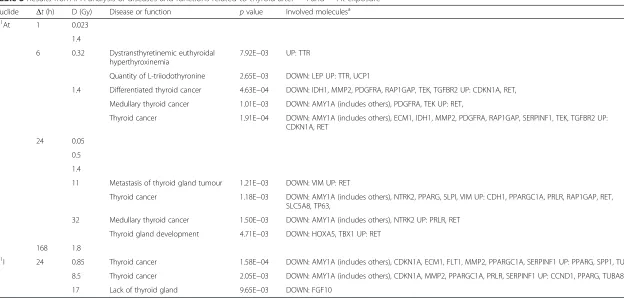
A diseases and function analysis related to thyroid was gen-erated with IPA (Table 3). At 6 (1.4 Gy) and 24 h (11 and 32 Gy) after 211At administration, a relation between the transcriptional response and various thyroid cancer types was identified. Such a relation was also identified 24 h after 131
I administration; however, at a lower absorbed dose level (0.85 and 8.5 Gy). Additionally, the transcriptional response was linked to altered T3 and T4 levels and to thyroid gland development at 0.32 and 32 Gy, 6 and 24 h after211At ad-ministration, respectively.
Upstream regulation of molecules related to peroxisomal proliferator-activated receptors
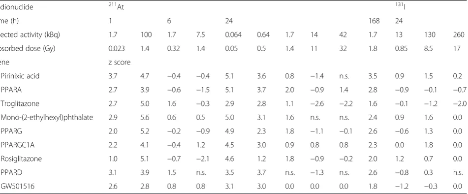
The upstream regulation analysis generated by IPA pre-dicted upstream regulation of various molecules related
to peroxisomal proliferator-activated receptors (PPARs) (Table 4). These included PPARA, PPARD, PPARG, PPARGC1A and several PPAR-targeting drugs and/or PPAR ligands such as GW501516, mono-(2-ethylhex-yl)phthalate, pirinixic acid, Rosiglitazone and Troglitazone. IPA predicted that these upstream regulators were generally activated at 1 h, for absorbed dose≤1.4 Gy at 24 h, and for 1.8 Gy at 168 h.
Discussion
In the present study, the values used to calculate absorbed dose were based on previously published131I and211At bio-distribution data, where mice were simultaneously injected with both131I and211At, which allows for direct compari-son of the absorbed dose per injected activity between131I and 211At in the same animal [26]. In the present study, radioactivity measurements of individual thyroid samples would have enhanced the certainty in absorbed dose calcu-lations but was not possible since all excised thyroid tissue was needed to ensure sufficient amount of RNA for micro-array analysis. There are several important differences in the characteristics of211At and131I exposure: i) the differ-ence in mean range of the α and βparticles emitted (65 and 400μm), ii) the much higher mean energy released per decay from211At compared with131I (7000 and 190 keV), iii) the difference in LET of particles emitted from131I and 211
At (0.25 and 98.8 keV/μm, respectively) and iv) much shorter half-life for 211At than 131I (7.2 h and 8.0 day, re-spectively). Taken together,211At irradiates more heteroge-neously and with higher dose rates at similar absorbed dose levels compared with 131I (and the dose rate will decline faster for 211At compared to 131I). The effects of radiation quality on global gene expression should be further studied, and to our knowledge, the present study is the first to in-vestigate such differences between131I and211At.
RNA microarray analysis was used to evaluate the im-pact of131I and211At exposure on global transcriptional regulation in normal mouse thyroid tissue. Regulated genes were associated with biological functions using previously published literature reports and various data-bases, in addition to upstream and downstream regula-tion analysis and canonical pathway analysis generated by Ingenuity Pathway Analysis (IPA) software. In total, 1144 genes were differentially regulated showing a large variation in number of genes per group. In general, hier-archical clustering divided groups that received high absorbed doses into one branch and groups receiving low absorbed doses into another. Thus, we hypothesize that the transcriptional profiles presented here may re-flect intrinsic biological properties predictive of131I and 211
At absorbed dose levels at various time points. At both 1 and 6 h, the number of regulated genes in-creased with absorbed dose. This was not the case for 131
I or211At exposure at 24 h, where a broader range of
Table 2Top three Ingenuity canonical pathways enriched by genes regulated after131I or211At exposure
Canonical pathway Nuclide Δt(h) D (Gy) pvalue Involved moleculesa
Calcium signalling 211At 1 0.023 0.035 DOWN: MYL1, TNNC2, TNNI2, TNNT3, TPM2
1.4 0.001 DOWN: ACTA1, ATP2A1, ATP2A3, Calm1 (includes others), MYH1, MYH2, MYH4, MYL1, TNNC2, TNNI2, TNNT3, TPM2 UP: LETM1, PRKACA
6 0.32 <0.0005 DOWN: ACTA1, ATP2A1, MYH1, MYL1, TNNC2, TNNI2, TNNT3, TPM2
1.4 <0.0005 DOWN: Calm1 (includes others) UP: ACTA, ATP2A1, ATP2A3, MYH, MYH2, MYH4, RYR1, TNNC2, TNNI2, TNNT3, TPM2
24 0.05 0.180 UP: ATP2A1, LETM1, MYH2, RYR1, TNNT3
0.5 0.390 UP: ATP2A1, MYH2
1.4 0.009 DOWN: ATP2A3 UP: ACTA1, ATP2A1, MYH4, TNNC2, TNNI2, TNNT3, TPM2
11 <0.0005 DOWN: Camk2b, LETM1 UP: ACTA1, ATP2A1, ATP2A3, Calm1 (includes others), CREB3L4, MYH1, MYH2, MYH4, MYL1, RYR1, TNNC2, TNNI2, TNNT3, TP63, TPM2
32 <0.0005 DOWN: Camk2b, LETM1 UP: ACTA1, ATP2A1, ATP2A3, CREB3L4, MYH1, MYH2, MYH4, MYL1, RYR1, TNNC2, TNNI2, TNNT3, TPM2
168 1.8 0.027 DOWN: TNNC2, TNNI2, TNNT3 UP: MYL1
131I 24 0.85 – –
8.5 <0.0005 DOWN: MEF2C UP: ACTA1, ATP2A1, MYH1, MYH4, MYL1, RYR1, TNNC2, TNNI2, TNNT3, TPM2
17 0.071 UP: ACTA1, ATP2A1
Integrin-linked kinase signalling
211
At 1 0.023 0.291 DOWN: MYL1 UP: DSP, IRS2
1.4 <0.0005 DOWN: ACTA1, ACTB, Actn3, CCND1, ITGB6, KRT18, MYH1, MYH2, MYH4, MYL1, VIM UP: IRS2, LIMS2, SH2B2, VIM, ITGB6 UP/DOWN: PPP2R5A
6 0.32 0.004 DOWN: ACTA1, MYH1, MYL1 UP: DSP, IRS2, RHOU
1.4 0.001 DOWN: PPAP2B UP: ACTA1, ACTN2, Actn3, DSP, MYH1, MYH2, MYH4, RHOU
24 0.05 0.203 UP: ACTN2, LIMS2, MYH2, RHOT2 UP/DOWN: PPP2R5A
0.5 0.413 UP: ACTN2, MYH2
1.4 0.001 DOWN: ACTB, CCND1, CDH1, CTNNB1, ITGB4, KRT18 UP: ACTA1, FOS, IRS2, MYH4
11 <0.0005 DOWN: Irs3, VIM UP: ACTA1, Actn3, CDH1, CREB3L4, DSP, IRS2, ITGB4, ITGB6, MYH1, MYH2, MYH4, MYL1, KRT18
32 <0.0005 UP: ACTA1, ACTN2, Actn3, CDH1, CREB3L4, DSP, IRS2, ITGB4, ITGB6, KRT18, MYH1, MYH2, MYH4, MYL1
168 1.8 – –
131I 24 0.85
0.169 DOWN: ACTB, IRS2, PPAP2B UP: FOS
8.5 0.001 DOWN: IRS2 UP: ACTA1, ACTN2, Actn3, CCND1, FOS, MYH1, MYH4, MYL1
17 0.076 UP: ACTA1, CCND1
Thyroid cancer signalling
211At 1 0.023
0.291 DOWN: MYL1 UP: DSP, IRS2
1.4 0.029 DOWN: CCND1, KLK3, NGF UP: PPARG
6 0.32 0.040 DOWN: NGF DOWN/UP: KLK3
1.4 0.019 UP: NGF, KLK3, RET
24 0.05 0.490 DOWN/UP: KLK3
0.5 0.036 DOWN: KLK3, UP: NGF
Rudqvist
et
al.
EJNMMI
Research
(2015) 5:59
Page
7
of
Table 2Top three Ingenuity canonical pathways enriched by genes regulated after131I or211At exposure(Continued)
1.4 0.004 DOWN: CCND1, CDH1, CTNNB1 UP: KLK3
11 <0.0005 DOWN: NTRK2, NTRK3, PPARG UP: CDH1, KLK3, NGF, RET
32 <0.0005 DOWN: NTRK2, NTRK3 UP: CDH1, KLK3, NGF, RET
168 1.8 0.026 UP: KLK3, NGF
131I 24 0.85
0.370 UP: PPARG
8.5 0.100 UP: CCND1, PPARG,
17 0.004 UP: CCND1, KLK3
DOWN and UP indicate down- and upregulation, respectively. Italics indicates no statistically significant effect on the specific canonical pathway Abbreviations:Δtexposure time,Dabsorbed dose
a
IPA predicts involved molecules in the form of human proteins
Rudqvist
et
al.
EJNMMI
Research
(2015) 5:59
Page
8
of
Table 3Results from IPA analysis of diseases and functions related to thyroid after131I and211At exposure
Nuclide Δt(h) D (Gy) Disease or function pvalue Involved moleculesa 211At 1 0.023
1.4
6 0.32 Dystransthyretinemic euthyroidal hyperthyroxinemia
7.92E−03 UP: TTR
Quantity of L-triiodothyronine 2.65E−03 DOWN: LEP UP: TTR, UCP1
1.4 Differentiated thyroid cancer 4.63E−04 DOWN: IDH1, MMP2, PDGFRA, RAP1GAP, TEK, TGFBR2 UP: CDKN1A, RET,
Medullary thyroid cancer 1.01E−03 DOWN: AMY1A (includes others), PDGFRA, TEK UP: RET,
Thyroid cancer 1.91E−04 DOWN: AMY1A (includes others), ECM1, IDH1, MMP2, PDGFRA, RAP1GAP, SERPINF1, TEK, TGFBR2 UP: CDKN1A, RET
24 0.05
0.5
1.4
11 Metastasis of thyroid gland tumour 1.21E−03 DOWN: VIM UP: RET
Thyroid cancer 1.18E−03 DOWN: AMY1A (includes others), NTRK2, PPARG, SLPI, VIM UP: CDH1, PPARGC1A, PRLR, RAP1GAP, RET, SLC5A8, TP63,
32 Medullary thyroid cancer 1.50E−03 DOWN: AMY1A (includes others), NTRK2 UP: PRLR, RET
Thyroid gland development 4.71E−03 DOWN: HOXA5, TBX1 UP: RET
168 1.8 131
I 24 0.85 Thyroid cancer 1.58E−04 DOWN: AMY1A (includes others), CDKN1A, ECM1, FLT1, MMP2, PPARGC1A, SERPINF1 UP: PPARG, SPP1, TUBA8
8.5 Thyroid cancer 2.05E−03 DOWN: AMY1A (includes others), CDKN1A, MMP2, PPARGC1A, PRLR, SERPINF1 UP: CCND1, PPARG, TUBA8
17 Lack of thyroid gland 9.65E−03 DOWN: FGF10
DOWN and UP indicate down- and upregulation, respectively Abbreviations:Δtexposure time,Dabsorbed dose
a
IPA predicts involved molecules in the form of human proteins
Rudqvist
et
al.
EJNMMI
Research
(2015) 5:59
Page
9
of
absorbed dose was tested. Furthermore, hierarchical clus-tering revealed distinct differences between the transcrip-tional profiles of both similar and different exposures, e.g. the transcriptional profiles for 1.4 Gy at 1 and 6 h after 211
At administration were distinctly different although the absorbed dose was similar. This could in part be explained by the profound differences in dose rate; dose rate effects on the transcriptional response have previously been described in vivo following radionuclide administration [22, 25, 29]. These findings indicate that variations in the radiation-induced response with absorbed dose will be reflected in the number of regulated genes, in addition to which specific genes are regulated, although not in a clear dose-dependent manner. Instead it is likely that changes in transcriptional patterns in a specific tissue will depend with varying degree on, but not excluded to, the following parameters: exposure time, injected activity, absorbed dose, dose rate, dose distribution (e.g. frequency of non-, single- or multi-hit cells) and radiation quality. Each unique setup of these parameters may then yield a specific response in the target tissue. In addition, cells are dynamic systems with complex regulatory networks that activate cascades of downstream regulation that is sensi-tive to type and frequency of incoming stimulus.
It is valuable to identify genes with exposure-specific ex-pression as they may be used as biomarkers. Biomarkers are useful to better understand the mechanisms behind the radiation-induced response. A potential application of biomarkers for ionizing radiation exposure of the thyroid might be in biological dosimetry after exposure to rela-tively high doses, maybe in a triage setting. In the present study, kallikrein 1 (Klk1) and 12 of 13 kallikrein 1-related
(Klk1b) peptidases in the mouse genome were frequently regulated with fold change values between −3.8 and 110. The expression of these genes generally increased with absorbed dose and time after injection of 131I or 211At; however, 32 Gy resulted in less upregulation compared with 11 Gy (24 h, 211At) and the highest dose rate used (1.4 Gy, 1 h,211At) resulted in downregulation. It is likely that the expression of Klk1 and Klk1-related peptidases depends, to a different degree, on dose rate, absorbed dose and time after injection. In a study on the rat urine prote-ome 24 h after 10 Gy total body irradiation, the occur-rence of kallikrein 1-related peptidase b24 precursor protein increased while the kallikrein-binding serine pro-tease inhibitor A3K precursor decreased [30]. In another study, the plasma kallikrein levels decreased with absorbed dose (0–19 Gy) at 2–24 h after local irradiation of the hind legs in tumour-bearing rats and controls [31]. Additionally, we have shown that regulation of Klk1and Klk1-related genes in mouse thyroids after 131I exposure does not show a circadian variation [32]. We hypothesize that genes involved in the kallikrein network may be po-tential biomarkers of radiation exposure, but further re-search is warranted to elucidate the relationship between radiation exposure and kallikrein proteases and kallikrein inhibitor levels. The kallikrein genes have also been shown to contribute to the radiation-induced death of various species. After treatment with soy bean trypsin inhibitors (SBTI), the mortality rate in mice and chickens 14 days after exposure to 690 and 820 R (6.7 and 8 Gy to soft tis-sue), respectively, decreased from 100 to 50 % in mice and from 86 to 4 % in chickens [33]. The authors suggested that the decrease in mortality rate after administration of
Table 4Peroxisomal proliferator-activated receptor (PPAR)-related upstream regulators in thyroids exposed to131I or211At according to IPA upstream regulator analysis
Radionuclide 211At 131I
Time (h) 1 6 24 168 24
Injected activity (kBq) 1.7 100 1.7 7.5 0.064 0.64 1.7 14 42 1.7 13 130 260
Absorbed dose (Gy) 0.023 1.4 0.32 1.4 0.05 0.5 1.4 11 32 1.8 0.85 8.5 17
Gene z score
Pirinixic acid 3.7 4.7 −0.4 −0.4 5.1 3.6 0.8 −1.4 n.s. 3.5 0.9 1.5 0.2
PPARA 2.7 3.9 −0.6 −1.5 5.1 3.7 2.0 −0.9 1.4 2.8 −0.9 −0.1 −0.7
Troglitazone 2.7 5.0 1.6 −0.3 2.9 2.8 1.1 −2.6 −2.2 1.6 −0.1 −1.2 −2.0
Mono-(2-ethylhexyl)phthalate 2.9 5.6 0.6 0.5 5.0 3.1 1.6 n.s. n.s. 2.4 0.9 1.6 0.0
PPARG 2.0 5.2 −0.2 −0.9 4.9 2.3 1.8 −1.1 −0.1 2.6 −0.6 1.3 0.0
PPARGC1A 2.2 4.1 −0.4 1.2 4.5 3.0 0.9 0.8 0.8 2.3 0.0 1.8 0.0
Rosiglitazone 1.0 5.1 −0.7 −2.1 4.6 1.2 1.8 −0.9 −0.2 2.0 1.2 0.7 0.0
PPARD 3.1 3.9 1.5 n.s. 3.5 3.7 n.s. −1.3 n.s. 2.6 −0.8 0.3 n.s.
GW501516 2.6 2.8 0.8 0.8 3.1 3.0 0.0 0.0 0.0 1.8 −1.2 −0.3 0.0
A z score equal to or larger or less than 2 or−2 indicate activated or inhibited upstream regulator, respectively. A z score value between -2 and 2 is considered not statistically significant. n.s. indicates that IPA was not able to predict upstream regulation of that specific upstream regulator for the specific exposure condition
SBTI originated from a radioprotective effect on the vascu-lar system with less vascuvascu-lar leakage and that the protease inhibited was likely tissue pre-kallikrein. We suggest that the radioprotective role of SBTI should be further assessed. Recurrently regulated genes might be potential bio-markers and show how different exposure types influ-ence similar/related genes and biological functions, although maybe with different magnitude and/or direction of regulation. The 27 recurrently regulated genes in the present study were divided into six clusters according to the transcriptional pattern of each individual gene following a specific exposure. In cluster 1, 211At-induced regulation was dependent on both absorbed dose and time after ex-posure with monotonous change in regulation at 24 h, and 8.5 Gy 131I exposure resulted in very high upregulation. Genes in cluster 1 are related to muscular activity and/or calcium activity (Atp2a1, Eno3, Pvalb,Tnnc2,Tnni2 and Tnnt3). Notably, the thyroid gland contains parafollicular cells (C-cells) that produce the calcium homeostasis regu-lating hormone calcitonin. Cluster 2 contains genes related to various biological functions, e.g. cellular and tissue devel-opment and wound healing (Ctgf) [34], energy transduction (Coq10b) and circadian rhythm (Dbp,Per1) [35, 36]. These genes were generally upregulated and may be indicators of radiation exposure in general. Cluster 3 consisted of only one gene (Mfsd2) that was up- and downregulated after 211
At and131I exposure, respectively, indicating a difference between radiation qualities (Ctgf,Per1,S100a8,S100a9, also showed a radiation quality dependency). No clear connec-tion to the immune system, inflammaconnec-tion or the cytokine system was found for genes in clusters 1–3. However, the sole gene in cluster 4 (Ltf), all genes in cluster 5 (Ccl8, Ly6g6d,S100a8,S100a9) and a majority of genes in cluster 6 (Aoc3,Ccl9,Clec2d,Cpa3,Fstl1,Scara3) were related to the immune system in various ways, and many related to both inflammation and the cytokine network [37–47]. The cytokine encoded by Ccl9 is associated with systemic in-flammation and has increased expression in macrophages after exposure to triiodothyronine [43]. Lactoferrin—in mice encoded by theLtfgene, solely expressed in cluster 4 and generally downregulated—has been patented as a ra-dioprotective drug and increased survival in mice exposed to 10 Gy (whole-body, external irradiation) via an impact on, e.g., cytokine regulation [38]. In cluster 5, genes were oppositely regulated when comparing 131I and 211At exposure, indicating a radiation quality-dependent im-mune response. In cluster 6, genes were downregu-lated at low absorbed doses and upregudownregu-lated at high absorbed dose levels even though the shift from down-to upregulation occurred at a lower absorbed dose level for 211At compared with 131I. This suggests that the radiation-induced regulation of genes in cluster 6 is dependent on both radiation quality and absorbed dose. The 27 recurrently regulated genes can potentially be used
to discriminate between several different exposure parame-ters, e.g. radiation quality, absorbed dose levels and time after administration, and might be considered as potential biomarkers for at least131I and211At exposure of thyroid. These results indicate a connection between specific expo-sures and biological responses, especially for the genes in clusters 4–6 that were clearly associated with immuno-logical response, inflammation and the cytokine network. These recurrently regulated genes should be further stud-ied to better understand their impact on radiation-induced biological responses and in particular the local and sys-temic effects that involve inflammation, the immune system and the cytokine network.
To assess systemic effects from 131I and 211At expos-ure, regulation of the 27 recurring genes was compared with transcriptional changes in the lungs, spleen, liver and kidney cortex and medulla in the same mice dis-sected in the present study [23, 24]. These non-thyroidal tissues, that are exposed at a much lower absorbed dose level compared with thyroid, shared regulation of 19/27 and 6/27 recurring genes after 211At and 131I exposure, respectively. Additionally, we have previously shown that the transcriptional response in the lungs, spleen, liver and kidney cortex and medulla in mice administered131I and211At can partly be explained as a systemic response from radiation-induced effects on thyroid [32]. One gene with potential biomarker properties isDbp. TheDbpgene expression pattern changed in several non-thyroidal tis-sues after low absorbed dose level exposure to both 131I and211At [23, 24], in kidneys in mice both early and late after177Lu-octreotate administration [48], and in rat thy-roids after131I administration [49].
A connection to thyroid cancer was detected for 5/27 recurring genes. PVALB has been suggested as an ideal biomarker to discriminate between benign and malignant thyroid cancer [50].ENO3is another cancer-related gene, associated to the PAX8-PPARG fusion protein in thyroid follicular carcinomas, and upstream regulation of PPARs and PPAR-related pathways was detected in the present study [51]. The level of CTGF correlated with metastasis, tumour size and clinical stage for papillary thyroid carcin-oma in a previous study [52]. Furthermore, undifferenti-ated thyroid carcinomas have been shown to be S100A8/9 immunopositive and both genes were associated with, e.g., inflammation-associated cancer and aggressive breast can-cer [41, 53, 54].
According to the Ingenuity canonical pathway and dis-eases and functions analysis tool, the transcriptional re-sponse after both 131I and211At exposure was related to thyroid cancer signalling and various thyroid cancers, re-spectively. It is uncertain whether induction of thyroid cancer can be detected at the transcriptional level at these early times after initiation of radiation exposure. However, according to IPA, exposure to ionizing radiation activates
thyroid cancer signalling by rearrangements ofRETand/ or NTRK, both present in some of the exposed groups. Unfortunately, all parts of the thyroid samples from mice in the three studies this work is based on were used for microarray analysis, why further studies of genomic rear-rangements were not possible from the same samples.
Additionally, KLK3 (human denotation of KLK1 in mouse) is among the involved molecules in all groups that show an impact on thyroid cancer signalling. Since IPA uses human protein nomenclature for annotation of genes, the presence of KLK3 in the IPA analysis is likely a result of regulation of mouse Klk1 and Klk1-related genes in the present study.
The number of annotated genes involved in calcium signalling generally increased with absorbed dose for 211
At exposure. Several of the genes associated with these molecules could be found in cluster 1 among the 27 recurrently regulated genes (Tnni2, Tnnc2, Tnnt3 and Atp2a1). Additionally, gene products of several other recurrently regulated genes are also calcium-related according to literature reports, but not associ-ated with calcium signalling in the IPA canonical path-way analysis. For example, the gene products ofClec2d and Ogn both inhibit osteoclasts that can release Ca2+ into the blood [55, 56]. As previously mentioned, the thyroid gland also contains parafollicular cells that pro-duce calcitonin, a hormone partly responsible for calcium homeostasis and an inhibitor of osteoclast activity. How-ever, the impact on calcitonin levels from 131I and 211At exposure was not investigated in the present study. A rela-tionship between calcium and radiation-induced response has been previously reported and it was shown that cal-cium was required for bystander-induced apoptosis in un-irradiated keratinocytes [57].
An impact on integrin-linked kinase (ILK) signalling was identified using an IPA canonical pathway analysis, and some genes associated with calcium signalling were also associated with ILK signalling in the present study. For211At, a higher absorbed dose generally resulted in a response involving a higher number of annotated genes. For 131I exposure, ILK signalling was only statistically significant after 8.5 Gy, suggesting a difference in response due to radiation quality. In blood from mice administered with137Cs, genes associated with integrin-signalling were found upregulated at days 2 and 3 and downregulated at days 20 and 30 (transcriptional level) [58]. No clear tem-poral effect on integrin-signalling was seen during the somewhat shorter time range used in the present study. Interestingly, however, in the present study, we demon-strate that genes, e.g., associated with ILK signalling were generally upregulated at higher absorbed dose levels and downregulated at lower absorbed dose levels, suggesting different involvement of this signalling pathway at differ-ent absorbed dose levels. This was especially the case 24 h
after 211At administration, but a similar trend was also seen after131I administration. ILK signalling and radiation damage have been previously connected, and furthermore, ILK signalling partly controls cell adhesion and mediates prosurvival and antiapoptotic signalling after exposure to ionizing radiation [59]. Additionally, several cell adhesion GO terms were identified when performing in-depth sep-arate analysis of the transcriptional response of thyroid tis-sue from animals in the three experiments that constitute the present study [20–22].
In the present study, the predicted upstream regula-tion of several peroxisomal proliferator-activated re-ceptors (PPARs), and PPAR ligands and agonists was found. The PPARs are of interest in the radiation-induced biological response. In one study, administra-tion of the PPARα agonist fenofibrate prevented some cognitive function impairment in young rats exposed to 40 Gy fractionated whole-brain irradiation [60]. In another study, knockout of PPARαresulted in inhibition of radiation-induced apoptosis in the mouse kidney through regulation ofNfkband anti-apoptosis factors [61]. Together, these results indicate that the PPAR network may play a role in radiation-induced biological response and that it may be targeted to modulate radiation damage in vari-ous tissues.
Conclusions
A profound effect on gene expression in mouse thyroid tissue after131I and211At exposure was detected, and 27 genes, of which many are associated with immune re-sponse, were identified as potential biomarkers for 131I and 211At exposure, and the biomarker applicability of these genes should be further studied. Hierarchical clus-tering revealed distinct differences between transcrip-tional profiles of both similar and different exposures, demonstrating the necessity for better understanding of radiation-induced changes in cellular activity. Addition-ally, the kallikrein network deserves further attention since literature shows how administration of protease in-hibitors, known to decrease kallikrein levels, drastically increased survival in various irradiated species. An effect on thyroid cancer signalling was identified, as well as regulation of several genes previously identified as bio-markers for thyroid cancer; however, it is unlikely that thyroid cancer can be manifested at the transcriptional level at these early times after initiation of radiation ex-posure. Furthermore, the present study supports that ionizing radiation may impact calcium-related biological processes. Taken together, we consider RNA microar-rays, especially in the in vivo setting, to be an important tool to gain further insight in the complex radiation-induced changes in cellular activity and affected path-ways, and for biomarker discovery.
Compliance with ethical standards
All applicable international, national, and/or institutional guidelines for the care and use of animals were followed. The study design was approved by the Ethical Committee on Animal Experiments in Gothenburg, Sweden. This art-icle does not contain any studies with human participants performed by any of the authors.
Competing interests
The authors declare that they have no competing interests.
Authors’contributions
NR and EFA designed the animal studies. NR, ES, and TZP carried out the animal experiments. TZP, ES, and NR performed the extraction of RNA and preprocessing of the data. NR, KH, JS, ES, and BL performed the data analysis. All authors contributed to the scientific and intellectual discussion and interpretation of the results. NR drafted the manuscript, and all authors participated with substantial input and revision of the manuscript. All authors read and approved the final manuscript.
Acknowledgements
This study was supported by grants from the European Commission FP7 Collaborative Project TARCC HEALTH-F2-2007-201962, the Swedish Research Council (grant no. 21073), the Swedish Cancer Society (grant no. 3427), BioCARE - a National Strategic Research Program at the University of Gothenburg, the Swedish Radiation Safety Authority, the King Gustav V Jubilee Clinic Cancer Research Foundation, the Sahlgrenska University Hospital Research Funds, and the Assar Gabrielsson Cancer Research Foundation.
Author details
1Department of Radiation Physics, Institute of Clinical Sciences, Sahlgrenska Cancer Center, Sahlgrenska Academy, University of Gothenburg, SE-413 45 Gothenburg, Sweden.2Department of Oncology, Institute of Clinical Sciences, Sahlgrenska Cancer Center, Sahlgrenska Academy, University of Gothenburg, SE-413 45 Gothenburg, Sweden.
Received: 31 March 2015 Accepted: 9 October 2015
References
1. Cooper DS, Doherty GM, Haugen BR, Kloos RT, Lee SL, Mandel SJ, et al. Revised American Thyroid Association management guidelines for patients with thyroid nodules and differentiated thyroid cancer: the American Thyroid Association (ATA) guidelines taskforce on thyroid nodules and differentiated thyroid cancer. Thyroid. 2009;19(11):1167–214.
2. Kaminski MS, Tuck M, Estes J, Kolstad A, Ross CW, Zasadny K, et al. 131I-tositumomab therapy as initial treatment for follicular lymphoma. New Engl J Med. 2005;352(5):441–9.
3. Bombardieri E, Giammarile F, Aktolun C, Baum RP, Delaloye AB, Maffioli L, et al. 131I/123I-metaiodobenzylguanidine (mIBG) scintigraphy: procedure guidelines for tumour imaging. EJNMMI. 2010;37(12):2436–46. 4. Reid JR, Wheeler SF. Hyperthyroidism: diagnosis and treatment. Am Fam
Physician. 2005;72(4):623–30.
5. Brown I. Astatine-211: its possible applications in cancer therapy. Int J Rad Appl Instrum A. 1986;37(8):789–98.
6. Andersson H, Cederkrantz E, Back T, Divgi C, Elgqvist J, Himmelman J, et al. Intraperitoneal alpha-particle radioimmunotherapy of ovarian cancer patients: pharmacokinetics and dosimetry of (211)At-MX35 F(ab’)2–a phase I study. J Nucl Med. 2009;50(7):1153–60. doi:10.2967/jnumed.109.062604. 7. Vaidyanathan G, Zalutsky MR. Applications of 211At and 223Ra in targeted
alpha-particle radiotherapy. Curr Radiopharm. 2011;4(4):283.
8. Zalutsky MR, Reardon DA, Pozzi OR, Vaidyanathan G, Bigner DD. Targeted alpha-particle radiotherapy with 211At-labeled monoclonal antibodies. Nucl Med Biol. 2007;34(7):779–85. doi:10.1016/j.nucmedbio.2007.03.007. 9. Spetz J, Rudqvist N, Forssell-Aronsson E. Biodistribution and dosimetry of free
211At, 125I−and 131I−in rats. Cancer Biother Radiopharm. 2013;28(9):657–64. 10. Lundh C, Lindencrona U, Schmitt A, Nilsson M, Forssell-Aronsson E. Biodistribution
of free 211At and 125I-in nude mice bearing tumors derived from anaplastic thyroid carcinoma cell lines. Cancer Biother Radiopharm. 2006;21(6):591–600.
11. Lindencrona U, Nilsson M, Forssell-Aronsson E. Similarities and differences between free 211 At and 125I−transport in porcine thyroid epithelial cells cultured in bicameral chambers. Nucl Med Biol. 2001;28(1):41–50. 12. Brans B, Monsieurs M, Laureys G, Kaufman JM, Thierens H, Dierckx RA.
Thyroidal uptake and radiation dose after repetitive I-131-MIBG treatments: influence of potassium iodide for thyroid blocking. Med Pediatr Oncol. 2002;38(1):41–6.
13. Annex J. Exposures and effects of the Chernobyl accident. Sources and Effects of Ionizing Radiation: The United Nations Scientific Committee on the Effects of Atomic Radiation UNSCEAR. 2000. p. 451–566.
14. Cardis E, Kesminiene A, Ivanov V, Malakhova I, Shibata Y, Khrouch V, et al. Risk of thyroid cancer after exposure to 131I in childhood. JNCI. 2005;97(10):724–32. 15. Danielsson A, Claesson K, Parris TZ, Helou K, Nemes S, Elmroth K, et al.
Differential gene expression in human fibroblasts after alpha-particle emitter (211)At compared with (60)Co irradiation. Int J Radiat Biol. 2013;89(4):250–8. doi:10.3109/09553002.2013.746751.
16. Seidl C, Port M, Apostolidis C, Bruchertseifer F, Schwaiger M, Senekowitsch-Schmidtke R, et al. Differential gene expression triggered by highly cytotoxic alpha-emitter-immunoconjugates in gastric cancer cells. Investig New Drugs. 2010;28(1):49–60. doi:10.1007/s10637-008-9214-4.
17. Seidl C, Port M, Gilbertz KP, Morgenstern A, Bruchertseifer F, Schwaiger M, et al. 213Bi-induced death of HSC45-M2 gastric cancer cells is characterized by G2 arrest and up-regulation of genes known to prevent apoptosis but induce necrosis and mitotic catastrophe. Mol Cancer Ther. 2007;6(8):2346–59. doi:10.1158/1535-7163.MCT-07-0132.
18. Yong KJ, Milenic DE, Baidoo KE, Brechbiel MW. Impact of alpha-targeted radiation therapy on gene expression in a pre-clinical model for disseminated peritoneal disease when combined with paclitaxel. PLoS ONE. 2014;9(9):e108511. doi:10.1371/journal.pone.0108511.
19. Yong KJ, Milenic DE, Baidoo KE, Kim YS, Brechbiel MW. Gene expression profiling upon (212) Pb-TCMC-trastuzumab treatment in the LS-174T i.p. xenograft model. Cancer Med. 2013;2(5):646–53. doi:10.1002/cam4.132. 20. Rudqvist N, Parris TZ, Schuler E, Helou K, Forssell-Aronsson E. Transcriptional
response of BALB/c mouse thyroids following in vivo astatine-211 exposure reveals distinct gene expression profiles. EJNMMI Res. 2012;2(1):32. doi:10.1186/ 2191-219X-2-32.
21. Rudqvist N, Schüler E, Parris TZ, Langen B, Helou K, Forssell-Aronsson E. Dose-specific transcriptional responses in thyroid tissue in mice after 131I administration. Nuclear Med and Biol. 2014; 42(3): 263-268. doi:10.1016/ j.nucmedbio.2014.11.006.
22. Rudqvist N, Spetz J, Schüler E, Parris TZ, Langen B, Helou K, et al. Transcriptional response in mouse thyroid tissue after 211 at administration: effects of absorbed dose, initial dose-rate and time after administration. PLoS ONE. 2015;10(7):e0131686.
23. Schuler E, Parris TZ, Rudqvist N, Helou K, Forssell-Aronsson E. Effects of internal low-dose irradiation from 131I on gene expression in normal tissues in Balb/c mice. EJNMMI Res. 2011;1(1):29. doi:10.1186/2191-219X-1-29. 24. Langen B, Rudqvist N, Parris TZ, Schuler E, Helou K, Forssell-Aronsson E.
Comparative analysis of transcriptional gene regulation indicates similar physiologic response in mouse tissues at low absorbed doses from intravenously administered 211At. J Nucl Med. 2013;54(6):990–8. doi:10.2967/jnumed.112.114462.
25. Langen B, Rudqvist N, Parris TZ, Schüler E, Spetz J, Helou K, et al. Transcriptional response in normal mouse tissues after iv 211At administration-response related to absorbed dose, dose rate, and time. EJNMMI Res. 2015;5(1):1–12.
26. Garg PK, Harrison CL, Zalutsky MR. Comparative tissue distribution in mice of the alpha-emitter 211At and 131I as labels of a monoclonal antibody and F(ab’)2 fragment. Cancer Res. 1990;50(12):3514–20.
27. Flynn AA, Green AJ, Pedley RB, Boxer GM, Boden R, Begent RH. A mouse model for calculating the absorbed beta-particle dose from (131)I- and (90)Y-labeled immunoconjugates, including a method for dealing with heterogeneity in kidney and tumor. Radiat Res. 2001;156(1):28–35. 28. Lance GN, Williams WT. A general theory of classificatory sorting strategies
II. Clustering systems. Comput J. 1967;10(3):271–7.
29. Schuler E, Rudqvist N, Parris TZ, Langen B, Spetz J, Helou K, et al. Time- and dose rate-related effects of internal (177)Lu exposure on gene expression in mouse kidney tissue. Nucl Med Biol. 2014;41(10):825–32. doi:10.1016/ j.nucmedbio.2014.07.010.
30. Sharma M, Halligan BD, Wakim BT, Savin VJ, Cohen EP, Moulder JE. The urine proteome as a biomarker of radiation injury: submitted to proteomics- clinical
applications special issue:“renal and urinary proteomics (Thongboonkerd)”. Proteomics Clin Appl. 2008;2(7–8):1065–86. doi:10.1002/prca.200780153. 31. Makoyo PO, West WL. Effect of focal irradiation on plasma kallikrein activity
in tumor bearing rats. Int J Radiat Oncol Biol Phys. 1977;2(9–10):945–8. 32. Langen B. Systemic effects after ionizing radiation exposure: Genome-wide
transcriptional analysis of mouse normal tissues exposed to (211) At,(131) I, or 4 MV photon beam: PhD thesis, Chalmers University of Technology, Gothenburg, Sweden. 2015.
33. Palladino MA, Galton JE, Troll W, Thorbecke GJ. Gamma-irradiation-induced mortality: protective effect of protease inhibitors in chickens and mice. Int J Radiat Biol Relat Stud Phys Chem Med. 1982;41(2):183–91.
34. Hall-Glenn F, Lyons KM. Roles for CCN2 in normal physiological processes. Cell Mol Life Sci. 2011;68(19):3209–17. doi:10.1007/s00018-011-0782-7. 35. Curie T, Mongrain V, Dorsaz S, Mang GM, Emmenegger Y, Franken P.
Homeostatic and circadian contribution to EEG and molecular state variables of sleep regulation. Sleep. 2013;36(3):311–23. doi:10.5665/sleep.2440. 36. Sun ZS, Albrecht U, Zhuchenko O, Bailey J, Eichele G, Lee CC. RIGUI, a
putative mammalian ortholog of the Drosophila period gene. Cell. 1997;90(6):1003–11.
37. Sanchez L, Calvo M, Brock JH. Biological role of lactoferrin. Arch Dis Child. 1992;67(5):657–61.
38. Varadhachary A, Petrak K, Blezinger P, inventors; Google Patents, assignee. Lactoferrin as a radioprotective agent. 2010.
39. Muller K, Meineke V. Radiation-induced mast cell mediators differentially modulate chemokine release from dermal fibroblasts. J Dermatol Sci. 2011;61(3):199–205. doi:10.1016/j.jdermsci.2011.01.003.
40. Mallya M, Campbell RD, Aguado B. Transcriptional analysis of a novel cluster of LY-6 family members in the human and mouse major histocompatibility complex: five genes with many splice forms. Genomics. 2002;80(1):113–23. 41. Gebhardt C, Nemeth J, Angel P, Hess J. S100A8 and S100A9 in inflammation
and cancer. Biochem Pharmacol. 2006;72(11):1622–31. doi:10.1016/ j.bcp.2006.05.017.
42. Smith DJ, Salmi M, Bono P, Hellman J, Leu T, Jalkanen S. Cloning of vascular adhesion protein 1 reveals a novel multifunctional adhesion molecule. J Exp Med. 1998;188(1):17–27.
43. Perrotta C, Buldorini M, Assi E, Cazzato D, De Palma C, Clementi E, et al. The thyroid hormone triiodothyronine controls macrophage maturation and functions: protective role during inflammation. Am J Pathol.
2014;184(1):230–47. doi:10.1016/j.ajpath.2013.10.006.
44. Carlyle JR, Jamieson AM, Gasser S, Clingan CS, Arase H, Raulet DH. Missing self-recognition of Ocil/Clr-b by inhibitory NKR-P1 natural killer cell receptors. Proc Natl Acad Sci U S A. 2004;101(10):3527–32. doi:10.1073/pnas.0308304101. 45. Pejler G, Knight SD, Henningsson F, Wernersson S. Novel insights into the
biological function of mast cell carboxypeptidase A. Trends Immunol. 2009;30(8):401–8. doi:10.1016/j.it.2009.04.008.
46. Chaly Y, Fu Y, Marinov A, Hostager B, Yan W, Campfield B, et al. Follistatin-like protein 1 enhances NLRP3 inflammasome-mediated IL-1beta secretion from monocytes and macrophages. Eur J Immunol. 2014;44(5):1467–79. doi:10.1002/eji.201344063.
47. Han HJ, Tokino T, Nakamura Y. CSR, a scavenger receptor-like protein with a protective role against cellular damage causedby UV irradiation and oxidative stress. Hum Mol Genet. 1998;7(6):1039–46.
48. Schuler E. Biomarker discovery and assessment for prediction of kidney response after 177Lu-octreotate therapy. PhD thesis, Department of Radiation Physics, Institution of Clinical Sciences, Sahlgrenska Academy, University of Gothenburg. 2014.
49. Rudqvist N. Radiobiological effects of the thyroid gland: Transcriptomic and proteomic responses to 131I and 211At exposure: PhD thesis, Department of Radiation Physics, Institution of Clinical Sciences, Sahlgrenska Academy, University of Gothenburg, Gothenburg, Sweden, 2015.
50. Cerutti JM, Oler G, Delcelo R, Gerardt R, Michaluart Jr P, de Souza SJ, et al. PVALB, a new Hurthle adenoma diagnostic marker identified through gene expression. J Clin Endocrinol Metab. 2011;96(1):E151–E60.
51. Giordano TJ, Au AY, Kuick R, Thomas DG, Rhodes DR, Wilhelm KG, et al. Delineation, functional validation, and bioinformatic evaluation of gene expression in thyroid follicular carcinomas with the PAX8-PPARG translocation. Clin Cancer Res. 2006;12(7):1983–93.
52. Cui L, Zhang Q, Mao Z, Chen J, Wang X, Qu J, et al. CTGF is overexpressed in papillary thyroid carcinoma and promotes the growth of papillary thyroid cancer cells. Tumour Biol. 2011;32(4):721–8. doi:10.1007/s13277-011-0173-6.
53. Ito Y, Arai K, Nozawa R, Yoshida H, Hirokawa M, Fukushima M, et al. S100A8 and S100A9 expression is a crucial factor for dedifferentiation in thyroid carcinoma. Anticancer Res. 2009;29(10):4157–61.
54. Parris TZ, Kovacs A, Aziz L, Hajizadeh S, Nemes S, Semaan M, et al. Additive effect of the AZGP1, PIP, S100A8 and UBE2C molecular biomarkers improves outcome prediction in breast carcinoma. Int J Cancer. 2014;134(7):1617–29. doi:10.1002/ijc.28497.
55. Kukita A, Bonewald L, Rosen D, Seyedin S, Mundy GR, Roodman GD. Osteoinductive factor inhibits formation of human osteoclast-like cells. Proc Natl Acad Sci U S A. 1990;87(8):3023–6.
56. Hu YS, Zhou H, Myers D, Quinn JM, Atkins GJ, Ly C, et al. Isolation of a human homolog of osteoclast inhibitory lectin that inhibits the formation and function of osteoclasts. J Bone Miner Res. 2004;19(1):89–99. doi:10.1359/ JBMR.0301215.
57. Lyng FM, Maguire P, McClean B, Seymour C, Mothersill C. The involvement of calcium and MAP kinase signaling pathways in the production of radiation-induced bystander effects. Radiat Res. 2006;165(4):400–9. 58. Paul S, Ghandhi SA, Weber W, Doyle-Eisele M, Melo D, Guilmette R, et al.
Gene expression response of mice after a single dose of 137CS as an internal emitter. Radiat Res. 2014;182(4):380–9. doi:10.1667/RR13466.1. 59. Cordes N. Overexpression of hyperactive integrin-linked kinase leads to
increased cellular radiosensitivity. Cancer Res. 2004;64(16):5683–92. doi:10.1158/0008-5472.CAN-04-1056.
60. Greene-Schloesser D, Payne V, Peiffer AM, Hsu FC, Riddle DR, Zhao W, et al. The peroxisomal proliferator-activated receptor (PPAR) alpha agonist, fenofibrate, prevents fractionated whole-brain irradiation-induced cognitive impairment. Radiat Res. 2014;181(1):33–44. doi:10.1667/RR13202.1. 61. Zhao W, Iskandar S, Kooshki M, Sharpe JG, Payne V, Robbins ME. Knocking out
peroxisome proliferator-activated receptor (PPAR) alpha inhibits radiation-induced apoptosis in the mouse kidney through activation of NF-kappaB and increased expression of IAPs. Radiat Res. 2007;167(5):581–91. doi:10.1667/RR0814.1.
Submit your manuscript to a
journal and benefi t from:
7Convenient online submission
7Rigorous peer review
7Immediate publication on acceptance
7Open access: articles freely available online
7High visibility within the fi eld
7Retaining the copyright to your article
Submit your next manuscript at 7 springeropen.com