R E V I E W A R T I C L E
Diagnostic criteria for CADASIL in the International
Classification of Headache Disorders (ICHD-II):
are they appropriate?
Simona Sacco
•Diana Degan
•Antonio Carolei
Received: 13 December 2009 / Accepted: 16 February 2010 / Published online: 12 March 2010
ÓSpringer-Verlag 2010
Abstract
We reviewed the characteristics of headache in
patients with cerebral autosomal dominant arteriopathy
with subcortical infarcts and leukoencephalopathy
(CA-DASIL), to verify the appropriateness of the International
Classification of Headache Disorders, second edition
(ICHD-II) criteria. Available data were found through
Medline/PubMed using the keyword ‘‘cerebral autosomal
dominant arteriopathy with subcortical infarcts and
leuko-encephalopathy (CADASIL)’’. The search was restricted to
studies published in English in the years between 1993 and
2008. We excluded studies that did not report original data
on CADASIL and information regarding the presence of
headache. We found 34 studies reporting data on 749
patients overall; 387 (51.7%) patients had headache.
According to the authors’ definition, 356 (92%) patients
were reported as having migraine and 31 (8%) as having
headache. Of the 356 patients who were defined as
mi-graineurs, 125 (35.1%) had migraine with aura, 7 (2%)
migraine without aura, 156 (43.8%) unspecified migraine
and 68 (19.1%) had more than one type of migraine.
Among the 31 patients reported as suffering from
head-ache, the headache was not further detailed in 18 (58.1%)
patients; it was defined as chronic in 6 (19.3%), as
resembling migraine with aura in 4 (12.9%), as resembling
migraine without aura in 2 (6.5%) and as tension type in 1
(3.2%) patient. In patients with CADASIL, the headache
was usually referred to as migraine and mostly as migraine
with aura. However, this referral is formally incorrect since
the diagnostic criteria for any type of migraine in the
ICHD-II require that the disturbance is not attributed to
another disorder. For this reason, we suggest updating the
ICHD-II in relation to CADASIL. Our suggestion is to
insert a new category referred to as
Headache attributed to
genetic
disorder
including
Headache
attributed
to
CADASIL.
Keywords
Migraine
Headache
CADASIL
International Classification of Headache Disorders
Introduction
Cerebral autosomal dominant arteriopathy with subcortical
infarcts and leukoencephalopathy (CADASIL) is an
adult-onset inherited disease due to mutations in the Notch3 gene
on chromosome 19p13 [
1
–
3
]. These mutations cause an
abnormal accumulation of Notch3 in the cytoplasmic
membrane of vascular smooth muscle cells both in cerebral
and extracerebral vessels, which appear as granular
osmi-ophilic deposits on electron microscopy [
4
]. The
neurora-diological hallmark of the disease is represented by
leukoencephalopathy with lacunaes in the basal ganglia,
which can become evident at any stage of the disease [
4
].
The clinical spectrum of CADASIL includes migraine with
or without aura, mood disturbances, transient ischemic
attacks or strokes (usually lacunar infarcts) and progressive
cognitive decline [
1
,
2
,
4
,
5
]. Other disturbances are also
reported, including epilepsy, acute reversible
encephalop-athy and myopencephalop-athy [
6
,
7
].
Headache in patients with CADASIL was first coded,
according to the Classification and Diagnostic Criteria for
Headache Disorders, Cranial Neuralgias and Facial Pain
(ICHD), among headaches associated with other vascular
disorders (code 6.9) [
8
]. Following the ICHD, second
edition (ICHD-II), headache in patients with CADASIL is
S. Sacco (&)D. DeganA. Carolei
Department of Neurology, University of L’Aquila, piazzale Salvatore Tommasi 1, 67100 L’Aquila, Italy e-mail: [email protected]
coded among the headaches attributed to cranial or cervical
vascular disorder (code 6.7.1) on the implicit assumption
that a vascular disorder is the cause of the headache [
9
], an
interpretation that, in our opinion, should be reconsidered
[
5
,
10
].
To verify the appropriateness of the ICHD-II criteria, we
deemed it to be of interest to review the studies reporting
any headache in patients with CADASIL.
Methods
Available data for this review were found through Medline/
PubMed using the keyword ‘‘cerebral autosomal dominant
arteriopathy with subcortical infarcts and
leukoencepha-lopathy (CADASIL)’’. For our study, we reviewed all
studies published in English between 1993 and 2008. All
the retrieved studies were examined by the two of us (SS
and DD). We excluded studies that did not report original
data on CADASIL such as reviews, meta-analyses,
com-ments and editorials. Among studies describing clinical
details of patients with CADASIL, we excluded those in
which there was no mention of headache or migraine. To
exclude overlapping of CADASIL cases by the same
research group, we considered what authors wrote in their
studies and, where necessary, contacted the corresponding
author.
Two of us (SS and DD) evaluated and classified all the
reported cases separately. Any possible disagreement was
resolved by a third opinion (AC). We maintained the
ter-minology used by the authors in their studies and,
conse-quently, where authors classified the headache according to
the ICHD-II we kept their classification and where they
used terms not contemplated in the ICHD or ICHD-II we
kept their terminology. We also considered whether the
headache was the first symptom or whether it was preceded
by an ischemic stroke, TIA or other symptoms such as
cognitive and psychiatric disturbances.
Mean age
±
standard deviation (SD) was calculated
with the method of weighted mean.
Results
The Medline/PubMed search identified 590 studies; 508
studies were excluded because they did not report clinical
data or because there was no mention of headache, and 48
because their data had been already included in other studies
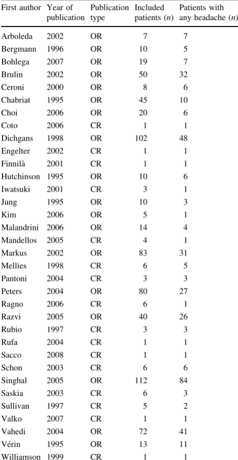
by the same authors. We finally reviewed 34 (5.8%) studies
including 16 case reports or letters (Table
1
) [
1
,
5
–
7
,
11
–
40
].
The reviewed studies provided data on 749 patients
including 387 (51.7%) who had headache. According to the
authors’ definition, 356 (92%) patients were reported as
having migraine and 31 (8%) as having headache
(Table
2
).
Out of the 356 patients described as migraineurs, 125
(35.1%) had migraine with aura, 7 (2%) migraine without
aura, 156 (43.8%) unspecified migraine, and 68 (19.1%)
had more than one type of migraine (Table
3
). Among the
125 patients who had migraine with aura, 24 (19.2%) had
migraine with typical aura, 7 (5.6%) migraine with
pro-longed aura, 5 (4.0%) familial hemiplegic migraine, 2
(1.6%) basilar migraine, while in 87 (69.6%) patients the
migraine type was not reported. The aura was visual in 10
Table 1 Studies included in the reviewFirst author Year of publication
Publication type
Included patients (n)
Patients with any headache (n) Arboleda 2002 OR 7 7
Bergmann 1996 OR 10 5 Bohlega 2007 OR 19 7 Brulin 2002 OR 50 32 Ceroni 2000 OR 8 6 Chabriat 1995 OR 45 10 Choi 2006 OR 20 6
Coto 2006 CR 1 1
Dichgans 1998 OR 102 48 Engelter 2002 CR 1 1 Finnila` 2001 CR 1 1 Hutchinson 1995 OR 10 6 Iwatsuki 2001 CR 3 1 Jung 1995 OR 10 3
Kim 2006 OR 5 1
Malandrini 2006 OR 14 4 Mandellos 2005 CR 4 1 Markus 2002 OR 83 31 Mellies 1998 CR 6 5 Pantoni 2004 CR 3 3 Peters 2004 OR 80 27 Ragno 2006 CR 6 1 Razvi 2005 OR 40 26 Rubio 1997 CR 3 3
Rufa 2004 CR 1 1
Sacco 2008 CR 1 1 Schon 2003 CR 6 6 Singhal 2005 OR 112 84 Saskia 2003 CR 6 3 Sullivan 1997 CR 5 2 Valko 2007 CR 1 1 Vahedi 2004 OR 72 41 Ve´rin 1995 OR 13 11 Williamson 1999 CR 1 1
(8%) patients, sensory in 7 (5.6%), motor in 5 (4%) and
included multiple symptoms in 25 (20%); the aura type was
not specified in 78 (62.4%) patients. Among the 68 patients
with more than one type of migraine, 50 (73.5%) had
migraine with aura, 38 (55.9%) migraine without aura, 18
(26.5%) migraine with aura and without headache, 13
(19.1%) basilar migraine, 11 (16.2%) familial hemiplegic
migraine, 11 (16.2%) migraine with unspecified atypical
aura, 4 (5.9%) acute onset aura without headache, 2 (2.9%)
unspecified migraine, 1 (1.5%) migraine with prolonged
aura, 1 (1.5%) migraine with acute onset aura, and 1 (1.5%)
had status migrainosus (Table
3
). At the onset of migraine,
the mean age
±
SD was 32.2
±
13.1 years.
In the 31 patients reported as having headache, the
headache was not further specified in 18 (58.1%) patients,
was defined as chronic in 6 (19.3%), as resembling
migraine with aura in 4 (12.9%), as resembling migraine
without aura in 2 (6.5%), and as tension type headache in 1
(3.2%) patient. At the onset of headache, mean age
±
SD
was 38.9
±
16.8 years.
We also analyzed whether the headache was the first
symptom or whether it was preceded by an ischemic stroke,
TIA or by other symptoms. After excluding patients for
whom data were not reported, 81 patients were described as
suffering from migraine and 19 from headache. While 9 of
the 81 patients (11.1%) had a previous stroke or TIA,
migraine was the first symptom in 66 patients (81.5%) and
in 6 (7.4%) it was preceded by other symptoms. While 3 of
the 19 patients (15.8%) had a previous stroke or TIA,
headache was the first symptom in 14 (73.7%) and in 2
(10.5%) it was preceded by other symptoms.
Discussion
From the 34 reviewed studies, we collected data on 387
patients [
1
,
5
–
40
]. In none of the cases, the headache was
coded according to the ICHD [
8
] or ICHD-II [
9
] criteria.
The ICHD-II diagnostic criteria for
Headache attributed to
CADASIL
required the following: (A) attacks of migraine
with aura, with or without other neurological signs; (B)
typical white matter changes on magnetic resonance
imaging
T2-weighted
sequences;
(C)
diagnostic
Table 2 Overall characteristics of patients with CADASILCharacteristics Migraine Headache Patients (n) 356 31 Gendera(%)
Men 36.7 36.4
Women 63.3 63.6
Mean age at onset (years) 32.2 38.9
a Where available
Table 3 Distribution of migraine subtypes in patients with CADASIL
a Includes one patient
diagnosed with migraine accompagne´e23
b Includes two cases of
migraine accompagne´e23
c Unspecified migraine includes
cases defined as migraine by authors without any further detail
Migraine subtype ICHD code ICHD-II code Patients
n %
Migraine without aura 1.1 1.1 7 2.0 Migraine with aura 1.2 1.2 125 35.1
Migraine with typical auraa 1.2.1 1.2.1 24 Migraine with prolonged aura 1.2.2 – 7 Familial hemiplegic migraine 1.2.3 1.2.4 5 Basilar migraine 1.2.4 1.2.6 2
Unspecified – – 87
Unspecified migrainec – – 156 43.8
More than one type of migraine – – 68 19.1 Migraine with aurab 1.2 1.2 50
Migraine without aura 1.1 1.1 38 Migraine aura without headache – 1.2.3 18
Basilar migraine – 1.2.6 13
Familial hemiplegic migraine 1.2.3 1.2.4 11 Migraine with unspecified atypical aura – – 11 Acute-onset aura without headache – – 4
Unspecified migrainec – – 2
Migraine with prolonged aura 1.2.2 – 1 Migraine with acute-onset aura – – 1
confirmation from skin biopsy evidence or genetic testing
(Notch3 mutations) [
9
]. The coding of headache according
to the ICHD or ICHD-II criteria might not have been done
due to the paucity of details reported in the previous and
present classification. Had authors correctly applied the
ICHD-II criteria, patients with CADASIL would have been
diagnosed as suffering from headache attributed to
CA-DASIL without any further detail. In the reviewed studies,
we found that 51.7% of patients with CADASIL were
reported to have suffered from headache. Anyhow, despite
that headache represents one of the major clinical features
of CADASIL, we cannot exclude that some patients
suf-fered from a primary headache. Besides, based on age of
onset and clinical characteristics of the headache in
CA-DASIL resembling those of some primary headaches, it
was not possible to establish the proportion of primary
headaches in the same patients. The headache in patients
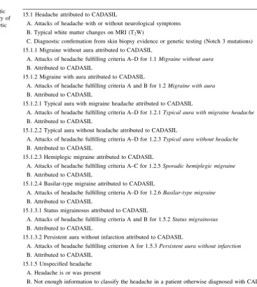
Table 4 Proposal for a new classification of headache in patientswith CADASIL based on available data 15 Headache attributed to genetic disorder
15.1 Headache attributed to CADASIL
15.1.1 Migraine without aura attributed to CADASIL 15.1.2 Migraine with aura attributed to CADASIL
15.1.2.1 Typical aura with migraine headache 15.1.2.2 Typical aura without headache 15.1.2.3 Hemiplegic migraine
15.1.2.4 Basilar-type migraine
15.1.3 Complications of migraine attributed to CADASIL 15.1.3.1 Status migrainosus
15.1.3.2 Persistent aura without infarction 15.1.5 Unspecified headache
15.2 Headache attributed to other genetic disordersa a This category is to be expanded and detailed
Table 5 Proposed diagnostic criteria for the new category of headache attributed to genetic disorder
15.1 Headache attributed to CADASIL
A. Attacks of headache with or without neurological symptoms B. Typical white matter changes on MRI (T2W)
C. Diagnostic confirmation from skin biopsy evidence or genetic testing (Notch 3 mutations) 15.1.1 Migraine without aura attributed to CADASIL
A. Attacks of headache fulfilling criteria A–D for 1.1Migraine without aura
B. Attributed to CADASIL
15.1.2 Migraine with aura attributed to CADASIL
A. Attacks of headache fulfilling criteria A and B for 1.2Migraine with aura
B. Attributed to CADASIL
15.1.2.1 Typical aura with migraine headache attributed to CADASIL
A. Attacks of headache fulfilling criteria A–D for 1.2.1Typical aura with migraine headache
B. Attributed to CADASIL
15.1.2.2 Typical aura without headache attributed to CADASIL
A. Attacks of headache fulfilling criteria A–D for 1.2.3Typical aura without headache
B. Attributed to CADASIL
15.1.2.3 Hemiplegic migraine attributed to CADASIL
A. Attacks of headache fulfilling criteria A–C for 1.2.5Sporadic hemiplegic migraine
B. Attributed to CADASIL
15.1.2.4 Basilar-type migraine attributed to CADASIL
A. Attacks of headache fulfilling criteria A–D for 1.2.6Basilar-type migraine
B. Attributed to CADASIL
15.1.3.1 Status migrainosus attributed to CADASIL
A. Attacks of headache fulfilling criteria A and B for 1.5.2Status migrainosus
B. Attributed to CADASIL
15.1.3.2 Persistent aura without infarction attributed to CADASIL
A. Attacks of headache fulfilling criterion A for 1.5.3Persistent aura without infarction
B. Attributed to CADASIL 15.1.5 Unspecified headache
A. Headache is or was present
with CADASIL was more frequently and clearly defined as
migraine and the majority of the migrainous patients were
reported as suffering from migraine with aura (35.1%).
Formally, this codification was inappropriate since
diag-nostic criteria for any type of migraine in the ICHD-II
require that the disturbance is not attributed to another
disorder. If the classification of CADASIL in the ICHD-II
was not revised, clinicians will be induced to commit a
formal error defining the headache as migraine or omit
useful clinical details attributing the headache to
CADA-SIL without giving any further characteristic. As much as
31 cases were diagnosed with disturbance other than
migraine; in those cases, the authors used a generic
ter-minology and possibly included patients suffering from
primary headaches such as tension-type headache.
The inclusion of headache in CADASIL, in chapter 6
of ICHD-II, among
Headache attributed to cranial or
cervical vascular disorder, implies that the headache is
caused by a vascular disease. This implication may rely
on two possibilities: the former is that the headache in
CADASIL might be considered secondary to the presence
of an organic vascular lesion and the latter is that a
dis-order of the vascular system might be considered as the
common underlying pathogenic mechanism that causes
both headache and stroke. Referring to the former
possi-bility, in CADASIL, the headache is usually the first
symptom of the disease (73.7–81.5% according to
reviewed data) preceding the onset of stroke or TIA.
Moreover, a diagnosis of secondary headache is usually
evident only when the headache resolves or greatly
improves within a specified time interval after its onset or
after the acute phase of the vascular disorder; however,
this evolution is not reported in patients with CADASIL
since the headache persists across the years.
Conse-quently, for all the above reported reasons, this possibility
is unlikely. Referring to the latter possibility, we have to
consider that the underlying pathology in CADASIL is
represented by an angiopathy with a unique type of
ultrastructural basal lamina deposits and by degeneration
of vascular smooth muscle cells, which are the major
source of the Notch3 expression. The evidence for a
functional impairment of vascular smooth cells is in line
with this latter hypothesis [
41
] and consequently the
pathogenic assumption reported in the ICHD-II is correct.
However, we would underscore that the primary
mecha-nism is not represented by the vascular damage, but by
the genetic alteration in the Notch3 expression. For all
these reasons, we suggest considering the possibility of
revising the ICHD-II when referring to CADASIL.
Spe-cifically, we suggest adding a new category that could be
named
Headache attributed to genetic disorder
including
Headache attributed to CADASIL
(Table
4
). The new
category might include also other genetic diseases in
which headache represents a major clinical feature.
Moreover, the new category should also report subtypes
according to specific clinical details to allow the precise
characterization
of
headache
in
the
single
patient
(Table
4
). The proposed diagnostic criteria for headache
attributed to CADASIL are reported in Table
5
. If our
proposal is shared by the experts in the field, we think
that it shall be easier from now on to characterize patients
with CADASIL uniformly and that a good step forward
will be realized.
Conflict of interest None.
References
1. Chabriat H, Vahedi K, Iba-Zizen MT, Joutel A, Nibbio A, Nagy TG, Krebs MO, Julien J, Dubois B, Ducrocq X, Levasseur M, Homeyer P, Mas JL, Lyon-Caen O, Tournier Lasserve E, Bousser MG (1995) Clinical spectrum of CADASIL: a study of 7 families. Cerebral autosomal dominant arteriopathy with subcortical infarcts and leukoencephalopathy. Lancet 346:934–939 2. Joutel A, Corpechot C, Ducros A, Vahedi K, Chabriat H, Mouton
P, Alamowitch S, Domenga V, Ce´cillion M, Marechai E, Mac-iazek J, Vayssiere C, Cruaud C, Cabanis EA, Ruchoux MM, Weissenbach J, Bach JF, Bousser MG, Tournier–Lasserve E (1996) Notch 3 mutations in CADASIL, a hereditary adult-onset condition causing stroke and dementia. Nature 383:707–710 3. Sacco S, Olivieri L, Bastianello S, Carolei A (2006) Comorbid
neuropathologies in migraine. J Headache Pain 7:222–230 4. Chabriat H, Joutel A, Dichgans M, Tournier-Lasserve E, Bousser
MG (2009) CADASIL. Lancet Neurol 8:643–653
5. Vahedi K, Chabriat H, Levy C, Joutel A, Tournier-Lasserve E, Bousser MG (2004) Migraine with aura and brain magnetic res-onance imaging abnormalities in patients with CADASIL. Arch Neurol 61:1237–1240
6. Schon F, Martin RJ, Prevett M, Clough C, Enevoldson TP, Markus HS (2003) ‘‘CADASIL coma’’: an underdiagnosed acute encephalopathy. J Neurol Neurosurg Psychiatry 74:249–252 7. Valko PO, Siccoli MM, Schiller A, Wieser HG, Jung HH (2007)
Non-convulsive status epilepticus causing focal neurological deficits in CADASIL. J Neurol Neurosurg Psychiatry 78:1287– 1289
8. Headache Classification Committee of the International Head-ache Society (1988) Classification and diagnostic criteria for headache disorders, cranial neuralgias and facial pain. Cepha-lalgia 8:1–96
9. Headache Classification Subcommittee of the International Headache Society (2004) The international headache classifica-tion of headache disorders, 2nd edn. Cephalalgia 24(Suppl 1):9– 160
10. Sacco S, Carolei A (2007) Migraine attributed to genetic disorder. Funct Neurol 22:117–118
11. Arboleda-Velasquez JF, Lopera F, Lopez E, Frosch MP, Sep-ulveda-Falla D, Gutierrez JE, Vargas S, Medina M, Martinez de Arrieta C, Lebo RV, Slaugenhaupt SA, Betensky RA, Villegas A, Arcos-Burgos M, Rivera D, Restrepo JC, Kosik KS (2002) C455R notc3 mutation in a Colombian CADASIL kindred with early onset of stroke. Neurology 59:277–279
morphological study of a German family. Acta Neuropathol 92:341–350
13. Bohlega S, Al Shubili A, Alreshaid A, Alkhairallah T, AlSous MW, Farah S, Abu-Amero KK (2007) CADASIL in Arabs: clinical and genetic findings. BMC Med Genet 8:67
14. Brulin P, Godfraind C, Leteurtre E, Ruchoux MM (2002) Mor-phometric analysis of ultrastructural vascular changes in CA-DASIL: analysis of 50 skin biopsy specimens and pathogenetic implications. Acta Neuropathol 104:241–248
15. Ceroni M, Poloni TE, Tonietti S, Fabozzi D, Uggetti C, Frediani F, Simonetti F, Malaspina A, Alimonti D, Celano M, Ferrari M, Carrera P (2000) Migraine with aura and white matter abnor-malities: Notch3 mutation. Neurology 54:1869–1871
16. Choi JC, Kang SY, Kang JH, Park JK (2006) Intracerebral hemorrhages in CADASIL. Neurology 67:2042–2044
17. Coto E, Mene´ndez M, Navarro R, Garcia-Castro M, Alvarez V (2006) A new de novo Notch3 mutation causing CADASIL. Eur J Neurol 13:628–631
18. Dichgans M, Mayer M, Uttner I, Bru¨ning R, Mu¨ller-Ho¨cker J, Rungger G, Ebke M, Klockgether T, Gasser T (1998) The phe-notypic spectrum of CADASIL: clinical findings in 102 cases. Ann Neurol 44:731–739
19. Engelter ST, Rueegg S, Kirsch EC, Fluri F, Probst A, Steck AJ, Lyrer PA (2002) CADASIL mimicking primary angiitis of the central nervous system. Arch Neurol 59:1480–1483
20. Finnila¨ S, Tuisku S, Herva R (2001) A novel mitochondrial DNA mutation and a mutation in the Notch3 gene in a patient with myopathy and CADASIL. J Mol Med 79:641–647
21. Hutchinson M, O’Riordan J, Javed M, Quin E, Macerlaine D, Wilcox T, Parfrey N, Nagy TG, Tournier-Lasserve E (1995) Familial hemiplegic migraine and autosomal dominant arteriop-athy with leukoencephalopathy (CADASIL). Ann Neurol 38:817–824
22. Iwatsuki K, Murakami T, Manabe Y, Narai H, Warita H, Hayashi T, Abe K (2001) Two cases of Japanese CADASIL with corpus callosum lesion. Tohoku J Exp Med 195:135–140
23. Jung H, Bassetti C, Tournier-Lasserve E, Vahedi K, Arnaboldi M, Arifi VB, Burgunder JM (1995) Cerebral autosomal dominant arteriopathy with subcortical infarcts and leukoencephalopathy: a clinicopathological and genetic study of a Swiss family. J Neurol Neurosurg Psychiatry 59:138–143
24. Kim Y, Kim JS, Kim G, No YJ, Yoo HW (2006) Two novel mutations of the NOTCH3 gene in Korean patients with CA-DASIL. Mutat Res 593:116–120
25. Lesnik Oberstein SA, van den Boom R, Middelkoop HA, Ferrari MD, Knaap YM, van Houwelingen HC, Breuning MH, van Bu-chem MA, Haan J (2003) Incipient CADASIL. Arch Neurol 60:707–712
26. Malandrini A, Carrera P, Palmeri S, Cavallaro T, Fabrizi GM, Villanova M, Fattaposta M, Vismara L, Brancolini V, Tanganelli P, Calı` A, Morocutti C, Zeviani M, Ferrari M, Guazzi GC (1996) Clinicopathological and genetic studies of two further Italian families with cerebral autosomal dominant arteriopathy. Acta Neuropathol 92:115–122
27. Mandellos D, Limbitaki G, Papadimitriou A, Anastasopoulos D (2005) Cerebral autosomal dominant arteriopathy with subcorti-cal infarcts and leukoencephalopathy (CADASIL) in a Greek family. Neurol Sci 26:278–281
28. Markus HS, Martin RJ, Simpson MA, Dong YB, Ali N, Crosby AH, Powell JF (2002) Diagnostic strategies in CADASIL. Neu-rology 59:1134–1138
29. Mellies JK, Ba¨umer T, Mu¨ller JA, Tournier-Lasserve E, Chabriat H, Knobloch O, Hackelo¨er HJ, Goebel HH, Wetzig L, Haller P (1998) SPECT study of a German CADASIL family. Neurology 50:1715–1721
30. Pantoni L, Sarti C, Pescini F, Bianchi S, Bartolini L, Nencini P, Basile AM, Lamassa M, Kalaria RN, Dotti MT, Federico A, Inzitari D (2004) Thrombophilic risk factors and unusual clinical features in three Italian CADASIL patients. Eur J Neurol 11:782– 787
31. Peters N, Herzog J, Opherk C, Dichgans M (2004) A two-year clinical follow-up study in 80 CADASIL subjects: progression patterns and implications for clinical trials. Stroke 35:1603–1608 32. Ragno M, Fabrizi GM, Cacchio` G, Scarcella M, Sirocchi G, Selvaggio F, Taioli F, Ferrarini M, Trojano L (2006) Two novel Italian CADASIL families from Central Italy with mutation CGC-TGC at codon 1006 in the exon 19 Notch3 gene. Neurol Sci 27:252–256
33. Razvi SSM, Davidson R, Bone I, Muir KW (2005) Is inadequate family history a barrier to diagnosis in CADASIL? Acta Neurol Scand 112:323–326
34. Rubio A, Rifkin D, Powers JM, Patel U, Stewart J, Faust P, Goldman JE, Mohr JP, Numaguchi Y, Jensen K (1997) Pheno-typic variability of CADASIL and novel morphologic findings. Acta Neuropathol 94:247–254
35. Rufa A, De Stefano N, Dotti MT, Bianchi S, Sicurelli F, Stro-millo ML, D’Aniello B, Federico A (2004) Acute unilateral visual loss as the first symptom of cerebral autosomal dominant arteriopathy with subcortical infarcts and leukoencephalopathy. Arch Neurol 61:577–580
36. Sacco S, Rasura M, Cao M, Bozzao A, Carolei A (2009) CA-DASIL presenting as status migrainosus and persisting aura without infarction. J Headache Pain 10:51–53
37. Singhal S, Rich P, Markus HS (2005) The spatial distribution of MR imaging abnormalities in cerebral autosomal dominant arteriopathy with subcortical infarcts and leukoencephalopathy and their relationship to age and clinical features. Am J Neuro-radiol 26:2481–2487
38. Sullivan AA, Teh BT, Jeavons S, Schalling M, Silburn P, Larsson C, Boyle R (1997) Cerebral autosomal dominant arteriopathy with subcortical infarcts and leucoencephalopathy. J Clin Neu-rosci 4:176–180
39. Ve´rin M, Rolland Y, Landgraf F, Chabriat H, Bompais B, Michel A, Vahedi K, Martinet JP, Tournier-Lasserve E, Lemaitre MH, Edan G (1995) New phenotype of the cerebral autosomal domi-nant arteriopathy mapped to chromosome 19: migraine as the prominent clinical feature. J Neurol Neurosurg Psychiatry 59:579–585
40. Williamson EE, Chukwudelunzu FE, Meschia JF, Witte RJ, Dickson DW, Cohen MD (1999) Distinguishing primary angiitis of the central nervous system from cerebral autosomal dominant arteriopathy with subcortical infarcts and leukoencephalopathy. Arthritis Rheum 42:2243–2248