REVIEW
A Review of Point Feature Based Medical
Image Registration
Shao‑Ya Guan
1, Tian‑Miao Wang
1,2, Cai Meng
2,3*and Jun‑Chen Wang
1,2Abstract
Point features, as the basis of lines, surfaces, and bodies, are commonly used in medical image registration. To obtain an elegant spatial transformation of extracted feature points, many point set matching algorithms (PMs) have been developed to match two point sets by optimizing multifarious distance functions. There are ample reviews related to medical image registration and PMs which summarize their basic principles and main algorithms separately. How‑ ever, to data, detailed summary of PMs used in medical image registration in different clinical environments has not been published. In this paper, we provide a comprehensive review of the existing key techniques of the PMs applied to medical image registration according to the basic principles and clinical applications. As the core technique of the PMs, geometric transformation models are elaborated in this paper, demonstrating the mechanism of point set registration. We also focus on the clinical applications of the PMs and propose a practical classification method according to their applications in different clinical surgeries. The aim of this paper is to provide a summary of point‑ feature‑based methods used in medical image registration and to guide doctors or researchers interested in this field to choose appropriate techniques in their research.
Keywords: Medical image registration, Point set matching, Optimization, Assessment, Application
© The Author(s) 2018. This article is distributed under the terms of the Creative Commons Attribution 4.0 International License (http://creat iveco mmons .org/licen ses/by/4.0/), which permits unrestricted use, distribution, and reproduction in any medium, provided you give appropriate credit to the original author(s) and the source, provide a link to the Creative Commons license, and indicate if changes were made.
1 Introduction
Medical image registration, i.e., aligning two medical images by finding an optimistic spatial transformation, plays a very important role in image-guided surgery and disease diagnosis. This alignment means that the same anatomical structures on two matching images corre-spond with each other spatially. One of the two purposes of medical image registration is to synthesize the associ-ated information contained in multiple modality images, and the other is to comprehend images obtained from the same instrument at different times and positions. Accord-ing to the nature of the registration bases, registration methods can be classified into two categories: extrinsic and intrinsic. The registration bases of extrinsic registra-tion methods are often features attached to patients or clinical instruments. Markers, frameworks and artificial implantations are widely used bases in extrinsic medical image registration. Intrinsic medical image registration
considers the information containing in the images as bases, such as the centerline of vessels and the outline of bones. Intrinsic medical image registration methods usu-ally fall into two categories: intensity-based methods and feature-based methods [1]. Intensity-based approaches employ difference measurements based on the pixel intensities, while feature-based methods use distance-based measures of the features extracted from the input images to be aligned. Feature-based algorithms are widely employed because of their briefness, simplicity and low computational complexity over intensity-based meth-ods. More importantly, multimodal registration using computer tomography (CT), magnetic resonance imag-ing (MR), positron emission tomography (PET), X-ray and other modality medical imaging methods is the main task of medical image registration. The intensity differ-ences among the multimodal images vary considerably due to the different imaging principles, which prohibit the development of intensity-based registration meth-ods. Feature similarities among the multimodal images are more explicit than the intensity differences, and as a
Open Access
*Correspondence: [email protected]
2 Beijing Advanced Innovation Center for Biomedical Engineering,
Beihang University, Beijing 100083, China
result that feature-based methods play an important role in medical image registration [2].
In this review, we focus on the point features used in medical image registration, because points are the most commonly used features in clinical applications. Point-feature-based registration means that the geometrical transformation between two images is only obtained through the characteristics of the feature points. The geo-metrical transformation can be classified into the rigid transformation and the non-rigid transformation accord-ing to whether there are feature deformations or not. Rigid geometric transformations are often used in the registration of rigid structures, such as bones. Another important application of a rigid transformation is to obtain a rough pre-registration result before a more com-plex and precise geometric transformation. Non-rigid geometrical transformations are usually used to finalize the precise registration or the registration of structures accompanied with deformations.
Point acquisition is the first step towards the PMs in medical image registration. The points extracted from the medical images can greatly reduce the amount of information to simplify the computational complexity. These extracted points are usually expressed as point sets to calculate the spatial transformation. The frame-work in Figure 1 shows the detailed process regarding how to a convert medical image registration to a point set matching problem. The acquisition of the transfor-mation model is a key component of point set matching. Transformation models are usually chosen according to different criteria, including the deformation of point sets, time and accuracy requirements of the registration, and the signal to noise ratio. Point set matching algorithms are the fundamental methods used to find the geometric transformation between two point sets, and have gained immense developments in image registration [3, 4].
As shown in Table 1, there have been several reviews of medical image registration methods and point set matching algorithms. Markelj et al. [1] surveyed the techniques used in 3D/2D registration methods for image-guided vascular interventions where CT, MRI, and X-ray images are the fundamental resources. In their paper in 2017, they also covered numerous avail-able PMs as well as methods to assess them [3]. Matl et al. [2] contributed a living review of vascular image registration in which abundant vessel based registration methods were discussed from variant aspects. Oliveira et al. [5] reviewed medical image registration method-ologies focusing on their geometric transformations, similarity measures, optimizers and accuracy assess-ment techniques. Sotiras et al. [6] conducted a survey regarding deformable medical image registration in which many nonrigid point set matching algorithms
were discussed deliberately. Multi-modality medical image registration is the most commonly applied field of feature-based medical image alignment. James et al. [7] provided a joint view of different image modali-ties applied in medical image fusion. Point sets are more familiar in image-guided vascular interventions in which segmented vascular bifurcations, centerlines, and edges can serve as the source of feature points.
Fixed image Floating image data
Feature extraction
Feature points sampling
Point set F Point set M
PSM:
Correspondence description Transformation calculation
Optimistic geometric transformation Medical image registration
Feature points sampling
Convert to the same dimension to fixed
image
Feature extraction
Aligned medical images
Figure 1 Procedure of the PMs in medical image registration
Table 1 Reviews of medical image registration and the PMs
Reference Year Content
[1] 2012 A review of 3D‑2D registration methods for image‑guided interventions
[2] 2017 Vascular image registration techniques: A living review
[3] 2017 Recent developments and trends in point set registration methods
[5] 2014 Medical image registration: a review
[6] 2013 Deformable Medical Image Registration: A Survey [7] 2014 Medical image fusion: A survey of the state of
The PMs in medical image registration are efficient and time-saving, which is crucial in clinical applica-tions. Nevertheless, a review focusing on point-feature-based medical image registration methods has not been published. The aim of this paper is to provide a sum-mary of the PMs used in medical image registration. The remainder of the paper is organized by the follow-ing: In Section 2, the related techniques of the PMs are discussed. In Section 3, Clinical applications of the PMs are classified by the type of clinical surgery. In Sec-tion 4, a discussion and a conclusion are given to sum-marize the paper.
2 Related Techniques of the PMs
PMs in medical image registration depend deeply on the acquisition of feature points. There are various point acquisition methods, which are discussed in detail in Section 2.1. When feature points are obtained, the aim of the registration between two medical images is deter-mined by the optimal geometric spatial transformation between the two feature point sets. Subsequently, three core steps of the geometric transformation are intro-duced in Sections 2.2 to 2.4, including transformation methods, optimizers, and an assessment of the transfor-mation methods.
2.1 Point Acquisition
Feature points applied in medical image registration fall into two categories: extrinsic and intrinsic points. Extrin-sic points are obtained by affixing certain artificial mark-ers to patients or clinical instruments, while intrinsic points are often acquired relying on anatomical struc-tures contained in the medical images.
2.1.1 Extrinsic Point Acquisition
Extrinsic points are usually markers attached to frames, skins, bones or organ surfaces. These points are com-monly used to distinguish soft tissues when there are not enough distinguishable structures to provide reliable ref-erences [8–12]. The number of extrinsic points used for registration is small and in most cases, the correspond-ences between the extrinsic points are usually known before registration. Precise closed-form solutions opti-mized by a least square method are usually used to obtain the spatial transformation. However, the focus of this paper is not on extrinsic points but on the intrinsic point sets where the number of points is large and the corre-spondence between two point sets are usually unknown.
2.1.2 Intrinsic Point Acquisition
Intrinsic points are subsampled from features, such as centerlines, outlines, corers, surfaces, and some special
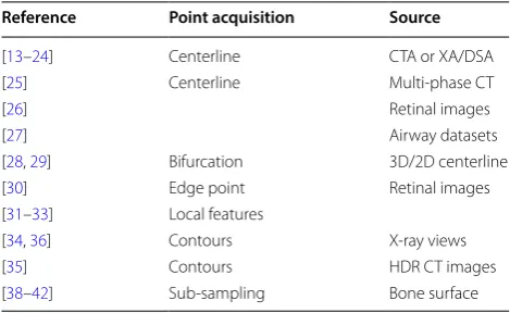
points, which are obtained from the medical images without attaching markers to the patients. Consequently, the intrinsic points cause less interference to patients and surgeries. As shown in Table 2, the centerline is one of the most widely used features to obtain feature points for the rising demand of vasculature registration in minimally invasive vascular interventions (MIVI). Many vessel thinning and centerline extracting meth-ods are used to obtain the proper centerlines. Methmeth-ods such as the vessel segmentation, vasculature filter, and centerline representation [13–16], have been employed to extract the centerlines in the registration of coronary or liver vessels [17–21]. In other interventions, such as an abdominal aortic aneurysm (AAA) intervention [22], graph-based segmentation [23] and a sequential topo-logical thinning process [24] are used to generate the centerline of the abdominal aorta. Centerlines also play an important role in the registration of multi-phase CT images [25], retinal images [26] and airway trees [27]. Bifurcations are special centerline points containing sig-nificant vascular information which makes bifurcation a vital component of centerline-based vasculature reg-istration [28, 29]. Retinal image registration is a special branch of vasculature-based registration because retinal image registration is restrained to 2D-2D image registra-tion in a relatively small and equal range. By embedding these particular properties of the retinal image into the registration process, many developed registration algo-rithms in computer vision and natural image registra-tion are introduced into retinal image registraregistra-tion. Edge points are extracted from notable features [30] embed-ding the geometry information into the registration. Local features, such as corners, speed-up robust feature (SURF) points and scale invariant feature transform (SIFT) points, are also extracted to improve registration accuracy by incorporating local features into the registra-tion [31–33].
Table 2 Intrinsic point sets acquisition
Reference Point acquisition Source
[13–24] Centerline CTA or XA/DSA
[25] Centerline Multi‑phase CT
[26] Retinal images
[27] Airway datasets
[28, 29] Bifurcation 3D/2D centerline
[30] Edge point Retinal images
[31–33] Local features
[34, 36] Contours X‑ray views
[35] Contours HDR CT images
For 3D-3D medical image registration, points are usu-ally extracted from surfaces and curves of 3D models.
Contours obtained from the cross sections of 3D mod-els are the most frequently used curves in 3D-3D medi-cal image registration [34–36]. Reconstructed surfaces, such as bone surfaces or other organ surfaces, cannot be used directly during registration because these surfaces contain abundant data, which will increase the com-putational complexity of the registration dramatically. Therefore subsampling methods are widely used in sur-face registrations to reduce the amount of data. Uniform sampling, random sampling, normal space sampling and relevance-based sampling [37] are the most commonly used subsampling methods to acquire point sets effi-ciently. The automatic surface sub-sampling method is extensively used in the US and CT bone surface registra-tion [38–41]. Manually picked sampling is an effective but time-consuming approach, which is also used in the registration of US, MR and CT bone surfaces [42].
2.2 Geometric Transformation Methods
The geometric method registers two images by minimiz-ing a distance calculation criterion. Before describminimiz-ing any methods, we introduce the known and unknown vari-ables of the problem. The rigid transformation between the target point set (M) and the source point set (S) can be expressed as follows:
where R and t represent the rotation and translation parameters, respectively, in the transformation. If M
and S are 3D point sets, then R and t represent the three rotation parameters and three translation parameters respectively. While in the 2D-2D rigid registration, there are only four parameters comprising two rotation param-eters and two translation paramparam-eters. The iterative clos-est point (ICP) algorithm (Section 2.2.1) is the most classical and prominent method for rigid point set reg-istration. However, it is difficult to describe the complex
(1)
M=R∗S+t
correspondence between M and S when there are defor-mations. The following formula only describes the crite-ria for non-rigid registration:
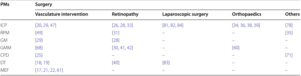
In this formula, f represents the transformation model, and θ represents parameters in the transforma-tion model. The robust point matching (RPM) algorithm (Section 2.2.2) is proposed using a spline function as the transformation model. The graph matching (GM) algo-rithm (Section 2.2.3) is proposed when f is described as a method to match two graphs. The minimized energy function (MEF) method (Section 2.2.4) is also widely used in describing deformations incorporating the energy function into the transformation model. The Gaussian mixture model (GMM) (Section 2.2.5) and the coherent point drift (CPD) algorithm (Section 2.2.6) are proposed using the distribution of two point sets to model the dif-ference between the two point sets. The distance trans-formation (DT) method (Section 2.2.7) shows several special distance measurements, which are more effec-tive than the Euler distance measurement, in describing the difference between two point sets. The application of these transformation models in clinical surgery is shown in Table 3.
2.2.1 ICP
The ICP algorithm, introduced by Besl and McKay [4], considers that the target point set (M) can be registered to the source point set (S) interactively after a rigid trans-formation. The fundamental principle of the ICP algo-rithm can be expressed as follows:
where mi∈M and si∈S . M is aligned to S, while dist(R,t) reaches the minimum value. The ICP algo-rithm has been significantly developed. Many variants of the ICP method have been proposed to affect all phases
(2)
M=f(S,θ )
(3)
dist(R,t)= 1 N
n
i=1
||Rmi+t−si||2
Table 3 Application of PMs in different surgery
PMs Surgery
Vasculature intervention Retinopathy Laparoscopic surgery Orthopaedics Others
ICP [20, 29, 47] [26, 28, 33] [81, 82, 84] [34, 36, 38, 39] [78]
RPM [49] [31] – – [35]
GM [29] [28] – – –
GMM [68] [30, 41, 42] – [40] –
CPD [25] – – – [71]
DT [18, 19] [40] [83] – –
of the algorithm. For error minimization, the classical ICP algorithm uses the Euclidean distance to construct a quadratic objective function subjected to a nonlinear constraint, while Sebastien developed a variant of the ICP algorithm called EM-ICP by applying the EM prin-ciple and an annealing scheme to avoid local minima [43]. Fitzgibbon proposed the LM-ICP method [44] where the objective function of the distance error was optimized with the Levenberg–Marquardt algorithm [45]. The ICP algorithm is simple and has a low compu-tational complexity, which also supports parallel com-puting. Nevertheless, the ICP algorithm suffers from its accurate demands of an initial estimation and limited application for only rigid transformations considerably. Another drawback that confines the application of the ICP approach is that the local minimum is obtained ulti-mately instead of a global minimum, even though Yang et al. [46] introduced a globally optimal solution Go-ICP based on the branch-and-bound algorithm under the L2-norm closest-point error metric. Another application is to combine the ICP algorithm with some global algo-rithms to obtain a global minimum [19, 29]. However, it is difficult to balance the speed and accuracy of the calculation.
Due to the characteristics of the ICP algorithm, it has been widely used in the rigid registration of medical images, such as the registration of 3D reconstructed rigid surfaces[34, 36, 38, 40] and vasculature without regarding the heartbeat and breathing[26, 28, 33, 47]. Nevertheless, the ICP method is not suitable for some non-rigid regis-tration especially with large deformations.
2.2.2 RPM
The robust point matching method (RPM) was initially put forward to simultaneously find the correspondence and transformation parameters between two point sets [48]. A deterministic annealing method was used to guar-antee the convexity of the error function of the RPM. The RPM uses a coordinate descent approach to calculate the transformation parameters and a soft-assign algorithm to estimate the correspondence between two point sets. Many variants of the RPM are proposed to modify the phases of the method from establishing a correspond-ence to optimizing the transformation parameters. Chui et al. developed the TPS-RPM [49] algorithm by using a thin plate spline (TPS) to parameterize the non-rigid mapping procedure because the TPS can describe affine and non-affine deformation elegantly. Zheng et al. [50] developed the RPM-LNS method by employing a simple graph to note the neighborhood structure of the point sets, which can transform the alignment of two point sets to the optimal matching between two graphs. The RPM-LNS method is generalized by the TPRL algorithm which
relaxes the labeling process with an optimal compatibil-ity coefficient. The Shape context is also an extensively employed descriptor to present the correspondence between points among the point sets [51–54].
As a non-rigid point set matching algorithm, the RPM has obtained several successes in the registration of medical images with small deformations, such as retinal images [31] and high-dose-rate (HDR) CT images for gynecological cancer detection [35]. As is well known, the quality of medical images is unsatisfactory due to the poor imaging mechanism, which substantially suffers from the noises of surgical instruments and the physi-ological activities of patients. The RPM performs poorly during the registration with high noise and outliers, which limited the application of RPM dramatically.
2.2.3 GM
Graph matching (GM) based approaches treat point sets as graphs. GM generally builds an adjacency matrix to present the correspondence between two graphs, where the nodes represent the correspondence between points and the weights stand for the pairwise agree-ments between potential correspondences [55]. The cor-rect assignments of the matching points are judged by whether the matching pairs are able to form a strongly connected cluster of the adjacency matrix. The graph shift (GS) method was proposed to eliminate the draw-backs of the GM approach to reduce its sensitivity to noises and outliers [56]. Subsequently, numerous algo-rithms have emerged to improve the robustness or computational complexity of the GM approach [57, 58]. Sousa et al. [59] combined the advantages of GM with the CPD method by adding the topological contribution of the GM into the CPD method to develop a stronger method.
GM is a proper approach to describe the topologi-cal structure of some vessels in which bifurcations can directly represent the nodes of a graph. Consequently, in medical image registration, GM has been mainly employed to register medical images with clear topologi-cal structures [28, 29].
2.2.4 MEF
Eexternal is generated by the transformation between the two images to be aligned, while the internal energy
Einternal represents the information of only one point set, including the displacement EDisp, the smoothness ESmooth and the myocardium constraint EMyocard. As mentioned above, the total energy function can be expressed as
where r is the transformation parameter, xi is one of the centerline points in the segmented fluoroscopy images, xˆi represents the centerline point of the model, H(x)∈ {0, 1} is used to reject the outliers and μ, β and γ are energy-balancing free parameters.
To describe the deformable registration of vascular structures, an energy function was proposed to map the deformable registration process [61]. The external item of this energy function is a difference measurement between two point sets, and the internal energy consists of a length preservation item and a smoothness preserva-tion item. This method is also used in an efficient graph-based 2D/3D deformable image registration algorithm, which is applied in abdominal aortic aneurysm interven-tions [62].
2.2.5 GMM
The Gaussian mixture model (GMM) algorithm for point set registration was proposed by Jian et al. [63]. These authors used the Gaussian mixture model to describe the distribution of the two given point sets M and S and treat the registration of two point sets as the alignment between two Gaussian mixtures by minimizing their L2-distance. The principle of GMM can be described as follows:
where θ stands for the transformation parameter of S
to align with M. The expectation maximization (EM) [64] method was employed to minimize the L2-distance and maximize the kernel correlation of the point sets. Although widely applied, the EM algorithm is quite sen-sitive to initial values, and its component number need to be pre-determined. To eliminate these drawbacks of EM, Yang et al. proposed a robust EM clustering algo-rithm which uses all the data points as the initialization. The authors have overcome the difficulties of choosing a
(4)
E(r)=Eexternal(r)+EDisp(r)+Esmooth(r)+EMyocard(r)
= K
i=1
H(xi)[ˆxi−(xi+ri)] +µ
K
i=1
|ri|2
+β
K
i=1
|˙ri|2+γ
{i,j∈K} |
ri−rj| |xi−xj|
2
(5)
dL2(S,M,θ )=
(gmm(S)−gmm(T(M,θ )))2dx
initial values and constructing a competition schema by embedding a penalty term into the EM algorithm [65]. To improve the accuracy of the GMM for 3D rigid/non-rigid registration, a convex hull indexed Gaussian mixture model (CH-GMM) [66] was proposed by incorporat-ing the proximity area conservation and projection con-sistency into a weighted GMM model. Dylan et al. [67] did not represent the distribution of the point set as a GMM directly but approximated the output function by training a one-class support vector machine (SVM) by a GMM. This method exploits the robust correspondence presentation of the SVM with sparse parametrization and occlusive outlier rejection to improve the robustness of the algorithm and minimize the L2-distance between two support vector-parameterized Gaussian mixtures.
As a probabilistic method, the GMM has achieved excel-lent results in medical image registration where the num-ber of feature points is considerable. The feature points in medical images of vessels are sufficient, because the cen-terlines and edges can be directly used as the source of the feature points. Consequently, the GMM is widely used in the registration of medical images of vasculature [68] or retinopathy [30, 40, 41]. However, the GMM relies greatly on the initial position of the GMM centers, which restrict the application of GMM and affect the registration result severely. As a global registration algorithm, another defi-ciency of the GMM is that it does not incorporate the detailed information among the feature points into reg-istration, thus the registration accuracy decreases greatly when the detailed information plays an important role in the alignment procedure. The most challenging task of the GMM is that a specific component number should be selected in advance, in which expert experience and a pre-analysis of the images to be aligned are needed.
2.2.6 CPD
The coherent point drift (CPD) algorithm, a probabilistic method extended from the GMM for both rigid and non-rigid registration of two point sets, considers the registra-tion of point sets as a maximum likelihood estimaregistra-tion problem which can be described as follows:
where σ2 is the equal isotropic covariance of the GMMs and θ represents the transformation parameters, X = (x1, …, xn)T is the data point set and Y = (y1, …, yn)T repre-sents the GMM centroids [69, 70]. The author regards the motion of the Gaussian centroids from the initial posi-tion to their final posiposi-tion as a temporal moposi-tion process.
(6)
Q(θ,σ2)= 1 2σ2
N
n=1 M
m=1
Pold(m|xn)||xn−T(ym,θ )||2
+ NPD 2 logσ
To preserve the topological structure of the point sets, the GMM centroids are restricted to move coherently as a group. Although the CPD approach performs more accurately and robustly than its competitors in the regis-tration of two point sets with noise, outliers and missing points, it also has two shortcomings. One issue is that the weight parameter w that estimates the level of noise and number of outliers in the GMMs is manually selected, and the other issue is that the CPD method does not take the neighborhood information of the points in the same point sets into account. To overcome these two short-comings of the original CPD algorithm, Peng et al. [71] proposed a robust CPD algorithm, which uses the shape context [51–54] to describe the neighborhood structure of the points and employs the EM framework to calculate and optimize w automatically. The robust CPD algorithm is a great improvement of the traditional CPD approach, where detailed information is incorporated into the reg-istration procedure. To accelerate or enhance the CPD algorithm, numerous variants of the CPD approach are proposed focusing on the different phases of the algo-rithm [72–75].
Similar to the GMM, CPD is also a probabilistic method in which it is difficult to describe the local neigh-borhood structure of the feature points. Although many local similarity descriptors have been proposed to elimi-nate the drawbacks of the CPD methods, there are still great difficulties in describing the complex anatomical structures as well as the local deformation of the medi-cal images. Despite the fact that applications in medimedi-cal image registration are restricted by the drawbacks men-tioned above, the CPD method is still successfully applied in the registration of multi-phase coronary CT images [25] and adjacent CT slices [71].
2.2.7 DT
The distance transformation (DT) method does not use the traditional L2-distance but some novel and ingen-ious distance measurements to calculate the difference between the two point sets. Here, we summarize two typical DT algorithms used in medical image registration.
To calculate the distance between the projected cor-onary CTA centerlines C and the X-ray images X, a sigmoid-like function was proposed by expanding the Euclidean distance [18]. The distance measure can be described as:
(7)
S(u,v)=
n−1
k=1
1 |x|
x∈X
h(D(X,C(u,v))),
(8)
h(d)=2− 2 1+exp(−d2
ha)
,
where u =(s, r) represents the offset and scaling param-eter, v represents the transformation parameters which vary according to the transformation types, ha is a con-trol parameter, and D represents the function to calculate the Euclidean distance. For the same purpose, Baka et al. proposed the G1/G2 distance measure for 2D-3D vascu-lature registration [17], in which the distance measure is a composition, including a 2D Euclidean measure, an ori-entation difference and a Frangi vasculature score Fj. the distance measure can be expressed as follows:
where Dij is the 2D Euclidean distance between the pro-jected vessel point i and the centerline point j, γij is the angle between the 2D centerline direction and the pro-jected 3D vessel direction, and α is a parameter deter-mining the influence of Fj. There are also several other DT methods modified from the Euclidean distance [19,
21] or other point set matching approaches [29].
2.3 Optimization of the Geometric Transformation
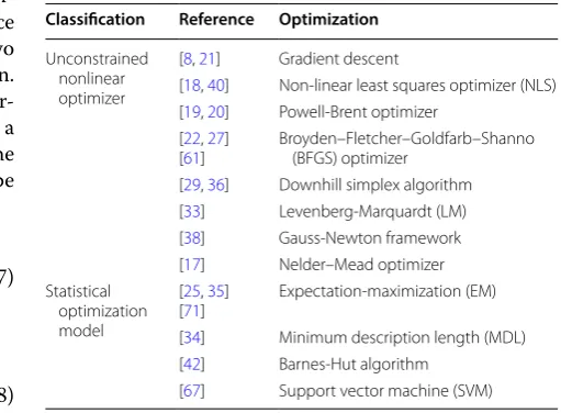
The iterative point set registration problem is a funda-mental optimization problem to search for the maximum or minimum value of the similarity measurements. As shown in Table 4, according to the objective function as elaborated in Section 2.2, we classify the optimization methods into two categories: an unconstrained nonlinear optimizer and a statistical optimization model.
The correspondence of the point-to-point registration is established based on each single point pair. Several
(9)
θij =α(1−Fj)+
1− |cosγij|exp
−D
2 ij
σ2
,
Table 4 Optimizers used in PSM-based medical image registration
Classification Reference Optimization
Unconstrained nonlinear optimizer
[8, 21] Gradient descent
[18, 40] Non‑linear least squares optimizer (NLS) [19, 20] Powell‑Brent optimizer
[22, 27]
[61] Broyden–Fletcher–Goldfarb–Shanno (BFGS) optimizer [29, 36] Downhill simplex algorithm [33] Levenberg‑Marquardt (LM) [38] Gauss‑Newton framework [17] Nelder–Mead optimizer Statistical
optimization model
[25, 35]
[71] Expectation‑maximization (EM) [34] Minimum description length (MDL) [42] Barnes‑Hut algorithm
unconstrained nonlinear optimizers have been used to determine the correspondence between two point sets, including the non-linear least squares optimizer (NLS) [17, 39], Levenberg–Marquardt (LM) algorithm [33], downhill simplex algorithm [29, 34], Nelder–Mead optimizer [21], Gauss-Newton framework [38], Gra-dient descent [8, 20], Powell-Brent optimizer [18, 19], and Broyden–Fletcher–Goldfarb–Shanno (BFGS) opti-mizer [22, 27, 61]. The references that contain detailed descriptions of these algorithms are displayed in Table 4. The applicable conditions and selection criteria of these optimize algorithms are elaborated in detail in the book by Press et al. [76] if the distance measure between two point sets has been modeled as a convex function. If the distance measure is non-convex, then Khoo et al. provide a proper method to convert a non-convex function into a convex function to improve the registration efficiency by relaxing some constrains [77].
Several probabilistic registration methods, whose cor-respondence is set up on the distribution of points, can-not be optimized by traditional optimization methods. Some statistical optimization models have been applied to optimize these point set matching algorithms such as the GMM and the CPD method. The minimum descrip-tion length (MDL) [36], Barnes-Hut algorithm [41], expectation-maximization (EM) [25, 35, 71] and support vector machine (SVM) [67] are presented as methods based on the point set distribution.
In summary, an optimization method satisfies a certain objective function. We only provide a general direction for the selection of optimization methods. Specific opti-mization method should be chosen after referring to the professional books [76].
2.4 Assessment of the Geometric Transformation
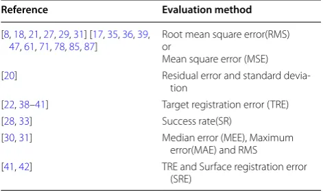
A registration has little value unless its accuracy can be evaluated. Several approaches have been proposed to assess the registration accuracy, which is shown in Table 5. Various distance measures have been used in
the PMs of medical image registration, so the evaluation method is usually a certain error function to assess the registration dissimilarity between the two point sets.
The root mean square error (RMS) or mean square error (MSE) has been the most commonly used approach to evaluate the registration results. These criterions effec-tively evaluate the registration between two point sets subsampled from the vessel centerlines or bifurcations in vasculature interventions [17, 20, 21, 29, 31, 47, 61], contour or surfaces of bone in orthopaedics [34, 42], edge points and some other local feature points of multiple kinds of medical images[8, 27, 35, 71, 78], because these criterions can take advantage of the distance measures directly without introducing additional errors. However, the drawback of the RMS is also its dependency on the distance measure. When the distance measure is not cho-sen properly, the RMS will rise dramatically, which will reduce the value of the evaluation criteria. The target reg-istration error (TRE) calculates the distance between the corresponding points that might represent surgically tar-geted tissues. These approach is an objective and accurate measurement, which is applied when there are known ground-truth positions of the validation points [22, 38,
39, 41, 42]. The success rate (SR) is a unique evaluation in the PMs by counting the successfully registered point pairs. The SR has been applied in retinal image registra-tions to assess the successful registration rate of center-line points or bifurcations of retinal vessels [28, 33].
Some methods use more than one evaluation method to obtain a more convincing result. The FRE and SRE are used to estimate registration accuracy between point sets subsampled from CT and US bone surfaces [40, 41]. The residual error and the standard deviation are chosen to evaluate registration between a 3D coronary projection and XA images, where the pixels of the vessel are consid-ered as the point sets to be aligned [19]. The MEE, MAE, and RMS work as standards when the authors compared their proposed method with many earlier algorithms to confirm that their method exhibited a more persuasive performance in the registration of retinal images [30, 31].
Robustness, speed, convergence, and stability are also frequently used to evaluate and compare the perfor-mance of numerous PMs [3]. The design of the evalua-tion image data sets, the definievalua-tion of the corresponding ground truth and accuracy, the selection of evaluation criteria, the design of evaluation metrics and the design of the evaluation protocol are prerequisites in the stand-ardization of medical image evaluation methodology [79]. However, a gold evaluation criterion to assess the performance of the PMs in medical image registration methods has not been published.
Table 5 Registration accuracy assessment methods
Reference Evaluation method
[8, 18, 21, 27, 29, 31] [17, 35, 36, 39,
47, 61, 71, 78, 85, 87] Root mean square error(RMS)or Mean square error (MSE)
[20] Residual error and standard devia‑
tion
[22, 38–41] Target registration error (TRE)
[28, 33] Success rate(SR)
[30, 31] Median error (MEE), Maximum
error(MAE) and RMS
3 Clinical Applications of the PMs
Medical images vary considerably in different clinical surgeries. The previous section provides a comprehen-sive view of the PMs used in medical image registra-tion. However, PMs are also influenced by the various circumstances in different clinical surgery. For vascular intervention (Section 3.1), the urgent requirement is the registration between 3D CTA/MRA models to the 2D X-ray/DSA images. For retinal image registration (Sec-tion 3.2), time and position are the prominent differences between two retinal images. Although the registration of bones is rigid, the modalities of the medical images are complex in orthopeadic surgery (Section 3.3), including ultrasound (US), CT and X-ray images. The PMs are also applied in some other clinical applications (Section 3.4) such as the registration of airways, ventricle surfaces, lung surfaces or a 3D reconstruction of CT or MR slices.
3.1 PMs in Vascular Intervention Surgeries
Enhancing interventional visualization is an essential part of image-guided interventions by integrating different modality information extracted from intraoperative and preoperative images into one single coordinate frame-work [1]. Combining complementary information taken from various imaging modalities or taken at the different times helps the surgeons obtain a comprehensive aware-ness of the vasculature and provides a better diagnosis of diseases. The 3D computed tomography (CT) or mag-netic resonance images (MRI) usually acquired as pre-interventional data, and 2D X-ray projected images are typically collected as the intro-interventional data. The alignment between CT/MRI and X-ray images forms a 3D/2D image registration scheme.
There are abundant classical methods to classify 3D/2D vasculature registration, as described in a living review developed by Stefan Matl et al. [2]. The most commonly used methods are pixel or voxel similarities based simi-larity measurements and feature based transformation estimations. The intensity information between the dif-ferent modality images diverges greatly from each other, while the anatomical structures of vessels, such as cen-terlines and bifurcation points can demonstrate the dis-similarity between the two images more competently. Meanwhile, features after segmentation contain less data, which can reduce the computational complexity dra-matically. Features used for vasculature registration are often geometrical entities such as isolated points or point sets sampled from curves, contours or surfaces. Con-sequently, the basis of most feature-based registration methods is point set registration. Point set registration of vasculature generally consists of the following five parts:
1) Segmentation and preprocessing of 3D and 2D images,
2) Centerlines or edges extraction,
3) Point resampling of the centerlines or the edges, 4) Point preprocessing,
5) Minimizing the distance between two point sets.
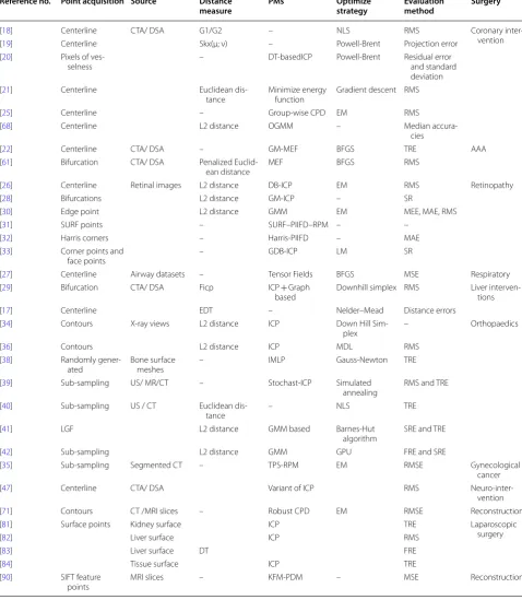
The first three steps of the five parts mentioned above are shown in Figure 2, which shows two approaches to convert the 3D-2D abdominal aorta image registration to align two 2D vascular centerlines that can be used as point sets directly in the PMs. The PMs applied in vas-culature interventions vary seriously in different clinical applications. Table 6 at the end of this paper shows an overview of the relationship between The PMs and their clinical applications.
In coronary intervention, the biggest challenge is that coronary vessels move along with a beating heart. There-fore the registration of coronary images must incor-porate the movement of the heart into the procedure. Nonrigid registration for large deformations exactly
fits this condition. Baka et al. [68] extended the original GMM algorithm for 3D-3D point set registration to the 3D-2DOGMM algorithm incorporating vascular orien-tation into the alignment of the preoperative computed
tomography angiography (CTA) and the intraoperative X-ray angiography of coronary interventions. The point sets in OGMM were obtained from the coronary center-line points, which appropriately represent the topological Table 6 Summary of PMs and their applications in medical image registration
Reference no. Point acquisition Source Distance
measure PMs Optimize strategy Evaluation method Surgery
[18] Centerline CTA/ DSA G1/G2 – NLS RMS Coronary inter‑
vention
[19] Centerline Skx(µ; ν) – Powell‑Brent Projection error
[20] Pixels of ves‑
selness – DT‑basedICP Powell‑Brent Residual error and standard
deviation
[21] Centerline Euclidean dis‑
tance Minimize energy function Gradient descent RMS
[25] Centerline – Group‑wise CPD EM RMS
[68] Centerline L2 distance OGMM – Median accura‑
cies
[22] Centerline CTA/ DSA – GM‑MEF BFGS TRE AAA
[61] Bifurcation CTA/ DSA Penalized Euclid‑
ean distance MEF BFGS RMS
[26] Centerline Retinal images L2 distance DB‑ICP EM RMS Retinopathy
[28] Bifurcations L2 distance GM‑ICP – SR
[30] Edge point L2 distance GMM EM MEE, MAE, RMS
[31] SURF points – SURF–PIIFD–RPM – –
[32] Harris corners – Harris‑PIIFD – MAE
[33] Corner points and
face points – GDB‑ICP LM SR
[27] Centerline Airway datasets – Tensor Fields BFGS MSE Respiratory
[29] Bifurcation CTA/ DSA Ficp ICP + Graph
based Downhill simplex RMS Liver interven‑tions
[17] Centerline EDT – Nelder–Mead Distance errors
[34] Contours X‑ray views L2 distance ICP Down Hill Sim‑
plex – Orthopaedics
[36] Contours L2 distance ICP MDL RMS
[38] Randomly gener‑
ated Bone surface meshes – IMLP Gauss‑Newton TRE
[39] Sub‑sampling US/ MR/CT – Stochast‑ICP Simulated
annealing RMS and TRE
[40] Sub‑sampling US / CT Euclidean dis‑
tance – NLS TRE
[41] LGF L2 distance GMM based Barnes‑Hut
algorithm SRE and TRE
[42] Sub‑sampling L2 distance GMM GPU FRE and SRE
[35] Sub‑sampling Segmented CT – TPS‑RPM EM RMSE Gynecological
cancer
[47] Centerline CTA/ DSA Variant of ICP RMS Neuro‑inter‑
vention
[71] Contours CT /MRI slices – Robust CPD EM RMSE Reconstruction
[81] Surface points Kidney surface ICP TRE Laparoscopic
surgery
[82] Liver surface ICP RMS
[83] Liver surface DT FRE
[84] Tissue surface ICP TRE
[90] SIFT feature
structure of vessels. The distance measurement calculat-ing the difference between two point sets is adjusted in this model to achieve a more accurate result. The OGMM algorithm first incorporates the distinctive features of vessels into the registration process. This improvement causes the OGMM to outperform the ICP algorithms, G1 and other distance transformation algorithms [17–20] in accuracy and convergence rate. Another procedure to improve the performance of the OGMM is to modify the optimization process. Khoo et al. [77] developed a convex programming framework to optimize the correspond-ence and the relative transformation between the point sets simultaneously. This effective framework can obtain a global optimum by transforming a non-convex process to a convex process by relaxing some restrictions of the optimization. By incorporating some priori information, such as transformation invariant features and gradient information, into the preprocessing of registration, the author concluded that the relaxed convex version con-verges to the solution of the original program despite the sharp sensitivity to noise. Combining this algorithm with some local information based methods takes advan-tages of both local and global methods to obtain a more accurate optimum. Motivated by the application of coro-nary matching in multi-phase cardiac spiral CT, a group-wise CPD algorithm [25] was proposed that embed the temporal consistency constraints between the point sets into registration to overcome one of the biggest prob-lems of multi-phase point set registration. The point sets are obtained from manually annotating centerlines and branch points. The coronary vessels deform sharply as the heart beats, so the registration between the multi-phase CT is a nonrigid process where the CPD algo-rithm performs effectively. Although the algoalgo-rithm has achieved elegant results in the registration of multi-phase coronary vessels, it is very restrictive with too many arti-ficial interventions.
Liver vasculature intervention is another vital surgery to detect liver cancer, liver biopsy, liver cirrhosis, portal hypertension and other liver diseases. Kim et al. pre-sented an EDT [80] based algorithm to register the cho-sen relevant 3D vessels of CTA and the given DSA image [21]. The EDT method was applied in this algorithm to calculate the distance between two point sets extracted from the projected CTA images and DSA images. The 3D vascular structure was divided into several segmented subtrees which ensure that the given DSA is aligned with the most similar subtree. Groher et al combined ICP with GM to obtain a novel 2D-3D liver vasculature registra-tion algorithm. This combinaregistra-tion takes advantage of the low computational complexity of ICP and the topological information of GM, which helps the registration process converge to an optimum result quickly and robustly [29].
Abdominal aortic aneurysm intervention (AAA) is also influenced severely by heart beating. The diameter of aorta is larger than the liver and the coronary vessels. Additionally, there are fewer branches in the aortic ves-sels. Consequently, the most commonly applied PMs in AAA intervention is MEF [22, 61], which not only describes the large deformation but also preserves the smoothness and length features of the aorta.
Deformation of cerebrovascular tissue in neuro- inter-vention is so slight that can be ignored, so medical image registration in neuro-intervention is generally considered to be a rigid registration. The variants of ICP are efficient to align two cerebrovascular images [47].
PMs utilized in vascular intervention surgery as men-tioned above, have gained significant achievements in some specific applications. Nevertheless, there are some deficiencies in these methods, which limit their clinical applications. First, the viewable range of CTA and DSA images differs greatly, which increases the rate of miss-ing points and noise of the registration and enlarges the dissimilarity between two point sets. Consequently, the fit of PMs for point sets with large number of differences should be developed to eliminate this restriction. Sec-ond, only the centerline points or bifurcation points are used in the registration process, other vessel information including diameters and curvatures are rarely considered in the proposed papers. Future algorithms should take these features into consideration. Finally, all the algo-rithms mentioned above could not describe the deforma-tion of vessels accurately, so future algorithms should be more capable of describing the deformation of the vascu-lar images.
3.2 PMs in Retinal Image Registration
Retinal images play an important role in the diagnosis of multiple eye diseases such as diabetic retinopathy, age-related macular, degeneration and glaucoma. Multiple imaging protocols have been applied to obtain images of the different parts of the fundus. Fundus angiography is a better technique to observe blood flow, obstruction and other vascular lesions [26]. It is necessary to register mul-tiple retinal image fragments or retinal images in multi-ple modes to obtain an entire retina image of a patient, to comprehend retinal image information in multiple modes and compared the vascular changes of a specific patient at different times.
above increase the difficulty of retinal image registration. Contraposing these problems, the above researchers have proposed a variety of methods.
The dual-bootstrap ICP [26] was proposed by Stew-art et al. which takes advantage of not only branches and intersections, but also some local information with high precision and similarity. Point sets are auto-matically abstracted from the given retinal images. The dual-bootstrap step calculates the covariance matrix of the estimated transformation to initialize and modify the transformation parameters, while the ICP step uti-lizes the robust ICP algorithm to minimize the distance between two point sets. The combination of the dual-bootstrap and ICP eliminates the drawback of the ICP that calls for an accurate initialization. The GM method is an excellent approach to make full use of the vascular topological structure information for registration. Deng et al. introduced the GM-ICP algorithm [28] which com-bined the GM and ICP to efficiently find the global corre-spondences between the vascular bifurcations. However, the GM-ICP is deeply affected by the sufficiency of con-sistent structures, which limits its application in medical image registration. Another technique [30] incorporating both local and global information of the retinal image is the Lo-GMM which integrates local image details into a GMM model (Lo-GMM). This method can effectively obtain a global result by aligning the probability den-sity distribution of the vascular edge points in two reti-nal images. The local image features contain abundant information which can improve the registration accuracy greatly, but the computational complexity of the algo-rithm increases sharply with embedding of too much local information.
Researchers attempted to use invariant features, such as the partial intensity invariant feature descriptor (PIIFD), and the speed up robust features (SURF), of reti-nal images to avoid the detection of vascular bifurcations. The PIIFD and Harris corners are used to obtain point sets in the PIIFD-Harris algorithm [32]. Corner points, instead of bifurcations, serve as control point candidates which reduce the computational complexity and improve the automation of the registration procedure. Wang et al. proposed the SURF–PIIFD–RPM method combining the invariant features with RPM algorithm to register retinal images with deformations automatically [31]. Poor qual-ity and nonvascular images, which are not considered in the PIIFD-Harris method, are also incorporated in the experiments of the SURF–PIIFD–RPM. This method takes advantage of the RPM in outlier rejection to improve the robustness of the algorithm, so that desired result can be obtained, even when the overlapping area between two retinal images is very small.
Retinal image alignment is restricted to 2D-2D registra-tion due to imaging principles and medical applicaregistra-tions. Different from vessel registration in vascular interven-tions, features applied in retinal image registration are not only branches, intersections, centerline points and edge points, but also invariant features, such as Har-ris and SURF features. The view range of the retinal images is exactly the same as each other, which facilitates the registration by ignoring scaling parameters. Future development should consider improving the registration accuracy as well as reduce the computational complexity when the overlapping areas are insufficient.
3.3 PMs in Orthopaedic Surgery
Registrations of bones in orthopaedic surgery are often rigid registrations. Feature points used for registration are usually derived from bone surfaces which are vis-ible in both the reconstructed bone model and the tar-get anatomy during surgery. Ultrasound (US) images, which do no harm to patients and surgeons, are gradu-ally used in orthopaedic surgeries to assist the registra-tion of preoperative and intraoperative images. US is safe and inexpensive and can be used in real-time. However, ultrasound also suffers from its sensitivity to noise and limited fields of view. High resolution and high signal to noise ratio of US make it the most commonly used meth-ods in clinical operations despite its high price. Register-ing tracked intraoperative US images with preoperative CT data has been proposed as an effective mechanism for computer assisted orthopaedic surgery (CAOS) [41]. Feature points extracted from bone surfaces are fre-quently used to generate data sets in the pre-procedure of the PMs. Consequently, the point matching algorithms play an important role in the registration of US and CT images. Other modalities of medical images are also applied to increase the amount of useful surgical infor-mation and decrease the cost of surgery.
a high calculation speed to meet real-time requirements and is a good descriptor of bone surfaces for incorporat-ing the local curvature of points. There are also many applications of the PMs in other bone-related surgery processes, such as 3D reconstruction of bones by fluoro-scopic images and alignment of X-ray bone slices [34, 36].
3.4 PMs in Laparoscopic Surgery
Laparoscopic surgery (LS) is another interventional tech-nique to access the abdominal anatomy of the patient. LS is minimally invasive in contrast to the traditional open surgery which involves cutting the skin and dividing the underlying tissues to gain a direct access to the surgical target. However, LS can only provide the surgeons with a restricted and small view of the surgical field, which abandoned the navigation of the surgery. To improve the visualization capability of LS, medical image registration is required to comprehend the various images taken in pre- or intra-laparoscopic surgery. Makers play an impor-tant role in the majority of automatic approaches for reg-istering the endoscopic images, where rigid PMs, such as ICP and its variants, are widely used to obtain an accu-rate registration. Benincasa et al. [81] used ICP to regis-ter the surface points of the kidney to enlarge the view of the intraoperative surface. In image guided liver sur-gery, Cash et al. [82] used the ICP algorithm to register the preoperative and intraoperative representation of the liver surface in the rigid registration procedure. Matching surface correspondences is one of the key components of shape-based guidance, and Santos proposed an approach based on the distance error to establish the surface cor-respondences of the liver surface reconstructed from volume scanners, such as CT and MRI, and 3D range scanners to initialize the fine surface matching algo-rithms for intra-operative soft tissue registration [83]. PMs also ensure that optical techniques for 3D surface reconstruction procedure are effective, even when there are thousands of feature points [84–87].
3.5 PMS in Other Surgical Applications
Registration of airway trees is a unique procedure where the orientation and thickness of the airway trees cannot be ignored. Wassermann et al. [27] introduced the ten-sor field into the registration of point sets collected from airway trees where each point is associated with a sym-metric matrix representing the orientation and thickness of the airway trees. The tensor field, which is represented sparsely, is transformed from a shape feature matrix to convert the alignment of two images to the registration of two tensor fields. The tensor field algorithm is tested in both synthetic and human airways to validate that its registration accuracy is superior to GMM and other up-to-date algorithms.
Almhdie [78] proposed a comprehensive ICP (CICP) algorithm to align two point sets collected from left ven-tricle and lung surfaces. A look-up matrix was introduced to timprove the performance of the original ICP. The pro-posed CICP algorithm performs better than the original ICP (OICP) and Picky ICP (PICP) methods in not only the precision of the point correspondence estimation but also the resistance to noise and outliers. TPS-RPM was employed for matching cavity surface points to facilitate the dose accumulation among the high-dose-rate treat-ment fractions [35].
approach in medical image registration still have room for improvement.
4 Conclusions
(1) In this paper, we focus on point-feature-based medical image registration which is extensively used in clinical applications. This paper is struc-tured around the two core aspects: registration techniques and clinical application. For each com-ponent, particular emphasis is placed on classifying the methods according to their theoretical founda-tions and applicafounda-tions.
(2) Independent of extrinsic points or intrinsic points, the precisely point acquisition is of great impor-tance in the pre-procedure of the PMs. The accu-racy of registration is affected severely by the pre-cision of point extraction, therefore future work should explore more accurate feature segmented algorithms and points extraction methods. In addi-tion, with the rapid development of the PMs, more precise and effective algorithms will be introduced into medical image registration.
(3) Choosing proper geometric transformation meth-ods is crucial after the feature points are acquired. If the correspondence between two point sets is known before registration, the least square method is sufficient to minimize the distance between two point sets. However, in most cases, the correspond-ence is unknown. Under these circumstances, parameters, such as the degree of deformation, the registration accuracy, and the signal-to-noise ratio, should be taken into consideration to choose an ele-gant geometric transformation. The RPM, GMM, and CPD are effective for non-rigid registration with large deformations, while ICP performs well at rigid and rough registration with low computa-tional complexity. If considerable noises are con-sisted among the extracted point sets, algorithms with a low sensitivity to noise such as CPD should be chosen.
(4) Clinical application is a new classification crite-rion that has been seldom related in the previous studies. As shown in Table 2 and Table 3, vascular interventional surgery is the most widely used field of the PMs. The main reason is that the centerlines, bifurcations and edge points of blood vessels can be used directly as the source of the feature points. Vascular interventional surgery of different vessels has different standards in the acquisition of feature points. For abdominal aortic vasculature, the vascu-lature diameter is very large, and centerline cannot describe the vessel structure precisely, so the edges of vessels are usually extracted as the source of the
feature points. For liver vessels and vessels of other organs, diameters of these vessels are narrow and have limited influence on the registration accuracy, so the centerline of the vessel can directly serve as the origin of feature point sets.
(5) In summary, feature-based medical image registra-tion has greatly benefitted from its real-time per-formance and registration accuracy. With the rapid development of feature extraction technology, fea-ture-based methods will experience an upcoming explosion.
Authors’ Contributions
Cai Meng was directed the entire trial; Shao‑Ya Guan wrote the manuscript; Tian‑Miao Wang and Jun‑Chen Wang assisted in modifying the structure and content of this paper. All authors read and approved the final manuscript.
Author details
1 School of Mechanical Engineering and Automation, Beihang University,
Beijing 100191, China. 2 Beijing Advanced Innovation Center for Biomedical
Engineering, Beihang University, Beijing 100083, China. 3 School of Astronau‑
tics, Beihang University, Beijing 100191, China.
Authors’ Information
Shao‑Ya Guan, born in 1992, is currently a PhD candidate at School of Mechani-cal Engineering and Automation, Beihang University, China. She received her bachelor’s degree from Hebei University of Technology, China, in 2014. Her research interests include medical robotics and medical image processing.
Tian‑Miao Wang, born in 1960, is currently a professor at School of Mechan-ical Engineering and Automation, Beihang University, China. He received his doctor’s degree from Northwestern Polytechnical University, China, in 1990. His research interests include medical robotics, special robots and service robots.
Cai Meng, born in 1977, is currently an associate professor at School of Astronautics, Beihang University, China. He received his doctor’s degree from Beihang University, China, in 2004. His research interests include machine vision and its application in robotic intelligent systems.
Jun‑Chen Wang, born in 1984, is currently an associate professor at School of Mechanical Engineering and Automation, Beihang University, China. He received his doctor’s degree from Beihang University, China, in 2012. His research interests include medical robotics, medical image processing and minimally invasive surgery navigation.
Competing Interests
The authors declare no competing financial interests.
Funding
Supported by the National Natural Science Foundation of China (Grant No. 61533016)
Publisher’s Note
Springer Nature remains neutral with regard to jurisdictional claims in pub‑ lished maps and institutional affiliations.
Received: 19 September 2017 Accepted: 10 August 2018
References
[1] P Markelj, D Tomaževič, B Likar, et al. A review of 3D/2D registration meth‑ ods for image‑guided interventions. Medical Image Analysis, 2012, 16(3): 642–661.
[3] B Maiseli, Y Gu, H Gao. Recent developments and trends in point set registration methods. Journal of Visual Communication and Image Repre-sentation, 2017, 46: 95–106.
[4] P J Besl, N D McKay. A method for registration of 3‑D shapes. IEEE Transac-tions on Pattern Analysis and Machine Intelligence, 1992, 14(2): 239–256. [5] F P Oliveira, J M Tavares. Medical image registration: a review. Comput.
Methods Biomech. Biomed Engin., 2014, 17(2): 73‑93.
[6] A Sotiras, C Davatzikos, N Paragios. Deformable medical image reg‑ istration: A Survey. IEEE Transactions on Medical Imaging, 2013, 32(7): 1153–1190.
[7] A P James, B V Dasarathy. Medical image fusion: A survey of the state of the art. Information Fusion, 2014, 19: 4–19.
[8] T S Y Tang, R E Ellis, G Fichtinger. Fiducial registration from a single X‑Ray image: a new technique for fluoroscopic guidance and radiotherapy. International Conference on Medical Image Computing and Computer-Assisted Intervention, Pittsburgh, USA, October 11–14, 2000: 502–511. [9] A Schweikard, H Shiomi, J Adler. Respiration tracking in radiosurgery without fiducials. The International Journal of Medical Robotics and Com-puter Assisted Surgery, 2005, 1(2): 19–27.
[10] R I Whyte. Stereotactic radiosurgery for lung tumors. Seminars in Thoracic and Cardiovascular Surgery. 2010, 22(1): 59–66.
[11] Z Mu, D Fu, G Kuduvalli. A probabilistic framework based on hidden Markov model for fiducial identification in image‑guided radiation treat‑ ments. IEEE Transactions on Medical Imaging, 2008, 27(9): 1288–1300. [12] R Balachandran, R F Labadie, J M Fitzpatrick. Clinical determination of tar‑
get registration error of an image‑guided otologic surgical system using patients with bone‑anchored hearing aids. SPIE, California, United States, February 25‑March 2, 2007, 6509: 650930.
[13] S D Olabarriaga, M Breeuwer, W J Niessen. Minimum cost path algorithm for coronary artery central axis tracking in CT images. International Confer-ence on Medical Image Computing and Computer-Assisted Intervention, Montreal, Canada, November 15–18, 2003: 687–694.
[14] A F Frangi, W J Niessen, K L Vincken, et al. Multiscale vessel enhance‑ ment filtering. International Conference on Medical Image Computing and Computer-Assisted Intervention, Boston, USA, July 27–30, 1998: 130–137. [15] M Schaap, T Walsum, L Neefjes, et al. Robust shape regression for super‑ vised vessel segmentation and its application to coronary segmentation in CTA. IEEE Transactions on Medical Imaging. 2011, 30(11): 1974–1986. [16] M A Gülsün, H Tek. Robust vessel tree modeling. International Conference
on Medical Image Computing and Computer-Assisted Intervention, New York, USA, September 6–10, 2008: 602–611.
[17] N Baka, C T Metz, C Schultz, et al. Statistical coronary motion models for 2D+t/3D registration of X‑ray coronary angiography and CTA. Medical Image Analysis, 2013, 17(6): 698–709.
[18] C T Metz, M Schaap, S Klein, et al. Registration of 3D+t coronary CTA and monoplane 2D+t X‑ray angiography. IEEE Transactions on Medical Imag-ing, 2013, 32(5): 919–931.
[19] D Ruijters, B M Romeny, P Suetens. Vesselness‑based 2D‑3D registration of the coronary arteries. International Journal of Computer Assisted Radiol-ogy and Surgery, 2009, 4(4): 391–397.
[20] D Rivest‑Henault, H Sundar, M Cheriet. Non‑rigid 2D/3D registra‑ tion of coronary artery models with live fluoroscopy for guidance of cardiac interventions. IEEE Transactions on Medical Imaging, 2012, 31(8): 1557–1572.
[21] J Kim, J Lee, W C Jin, et al. Locally adaptive 2D–3D registration using vascular structure model for liver catheterization. Computers in Biology and Medicine, 2016, 70: 119–130.
[22] R Liao, Y Tan, H D Sun, et al. An efficient graph‑based deformable 2D/3D registration algorithm with applications for abdominal aortic aneurysm interventions. International Workshop on Medical Imaging and Virtual Real-ity, Beijing, China, September 19–20, 2010: 561–570.
[23] H Lombaert, Y Sun, L Grady, et al. A multilevel banded graph cuts method for fast image segmentation. Tenth IEEE International Conference on Com-puter Vision (ICCV), Beijing, China, October 17–20, 2005: 259–265. [24] K Palágyi, E Balogh, A Kuba, et al. A sequential 3D thinning algorithm and
its medical applications. Biennial International Conference on Information Processing in Medical Imaging, Davis, USA, June 18–22, 2001: 409–415. [25] S Habert, P Khurd, C H Chefd. Registration of multiple temporally related
point sets using a novel variant of the coherent point drift algorithm: application to coronary tree matching. Medical Imaging: Image Processing, Florida, USA, February 10–12, 2013: 86690M.
[26] C V Stewart, C L Tsai, B Roysam. The dual‑bootstrap iterative closest point algorithm with application to retinal mage registration. IEEE Transactions on Medical Imaging, 2003, 22(11): 1379–1394.
[27] D Wassermann, J Ross, G Washko, et al. Deformable registration of feature‑endowed point sets based on tensor fields. IEEE Conference on Computer Vision and Pattern Recognition (CVPR), Columbus, Ohio, USA, June 24–27, 2014: 2729–2735.
[28] K Deng, J Tian, J Zheng, et al. Retinal fundus image registration via vascu‑ lar structure graph matching. International Journal of Biomedical Imaging, 2010, 2010: 1–13.
[29] M Groher, T F Jakobs, N Padoy, et al. Planning and intraoperative visualization of liver catheterizations. Academic Radiology, 2007, 14(11): 1325–1340.
[30] C Liu, J Ma, Y Ma, et al. Retinal image registration via feature‑guided Gaussian mixture model. Journal of the Optical Society of America A, 2016, 37(7): 1267–1276.
[31] G Wang, Z Wang, Y Chen, et al. Robust point matching method for multi‑ modal retinal image registration. Biomedical Signal Processing and Control, 2015, 19: 68–76.
[32] J Chen, J Tian, N Lee, et al. A partial intensity invariant feature descriptor for multimodal retinal image registration. IEEE Transactions on Biomedical Engineering, 2010, 57(7): 1707–1718.
[33] G Yang, C V Stewart, M Sofka, et al. Registration of challenging image pairs: Initialization, estimation, and decision. IEEE Transactions on Pattern Analysis and Machine Intelligence, 2007, 29(11): 1973–1989.
[34] M Fleute, S Lavallée. Nonrigid 3‑D/2‑D registration of images using sta‑ tistical models. International Conference on Medical Image Computing and Computer-Assisted Intervention, Cambridge, UK, September 19–22, 1999: 138–147.
[35] X Zhen, H Chen, H Yan, et al. A segmentation and point‑matching enhanced efficient deformable image registration method for dose accumulation between HDR CT images. Physics in Medicine and Biology, 2015, 60(7): 2981–3002.
[36] G Zheng, M Á G Ballester, M Styner, et al. Reconstruction of patient‑ specific 3D bone surface from 2D calibrated fluoroscopic images and point distribution model. International Conference on Medical Image Computing and Computer-Assisted Intervention, Copenhagen, Denmark, October 1–6, 2006: 25–32.
[37] A Torsello, E Rodola, A Albarelli. Sampling relevant points for surface registration. International Conference on 3D Imaging, Modeling, Process-ing, Visualization and Transmission (3DIMPVT), Hangzhou, China, May 16–19, 2011: 290–295.
[38] S D Billings, E M Boctor, R H Taylor. Iterative most‑likely point registra‑ tion (IMLP): A robust algorithm for computing optimal shape align‑ ment, PLOS ONE, 2015, 10(3): e117688.
[39] D C Barratt, G Penney, C S Chan, et al. Self‑calibrating ultrasound‑to‑CT bone registration. International Conference on Medical Image Computing and Computer-Assisted Intervention(MICCAI), Palm Springs, USA, October 26–29, 2005: 605–612.
[40] A Brounstein, I Hacihaliloglu, P Guy, et al. Towards real‑time 3D US to CT bone image registration using phase and curvature feature based GMM matching. Medical Image Computing and Computer-Assisted Intervention(MICCAI), Toronto, Canada, September 18–22, 2011: 235–242.
[41] I Hacihaliloglu, B A Rounstein, P Guy, et al. 3D ultrasound‑CT registra‑ tion in orthopaedic trauma using GMM registration with optimized particle simulation‑based data reduction. Medical Image Computing and Computer-Assisted Intervention(MICCAI), Nice, France, October 1–5, 2012: 82–89.
[42] G Penney, P Edwards, A King, et al. A stochastic iterative closest point algorithm (stochastICP). Medical Image Computing and Computer-Assisted Intervention(MICCAI), Utrecht, Netherlands, October 14–17, 2001: 762–769.
[43] S Granger, X Pennec. Multi‑scale EM‑ICP: A fast and robust approach for surface registration. European Conference on Computer Vision(ECCV), Copenhagen, Denmark, Jan 1–4, 2002: 418–432.
[44] A W Fitzgibbon. Robust registration of 2D and 3D point sets. Image and Vision Computing, 2003, 21(13): 1145–1153.