Dentine Hypersensitivity - An Enigma?
Sumangali Anand1, Anu Priya S2, Anand Raju3, Arun Priya S4
Email for correspondence: [email protected]
INTRODUCTION
According to Addy et al. dentine hypersensitivity is defined as ‘pain derived from exposed dentine in response to chemical, thermal, tactile or osmotic stimuli which cannot be explained as arising from any other dental defect or pathology.’ A recent modification to this definition has been made to replace the term ‘pathology’ with the word ‘disease’.1, 2
The condition, etiology, and treatment of dentin hypersensitivity, have been reported in the literature for over 100 years. As early as 1884, Calvo stated that there is a great need of a medicament that, while lessening the sensibility of dentine, will not impair the vitality of the pulp.3
Article Info
Received: July 12, 2011
Review Completed:August, 15, 2011 Accepted: September, 17, 2011 Available Online: January, 2012 © NAD, 2011 - All rights reserved
R
EVIEWABSTRACT:
Tooth sensitivity is a very common clinical presentation which can cause considerable concern for patients. This condition is frequently encountered by dentists, endodontists, periodontists, hygienists and dental therapists. This condition generally involves the facial surfaces of teeth near the cervical aspect and is very common in premolars and canines.
The most widely accepted theory of how the pain occurs is Brannstrom's hydrodynamic theory, fluid movement within the dentinal tubules. The dental professional, using a variety of diagnostic techniques, will discern the condition from other conditions that may cause sensitive teeth. The management of this condition requires a good understanding of the complexity of the problem, as well as the variety of treatments available.
This review considers the mechanism, aetiology, diagnosis and management of dentinal hypersensitivity.
Key words: Dentinal hypersensitivity, hydrodynamic theory, desensitizing agents, cervical sensitivity.
Professor1
Department of Conservative Dentistry, Mamata Dental College, Khammam, A.P.
Senior lecturer2
AECS Maaruti College of Dental Sciences and Research Centre, Bangalore.
Dental Surgeon3
Bangalore
Senior Lecturer4
Dept. of Oral Surgery,
K.V.G. Dental College and Hospital, Sullia.
J o u r n a l h o m e p a g e : w w w. n a c d . i n
doi: 10.5866/3.4.659
Quick Response Code
MECHANISM FOR DENTINAL SENSATION
Although the morphological characteristics of the dentinal tubule have been described through numerous studies, the precise mechanism of pain transmission from the exposed dentin surface to the terminal nerve ending is only theorized. The theories that have been proposed include the transducer, neural, modulation, gate control, and hydrodynamic theory. 2
THEORIES FOR DENTINAL HYPERSENSITIVITY
Odontoblastic transduction theory
Fig: Schematic representation of odontoblast transduction theory. The odotoblastic process extends to the full length
of the dentinal tubule.
To date no neurotransmitters have been found to be produced or released by odontoblastic processes.
Neural theory
As an extension of the odontoblastic theory, this concept advocates that thermal, or mechanical stimuli, directly affect nerve endings within the dentinal tubules through direct communication with pulpal nerve fibres.
This theory has been supported by the observation of the presence of unmyelinated nerve fibres in the outer layer of root dentine and the presence of putative neurogenic polypeptides, this theory is still considered theoretical with little solid evidence to support it.
Fig: Schematic representation of Neural theory. Nerve endings within the dentinal tubules are directly triggered by the stimulus
Hydrodynamic theory
The most widely accepted theory for dentinal hypersensitivity is the hydrodynamic theory proposed by Brannstrom et al. This theory postulates that fluids within the dentinal tubules are disturbed either by thermal, physical or osmotic changes and these fluid changes or movements stimulate a baroreceptor which leads to neural discharge. The basis of this theory is that the fluid filled dentinal tubules are open to the oral cavity at the dentine surface as well as towards the pulp. The excitement of nerve fibres by different kinds of stimuli can be explained by the hydrodynamic theory.
Example:
1. Dehydration associated with desiccation following air movement over the exposed dentine surface results in outward movement of dentinal fluid towards the dehydrated surface, which triggers nerve fibres and results in a painful sensation.
2. Thermal changes can result in expansion or contraction of the dentinal tubules resulting in changes in dentinal fluid flow and associated excitation of nerve fibres causing pain.
3. High osmotic stimuli such as sugar, acid and salt can also result in fluid flow within the dentinal tubules and induce nerve stimulation and painful sensations.
4. Physical stimulation such as mechanical abrasion of the exposed dentine surface may be sufficient to induce unwanted fluid flow within the dentinal tubules with resulting pain from the stimulated nerve fibres.1, 4, 5, 6
Two phases in dentine hypersentivity development
There are two phases to the development of dentine hypersensitivity.
1. ‘Lesion localisation’ by exposure of dentine
2. ‘Lesion initiation’ by opening of dentinal tubules.
Given the buccal cervical site of predilection of the condition, lesion localisation will mainly result from abrasion and erosive influences to enamel and the gingivae. Clinical evidence indicates that gingival recession accounts for a much greater dentine area exposure than cervical enamel loss. Lesion initiation requires removal of cementum or smear layers. These could be achieved by abrasive or erosive agents. The evidences suggests that erosion is the more dominant factor but can be potentiated by abrasion.5, 7, 8
Etiology of Cervical Tooth Sensitivity
Scientific hypothesis of dentinhypersensitivity mentioned above assists us in understanding the condition andtreatment of cervical tooth sensitivity, the clinical cause is exposed and open dentinal tubules.
In the ideal anatomicalposition, only the enamel of most teeth is exposedto the oral environment, and dentin that is protected by enamel or cementum is not sensitive. Cervical tooth sensitivityoccurs when this protective layer is removed andthe underlying dentinal tubules are exposed. This exposurecan be the result of numerous etiologic factors thatinclude abrasion, erosion, attrition, abfraction, and gingival recession. These etiologic factors can be attributed to a multitude of conditions (eg, aging, improper oralhygiene habits, dietary habits, low pH mouth rinses).1, 3, 8
Diagnosis
The reasons for tubules to be exposed should be assessed during a visual examination of the teeth as well as a detailed dietary history should be elicited. Useful diagnostic tools are the air/water syringe (thermal), dental explorer (touch), percussion testing, bite stress tests, and other thermal tests such as an ice cube and assessment of occlusion.
A comprehensive dental examination will ultimately rule out other underlying conditions for which sensitivity is a symptom such as cracked tooth, fractured restoration, chipped teeth, dental caries, gingival inflammation, post-restorative sensitivity, marginal leakage, and pulpitis. Excessive dietary acids such as citrus juices and fruits, carbonated drinks, wines, and ciders have been identified as potential risk factors for dental hypersensitivity.
The dietary history provided by the patient will assist in identifying the risk factors the patient may have for tooth sensitivity.
In addition other risk factors will be ferreted out during an examination such as toothbrush abrasion, chemical erosion, thin enamel, gingival recession, exposed dentin, and eating disorders. The patient will be able to assist in diagnosis by identifying the pain inciting stimuli, i.e., thermal, tactile, etc., as well as describing the pain. The response to stimuli varies from patient to patient. Factors such as individual pain tolerance, emotional state, and environment can contribute to the variety of responses between and among patients.
The most common cited reason for exposed dentinal tubules is gingival recession (predisposing factor). Chronic exposure to bacterial plaque, toothbrush abrasion, gingival laceration from oral habits such as toothpick use, excessive flossing, crown preparation, inadequate attached gingiva, and gingival loss secondary to disease or surgery are some but not all causes of gingival recession. Gingival recession is the reduction of the height of the marginal gingival to a location apical to the CEJ. Recessed areas may become sensitive due to the loss of cementum, ultimately exposing dentin. Probing depths, recessed areas, and sensitivity reported by the patient must be accurately recorded and monitored to provide a reference for the patient’s disease activity over time.6, 7, 8, 9, 10
Differential diagnosis
Factors to consider in the differential diagnosis of dentine hypersensitivity
canal, with vital tissue elsewhere (in which case tooth tests vital to stimuli). Pain typically occurs spontaneously or upon occlusion or tapping.
2. Cracked tooth showing vertical fracture or single cusp partial fracture. Pain typically occurs on release of biting or tapping of a single cusp.
Fig: Schematic representation of a cracked tooth
3. Dental caries. Greatest degree of sensitivity experienced when dental decay passes the dentine-enamel junction. As caries penetrates further into the tooth, sensitivity lessens until pulp becomes involved.
Fig : Pit and fissure caries
4. Gingival recession. Often occurs post-periodontal surgery, when a large portion of the root is exposed, or due to ageing, mechanical trauma or occlusal trauma.
Fig: Gingival recession seen with respsect to the mandibular central and lateral incisors.
5. Toothbrush abrasion. Caused by use of a hard toothbrush or a soft toothbrush with abrasive toothpaste or by aggressive brushing, and generally located on the side opposite the dominant hand. Abrasion may either instigate gingival recession or stem from greater accessibility to softer root surfaces from recession.
Fig: Abrasion of the tooth roots associated with tooth brushing
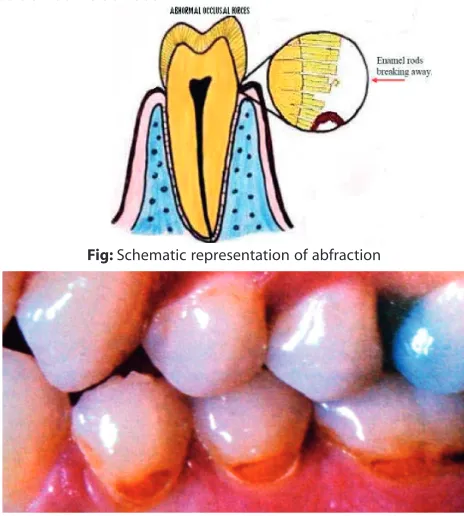
6. Abfraction lesions. Generally associated with occlusal trauma where the anatomic crown of the tooth has flexure. Although non-carious, these lesions can become very sensitive and even progress into the pulp. They may be multifactorial where abrasion and erosive forces combine to produce tooth surface loss.
Fig: Schematic representation of abfraction
7. Erosive lesions. Associated with acid reflux, hiatus hernia, purging, bulimia (intrinsic causes), and diet (extrinsic causes). Intrinsic acid lesions typically occur on the palatal surfaces, while extrinsic acid lesions tend to occur on the buccal surfaces. Consuming large quantities of carbonated cola drinks and fruit drinks, which have a very low pH, causes tooth surface loss, as does toothbrushing following an acidic assault, which removes the acid-softened enamel or dentine.
Fig: Erosion: buccal surfaces of right maxillary cuspid and first premolar tooth showing loss of enamel. Both central and right lateral
incisor teeth show white areas at their labial surfaces indicating incipient decalcificatio
Fig: Erosion related to acid in soft drinks
8. Diet sensitivity. Generally associated with a low pH material, such as fresh tomatoes, orange juice, cola drinks. Areas with exposed dentine are etched, causing sudden sensitivity. Diet choices may aggravate sensitivity from erosion.
9. Genetic sensitivity. Patients will be reporting with history of sensitive teeth and it is not known whether sensitivity correlates to the 10 per cent of teeth that do not have cementum covering all the dentine at the DEJ, or is a factor of lower overall patient pain threshold values.
10. Restorativesensitivity. Triggered following placement of a restoration for several possible reasons: certain amalgams having a history of 24-48 hours sensitivity due to shrinkage, rather than the usual expansion, during setting; contamination of composites during placement or improper etching of the tooth on composites, which results in micro-leakage; improper tooth-drying technique; incorrect preparation of glass ionomer or zinc phosphate cements; general pulpal insult from cavity preparation technique; thermal or occlusal causes; galvanic reaction to dissimilar metals that creates a sudden shock or ‘tin foil’ taste in the mouth.
11. Medication sensitivity is cause due to medications that dry the mouth (e.g. antihistamines, high blood pressure medication), thereby compromising the protective effects of saliva and aggravating diet-related trauma or proliferating plaque. Even a reduction in salivary flow due to ageing or medications can lower the pH of the saliva below the level at which caries occurs (6.0-6.8 for Dentine caries; < 5.5 for enamel caries) and increase erosive lesions to exposed dentine.
12. Bleaching sensitivity. Commonly associated with carbamide peroxide vital tooth bleaching and thought to be due to the by-products of 10 per cent carbamide peroxide (3 per cent hydrogen peroxide and 7 per cent urea) readily passing through the enamel and dentine into the pulp in a matter of minutes. Sensitivity takes the form of a reversible pulpitis caused from the dentine fluid flow and pulpal contact of the material, which changes osmolarity, without apparent harm to the pulp. Sensitivity is caused by all other forms of bleaching (in-office, with or without light activation, and new, over-the-counter) and depends on peroxide concentration.6, 7, 8, 9, 10, 11, 12, 13, 14, 15
Treatments
Treating dentinal hypersensitivity is challenging task for the dental professional because of the difficulty related to measuring the pain response since the response varies from patient to patient. In addition if the dentin exposure is due to personal habits, it may be difficult for patients to change their behavior.1, 5, 7, 8, 10 If the diagnosis confirms dentinal hypersensitivity in the absence of underlying diseases or structural problems, then the following steps can be initiated:
(1) Identify the etiologic factor. Remove, reduce or modify the risk factors by educating the patient about dietary acids and other oral care habits (oral hygiene habits- frequency, brushing, timing of brushing, technique of brushing, force exerted while brushing and the abrasives used in the tooth paste)
(2) Recommend different tooth brushing methods, if appropriate
(3) Initiate treatment by recommending a desensitizing agent for home use
(4) Applying topical desensitizing agents professionally.
Treatment can be invasive in nature or noninvasive
Non-invasive treatment includes:
a) Topical agents
b) Dentifrices that contain a desensitizing active ingredient. (potassium nitrate, stannous fluoride, strontium chloride hexahydrate, and aluminum, potassium or ferric oxalates and fluorides
c) Anti-inflammatory agents: Corticosteroids
Invasive procedures include:
a) Dentine sealers
Glass ionomer cements
Composites
Resins
Varnishes
Sealants
Methyl methacrylate
b) Pulpectomy
c) Gingival surgery/Periodontal soft tissue grafting
d) Crown placement/restorative material
e) Lasers
Other active agents that have been proven to be effective as a desensitizing agents are Protein precipitants, Formaldehyde, Glutaraldehyde, Silver nitrate, Strontium chloride hexahydrate, Casein phosphopeptides, Burnishing, Fluoride iontophoresis.1, 5, 7, 8, 10
Topical desensitizing agents:
These topical desensitizing interfere with the transmission of pain stimulus at the level of the A-delta fibers around the odontoblasts or exert a blocking effect on the open dentine tubules. Some protein precipitants may act in the both the ways.15
Potassium nitrate
Hodosh study was the first to report that potassium nitrate was a “superior desensitizer”.
The mechanism of action of potassium nitrate is unknown, although an oxidizing effect or blocking of tubules by crystallization has been proposed, but not proven. A more likely explanation is that the potassium ions are the active component and that potassium nitrate reduces dentinal sensory nerve activity due to the depolarizing activity of the K+ ion and it aso prevents the repolarization of the nerve. Potassium nitrate does not induce any pulpal changes.1, 5, 6, 9, 10, 11, 15
Calcium hydroxide
Several studies have reported on the effectiveness of calcium hydroxide in managing dentinal hypersensitivity. Its mode of action has been proposed to be via occlusion of dentinal tubules through the binding of loose protein radicals by calcium ions and increasing mineralization of the exposed dentine. Although immediately effective, the action of calcium hydroxide diminishes rapidly requiring multiple applications to maintain its effect.1, 5, 6, 9, 10, 11
Casein phosphopeptides
A relatively new product on the market composed of casein phosphopeptides has been used for the management of dentinal hyperersensitivity.
Sodium monofluorophosphate
Toothpastes containing sodium monofluorophosphate have been shown to be effective in managing dentinal hypersensitivity. The mechanism of action of sodium monofluorophosphate is unclear. It does not appear to act by occluding dentinal tubules since scanning electronmicroscopic studies have failed to demonstrate any visual changes to the dentine surface treated with sodium monofluorophosphate. 1, 5, 6, 9, 10, 11
Sodium fluoride
Many clinical studies have shown that treatment of exposed root surfaces with fluoride toothpaste and concentrated fluoride solutions is very efficient in managing dentinal hypersensitivity. The improvement appears to be due to an increase in the resistance of dentine to acid decalcification as well as to precipitations in the exposed dentinal tubules.
Tal et al. suggested that the probable desensitizing effects of fluoride are related to precipitated fluoride compounds mechanically blocking exposed dentinal tubules or fluoride within the tubules blocking transmission of stimuli.1, 5, 6, 9, 10, 11
Stannous fluoride
Stannous fluoride in either an aqueous solution or in glycerine gelled with carboxymethyl cellulose is effective in controlling dentinal hypersensitivity.
The mode of action appears to be through the induction of a high mineral content which creates a calcific barrier blocking the tubular openings on the dentine surface. Alternatively, stannous fluoride may precipitate on the dentine surface leading to occlusion of the exposed dentinal tubules and reducing the fluid flow to the pulp.1, 5, 6, 9, 10, 11 14, 15
Figure: Open tubules following treatment with non-sensitivity fluoride toothpaste.
Figure: Closed tubules following treatment with SnF2 dentifrice.
Oxalate salts
Oxalate salts generally used are potassium oxalate and ferric oxalate. These salts are applied by rubbing or burnishing. They act via occlusion of dentinal tubules and reducing the tubule fluid flow in either direction.15
Arginine-calcium carbonate
paste to sensitive tooth surfaces by burnishing it in with a slowly rotating soft prophy cup. Paste can also be applied to accessible spots by massaging thoroughly with a cotton-tipped applicator and to furcations and other hard-to-reach areas with a microbrush. The dental professional should carefully burnish the arginine-calcium carbonate desensitizing paste into all sensitive areas, focusing on the CEJ and exposed cementum and dentin.1, 5, 6, 9, 10, 11
Fluoride iontophoresis
Iontophoresis is the process of influencing ionic motion by an electric current and has been used as a desensitizing procedure in conjunction with sodium fluoride. Studies report that there is an immediate reduction in sensitivity after treatment with iontophoresis, but the symptoms gradually return over the ensuing six months.1, 5, 6, 9, 10, 11
Formaldehyde or glutaraldehyde
Formaldehyde and glutaraldehyde, act through their ability to precipitate salivary proteins in dentinal tubules. This effect has been questioned since various formulations have been found to have little or no effect on dentinal hypersensitivity. Given that these agents are very strong tissue fixatives, they should be used with extreme caution to ensure they do not come in contact with the vital gingival tissues. 1, 5, 6, 9, 10, 11
Anti-inflammatory agents
Corticosteroids
Anti-inflammatory agents such as corticosteroids have been proposed for use to manage dentine hypersensitivity.
However, trials have not found them to be particularly useful. While it is presumed that these agents may induce mineralization leading to tubule occlusion, this view has yet to be validated and the validity of using such agents has been questioned. 1, 5, 6, 9, 10, 11
Dentine sealers
Resins and adhesives
Sealing of dentinal tubules with resins and adhesives has been advocated for many years as a
means of managing dentinal hypersensitivity. This technique is generally reserved for cases of specific and localized dentinal hypersensitivity rather than generalized dentinal pain.1, 5, 6, 9, 10, 11
Fig: Schematic representation of composite resin restoration
Periodontal surgery
There are numerous soft tissue grafting procedures which can be carried out to cover exposed root surfaces including lateral sliding grafts, free gingival grafts, connective tissue grafts, coronally repositioned flaps and in some cases guided tissue regeneration for treatment for the treatment of localized gingival recession using bioresorbable membrane or skin allograft. Soft tissue grafting for localized recession defects requires careful planning and an understanding of the anatomical defect to be treated. In general, soft tissue grafting for the management of sensitivity is not regarded as a very predictable treatment strategy.1, 5, 6, 9, 10, 11
Lasers
Both the Nd:YAG and CO2 lasers have been studied for their use in managing dentinal hypersensitivity.11, 14, 23
Both applications rely on their ability to occlude the dentinal tubules. The Nd:YAG laser has been used in conjunction with sodium fluoride varnish with encouraging results showing up to 90 per cent of the dentinal tubules being occluded through use of this combined therapy.
CO2 laser irradiation and stannous fluoride gel has also been shown to be effective for inducing tubule occlusion for up to six months after treatment. While still largely experimental, this technique requires further scientific investigation before it becomes a clinically acceptable means of treatment. 1, 5, 6, 9, 10, 11, 12, 13
Future therapies
Gene therapy may further be included in the treatment of dentinal hypersensitivity. It includes the treatment of sensory nerves to dental restorative procedures as well as to surgical and non surgical debridement that elicit dentine hypersensitivity. One such method is blocking the increased production of nerve growth factor by pulpal fibroblasts near the lesion thought to contribute to tooth hypersensitivity after restorative procedures.14
Conclusion
The ultimate goal in the treatment of dentine hypersensitivity is the immediate and permanent relief of pain. For many patients, conventional desensitising agents often produce unsatisfactory results, since the action is usually directed toward the occlusion of the opened dentinal tubules without considering the causative factors that created the problem. Once a definitive diagnosis of dentine hypersensitivity has been made, after considering a differential diagnosis, a careful assessment of the aetiologic factors must be considered, which in turn if identified and correctly managed may enhance the outcome of the currently used desensitizing agents and ensure more successful management.
References
1. PM Bartold: Dentinal Hypersensitivity: A Review. Australian Dental Journal 2006;51:(3):212-218.
2. D.G. Gillam & R. Orchardson: Advances In The Treatment Of Root Dentine Sensitivity: Mechanisms And Treatment Principles: Endodontic Topics 2006;13:13-33.
3. Douglas A. Terry: Dentin Hypersensitivity: Part I Fundamentals Of Adhesion: http:// w w w . d e n t a l i n s t i t u t e . c o m / A r t i c l e s / P P A D / Dentin%20Hypersensitivity.pdf.
4. Howard E. Strassler & Francis G. Serio : Dentinal Hypersensitivity: Etiology, Diagnosis And Management A Peer-Reviewed Publication : www.ineedce.com
5. Martin Addy Bristol: Dentine Hypersensitivity: New Perspectives On An Old Problem. International Dental Journal 2002;52:367-375.
6. Chun-Hung Chu, Edward Chin-Man Lo: Dentin Hypersensitivity: A Review: Hong Kong Dent J 2010;7 :15-22.
7. R. H. Dababneh, A. T. Khouri, & M. Addy :Dentinal hypersensitivity- an enigma? A Review Of Terminology, Epidemiology, Mechanisms, Aetiology And Management: British Dental Journal 1999;187: 606-611.
8. Cummins D: Dentin Hypersensitivity: From Diagnosis To A Breakthrough Therapy For Everyday Sensitivity Relief: J Clin Dent 2009; 20 (Spec Iss):1-9.
9. Isabel. C. C. M. Protp, Ana K. M. Andrade, M. A. J. R. Montes: Diagnosis And Treatment Of Dentinal Hypersensitivity: J Of Oral Sci 2009; 51(3):323- 332.
10. Patricia A. Walters: Dentinal Hypersensitivity: A Review : J Contemp Dent Pract 2005 May; (6)2:107-117.
11. César Augusto Galvão, Daniel Chi Ngai , Marcelo: Effects Of Desensitizing Agents On Dentinal Tubule Occlusion: J Appl Oral Sci 2004; 12(2):144-148.
12. Frank Schwarz, Nicole Arweiler Georg & Elmar Reich: Desensitizing Effects Of An Er:YAG Laser On Hypersensitive Dentine: A Controlled, Prospective Clinical Study: J Clin Periodontol 2002;29: 211-215.
13. Aldo Brugnera Junior, Ana Eliza Garrini, Antonio Pinheiro, Dilma Helena, Souza Campos, Elisângela Donamaria: Laser Therapy In The Treatment Of Dental Hypersensitivity~A Histologic Study And Clinical Application: Laser Therapy
12:16-21.
14. Connie Hastings Drisko: Dentine hypersensitivity- dental hygiene and periodontal considerations: International Dent J 2002;52: 385-393.
15. Van B. Haywood : dentine hypersensitivity : Bleaching and restorative considerations for successful management: