ISSN 0015–5659 www.fm.viamedica.pl
Address for correspondence: Dr T. Szmuda, Department of Neurosurgery, Medical University of Gdansk, ul. Dębinki 7, 80–952 Gdańsk, Poland, tel: +48 58 349 33 20, +48 58 349 33 39, fax: +48 58 349 33 30, e-mail: [email protected]
Quantification of white matter fibre pathways
disruption in frontal transcortical approach
to the lateral ventricle or the interventricular
foramen in diffusion tensor tractography
T. Szmuda
1, P. Słoniewski
1, M. Szmuda
2, P.M. Waszak
3, A. Starzyńska
4 1Neurosurgery Department, Medical University of Gdansk, Poland2Developmental Neurology Department, Medical University of Gdansk, Poland
3Students Scientific Association of Neurosurgery Department, Medical University of Gdansk, Poland 4Department of Maxillofacial and Oral Surgery, Medical University of Gdansk, Poland
[Received 10 June 2013; Accepted 31 August 2013]
Pathologies occupying the interventricular foramen (foramen of Monro — FM) or the anterior part of lateral ventricle (LV) are accessed by the transcortical or
transcallosal route. As severing of rostral corpus callosum has been deemed inferior
to cortical incision, the approaches through various points of frontal lobe have been developed. Superior (F1), middle (F2) frontal gyrus or occasionally superior frontal sulcus are used as an entry of neurosurgical corridor. In spite of the fact that every approach to LV or FM causes its characteristic irreversible damage to white matter, to date all of transcortical routes are regarded as equivalent. The current study compared the damage of main neural bundles between virtual trans-F1 and trans-F2 corridors by means of diffusion tensor tractography method (DTT) in 11 magnetic resonance imaging (MRI) exams from clinical series (22 he-mispheres, regardless of dominance). Corpus callosum, cingulum, subdivisions I and II of superior longitudinal fasciculus (SLF I and SLF II), corticoreticular as well as pyramidal tracts crossing both approaches were subjected to surgical violation.
Both approaches served a similar total number of fibres (0.94 to 1.78 [× 103]). Trans-F1 route caused significantly greater damage of total white matter volume (F1: 8.26 vs. F2: 7.16 mL), percentage of SLF I fibres (F1: 78.6% vs. F2: 28.6%) and cingulum (F1: 49.4% vs. F2: 10.6%), whereas trans-F2 route interrupted more corticoreticular fibres (F1: 4.5% vs. F2: 30.7%). Pyramidal tract (F1: 0.6% vs. F2: 1.3%) and SLF II (F1: 15.9% vs. F2: 26.2%) were marginally more vulnerable in case of the access via middle frontal gyrus. Both approaches destroyed 7% of callosal fibres. Summarising the above DTT findings, trans-F2 route disrupted a greater number of fibres from eloquent neural bundles (SLF II, pyramidal and
corticoreticular tracts), therefore is regarded as inferior to trans-F1 one. Due to lack of up-to-date guidelines with recommendations of the approaches to LV or
FM, an individual preoperative planning based on DTT should precede a surgery. (Folia Morphol 2014; 73, 2: 129–138)
INTRODUCTION
The body of lateral ventricle (LV) extends rostrally into the frontal lobe, composing the anterior (frontal) horn. The rostral part of the greatest chamber of the brain connects ventrally with 3rd ventricle by the
interventricular foramen (foramen of Monro — FM) [36]. Numerous neural fibres (corpus callosum, subdivisions of superior longitudinal fasciculus — SLF I and II, corticoreticular pathway, pyramidal tract, short association fibres) and brain cortex of frontal lobe (Brodmann areas 6, 8, 9, supplementary motor cortex — SMA and pre-SMA — preSMA) cover superior rostral part of LV. Therefore, historical or contemporary surgical route to LV and FM, to some extent involves vital fibres.
A variety of neurosurgical approaches to the pathologies originating from these locations has been developed over the years. Walter Dandy was the first who described anterior transcortical ex-posure of tumours occupying LV or FM [40]. Then, an extended frontal lobe incision (oval resection or transverse section on non-dominant side) resulted in frequent occurrence of post-operative seizures [13].
Contemporarily, 2 different, nonetheless equivalent, transcortical trajectories lead to anterior horn of LV or FM: one passes through the upper frontal gyrus (trans-F1) and the second through the middle frontal lobe (trans-F2) [14, 52]. Others consider transcallosal interhemispheric route as principal [53, 55]. To date, a neurosurgeon’s preference dictated the choice of the above approaches, as no guidelines regarding this issue were reported. Moreover, modern keyhole approaches concern relatively small cortical incision and thus a funnel-like operating corridor. The above--mentioned strategy has decreased the violation of brain cortex, but parallel to the expanding of subcor-tical disruption (Fig. 1).
A variety of complications related to access to LV or FM can occur. Increased seizure occurrence rate is related to the transcallosal interhemispheric appro -ach, where paradoxically anatomic considerations would be conductive to that route [5]. However, a great callosal incision, coagulation of medial drai-ning veins on routine basis and unintended perical-losal artery injury are regarded as negative aspects of transcallosal access in some reports [23, 24]. In
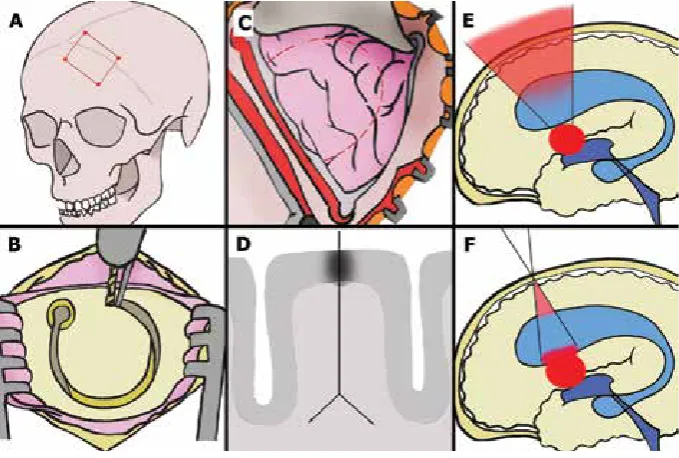
Figure 1. The placement of craniotomy, cortical incision and the idea of keyhole approach to lateral ventricle and the interventricular foramen;
A. The historical method of the frontal approach to the 3rd ventricle lesions. This exposure was used in an access to the ventricular system in
hydrocephalic state. Primarily it was described by Apuzzo [4]; B. Contemporary craniotomy is limited to 1 burr hole (3 cm in diameter). Its proper placement and range are planned preoperatively using navigation systems; C, D. Most of the ventricular lesions are deep-seated. Vast cortical and white matter dissections, supported by an extended retraction of the frontal lobe, were related to high mortality and morbidity when
a historical transcortical approach was applied (C). On the other hand, that type of the neurosurgical approach can provide an opportunity to visualise large ventricular lesions, which facilitate its removal. Contemporary small cortical incision and transgyral white matter dissection (D) preserve more vital neural structures than the historical approach to the ventricles; E, F. The keyhole transcortical approach (F) is used for the access to the 3rd ventricle. a minimal craniotomy and limited frontal lobe incision are required. However, the keyhole access produces similar
transcortical route, up to 20% of patients may deve-lop hemiparesis, which is attributed to the violation of centrum semiovale [3, 15, 39]. Memory deficits occur after the damage of nucleus caudatus or thalamo -cortical fibres [20]. Alexia, agraphia or spatial neglect are another complications of transcortical approach caused by an interruption of subcortical SLF [44, 45].
The application of the preoperative planning, using the preoperative magnetic resonance imaging (MRI) and the intraoperative navigation systems, were milestones in neurosurgery. It finally facilitated the optimum anatomical placement of a craniotomy, cortical incision, as well as exclusive exposing a tar-geted region [48]. Since further introduction of the in vivo visualisation and localisation of white matter pathways by diffusion tensor tractography method (DTT), the strategy of either targeted craniotomy or tumour resection has changed [12]. DTT is the MRI-based, non-invasive method that measures di-rectional anisotropy of water [7]. By following the directions of diffusion (diffusion tensor imaging — DTI), DTT estimates the trajectories of fibre tracts and displays a detailed map of their configuration as a portrayal of anatomical structures 3-dimensionally. That method is limited to gross fibre architecture. The whole planning can be then incorporated into a navigation system. Generally, DTT facilitates the resection of lesions infiltrating eloquent areas con-cerning language and motor function, and is also preclinically used in patients with neurological or psychiatric diseases [31, 43].
Two general principles of every transcortical approach to LV or FM, namely craniotomy anterior to coronal suture and access through wider LV, ar-gue with recent technological capabilities [40]. To the best of our knowledge, neither quantification of white matter pathways in any frontal approach nor comparison of incised fibres between trans-F1 nor trans-F2 route have been published yet. The presented study, which applies modern neural fibres visualisation technique, seems to be of interest to researchers in the neuroscience field and neurosur-geons alike.
MATERIALS AND METHODS
MRI of the brain (as follows: T1, T2 and DTI with single-shot echo-planar imaging) were acquired using a 20-channel Head/Neck coil on a 1.5 T Siemens Mag-netom Aera scanner (Erlangen, Germany). Three repetitions of 20 non-collinear diffusion gradient
directions for contiguous slices were used. All slices were acquired parallel to the anterior commissure– posterior commissure line. Imaging parameters of DTI acquisition: 5.0 mm slice thickness, 128 × 128 matrix, 240 mm × 240 mm field of view, b = 1000 s/mm2,
repetition time = 3500 ms, echo time = 83.0 ms and weak fat suppression, were used in the study. DTI output volumes of tensors were computed from the derived tensor information by means of Fractional Anisotropy (FA), Directionally Encoded Colours (DEC), Apparent Diffusion Coefficient (ADC) and Mean Diffu-sivity (MD). Output volumes were co-registered with T1 exam as an anatomical reference. Fibre tracking was performed using a probabilistic method of multi--fibre model. FA level of 0.2 was set, seed density was held at 5.0, minimal fibre length was set to 20 mm, curvature threshold of 0.2, step length of 0.5 mm and maximal directional change of fibres was chosen between 45 and 50 degrees. Each neural tract group (neural bundle), which passed the hypothetic trans--F1 and trans-F2 corridor to LV, was determined by seeding start and end regions of interest in a specific location. Corpus callosum, cingulum, pyramidal and corticoreticular tracts, short association fibres, SLF I, as well as SLF II were respectively segmented by both selecting and excluding different anatomical landmarks according to the methods proposed in pre -vious reports (for corpus callosum [1], for cingulum, for pyramidal tract [9], for short association fibres, for SLF I [27], for SLF II [17]). FA, ADC, total crossing quantity and volume of fibres were calculated for hy-pothetically devastating trans-F1 and trans-F2 appro-aches. Region of interest (ROI) was set at the entire neurosurgical approach; the mean value of separate measurements of diffusion was drawn.
MRI exams were performed in 11 patients from clinical series (7 males and 5 females), which allowed the analysis of 22 hemispheres. The age of subjects varied between 36 and 76 (mean age was 55.6; stan-dard deviation [SD] = 11.2). Volumetric asymmetry and symmetry coefficients between hemispheres were not calculated; hemisphere dominance was not evaluated. Nine of the presented MRIs were of brains with tumours in other locations of the brains that were not affecting neural fibres assessment of hypothetical frontal transcortical approach to LV or FM. One MRI showed tumour located in the anterior part of the body of LV, another in FM. In those patients harbouring ventricular tumour, the evaluation of whi-te matwhi-ter fibre bundles was validawhi-ted by the clinical use. The current study data has been retrospectively collected in years 2012 and 2013 at the Neurosurgery Department. Due to the observational character of our study and blinded patients’ data, the local Ethical Committee approval was not obligatory.
Descriptive statistics, including mean value, SD and range were included. An independent t-test or Mann-Whitney test (for not normally distributed data) was applied to determine the variances between 2 analysed approaches. Statistica v.10.0 (StatSoft, Tulsa Inc., USA) was used for statistical analysis, with the significance level (p) set at 0.05.
RESULTS
The extension of white matter disruption, measu-red in terms of total volumes, was greater in case of trans-F1 approach. Total volume of all fibres, of which connectivity was potentially destroyed by a corridor, did not differ between the approaches. Mean ADC
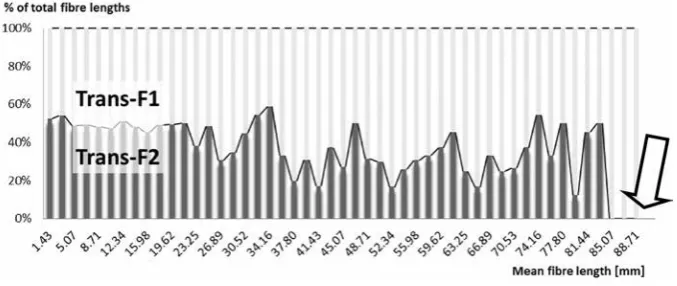
of all fibres crossing the ROI (region of interest) of trans-F1 occurred significantly higher than the ADC of trans-F2. Similarly, mean FA of all fibres was increased if ROI was set on trans-F2, but the comparison occur-red insignificant. The approach through superior fron-tal gyrus destroyed insignificantly longer DTI fibres than trans-F2 approach. Fibres longer than 83.3 mm did not cross trans-F2 trajectory at all, whereas there were 11 DTI fibres on average in the area of trans-F1 corridor (Fig. 2, Table 1).
The qualitative evaluation of white matter bundles crossing frontal transcortical approaches revealed that the access through either superior or medial frontal gyrus at least partially sectioned following tracts: division I and II of SLF, 2 sub-volumes of ro-stral corpus callosum (CC1/CC2 [22]), pyramidal and corticoreticular tracts. The presented result was con-sistently noted in every analysed hemisphere (22 of 22; 100%). The exception was cingulum, which was not violated during trans-F2 approach in 20 of the 22 investigated cases (90.9%). The disruption of short association fibres depended on a specific gyrus, where the cortical incision was hypothetically set off. The constraint regarding different short association fibres of superior and middle frontal gyri prevented their qualitative comparison (Fig. 3).
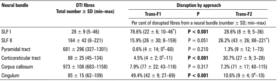
Total number of fibres composing a whole bundle was quantified. A part of disrupted SLF I/II, pyramidal and corticoreticular tracts, cingulum, and corpus cal -losum were outlined during both frontal transcortical approaches as well as its fibres were quantified. The percentage of fibres of a given neural bundle that was cut during trans-F1 and trans-F2 approach was calculated (Fig. 4).
Figure 2. The 100% column chart depicting relative proportion between trans-F1 and trans-F2 approach in terms of violated diffusion tensor
Lower number of DTI fibres of SLF I, corpus cal-losum and cingulum was interrupted by trans-F2 corridor. On the contrary, trans-F1 trajectory was responsible for lesser damage of DTI fibres of SLF II, corticoreticular and pyramidal tract. The signifi-cant differences in the extension of fibres’ damage between both approaches regarded SLF I and cingu-lum (the results supported trans-F2 access), as well as corticoreticular tract (trans-F1 was supported) (Table 2).
The analysed material included 2 clinical cases of ventricular tumours. They were operated via trans-F1 approach, followed by planning of trajectory to LV
using DTT. Those patients did not present any appro -ach-related complications after the surgery.
Beyond white matter disruption, approaches to LV and FM damage brain cortex. Trans-F1 and trans-F2 requires incision of superior and middle frontal gyrus respectively. In this work, we have observed that cor-tex within cingulate sulcus (a part of limbic system) might be partially interrupted by trans-F1 corridor. That was noted in 13.6% of theoretically delineated trajectories starting from superior frontal gyrus (3 of 22). However, shifting the middle boundary of the corridor laterally by at least 4 mm decreased the risk of violation of frontal midline cortex (Fig. 5).
Table 1. Total volume of white matter; number, length and volume of diffusion tensor imaging (DTI) fibres; fractional anisotropy (FA) and apparent diffusion coefficient (ADC) of DTI fibres, which were destroyed by a hypothetical trans-F1 and trans-F2 approach
Hypothetical
approach Total volume of white matter damage by the
surgery [mL]
Total fibre track volume being disrupted [mL]
Total number of DTI fibres being disrupted [× 103]
Length of all DTI fibres crossing the approach [mm]
FA of all fibres crossing the
approach [scalar value 0–1]
ADC of all fibres crossing the approach [µm2/s]
Trans-F1 8.26 ± 1.24
(6.15–10.34) (65.30–112.0)86.2 ± 17.7 1.17 ± 0.35 (0.82–1.69) 24.30 ± 21.06 (1.44–90.50) 0.41 ± 0.13 (0.06–0.59) (672.6–1622.7)897.3 ± 181.8
Trans-F2 7.16 ± 0.98
(4.95–8.32) (40.2–118.6)91.0 ± 25.4 1.32 ± 0.33 (0.94–1.78) 19.18 ±18.64 (1.62–84.42) 0.39 ± 0.13 (0.08–0.61) (672.6–1659.6)801.1 ± 88.1
P < 0.01 0.47 0.15 0.40 0.61 0.03
Data are expressed as mean ± SD (min–max)
Figure 3. White matter fibres presented in Directionally Encoded Colours (DEC). Two different approaches to lateral ventricle (LV) and foramen of Monro (FM) are indicated: trans-F1 (marked by thin dark purple line in every picture and by blue arrow in lower 3A figure) and trans-F2 (marked by thin light purple line in every picture and with red arrow in upper 3A figure). Top row presents DEC axial planes. The colours of
directions are coded with the XYZ-RGB principal eigenvector colour orientation: red — lateral ´ medial; green — rostral ´ caudal; blue — ventral ´ dorsal. Bottom row visualises T1 coronal planes. Blue thin line in each picture of the bottom row corresponds to the plane of each
of the top row DEC axial section. A. Red arrow in DEC axial plane indicates the disruption of callosal fibres caused by trans-F1 and partially
by trans-F2 corridor. Superior fronto-occipital fasciculus (not analysed in the current study; marked with green arrow) hypothetically would be cut if trans-F2 approach projected laterally by incidence. Intraoperative use of navigation is to prevent this mistake from happening.
B. Superior longitudinal fascicullus I and short association fibres (green arrow) lay in the way of trans-F2 trajectory. There is a hazard of cin -gulum interruption (yellow arrow) mainly during the access via superior frontal lobe. C. Pyramidal or corticoreticular tract (blue arrow) may
be disrupted by both of the frontal transcortical approaches to LV or FM. Only diffusion tensor tractography supports the exact distinction
DISCUSSION
Two different routes to LV cause various traumati-sation by surgical manipulation: densely packed fibres of corpus callosum are traversed in interhemispheric approach [34], whereas transcortical approach da-mages the eloquent grey and white matter areas of frontal lobe [50, 52]. Some authors believed that post-operative seizure rate is higher when using tran-scortical approach [6], but others imputed increased
seizure occurrence to interhemispheric access [5]. However, the most recent studies provide opposite suggestions [2]. Among transcortical avenues to LV and FM, either the access via superior (trans-F1), middle frontal gyrus (trans-F2) or superior frontal sulcus is also deemed equivalent. Nowadays, the application of emerging techniques, including DTT and its adoption to intraoperative navigation, allows to tailor the craniotomy, minimalise cortical incision Figure 4. Three-dimensional visualisation of diffusion tensor tractography. Picture a and B (left column): trans-F1 and trans-F2 are marked by blue and red arrow respectively. The corridors of both frontal transcortical approaches are outlined by thin purple line. Pictures C and E (top):
all diffusion tensor imaging (DTI) fibres crossing trans-F1 approach. Pictures D and F (bottom): all DTI fibres crossing trans-F2 approach. Pic -tures C and D (middle column): anterior view. Pictures E and F (right column): lateral view. A. (Anterior view) lateral ventricles are segmented and coloured yellow. B. Axial plane with both transcortical approaches. T1 magnetic resonance imaging exam and Directionally Encoded Colours are merged, hence the relative position of DTI fibres and anatomical structures is visualised. C. The approach through superior frontal gyrus violates cingulum (green arrow). D. Trans-F2 corridor damages a part of pyramidal/tract (blue arrow), superior longitudinal fascicullus (SLF) I (green arrow) as well as cortical connections with pars triangularis and opercularis of inferior frontal gyrus (red arrow). Extensive pro
-jection of the frontal aslant tract (linking superior and inferior frontal gyrus), which might be violated by a lateral aspect of trans-F2 corridor, is
marked by red curly bracket. E. Trajectory to lateral ventricle or foramen of Monro through superior frontal gyrus interrupts cingulum (green arrow). F. Partial damage of pyramidal and extrapyramidal tracts (blue arrow). The severing superior part of SLF I (green arrow).
Table 2. Mean quantity of fibres detected by diffusion tensor imaging (DTI) method; neural bundles are specified. The percentages of disrupted fibres of each bundle were compared between 2 frontal transcortical approaches
Neural bundle DTI fibres
Total number ± SD (min–max) Trans-F1 Disruption by approachP Trans-F2
Per cent of disrupted fibres from a neural bundle (number ± SD; min–max)
SLF I 28 ± 9 (5–46) 78.6% (22 ± 6; 10–46*) P < 0.001 28.6% (8 ± 9; 5–36)
SLF II 164 ± 42 (8–221) 15.9% (26 ± 30; 8–159) P = 0.051 26.2% (43 ± 26; 88–221*)
Pyramidal tract 681 ± 296 (327–1301) 0.6% (4 ± 14; 0†–60) P = 0.210 1.3% (9 ± 12; 1–73)
Corticoreticular tract 88 ± 25 (45–134) 4.5% (4 ± 2; 0†–11) P < 0.001 30.7% (27 ± 9; 3–28)
Corpus callosum 973 ± 108 (683–1158) 7.9% (77 ± 22; 43–118) P = 0.317 7.3% (71 ± 17; 40–115)
Cingulum 85 ± 15 (62–109) 49.4% (42 ± 9; 27–69) P < 0.001 10.6% (9 ± 4; 0†–10)
†There was at least 1 hemisphere, when a specific neural bundle remained intact by the corridor of trans-F1 or trans-F2; *There was at least 1 hemisphere, when a specific neural bundle
as well as assess fibres that are damaged by potential corridor [40, 52]. DTI was originally proposed by Bas-ser et al. in 1994 [7, 8]. That method was developed from diffusion MRI and has been applied to clinical use and tremendous variety of neuroscientific studies [30]. DTT can be used to explore an individual anato-my and provides the opportunity to stipulate various approaches before the surgery [52, 56]. However, that technique at present is not used routinely in planning of the ideal trajectory to ventricles. Many years ago Yasargil and Abdulrauf [53] introduced the shift from transcerebral to transventricular access. Their philosophy was to decrease the damage of cortical and subcortical structures. The comparison of trans-F1 and trans-F2 approaches in terms of the extension of white matter disruption has not been discussed so far. Moreover, the quantitive analysis of neural bundles crossing these 2 routes to LV or FM has not been performed either.
In this study, the route through superior frontal gyrus caused significantly greater volume of white matter damage than the access via middle frontal
gyrus. However, total fibre track volume, count or length of fibres severed by trans-F1 and trans-F2 were similar. These qualitative data do not reflect potential clinical consequences for the patients. Numerous neural bundles can be at least partially destroyed by microsurgical manipulation during both frontal transcortical approaches, all of which could poten -tially lead to a surgery-related morbidity [52]. Post--operative complications of any of the transcortical approaches would depend on the specific neural bundle disruption, the damage of which is known to produce unique symptoms. Therefore, the analysis of each neural pathway violated by trans-F1 or trans-F2 working corridor is validated.
The most important motor pathway in the human brain is the pyramidal tract. It arises from the motor cortex and travels through the corona radiata and posterior limb of internal capsule [26, 42]. The current study have proven, that both trans-F1 and trans-F2 corridors violate only 1% of upper motor neuron axons, causing theoretically at most minor focal pa-resis of lower extremity. Relatively high hemipapa-resis Figure 5. The examples of medial frontal cortex damage caused by trans-F1 trajectory. The trajectory to lateral ventricle (LV) via superior fron -tal gyrus violates grey matter of cingulate sulcus (marked by red arrow in every picture). A. (T1 magnetic resonance imaging [MRI]; coronal plane) standard, 15 mm width, hypothetical corridor of trans-F1 approach (marked blue). B. (T1 MRI; coronal plane, modified by trajectory) the patient was operated due to neurocytoma of LV. Coexistence of hydrocephalus theoretically facilitates the approach via medial frontal
rate after the removal of ventricular tumour in clinical series disagrees with the low rate of fibres converging into corona radiata disruption in our study. It might reflect the principal pathogenic role of surgical ma-nipulation within thalamus, basal ganglia, internal capsule or vasogenic oedema in hemiparesis evolve -ment [34]. Corticobulbar and corticospinal projection fibres run together and are not discriminated on DTT colour maps [28]. Corticoreticular fibres originate mainly from premotor cortex (Brodmann’s area 6) and are involved in postural control and locomotor function [33]. The fibres of rostral premotor cortex origin are longer than corticospinal fibres among whole head scan area. It can explain our observation that the longest fibres were severed only by trans-F1 corridor. The virtual incision of middle frontal gyrus in the approach to LV concerned the violation of 1/3 of corticoreticular fibres in significantly greater exten-sion than the access through superior frontal gyrus did. Adversely, significantly lesser damage of 2 asso-ciation fibres was observed when trans-F2 approach was applied. Cingulum projects from the cingulate gyrus to the entorhinal cortex, runs arching around the corpus callosum and ensures the communication between the components of the limbic system. That neural bundle integrates disparate systems involved in emotion, attention and memory [16, 29, 35]. It has been used as a target for lesion in psychoneurosur-gery, it has also been stimulated in intractable pain syndromes [11, 46]. In our work, trans-F1 corridor was responsible for the interruption of approxima-tely half of all cingulum fibres. The second route to LV or FM severed only 10% of that bundle. However, the vulnerability to unilateral anterior cingulotomy is not thoroughly documented. Cognition impairment, personality disruption, akinetic mutism and response inhibition are ascribed to bilateral cingulotomy even in long-term follow-up [21]. Other results confirm that unilateral cingulum disruption in the access to LV results in similar rates of neglecting, memory de-ficits and naming disturbances as the transcallosal approach [19, 34, 55]. SLF is the major association interhemispheric collection of fibres. Subdivision I of SLF contributes to the regulation of higher aspects of motor behaviour; bidirectional SLF II relates to spatial working memory [17, 27, 32]. In the current study, three quarters of SLF I fibres were interrupted by trans-F1 corridor, nevertheless, in 1 case the whole SLF I bundle was cut. The disruption of SLF I might be unnoticed after the frontal transcortical approach is
used, as its clinical significance has not been elucida-ted [27]. Greater functional role is ascribed to SLF II. Articulate fasciculus, which connects Wernicke’s with Broca’s area, runs contiguously with SLF II [57]. One third of the patients operated with either transcallosal (far from articulate fasciculus) or frontal transcortical approach are affected by aphasia [34]. In 1 virtual trans-F2 corridor planning from our series, the who-le SLF II was cut. Therefore, post-operative speech disturbances cannot be simply explained by the partial damage of SLF II [19]. Corpus callosum was the only commissural fibre pathway violated by the frontal transcortical route. Less than 10% of all callosal com-missure fibres were discovered in the corridor to LV regardless of the route. The transcallosal approach to LV or FM produces higher post-operative seizure rate than the transcortical route [18, 34, 49]. Controversial theories, regarding pathophysiology of seizures after the removal of tumours occupying LV or FM, hold that normal callosal white matter function is implicated in the synchronisation of potential epileptiform foci and thus limits the spread of seizure activity [41]. Equal damage of callosal fibres produced by trans-F1 and trans-F2 proves that similar seizure occurrence might be related to these approaches, although it has not been clinically compared.
FA and ADC are indirect markers of white matter microstructural properties. Both are influenced by nu-merous factors and both describe a different feature of the fibres [10]. FA indicates the directionality of the diffusion process and reflects the degree of axonal alig-nment within the voxel of a brain tissue [51]. Hence, it describes fibre density, axonal diameter and myelination in white matter. Furthermore, the mean FA is relatively si-milar in each structure across patients, yet differs across the structures [38]. ADC is increased in neural bundles that are coherently oriented to gradient direction, in older patients and also in immature brain, regions of oedema or hypoxia [37, 47]. In our material, trans-F2 corridor consisted of slightly more DTI fibres, while ADC was significantly decreased. FA did not vary among fron-tal transcortical routes. The above-mentioned relations between anisotropy, diffusion and fibre count maintain, that both approaches comprise similar number and density of fibres, but present distinct overall direction of fibres (Fig. 4C–F). Several limitations of the above conclusion result from unexplored scientific value of FA and ADC [12, 25, 37, 51].
interpretation of fibre tracking results. DTT is strongly dependent on MRI acquisition, patient’s movement, the technique of DTI preparation, software, operator, fusion accuracy, exact placement of ROI, employed method of data analysis, etc. [9, 12]. Furthermore, DTT does not reflect small neural pathways and is restricted to regions of non-crossing bundles [54]. It seems highly probable that the use of more precise analytic algorithms, including cross fibres and small neural bundles, would alter the results of that study. However, DTT is a powerful anatomic imaging tool of demonstrating gross fibre architecture, in particular major neural bundles.
CONCLUSIONS
We have proven that the trans-F2 and trans-F1 approaches cause greater damage of respectively: projection and association fibres. In conclusion, pre-sented results do favour a frontal transcortical route to LV and FM via superior frontal gyrus. SLF II, pyra-midal and corticoreticular tracts are more vulnerable when a route starts at the middle frontal gyrus. Hen-ce, a violation of these fibres by trans-F2 corridor, even insignificantly greater number than trans-F1 route, is likely to result in a serious disability. Based on DTI findings, the route through superior frontal gyrus is considered more beneficial. However, only a prospec-tive randomised clinical trial concerning these 2 routes would resolve that debate. Lack of contemporary guidelines reflects current decision hesitancy before surgery. A detailed and individual-based preoperative planning, taking advantage of DTI, as well as applying intraoperative neuronavigation, should precede every LV or FM tumour surgery.
ACKNOWLEDGEMENTS
Special thanks to Agnieszka Sabisz (medical phy-sicist from Radiology Department, Medical University of Gdansk) for the description of magnetic resonance imaging acquisition parameters, Marta Adamska for preparing Figure 1 and Agnieszka Prylińska for gram-mar and vocabulary improvements.
REFERENCES
1. Abe O, Masutani Y, Aoki S, Yamasue H, Yamada H, Kasai K, Mori H, Hayashi N, Masumoto T, Ohtomo K (2004)
Topo-graphy of the human corpus callosum using diffusion ten -sor tractography. J Comput Assist Tomogr, 28: 533–539. 2. Aicardi J (2008) Epilepsy syndromes. In: Engel J, Pedley T
eds. Epilepsy: a comprehensive textbook. Lippincott Wil-liams & Wilkins, New York, pp. 2283–2695.
3. Antunes JL, Louis KM, Ganti SR (1980) Colloid cysts of the third ventricle. Neurosurgery, 7: 450–455.
4. Apuzzo ML (1988) Surgery of masses affecting the third ventricular chamber: techniques and strategies. Clin Neu-rosurg, 34: 499–522.
5. Aryan HE, Ozgur BM, Jandial R, Levy ML (2006)
Com-plications of interhemispheric transcallosal approach
in children: review of 15 years experience. Clin Neurol Neurosurg, 108: 790–793.
6. Asgari S, Engelhorn T, Brondics A, Sandalcioglu IE, Stolke D (2003) ‘Transcortical or transcallosal approach to ventricle--associated lesions: a clinical study on the prognostic role of surgical approach. Neurosurg Rev, 26: 192–197. 7. Basser PJ, Mattiello J, LeBihan D (1994) Estimation of the
effective self-diffusion tensor from the NMR spin echo. J Magn Reson B, 103: 247–254.
8. Basser PJ, Mattiello J, LeBihan D (1994) MR diffusion tensor spectroscopy and imaging. Biophys J, 66: 259–267. 9. Basser PJ, Pajevic S, Pierpaoli C, Duda J, Aldroubi A (2000)
In vivo fiber tractography using DT-MRI data. Magn Reson Med, 44: 625–632.
10. Beaulieu C (2002) The basis of anisotropic water diffusion in the nervous system: a technical review. NMR Biomed, 15: 435–455.
11. Broggi G (2008) Pain and psycho-affective disorders. Neurosurgery, 62: 901–919 (discussion 919–920). 12. Bull JG (2010) Diffusion imaging and tractography. In: The
paediatric neurosurgical population. University College,
London.
13. Cairns H (1971) Who’s in charge here?. Lancet, 2: 318–319. 14. Carrasco EM (2011) Left intraventricular tumour resected
via superior frontal transulcal transcortical approach.
Neurosurgic blog and news channel. NEUROSURGIC.com, Santa Cruz, Bolivia.
15. Casmiro M, Sintini M, Martinelli P, Leonardi M, D’alessandro R (1992) Chronic spastic atrophic hemiparesis with benign course. Ital J Neurol Sci, 13: 611–616.
16. Catani M, Dell’acqua F, Vergani F, Malik F, Hodge H, Roy P, Valabregue R, Thiebaut De Schotten M (2012) Short frontal lobe connections of the human brain. Cortex, 48: 273–291.
17. Catani M, Howard RJ, Pajevic S, Jones DK (2002) Virtual in vivo interactive dissection of white matter fasciculi in the human brain. Neuroimage, 17: 77–94.
18. Ellenbogen RG (2001) Transcortical surgery for lateral ventricular tumors. Neurosurg Focus, 10: E2.
19. Friedman MA, Meyers CA, Sawaya R (2003)
Neuropsycho-logical effects of third ventricle tumor surgery. Neurosur -gery, 52: 791–798 (discussion 798).
20. Fuh JL, Wang SJ (1995) Caudate hemorrhage: clinical
fe-atures, neuropsychological assessments and radiological
findings. Clin Neurol Neurosurg, 97: 296–299.
21. Gasquoine PG (2013) Localization of function in anterior cingulate cortex: from psychosurgery to functional neu-roimaging. Neurosci Biobehav Rev, 37: 340–348. 22. Hasan KM, Kamali A, Iftikhar A, Kramer LA, Papanicolaou AC,
Fletcher JM, Ewing-Cobbs L (2009) Diffusion tensor tracto-graphy quantification of the human corpus callosum fiber pathways across the lifespan. Brain Res, 1249: 91–100. 23. Hassaneen W, Suki D, Salaskar AL, Levine NB, Demonte F,
Lang FF, Mccutcheon IE, Dorai Z, Feiz-Erfan I, Wildrick DM, Sawaya R (2010) Immediate morbidity and mortality associated with transcallosal resection of tumors of the third ventricle. J Clin Neurosci, 17: 830–836.
cysts by interhemispheric far lateral transcallosal appro-ach: experience of 134 patients. Surg Neurol, 69: 447–453 (discussion 453–446).
25. Hoeft F, Barnea-Goraly N, Haas BW, Golarai G, Ng D, Mills D, Korenberg J, Bellugi U, Galaburda A, Reiss AL (2007) More is not always better: increased fractional anisotropy of superior longitudinal fasciculus associated with poor visuospatial abilities in Williams syndrome. J Neurosci, 27: 11960–11965. 26. Ino T, Nakai R, Azuma T, Yamamoto T, Tsutsumi S, Fukuyama H
(2007) Somatotopy of corticospinal tract in the internal capsule shown by functional MRI and diffusion tensor images. Neuroreport, 18: 665–668.
27. Jang SH, Hong JH (2012) The anatomical characteristics of superior longitudinal fasciculus I in human brain: Diffusion tensor tractography study. Neurosci Lett, 506: 146–148. 28. Jellison BJ, Field AS, Medow J, Lazar M, Salamat MS, Ale-xander AL (2004) Diffusion tensor imaging of cerebral white matter: a pictorial review of physics, fiber tract anatomy, and tumor imaging patterns. Am J Neuroradiol, 25: 356–369. 29. Keedwell PA, Chapman R, Christiansen K, Richardson H,
Evans J, Jones DK (2012) Cingulum white matter in young women at risk of depression: the effect of family history and anhedonia. Biol Psychiatry, 72: 296–302.
30. LeBihan D, Mangin JF, Poupon C, Clark CA, Pappata S, Molko N, Chabriat H (2001) Diffusion tensor imaging: concepts and applications. J Magn Reson Imaging, 13: 534–546.
31. Lin F, Yu C, Jiang T, Li K, Li X, Qin W, Sun H, Chan P (2006) Quantitative analysis along the pyramidal tract by length--normalized parameterization based on diffusion tensor tractography: application to patients with relapsing neu-romyelitis optica. Neuroimage, 33: 154–160.
32. Makris N, Kennedy DN, Mcinerney S, Sorensen AG, Wang R, Caviness VS, Pandya DN (2005) Segmentation of sub-components within the superior longitudinal fascicle in humans: a quantitative, in vivo, DT-MRI study. Cereb Cortex, 15: 854–869.
33. Matsuyama K, Mori F, Nakajima K, Drew T, Aoki M, Mori S (2004) Locomotor role of the corticoreticular-reticulospinal--spinal interneuronal system. Prog Brain Res, 143: 239–249. 34. Milligan BD, Meyer FB (2010) Morbidity of transcallosal
and transcortical approaches to lesions in and around the
lateral and third ventricles: a single-institution experience. Neurosurgery, 67: 1483–1496 (discussion 1496). 35. Morgane PJ, Galler JR, Mokler DJ (2005) A review of
systems and networks of the limbic forebrain/limbic midbrain. Prog Neurobiol, 75: 143–160.
36. Moore AJ, Newell DW (2005) Neurosurgery: principles and practice. Springer, London.
37. Mukherjee P, Berman JI, Chung SW, Hess CP, Henry RG (2008) Diffusion tensor MR imaging and fiber tracto-graphy: theoretic underpinnings. Am J Neuroradiol, 29: 632–641.
38. O’donnell L, Haker S, Westin C (2002) New approaches to estimation of white matter connectivity in diffusion tensor MRI: Elliptic PDE’s and geodesics in tensor-warped space. In: Proc. of Medical Image Computing and Computer--Assisted Intervention (MICCAI), Springer, Berlin Heidel-berg, pp. 459–466.
39. Park ES, Cho YH, Kim JH, Kim SJ, Khang SK, Kim CJ (2012) Frontal transcortical approach in 12 central neurocytomas. Acta Neurochir, 154: 1961–1971 (discussion 1972). 40. Perneczky A, Reisch R (2008) Transcortical approach.
Keyhole approaches in neurosurgery. Vol. 1: Concept and surgical technique. Springer-Verlag, Wien, pp. 267–282.
41. Prince DA (1985) Physiological mechanisms of focal epi-leptogenesis. Epilepsia, 26 (suppl. 1): S3–S14.
42. Qazi AA, Radmanesh A, O’donnell L, Kindlmann G, Peled S, Whalen S, Westin CF, Golby AJ (2009) Resolving crossings in the corticospinal tract by two-tensor streamline trac-tography: Method and clinical assessment using fMRI. Neuroimage, 47: Suppl 2 T98–106.
43. Raj A, Kuceyeski A, Weiner M (2012) A network diffusion model of disease progression in dementia. Neuron, 73: 1204–1215.
44. Shinoura N, Onodera T, Kurokawa K, Tsukada M, Yamada R, Tabei Y, Koizumi T, Yagi K (2010) Damage to the upper por-tion of area 19 and the deep white matter in the left inferior parietal lobe, including the superior longitudinal fasciculus, results in alexia with agraphia. Eur Neurol, 64: 224–229. 45. Shinoura N, Suzuki Y, Yamada R, Tabei Y, Saito K, Yagi K
(2009) Damage to the right superior longitudinal fasci-culus in the inferior parietal lobe plays a role in spatial neglect. Neuropsychologia, 47: 2600–2603.
46. Spooner J, Yu H, Kao C, Sillay K, Konrad P (2007)
Neu-romodulation of the cingulum for neuropathic pain
after spinal cord injury. Case report. J Neurosurg, 107: 169–172.
47. Sullivan EV, Adalsteinsson E, Pfefferbaum A (2006) Se-lective age-related degradation of anterior callosal fiber bundles quantified in vivo with fiber tracking. Cereb Cortex, 16: 1030–1039.
48. Vigneron LM, Warfield SK, Robe PA, Verly JG (2011) 3D XFEM-based modeling of retraction for preoperative image update. Comput Aided Surg, 16: 121–134. 49. Villani R, Tomei G (2006) Approach to tumors of the third
ventricle. In: Schmidek H, Roberts D eds. Schmidek and Sweet’s operative neurosurgical techniques: indications, methods, and results. Saunders Elsevier, Philadelphia, pp. 772–785.
50. Wen HT, Mussi ACM, Rhoton AL Jr., Oliveira ED, Tedeschi H (2006) Surgical approaches to lesions located in the lateral, third, and fourth ventricles. In: Sekhar LN, Fessler RG eds. Atlas of neurosurgical techniques: brain. Thieme, New York, pp. 527–528.
51. Westerhausen R, Huster RJ, Kreuder F, Wittling W, Schwe-iger E (2007) Corticospinal tract asymmetries at the level of the internal capsule: is there an association with han-dedness? Neuroimage, 37: 379–386.
52. Winn HR (2011) Youmans neurological surgery. Elsevier Saunders, Philadelphia.
53. Yaşargil MG, Abdulrauf SI (2008) Surgery of intraventri-cular tumors. Neurosurgery, 62: 1029–1040 (discussion 1040–1021).
54. Yeo SS, Chang MC, Kwon YH, Jung YJ, Jang SH (2012) Corticoreticular pathway in the human brain: diffusion tensor tractography study. Neurosci Lett, 508: 9–12. 55. Yonekawa T, Nakagawa E, Takeshita E, Inoue Y, Inagaki M,
Kaga M, Sugai K, Sasaki M, Kaido T, Takahashi A, Otsuki T (2011) Effect of corpus callosotomy on attention deficit and behavioral problems in pediatric patients with intrac-table epilepsy. Epilepsy Behav, 22: 697–704.
56. Yu CS, Li KC, Xuan Y, Ji XM, Qin W (2005) Diffusion tensor tractography in patients with cerebral tumors: a helpful
technique for neurosurgical planning and postoperative
assessment. Eur J Radiol, 56: 197–204.