Washington University in St. Louis
Washington University Open Scholarship
All Theses and Dissertations (ETDs)
Spring 4-29-2013
Characterization of the Placenta-Specific 8.1 Gene
Function during Zebrafish Embryogenesis
Haiting Ma
Washington University in St. Louis
Follow this and additional works at:
https://openscholarship.wustl.edu/etd
Part of the
Biology Commons
This Dissertation is brought to you for free and open access by Washington University Open Scholarship. It has been accepted for inclusion in All Theses and Dissertations (ETDs) by an authorized administrator of Washington University Open Scholarship. For more information, please contact
Recommended Citation
Ma, Haiting, "Characterization of the Placenta-Specific 8.1 Gene Function during Zebrafish Embryogenesis" (2013).All Theses and Dissertations (ETDs). 1051.
WASHINGTON UNIVERSITY IN ST. LOUIS
Division of Biology and Biomedical Sciences
Developmental, Regenerative and Stem Cell Biology
Dissertation Examination Committee:
Lilianna Solnica-Krezel, Chair
Philip V. Bayly
Robert J. Coffey
Susan K. Dutcher
Raphael Kopan
Gregory D. Longmore
Characterization of the Placenta-Specific 8.1 Gene Function during
Zebrafish Embryogenesis
by
Haiting Ma
A dissertation presented to the
Graduate School of Arts and Sciences
of Washington University in
partial fulfillment of the
requirements for the degree
of Doctor of Philosophy
May 2013
ii
TABLE OF CONTENTS
PAGE
List of Figures ... vi
List of Tables ... viii
List of Abbreviations ... ix
Acknowledgements ... xiii
Abstract ... xv
Chapter I. Introduction ... 1
A brief overview of cancer as a multigenic disease ... 1
Genetic studies in model organisms ... 6
Muller’s morphs offer a framework to understand the nature of mutations ... 8
Zebrafish as a model system to study embryo development ... 12
Cell fate specification and morphogenetic cell movements during zebrafish gastrulation ... 15
PLAC8 family of proteins and its implication in carcinomas ... 19
II. Excess PLAC8 promotes ERK2-dependent EMT in colon cancer ... 24
Abstract ... 25
Introduction ... 25
Results ... 27
Endogenous PLAC8 protein localizes to the apical domain of terminally differentiated human colonic epithelium. ... 27
Cytosolic PLAC8 is correlated to tumor grade and linked to mucinous and medullary CRC ... 27
Increased levels of PLAC8 are linked to tumor progression ... 28
Identification and characterization of zebrafish plac8 homologs ... 38
iii
Plac8.1 overexpression caused cell-autonomous, post-transcriptional downregulation of Cdh1 . 48 Overexpression of PLAC8 in HCA-7 cells reduces cell surface CDH1 and confers an EMT
phenotype ... 49
HCA-7P8 cells exhibit an EMT signature ... 54
PLAC8 enhances phosphorylation of ERK2 ... 58
Knockdown of endogenous PLAC8 in SC cells restores cell surface CDH1 in 3D culture and in xenografts ... 59
Discussion ... 64
Methods ... 67
Plasmids, transfection and infection of human cell lines ... 67
Antibody generation and other immunoassaying reagents ... 68
Immunoblotting ... 68
Immunocytochemistry ... 69
MAtS invasion assay ... 70
Patients, TMA Construction and TMA Slide Preparation ... 70
Mouse xenografts ... 70
Zebrafish strain maintenance and embryo staging ... 70
Identification and cloning of zebrafish plac8 homologs ... 70
Zebrafish embryo injection ... 71
Whole-mount in situ hybridization of zebrafish ... 71
Time-lapse imaging and analysis ... 72
RNA isolation and quantitative RT-PCR analysis ... 72
Genotyping of the cdh1vu44 allele ... 72
Pharmacological treatment using MG-132 ... 73
Acknowledgements ... 73
III. Zebrafish Plac8.1 links ubiquitination regulating protein Cops4 to cilia formation and function ... 75
iv
Introduction ... 76
Experimental procedures ... 78
Zebrafish strains, maintenance and embryo staging ... 78
Generation of plac8.1 mutant with transcription activator-like effector nucleases (TALENs) ... 78
Antisense MOs and synthetic RNA injections ... 79
Antibody generation, western blotting, and immunofluorescence ... 79
Whole-mount in-situ hybridization ... 81
Cryosection and histology staining ... 81
Kupffer’s vesicle flow analysis ... 81
Transmission electron microscopy ... 82
High-speed time-lapse imaging analysis ... 82
Acridine orange staining ... 82
Statistical analysis ... 83
Results ... 83
Expression of plac8.1 in epithelial tissues with motile cilia ... 83
Plac8.1 protein localized close to the cilia basal body at the apical domain of ciliated epithelial cells ... 83
Reduction of plac8.1 function led to ventrally curved body axes and kidney cyst ... 89
Reduction of plac8.1 function led to left-right asymmetry defects ... 93
Reduction of plac8.1 function led to defects in the formation and function of cilia in the Kupffer’s vesicle ... 93
Interference with plac8.1 expression impaired formation and function of cilia in the kidney ducts ... 96
Reduction of plac8.1 function did not affect Hh signaling ... 98
Reduction of plac8.1 function resulted in dampened cilia beating activity in the olfactory placode ... 100
Plac8.1 interacted with Cops4 to regulate cilia formation and beating ... 100 Targeted disruption of plac8.1 using transcription activator-like effector nucleases (TALENs) . 108
v
Discussion ... 115
Localization of Plac8.1 protein ... 116
No evidence of altered cell fates upon reduction of plac8.1 function ... 116
Comparison between functions of zebrafish plac8.1 and mouse Plac8 ... 117
Possible function of Plac8.1 in ciliogenesis and cilia beating ... 117
Plac8.1 and signaling events connected with cilia ... 119
Cops4 implicated ubiquitination pathway in ciliogenesis and function ... 120
Plac8.1 and left-right asymmetry formation ... 121
Plac8.1 and kidney cysts formation ... 121
Acknowledgements ... 123
IV. Discussion and future directions ... 124
Overexpression of zebrafish Plac8.1 leads to E-cadherin degradation and gastrulation movements defects ... 128
Zebrafish plac8.1 is required for cilia morphogenesis and function ... 129
Understanding the function of Plac8.1 according to Muller’s morphs ... 130
Future directions ... 131
Reference ... 132
vi
LIST OF FIGURES
Figure Page
1-1 A simplified step-wise model of tumorigenesis ... 2
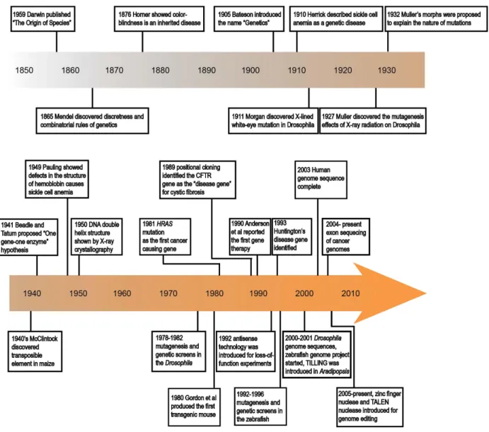
1-2 Landmarks of genetic studies in humans and model organisms ... 8
1-3 Muller’s morphs ... 10
1-4 Zebrafish as a genetic model system with forward genetic and reverse genetic approaches ... 14
1-5 Cell fate specifications and morphogenetic cell movements together shape zebrafish body plan during embryonic development ... 16
1-6 PLAC8 family of cysteine-rich proteins ... 22
2-1 PLAC8 immunofluorescence in human normal colon and CRC ... 29
2-2 PLAC8 extends into the neoplastic crypts of adenocarcinoma ... 31
2-3 Characterization of anti-PLAC8 antibodies ... 34
2-4 Increased PLAC8 protein is linked to tumor progression ... 36
2-5 Identification of zebrafish plac8 homologs ... 40
2-6 Characterization of plac8.1 RNA expression and protein localization in zebrafish embryos ... 42
2-7 Overexpression of plac8.1 results in cell-autonomous downregulation of E-cadherin and multiple developmental defects that phenocopy cdh1 loss-of-function ... 44
2-8 The effect of Plac8.1 overexpression on zebrafish embryonic development ... 46
2-9 Expression of PLAC8 enhances HCA-7 cell invasion and alters CDH1 subcellular localization .. 50
2-10 Cell migration measurement ... 52
2-11 HCA-7P8 cells exhibit features of EMT ... 56
2-12 PLAC8 induces EMT through ERK2 activation ... 60
2-13 Depletion of endogenous PLAC8 in SC cells restores cell surface CDH1 in 3D culture and xenografts ... 62
3-1 Zebrafish plac8.1 was expressed in ciliated tissues ... 85
3-2 Zebrafish Plac8.1 protein enriched at the apical sided of ciliated epithelium ... 87
vii
3-4 Reduction of plac8.1 function resulted in body curvature and kidney cysts ... 91
3-5 Reduction of plac8.1 function resulted in generalized left-right asymmetry defects ... 94
3-6 Reduction of plac8.1 function impaired motile cilia morphology and motility in the Kupffer’s vesicle. ... 97
3-7 Reduction of plac8.1 function impaired motile cilia morphology in the kidney duct ... 99
3-8 No apparent Hedgehog signaling defects observed in plac8.1 morphants ... 101
3-9 Reduction of cops4 function showed similar phenotype to reduction of plac8.1 function ... 105
3-10 Plac8.1 bound and cooperated with Cops4 to regulate motile cilia morphology and motility ... 106
3-11 Summary of the reduction of plac8.1 function study ... 107
3-12 Generating and testing a pair of TALEN nucleases to target plac8.1 ... 110
3-13 TALEN-induced plac8.1stl33 mutant allele is a five-nucleotide insertion in plac8.1 gene ... 111
3-14 Characterization of plac8.1stl33 mutant embryos ... 112
3-15 Transcription regulation of Plac8: perspective from in silico analysis ... 113
4-1 Schematic overview of findings from this study ... 127
viii LIST OF TABLES
Table Page
2-1 Correlation between advanced cancer grade and cytoplasmic intensity of PLAC8 ... 33 2-2 Quantitative RT-PCR array analysis of HCA-7 cells with PLAC8 overexpression (HCA-7P8) and
HCA-7 control cells (HCA-7C) ... 55 2-3 Sequences of primers used in quantitative RT-PCR ... 74
ix LIST OF ABBREVIATIONS µg Microgram µm Micrometer µM Micromole Am Axial mesoderm Ap Animal pole AP Anteroposterior Ap Animal pole
APC ADENOMATOUS POLYPOSIS COLI
Bmp Bone morphogenetic protein
bp Base pair
C&E Convergence and Extension CC Cystic colonies
CFTR CYSTIC FIBROSIS TRANSMEMBRANE CONDUCTANCE REGULATOR
COP Constitutive photomorphogenesis CRC Colorectal cancer
cyc cyclops
DIC differential interference contrast
dkk Dickkopf (Wnt signaling antagonist)
dpf Days post fertilization Dsh Dishevelled
DV dorsal-ventral ECM Extracellular matrix
EGFP Enhanced green fluorescent protein EGFR Epithelial growth factor receptor EMS Ethyl methanesulfonate
x EMT Epithelial-mesenchymal transition ENU N-ethyl N-nitrosourea
EVL Enveloping layer FGF Fibroblast growth factor
G protein Guanine nucleotide-binding proteins GFP Green fluorescent protein
GOF gain-of-function
GPCR G protein coupled receptor GPCR G protein-coupled receptors GTP Guanosine triphosphate GTPase guanosine triphosophatase H&E Hematoxylin and eosin
HEK293T Human embryonic kidney 293T cells Hg Hatching gland
Hh Hedgehog
hpf Hours post fertilization
HRAS Harvey rat sarcoma viral oncogene homolog
IFT Intraflagellar transport JAK Janus kinases
JNK Jun N-terminal kinase KLH Keyhole limpet hemocyanin
kny knypek/glypican4 KV Kupffer’s vesicle L/R Left-right LOF loss-of-function LWR Length-to-width ratio MBT Mid-blastrula transition
xi ML Mediolateral MO Morpholino oligonucleotide NC Notochord ng nanogram NT Neural tube ntl no tail
papc paraxial protocadherin
PBS phosphate buffered saline PCP Planar cell polarity
PDC plasmacytoid dendritic cells PFA paraformaldehyde
pg picogram
PGE2 prostaglandin E2 PKC Protein kinase C Plac8 Placenta-specific gene 8 Pp Prechordal plate PSM Presomitic mesoderm
RAS rat sarcoma viral oncogene homolog
RNA ribonucleatic acid Sc Spinal Cord SC spiky colonies
So Somite
spw southpaw
sqt squint
Stat3 Signal transducer and activator of transcription 3 TALENs Transcription Activator-Like Effector Nucleases
TB Tailbud
xii TMA Tissue microarray
TILLING Targeting Induced Local Lesions in Genomes
tri trilobite
Vg Vegetal pole
WT Wild-type
Y Tyrosine
YAMC Young adult mice colon cells YSL Yolk syncytial layer
xiii
ACKNOWLEDGEMENTS
This work would have been impossible without the tremendous supports from my mentors Dr. Lilianna Solnica-Krezel and Dr. Robert Coffey. Thank you both for providing the best possible PhD experience I could ever hope for. It is a great privilege to be a graduate student, and it is more so to be a student under the guidance of the two great mentors. I would like to thank Dr. Solnica-Krezel for giving me the opportunity to pursue a risky project. She leads by example as a brilliant and persistent scientist, a visionary leader, and a kind, generous, and supportive person. She always inspires me and other lab members to carry out careful research on important biological questions, to pursue better results with better approaches. She teaches me to be a careful, critical thinker and experimentalist, which is a life-long lesson for me to appreciate. I also would like to thank Dr. Coffey who is inspiring as a dedicated physician scientist leading cutting edge translational research. I learnt from their untiring contributions to science that what scientists ought to do is to push the boundaries on the frontiers for knowledge, insights, and advancement of human health. I am indebted to their coordinated efforts to make the transition from Vanderbilt University to Washington University smooth, and their continuing efforts and commitments to make this collaborative thesis project continue as if we had not moved.
I am also extremely grateful to my committee members Dr. Greg Longmore, Dr. Susan Dutcher, Dr. Philip Bayly, and Dr. Raphael Kopan at Washington University, and Dr. Jim Patton, Dr. Joshua Gamse, and Dr. Albert Reynolds at Vanderbilt University for their constructively critical comments and insightful discussions in my continual journal as a scientist. In addition, I would like to express my deep appreciation to Dr. Andrzej Krezel for sharing his knowledge, expertise, and equipment for expression and purification of the Plac8.1 protein.
The friendly and knowledgeable colleagues in both Dr. Solnica-Krezel’s laboratory and Dr. Coffey’s laboratory make me feel exceptional fortunate and resourceful. My deep gratitude goes to all former and current members of both laboratories. In particular, I would like to thank Dr. Terry Van Raay, Dr. Cunxi Li, and Dr. Zheng Cao for bringing me the wonderful rotation student’s experiences in both laboratories. Also I wound like to thank Dr. Cunxi Li for being a long-time collaborator and friend. The boundary-free data sharing, and your patience for endless discussions and communications are essential for this collaborative project. Also I would like to thank Dr. Diane Sepich for all the discussions and help
xiv
with experiments, thank Jiakun Chen, Dr. Jimman Shin, and Yinzi Liu for their help with the TALEN experiments, thank Robert Zhang for his help with protein purification, thank Anna Hindes for her efforts to look for plac8.1 mutant fish and to order reagents and equipment, and thank Eric Sanders, Steve Canters, Amy Bradshaw, Heidi Beck, and so many people for taking care of the fish.
I am fortunate to experience the great training opportunities at Washington University and Vanderbilt University. I would like to thank Dr. Jim Skeath, Dr. Tim Schedl, and Dr. Jim Patton for their help. I would also like to thank the supporting staff: Kim Smith, Stacy Kiel, Linda Lobos, Leslie Maxwell, and many others.
Finally, I specially thank my wife who is my soul mate in life and science, and our parents for being the caring, understanding, patient, and perpetual providers of the unconditional love that makes this and beyond possible.
xv
ABSTRACT OF THE DISSERTATION
Characterization of the Placenta-Specific 8.1 Gene Function during
Zebrafish Embryogenesis
Haiting Ma
Doctor of Philosophy in Developmental, Regenerative and Stem Cell Biology
Washington University in St. Louis, 2013
Dr. Lilianna Solnica-Krezel, Chair
The PLAC8 gene encodes for a small, cysteine-rich protein conserved in vertebrates that is a
member of a large family of PLAC8-motif containing proteins with diverse functions in animals, plants, and algae. Recently, high levels of PLAC8 expression have been detected in aggressive colorectal cancer and invasive breast cancer, and potentially contributing to the cancer pathogenesis. However, the molecular and cellular functions of PLAC8 in vertebrate development, homeostasis, and disease remain unclear. To determine the function of PLAC8 under disease and normal conditions, in this thesis work, I identified plac8.1 as a PLAC8 homolog in zebrafish, a vertebrate model system amenable to various embryologic and genetic approaches. Zebrafish plac8.1 was maternally and ubiquitously expressed until four days post fertilization when its transcript became enriched in the gut. During the process of gastrulation, Plac8.1 protein distribution gradually shifted from the cytosol to the cell membrane. At larval stages, Plac8.1 accumulated at the apical region of epithelial cells of the gut and the kidney. This dynamic gene expression and protein localization patterns suggest that Plac8.1 may have functions during zebrafish embryogenesis and organogenesis.
In the first part of this thesis, I aimed to address questions concerning the effects of high levels
of PLAC8 on cell behavior by overexpressingPlac8.1 during early embryogenesis. Embryos
xvi
well as convergence and extension gastrulation movements were delayed in plac8.1-overexpressing embryos, a spectrum of phenotypes resembling impaired E-cadherin function in zebrafish. Indeed, E- cadherin levels were significantly reduced in plac8.1-overexpressing embryos by a cell-autonomous and post-transcriptional mechanism. Similarly, PLAC8-overexpressing colon cancer cells exhibited reduced cell surface E-cadherin and many features of epithelial-mesenchymal transition (EMT). Furthermore, knockdown of PLAC8 in colon cancer cells resulted in increased level of E-cadherin.
In the second part of this thesis, I aimed to determine the requirement for plac8.1 function during development by employing two loss-of-function approaches. Upon injection of either of two non-overlapping antisense morpholino oligonucleotides (MOs) that effectively reduced Plac8.1 protein levels, embryos displayed an array of defects that phenocopied the class of mutants with defective cilia. Consistently, the motile cilia morphology and motility were impaired in plac8.1 morphants. Moreover, in immunoprecipitation experiments, Plac8.1 bound Cops4, a component involved in ubiquitination regulation. Also Plac8.1 and Cops4 cooperated to regulate motile cilia morphology and motility. As a second loss-of-function approach, I generated loss-of-function allele plac8.1stl33 using Transcription Activator-Like Effector Nucleases (TALENs), a method that utilizes sequence-specific nucleases. Similar to plac8.1 morphants, plac8.1stl33 embryos showed motile cilia morphology and beating function defects. We hypothesize that zebrafish Plac8.1 functions at the ciliary base, possibly by modulating intraflagellar transport through ubiquitination modifications.
Our in vivo studies of zebrafish plac8.1 have uncovered pleiotropic functions of plac8.1 during
development. In addition, the overexpression experiments shed light on mechanisms underlying human disease. Based on the above results, I propose that Plac8.1 overexpression interferes with regulation of protein stability, whereas Plac8.1 is required for motile cilia morphogenesis and function. Common denominators of these seemingly unrelated phenotypes include the potential connection with the process of ubiquitination, and the enriched apical localization of Plac8.1. Our work can also inform studies of other members of the PLAC8 family of proteins.
1
CHAPTER I
INTRODUCTION
A brief overview of cancer as a multigenic disease
Cancer represents one of leading causes of death worldwide (Jemal et al., 2006; Meetoo, 2008). Carcinomas (malignant solid tumors that originate from epithelial tissues) account for over 80% of the total cancer burden in patients (statistical data from “Cancer facts and figures 2012” by American Cancer Society), and will be focused in this introduction. Environmental risk factors such as ionizing radiations and certain chemicals contribute to cancer formation (Figure 1-1, top panel). For example, tobacco smoking is closely associated with lung cancer. In fact, many tobacco combustion products and their metabolites act as mutagens to alter DNA sequences inside the cells to cause lung cancer (Hecht, 1999). In contrast to lung cancer that is closely associated with tobacco smoking, many of human cancers are not traceable to a particular environmental risk factor, suggesting other factors may also be part of carcinoma etiology (Fontham et al., 2009).
Indeed, inherited and spontaneous mutations in germ cells and somatic cells are common factors that contribute to tumorigenesis (Bertram, 2000). Despite the diversity of causes of cancers, and that cancers have been recognized as a collection of heterogeneous disorders, an abiding theme of cancer research has demonstrated that all cancer cells have multiple gene mutations (Hornberg et al., 2006; Hanahan and Weinberg, 2011). Although not all mutations in cancer cells contribute to cancer formation, highly mutated cancer genomes suggest cancer as a multigenic disease (Welch et al., 2012). Extensive studies on molecular mechanisms that create cancer over the past decades have accumulated an enormous amount of knowledge about the process of mutation accumulation as cancer develops (Rajagopalan et al., 2003; Vogelstein and Kinzler, 2004).
The malignant transformation process that converts normal cells to highly malignant cancer cells has been likened to a micro-scale evolution process (Figure 1-1). In this process, normal cells acquire mutations in various genes in a step-wise way. Each step is likely to give slight selection advantages to the cells with the mutated genes over the cells without this mutation. Then new mutations may occur within the daughter cells that derived from the previously mutated ancestors. After accumulation of
2 Fi gu re 1 -1. A si m p li fi ed st ep -wi s e m o d e l o f tu m o ri g e n e s is . To p pan el : env ir o nm ent a l f ac tor s and ac cum ul at io n of m ut at ions c ont ri but e to the pa thog enes is of c anc er ( a dapt ed fr om Ra ja g o p a la n et al ., 200 3) . Bo tt o m p a n e l: a s im p lif ie d m o d e l o f ca n ce r ce ll e vo lut ion and pr ogr es si o n. C lon al ev ol ut ion, pos si bl e cont ri but ion of c anc e r st e m c el ls ( red cel ls ), and s ign al s fr om t he ni che (deno ted by t he c yan band) ar e in co rp o ra te d to a cc o u n t f o r th e h e te ro g e n e ity d u ri n g th e p a th o f m u ta tio n ac cum ul at ion , and th e he ter oge nei ty in t he fin al f lo ck of hi ghl y m a ligna nt t um o r cel ls ( adap ted fr o m M agee et al ., 2 012) .
3
enough genetic changes in the process spanning several years or even decades, malignant tumors may occur. For example, in colorectal cancer (one of the most frequent types of cancer in the industrialized society), initiation events are often mutations that result in the activation of the Wnt/β-catenin pathway. Loss-of-function mutations in the ADENOMATOUS POLYPOSIS COLI (APC) gene, a crucial negative regulator of the Wnt/β-catenin pathway, have been observed in about 85% of sporadic and hereditary colorectal tumors (Kinzler and Vogelstein 1996). APC is thought to be a gatekeeper gene for colorectal cells, and its inactivation or impairment accelerates accumulation of additional mutations. These mutations include gain-of-function mutations in the proto-oncogene K-RAS, and growth factor receptor and proliferation regulatory gene EGFR, loss-of-function mutations in genes encoding type II TGF-β receptor, and tumor suppressor gene TP53 (Rajagopalan 2003).
During the process of cancer development, tumors are often heterogeneous, containing a mixture of cancer cells of phenotypic, functional and genetic heterogeneity (Fidler and Kripke, 1977; Fidler and Hart, 1982). These heterogeneities may be explained by at least three possibilities (Figure 1-1, bottom panel). First, several independent microevolution processes from multiple ancestor cells may take place in parallel during tumor progression. This multitude of evolution processes may result in a mixture of cells from several tribes with diverse genetic constitution (Nowell, 1976). Second, differences in
microenvironments experienced by cancer cells contribute to functional and phenotypic differences even for genetically identical cells (Bissell and Hines, 2011). Third, in cancers like myeloid leukemia, certain cells referred to as cancer stem cells possess the characteristic of normal stem cells, and can ultimately give rise to diverse differentiated cancer cells (Ogawa et al., 1970; Becker and Jordan, 2011). However, for solid tumors, the cancer stem cell model is still under investigation (Quintana et al., 2008). It remains possible that all three events can contribute to the diversity and heterogeneity of cancer cells.
Despite the heterogeneity of cancer cells, one of the defining effects of accumulated mutations is deregulated growth and proliferation. Generally speaking, gain-of-function mutations in proto-oncogenes and loss-of-function mutations in tumor suppressor genes are largely responsible for deregulated growth and proliferation (Vogelstein et al., 2000; Vogelstein and Kinzler, 2004).
Normal epithelial cells respond to extracellular signals that promote or inhibit growth and proliferation, thereby maintaining tissue integrity (Cross and Dexter, 1991). In contrast, many abnormal
4
cells including those cells at early stages of malignant transformation often grow and proliferate
independent of extracellular regulatory signals (Vogelstein and Kinzler, 2004). Gain-of-function mutations that convert proto-oncogenes to oncogenes account for multiple aspects of deregulated growth and proliferation. Many of proto-oncogenes are components of the growth factor signaling pathways (Basergar, 1994). For example, RAS (one of the most frequently mutated genes in cancer) encodes a cytoplasmic or membrane-tethered protein responsible for relaying growth factor signaling from the outside of cells to the cytosol (Boguski and McCormick, 1993). Gain-of-function mutations can lock the changed RAS protein in GTP bound state, a state that activates the growth factor signaling constitutively (Tabin et al., 1982; Parada et al., 1982). This unabated signaling by the constitutively active RAS protein promotes growth and proliferation even without exogenous stimuli, thereby contributing to the
enlargement of tumors (Bos, 1989).
In addition to gain-of-function mutations in proto-oncogenes, loss-of-function mutations in tumor suppressor genes are also important for cancer cell growth and proliferation. Tumor suppressors genes generally function as negative regulators of cell growth and proliferation. For example, APC tumor suppressor is a negative regulator of the Wnt/β-catenin signaling that promotes proliferation in intestinal epithelia (Huang, et al., 1996; Kinzler and Vogelstein, 1996). TP53 is another tumor suppressor that constitutes a crucial defense mechanism against cancer (Vogelstein et al., 2000). TP53 encodes a ubiquitously expressed transcription factor p53 that is constantly produced, and degraded, to achieve a relatively low steady state level in normal cells (Kruse and Gu, 2009). When a cell experiences
unchecked growth stimuli including those caused by RAS mutations, p53 degradation is blocked, giving rise to high levels of p53 proteins that translocate to the nucleus to activate responsive genes (Prives and Hall, 1999; Kruse and Gu, 2009). Depending on the nature of the responsive genes, p53 protein may activate apoptosis, the cell suicide program that is able to eliminate over-proliferating cells in a short period of time (Oren, 2003). In such a way, gain-of-function mutations in proto-oncogenes alone have limited ability to promote unrestrained proliferation in cells with properly functioning p53 (Vogelstein and Kinzler, 2004). Therefore, gain-of-function mutations in proto-oncogenes and loss-of-function mutations in tumor suppressor genes cooperate to promote growth and proliferation, which drive early phases of cancer pathological progression and formation of local adenomas or carcinomas (Figure 1-1 top panel).
5
At the advanced phases of cancer pathological progression, cancer cells acquire other properties in addition to proliferation and growth (Figure 1-1, top panel). These properties include invasion and metastasis that are responsible for majority of mortalities in patients with cancer (Hornberg et al., 2006; Hanahan and Weinberg, 2011). The process of invasion and metastasis entails downregulation of adhesion molecules and ensuing detachment from neighbor cells in the epithelia sheet, losing epithelial transcription program while gaining properties of mesenchymal cells to change cells shape and acquire motility (undergoing epithelial-mesenchymal transition, or EMT), migrating to new locations and
establishing tumor growth (Thiery et al., 2009; Valastyan and Weinberg, 2011). The cell behaviors underlying invasion and metastasis are caused by additional mutations or altered expression in a class of cancer-promoting genes that is difficult to be classified as proto-oncogenes or tumor suppressor genes (Luo et al., 2009). Increasing evidence has indicated that cancer cells execute many steps in the invasion and metastasis process by activating or overexpressing genes that are normally used to mediate critical steps in early embryonic development (Thiery et al., 2009). For example, upregulation of genes encoding transcription factors from the Snail family mediates both normal gastrulation cell movements and cancer metastasis (Thiery et al., 2009).
Not all genes mutated or deregulated in cancer cells have clearly defined biological functions. For example, elevated expression of PLAC8 (PLACENTA-SPECIFIC 8) has been found in invasive colorectal cancer cells in human and in mice, and it is required for cancer formation in xenograft experiments (McMurray et al., 2008). However, the molecular mechanisms underlying how elevated PLAC8 supports tumorigenesis, and the normal function of PLAC8 remain unclear. The discovery of mutations in cancer genomes and modified gene transcription profiles in cancer cells leave open the question as to the function of the mutated or abnormally expressed genes. Understanding the function of the mutated or deregulated genes is necessary to understand mechanisms that create cancer, and to devise potential treatments for cancer patients.
In summary, cancer is a multigenic disease (Vogelstein and Kinzler, 2004). The overlap between genes crucial for embryonic development and those underlying the etiology of cancer suggests a close connection between cancer biology and developmental biology. In some cases, cancer can also be understood as developmental molecular mechanisms gone awry. Moreover, insights into the mechanisms
6
of tumorigenesis can be obtained from studies of normal embryonic development. Genetic studies in models organisms amenable to genetic approaches can bridge the knowledge gap between the detection of genes implicated in cancer and understanding the molecular function of these genes.
Genetic studies in model organisms
Genetics proved over and over again to be a crucial approach to understand the molecular mechanisms underlying normal development and physiology, and pathogenesis of human disease (Figure 1-2). In particular, genetic studies in model organisms can provide key insights into functions of disease genes, including genes overexpressed in cancer tissues. Both human genetics, and experimental genetics in model organisms are valuable approaches that compliment each other in important ways. Classical human genetic studies provide direct evidence of gene-disease association. CFTR is one of the first disease genes cloned on the basis of linkage analysis (Riordan et al., 1989). In addition, cell
transformation assays and molecular approaches enabled the identification of HRAS mutation as the first somatic mutation found in a human bladder carcinoma cell line (Parada et al., 1982; Der, 1982; Tabin et al., 1982). These discoveries motivated functional studies on CFTR and HRAS in human cultured cells and model organisms, and disease genes-targeted therapeutics. However, due to the limited availability of naturally occurring mutations in the human population, it is challenging to understand molecular mechanism or conclusive causal relationship between genotypes and phenotypes with human genetics approaches.
In comparison, analyses with forward and reverse genetic approaches in model organisms make it possible to characterize gene functions and the underlying molecular mechanisms of disease, to establish causal relationship between genotypes and phenotypes, and to create disease models (Figure 1-2). Forward genetics is driven by phenotypes resulting from spontaneous or induced mutations, followed by gene identification via positional cloning and sequencing of candidate genes, in a way essentially similar to how human genetics approaches the phenotype-to-gene questions. While
spontaneous mutations occur at low rates, Hermann Muller’s discovery of the mutagenic effects of X-ray radiation in Drosophila alleviated the reliance on spontaneous mutations (Muller, 1946). The mutagenic effects of X-ray and other ionizing radiation, together with chemical mutagens were used in genetic
7
studies of Drosophila, bacteria, λ phage, and yeast, together establishing the forward genetics paradigm (Muller 1946; Beadle and Tatum, 1958; Jacob and Monod, 1961; Hartwell, 1978). Subsequently, using alkylating reagent ethyl methanesulfonate (EMS) based mutagenesis screens in Drosophila, Christiane Nüsslein-Volhard and Eric Wieschaus demonstrated the power of forward genetic screens to the complex problem of animal embryonic development. In addition to forward genetic screens in Drosophila, an alkylating reagent N-ethyl-N-nitrosourea (ENU) based mutagenesis and genetic screens were carried out in the zebrafish, the first vertebrate species to be employed in such large-scale forward genetic approach (Mullins, et al., 1994; Solnica-Krezel et al., 1994; Driever et al. 1996). The results of these screens were reported in the 1996 Development zebrafish issue, and they ushered zebrafish as an important genetic vertebrate model system.
Reverse genetics entails generating mutations in genes-of-interest, followed by analyses of the resulting mutant phenotypes. In model organisms such as the mouse, homologous recombination between the introduced and endogenous genomic DNA in embryonic stem cells affords replacing wild-type alleles with engineered constructs to perturb target gene function (Doetschman et al., 1987; Snouwaert et al., 1992). Homologous recombination based gene targeting has only recently been successfully demonstrated in the zebrafish (Bedell et al., 2012a). Another approach, known as Targeting
Induced Local Lesions InGenomes (TILLING) can be used to obtain mutations in genes of interest. ENU,
the mutagen of choice in forward genetic screens, is used to mutagenize spermatogonia in male fish. When such mutagenized males are crossed with wild-type females, F1 fish are generated that are heterozygous for the newly induced mutations. Then DNA samples from thousands of F1 fish are collected to construct a screening library, and target gene specific primers are used to generate amplicons. Mutations in specific genes can then be detected by CEL-1 enzyme based heteroduplex detection or by next generation sequencing. Then individual samples carrying mutations in the genes are recovered (Wienholds et al., 2003). The recovered mutations are generally point mutations including missense mutation or nonsense mutations.
8
Figure 1-2. Landmarks of genetic studies in humans and model organisms
Muller’s morphs offer a framework to understand the nature of mutations
In addition to pioneering X-ray induced mutagenesis, another contribution of Muller to genetics comes from the “Muller’s morphs”, a conceptual framework to classify different Drosophila mutations (Muller, 1932). Muller’s morphs provided an advanced framework to interpret classical genetics, and to understand the effects of various mutations on the function of their wild-type genes. However, it must be recognized that that Muller’s morphs do not form a one-to-one relationship with the underlying molecular
9
mechanisms (Wilkie, 1994). Therefore, it is important to understand gene function by taking perspectives from Muller’s morphs, and from gene functions at the molecular level (Figure 1-3 B).
Our understanding of gene functions at the molecular level has evolved from the initial “one gene-one enzyme” hypothesis (Beadle and Tatum, 1941), to a broader view. The functions of genes are carried out by the products they encode, such as RNAs for non-protein coding genes (tRNA, rRNA, snoRNAs, microRNAs, siRNAs, piRNAs, long non-coding RNA such as Xist), or proteins of protein-coding genes. Here we only discuss protein-encoding genes, for which proteins are the units of function under many circumstances. In multicellular eukaryotes, many mechanisms regulate the form and amount of protein encoded by a gene, and thus its function. Spatio-temporally regulated gene transcription through genetic and epigenetic mechanisms, regulation of splicing isoforms, modulation of transcript stability by micro RNAs are among the factors that determine where, when, and how much protein (gene function) will be produced. Posttranslational regulations such as directed transport and localization, binding to other molecules to form complexes, post-transcriptional modification by lipid or small protein tags including ubiquitin, and stability modulation also determine the subcellular localization and amount of proteins (Walsh, 2005). In summary, the molecular function of a gene comprises its spatio-temporal expression, intracellular localization, and the molecular activity of its products (RNA or protein).
According to Muller’s morphs, loss-of-function mutations are classified as amorphic or
hypomorphic mutations (Figure 1-3 A). An amorphic mutation completely eliminates the function of the gene, whereas a hypomorphic mutation partially ablates the gene function to various degrees. From the molecular biology point of view, an amorphic mutation of a gene or equivalent condition can be generated when the RNA (for non-protein coding genes), or protein (for protein-coding genes) are completely eliminated, or when the resultant RNA (for non-protein coding genes), or protein (for protein-coding genes) are completely inactive. Similarly, a hypomorphic mutation of a gene or equivalent conditions can be generated when the RNA (for non-protein coding genes), or protein (for protein-coding genes) are present but at reduced quantity compared to wild-type conditions, or when the resultant RNA (for non-protein coding genes), or non-protein (for non-protein-coding genes) are only partially inactive. On the other hand, gain-of-function mutations are classified as hypermorphic, antimorphic, or neomorphic mutations (Figure 1-3 A). A hypermorphic mutation increases the normal function of the gene, by molecular mechanisms
11
Figure 1-3. Muller’s morphs.
A. Types of mutant alleles according to Muller’s morphs.
B. Relationship between Muller’s morphs and molecular mechanisms. Solid lines denote common connections, and dashed lines show less frequent connections (adapted from Wilkie, 1994).
Bottom panel: A table summarizing Muller’s morphs, and the genetic phenotypes, and possible molecular mechanisms.
12
such as increasing RNA or protein expression levels, or increasing RNA or protein activity. An antimorphic mutation is a mutation that antagonizes the normal gene function. At the molecular level, antimorphic mutations can also be referred to as dominant negative mutations. These mutations interfere with normal gene functions by various mechanisms such as forming functionally inactive complexes with wild-type gene-encoded proteins. A neomorphic mutation causes gain-of-functions that are distinct from the normal function of the wild-type gene through ectopic RNA or protein expression, increased RNA or protein expression levels, or acquired protein functions because of altered structure. While antimorphic, amorphic, hypomorphic, and hypermorphic mutations form a quantitative series regarding to the function of the wild-type gene, neomorphic mutations indicate qualitatively new functions of the wild-type gene (Figure 1-3 A). In addition to the five mutations types, Muller commented that a combination of mutations of pleiotropic genes, genes that influence multiple phenotypes, might form complex series of phenotype spectra that are challenging to interpret with simple quantitative analyses (Muller, 1932). Proposed 12 years prior to the identification of DNA as the genetic material, the framework of Muller’s morphs is still widely uses, and is applicable to diverse genetic model organisms including zebrafish.
Zebrafish as a model system to study embryonic development
During the past two decades, zebrafish has emerged as a powerful model organism for studying vertebrate development and disease (Driever et al., 1994; King, 2009; Santoriello and Zon, 2012). Adult zebrafish are small in size, and can be economically maintained in large number with fresh water aquaculture systems applicable in relatively small amounts of space. Zebrafish embryos are fertilized externally, and the rapid external development and the optical transparency of the embryos enable convenient cell labeling, in vivo and in vitro imaging (King, 2009). In addition, high fecundity (hundreds embryos from a single pair per week) and short generation time (about 2-3 months) make zebrafish a unique vertebrate system amenable to large-scale forward and reverse genetic screens. Pioneering forward genetic screens on zebrafish have uncovered many important mutants by inducing point mutations in zebrafish with ENU, followed by F2 classical genetic screens for morphologically abnormal embryos and gene identification with positional cloning (Solnica-Krezel et al., 1994; Mullins et al., 1994) (Figure 1-4). In addition to chemical based screens, retroviral insertion based screens can be performed
13
to identify a myriad of mutations (Lin et al., 1994; Gaiano et al., 1996). Alternatively, point mutations in genes-of-interest can be identified with reverse genetics approaches such as TILLING (Wienholds et al., 2003). Recent improvements in genome editing technology such as zinc finger nuclease, and
Transcription Activator-Like Effector Nucleases (TALENs) provide additional methods to induce mutations in genes-of-interest followed by screening founder fish for germ line mutations (Meng et al., 2008;
Cermak et al., 2011).
Besides forward and reverse genetic screens, injection of antisense morpholino oligonucleotides (MOs) designed to target specific gene sequences into one-cell stage zebrafish embryos represents another loss-of-function approach to interfere with gene translation or splicing (Nasevicius and Ekker, 2000). In MOs, the backbone of the nucleotide is engineered so that the morpholine ring substitute the ribose, thereby extending the stability of MO to several days in vivo (Hudziak et al., 1996; Moulton and Jiang, 2009). In addition, the change in the backbone renders high affinity of MO to its cognate target in a sequence specific manner, therefore having reasonably high specificity of gene interference. MOs designed to block target mRNA translation may contain sequence complementary to the 5’ UTR region and/or regions spanning the translation initiation AUG site. The steric hindrance caused by tight binding of MOs to the target mRNA interferes with its translation. Alternatively, MOs can be designed to bind to exon-intron junctions of mRNA, impairing correct intron splicing, and consequently leading to frame-shifts and premature stop codons (Corey and Abrams, 2001).
Complementary to the loss-of-function experiments, gain-of-function experiments can be
performed by injection of synthetic RNAs that encode proteins of interest. The injections are carried out at one-cell stage to achieve ubiquitous overexpression. Alternatively, an injection into a single cell at or after 32-cell stage can produce a mosaic expression. In addition to the transient experiments by RNA injection, transgenic fish can be generated with systems such as the Tol2 transposase system (Kwan et al., 2007). Based on the selection of promoters and drivers, expression of the transgene can be regulated temporally and spatially (Kwan et al., 2007; Villefranc et al., 2007). Besides the genetic approaches, effects of small molecules on zebrafish development can be conveniently assessed by adding small molecules to the zebrafish larvae culture medium (Hong, 2009; Zhong and Lin et al., 2011). These research approaches
14 Fi gu re 1 -4. Z eb ra fi s h a s a g e n e ti c m o d e l s y s te m w ith fo rw a rd g e n e ti c a n d r e v e rs e g e n e ti c a p p ro a c h e s ( ad ap ted f ro m S an to ri el lo a n d Zo n , 2 0 1 2 ).
15
make zebrafish a model system particularly suitable to study early developmental processes such as gastrulation. Again provide a logical lead to the next section.
Cell fate specification and morphogenetic cell movements during zebrafish gastrulation
Gastrulation is a highly orchestrated process that entails both extensive cell movements and inductive events that specify cell fates. During gastrulation, embryos form three germ layers, establish the body plans, and specify organ primordia and place them at proper locations. The combination of optical clarity of externally developing zebrafish embryos with the powerful genetic approaches makes zebrafish particularly suitable for studying the molecular genetic basis of the cell movements during gastrulation (Kimmel et al., 1989; Solnica-Krezel et al., 1996; Hammerschmidt et al., 1996). Extensive studies in zebrafish have identified three classes of gastrulation mutants: those specifically affecting cell fate
specification, those specifically affecting cell migration, and those affecting both cell fates and movements. These studies, suggest that cell fate specification and cell migration can be uncoupled in zebrafish, but also uncovered genetic programs that coordinate cell fate specification and movements (Kimmel et al., 1989; Solnica-Krezel et al., 1996; Hammerschmidt et al., 1996; Mullins et al., 1996; Myers et al., 2002a,b)
Within 24 hours, zebrafish embryos develop from radial symmetric zygotes to larvae with dorsal-ventral, anterior-posterior, and left-right axes (Bisgrove et al., 1999; Long et al., 2003; Amack and Yost, 2004; Schier and Talbot, 2005). Regulated by maternally deposited protein and RNA transcripts, dorsal-ventral axis is the earliest breach of symmetry. Syntabulin-dependent transportation of maternally
deposited dorsal determinant to the future dorsal side leads to stabilization of β-catenin in the nuclei at the future dorsal side (Schneider et al., 1996; Solnica-Krezel, 1999; Nojima et al., 2010). At about 3 hpf (512/1000 cell stage), zygotic transcription starts (Kane and Kimmel, 1993), and nuclear β-catenin initiates the Wnt/β-catenin signaling cascade that is essential for the formation of the Nieuwkoop center which is required for the formation of the future dorsal organizer in zebrafish (Schneider et al., 1996) (Figure 1-5 A).
At the onset of gastrulation at the shield stage, Nodal ligand gene squint is expressed at the margin (Dougan et al., 2003). Squint proteins relay Nodal signaling-dependent mesendodermal program across a distance of several cells, and are not just confined to the immediate neighbors (Chen and Schier,
17
Figure 1-5. Cell fate specification and morphogenetic cell movements together shape zebrafish body plan during embryonic development (diagram adapted from Solnica-Krezel, 2005, and Yin et al., 2009).
A. The maternal dorsal determinant is transported in a microtubule dependent manner to the future dorsal side, and mediating Wnt/β-catenin signaling to specify the dorsal Spemann organizer. Then the dorsal organizer antagonizes the ventral posteriorization signaling of zygotic BMP and Wnt/β-catenin molecules, contributing to the establishment of dorsal ventral patterning and cell fate specification. Other signaling events such as Nodal, FGF, and retinoid acid signaling events are omitted in the simplified diagram. B. Morphogenetic movements in a mid-gastrulation stage zebrafish embryo.
C. Possible cellular behaviors responsible for the gastrulation cell movements.
D. A diagram depicting the role of Kupffer’s vesicle and Nodal signaling molecules, and transcription factor Pitx2 in the left-right axis formation in zebrafish early embryogenesis.
18
2001). Thereby, the Nodal gradient along the margin-animal pole contributes to the specification of germ layers: low or no Nodal activity for the cells located far from the margin constitute the ectoderm, whereas the marginal cells that experience relatively high Nodal activity become constitute the mesendoderm, precursors of both mesoderm and endoderm (Chen and Schier, 2001; Dougan et al., 2003). On the other hand, the dorsal organizers express FGF ligands, and multiple secreted molecules that inhibit the ventral BMP and zygotic Wnt/β-catenin signaling molecules, setting up the dorsal-ventral, and anterior-posterior axes (Solnica-Krezel et al., 1995; Schneider et al., 1996; Schulte-Merker et al., 1997; Fekany-Lee et al., 2000; Hashimoto et al., 2000; Furthauer et al., 2004;). Cells located far from the dorsal organizer received high levels of BMP and Wnt/β-catenin activity, becoming the future ventral posterior mesoderm (Langdon and Mullins, 2011; Robertis, 2006) (Figure 1-5 A). The dorsal gastrula Spemann-Mangold organizer gives rise to the future dorsal and anterior midline tissues including the notochord, prechordal plate, and floor plate (Kimmel et al., 1990). Non-axial marginal cells form other types of mesoderm based on their position to the dorsal organizer and the ventral organizer (Agathon et al., 2003). The left-right axis is determined by expression of Nodal ligand southpaw (spw) specifically at the left side of lateral plate mesoderm (Essner et al., 2002). The beating of cilia in the Kupffer’s vesicle, a transiently ciliated epithelium in zebrafish analogous to the node in mouse embryo, is thought to set up the lateral expression of southpaw (Stern, 2002; Long et al., 2003; Essner et al., 2005) (Figure 1-5 D).
During zebrafish gastrulation, four morphogenetic movements (epiboly, internalization, convergence and extension movements) cooperate with the cell fate specification to form zebrafish embryos with a typical vertebrate body plan and properly placed organ primodia (Solnica-Krezel, 2005; Yin et al., 2009; Roszko et al., 2009). Epiboly is the animal pole to vegetal pole movements to thin three germ layers, and to spread three germ layers to cover the yolk ball. Internalization tucks mesendodermal cells underneath the ectodermal cells. Convergence and extension movements narrow the embryo media-laterally, and elongate the embryo anterior-posteriorly (Figure 1-5, B).
Multiple mutations have been identified to affects cells movements. For example, E-cadherin is needed for epiboly, convergence and extension movements (Kane et al., 2005). In addition, G protein-coupled receptors (GPCR) signaling by Gα12/13, and prostaglandin also regulate epiboly indirectly
19
through E-cadherin (Lin et al., 2005; Lin et al., 2009; Speirs et al., 2010). These results suggest that balance E-cadherin is required for proper gastrulation movements.
Several signaling pathways also closely regulate cell movements. For example, the ventral to dorsal gradient of BMP activity coordinate convergence and extension movements (Myers et al., 2002a). Also the ventral BMP signaling-mediated cell-cell adhesion regulation is critical for lateral mesodermal cells migration (von der Hardt et al., 2007). Nodal signaling that patterns the formation of mesendoderm regulates internalization (Carmany-Rampey and Schier, 2001; Feldman et al., 2002; Schier and Talbot, 2005). Loss-of-function of Wnt/β-catenin pathway regulator Dkk1 accelerates internalization in a Wnt/β -catenin independent manner (Caneparo et al., 2007). Proper levels of Wnt/planar cell polarity (PCP) signaling is crucial for efficient convergent and extension (Topczewski et al., 2001; Jessen et al., 2002). In addition, Stat3 is hypothesized to secrete signaling molecules to regulate convergence and extension (Yamashita et al., 2002; Miyagi et al., 2004).
PLAC8 family of proteins and its implication in carcinomas
The human PLAC8 gene was identified as encoding a putative secreted cytokine expressed in the plasmacytoid dendritic cells (PDCs) (Rissoan et al., 2002), a type of migratory antigen-presenting cells that may link innate and adaptive immunity (Colonna et al., 2004). A cysteine rich protein (16 of 117 amino acid residues are cysteine), human PLAC8 was initially named as C-15 (Rissoan et al., 2002). Similarly, the mouse Plac8, also known asOnzin, is a cysteine-rich protein with 80% sequence identity to the human PLAC8 protein. Mouse Plac8 gene was shown to be highly expressed in the placenta
(Galaviz-Hernandez et al., 2003). In addition, Plac8 is also expressed in other tissues including the gut, lung, kidney, and the immune system (Rogulski et al., 2005; Ledford et al., 2007). Plac8-/- mice displayed defects in innate immunity (Ledford et al., 2007; Johnson et al., 2012), and brown fat differentiation anomalies (Jimenez-Preitner et al., 2011).
Several lines of evidence from cancer research have implicated elevated levels of PLAC8 in cancer pathology. For example, Plac8 is one of the most upregulated genes in young adult mice colon (YAMC) cells transformed by active HRAS and defective p53. Knockdown of Plac8 in these cells or in HT29, a human colorectal cancer (CRC) cell line with mutated TP53, significantly reduced tumor growth
20
(McMurray et al., 2008), suggesting PLAC8 was required for tumor formation. In a separate study, three-dimensional cell culture experiments with human colorectal cancer cells HCA-7 also correlated the association of high levels of PLAC8 with cellular behavior resembling cancer invasion (Coffey and Li, Vanderbilt University, unpublished results). Moreover, between SW480 and SW620 colorectal cancer cells separated from the same patient, levels of PLAC8 transcript and levels of PLAC8 protein were higher in SW620 cells (derived from lymph node metastasis) than in SW480 cells (derived from the primary tumor) (Cunxi Li and Robert J. Coffey, unpublished results). In addition to colorectal cancer cells, invasive clones derived from the human Hs578T breast cancer cells had higher expression of PLAC8 compared to less invasive counterparts derived from the same parental cells (Hughes et al., 2007). The transcription of Plac8 was inhibited by Myc (Rogulski et al., 2005), and PKCε-Erk (Wu et al., 2010), and was activated by hematopoietic stem cell regulator Mies1 (Cai et al., 2012). The proposed molecular functions of PLAC8 in tumorigenesis included promoting growth, inhibiting apoptosis through Akt-Mdm2-p53 pathway (Rogulski et al., 2005), and through interaction with Phospholipid Scramblase 1 (Li et al., 2006) (Figure 1-6 D). However, the effect of PLAC8 on apoptosis varied depending on the cell types (Mourtada-Maarabouni et al., 2012), indicating the molecular function of overexpressed PLAC8 in cancer cells remains incomplete and poorly defined.
Various phylogenetic analyses showed that homologs of PLAC8 could be identified in
vertebrates, but not in invertebrates (Guo et al., 2010; Song et al., 2011; Bedell et al. 2012b) (Figure 1-6 A). On the other hand, motif analyses indicated that PLAC8 belong to a large family of proteins widely distributed in animals, plants, and algae. These proteins contain cysteine-concentrated motif such as CCXXXXCPC or CLXXXXCPC (C=cysteine, L=leucine, P=proline, and X represent any other amino acid residues), and are referred to as the PLAC8 motif-containing proteins (Song et al., 2011). One difference between PLAC8 proteins and PLAC8 motif-containing proteins is that PLAC8 motif-containing proteins tend to have a linker of variable lengths between the two PLAC8 motifs, whereas PLAC8 proteins tend to lack such linkers, and are usually of little more than 100 amino acid residues in length (Figure 1-6 B, C).
Although no developmental defects have been reported in the Plac8-/- mice (Ledford et al., 2007), the expression levels of PLAC8 in in vitro fertilized cattle embryos were used to predict implantation success in cattle breeding practices (El-Sayed et al., 2006), suggesting that PLAC8 and its related genes
21
might be important for embryogenesis in other species. Indeed, in zebrafish, ponzr1, a gene encoding for a PLAC8 motif-containing protein, is essential for kidney and pharyngeal arch development (Bedell et al., 2012b). In tomato and in maize, a PLAC8 related gene fruit weight 2.2 (fw2.2) regulates fruit cell number and fruit size (Guo et al., 2010; Libault and Stacey, 2010). AtPCR1 and AtPCR2 in Arabidopsis thaliana extrude metal ion including cadmium and zinc ions from plant cells (Song et al. 2011). In the algae Chlamydomonas reinhardtii, a PLAC8 motif-containing protein Agg2p localizes to the flagellar membrane close to the base of the flagella, and regulates phototaxis (Iomini et al., 2006). These observations suggest that these PLAC8-motif containing proteins constitute an emerging family, of which the diverse functions need to be uncovered. It is possible that in a cell type-, protein subcellular localization-, and species-dependent manner, the cysteine clusters of PLAC8-motifs contribute to diverse molecular functions by forming cysteine-divalent ion interactions, protein-protein interactions, and forming thiol-based catalytic centers (Brüne and Mohr, 2001; Leichert and Jakob, 2006; Kodali and Thorpe, 2010).
This thesis focuses on a two-pronged approach to the functional characterization of zebrafish Plac8.1, the zebrafish protein of functional similarity and highest sequence identify to human PLAC8. First, I modeled elevated levels of PLAC8 expression in cancer by gain-of-function experiments of plac8.1 during zebrafish embryonic development, and to provide insights in cancer cells with high levels of PLAC8. Second, I investigate the function of plac8.1 in zebrafish embryogenesis by reduction of function experiments.
23
Figure 1-6. PLAC8 family of cysteine-rich proteins
A. Phylogenetic analysis of the Plac8 family genes in human, mouse and zebrafish. The vertical red bar indicates the human PLAC8, mouse Plac8, and zebrafish plac8.1, plac8.2 that cluster together. B. Typical domain arrangement of Plac8 like proteins (upper panel) and members of the Plac8 family of proteins (lower panel). C. Amino acid sequence alignment of the Plac8 family of proteins in human. D. A diagram summarizing the transcriptional regulation, protein-protein interaction, and downstream signaling of Plac8 from the literature.
24
CHAPTER II
Excess PLAC8 promotes ERK2-dependent EMT in colon cancer
Cunxi Li1†, Haiting Ma2†, Zheng Cao1, Yang Wang1, Alina Starchenko3, Anne Powell1, Ramona Graves-Deal1, Gregory D Ayers4, Mary Kay Washington, Michael Gerdes, Lila Solnica-Krezel2 and Robert J. Coffey1,3,5*
1Department of Medicine, Vanderbilt University Medical Center, Nashville, Tennessee 37232, 2Department of Developmental Biology, Washington University School of Medicine, St. Louis, MO 63110, 3Department of Cell and Developmental Biology, 4Department of Statistics, Vanderbilt University Medical Center, Nashville, Tennessee 37232; 5Department of Veterans Affairs Medical Center, Nashville, TN 37232
Running Title: Implicating PLAC8 in EMT
Key words: PLAC8, invasion, EMT, colon cancer, zebrafish, gastrulation
25
Abstract
Epithelial to mesenchymal transition (EMT) is thought to be a transcriptionally driven program characterized by a cadherin switch (repression of CDH1 and expression of CDH2 mRNA), along with induction of mesenchymal genes like vimentin (VIM). PLAC8 has been implicated in colon cancer, but the mechanism is unclear and endogenous PLAC8 protein has not been studied. By combining analysis in zebrafish and humans, we show that endogenous PLAC8 localizes to the apical domain of differentiated intestinal epithelium. Excess PLAC8 in zebrafish embryos and colon cancer cells results in a post-transcriptional reduction in total and/or cell surface CDH1. PLAC8-overexpressing colon cancer cells exhibit many features of EMT, including aberrant cell motility and increased invasiveness. Despite increased CDH1 mRNA and lack of CDH2 mRNA, there is increased mRNA and protein expression of CDH3, VIM and ZEB1 in PLAC8-overexpressing colon cancer cells. The EMT phenotype in these cells is coupled to increased p-ERK2, and selective knockdown of ERK2 restores CDH1 levels while suppressing elevated levels of CDH3 and VIM. Knockdown of endogenous PLAC8 in colon cancer cells results in increased levels of CDH1 and decreased levels of CDH3 and p-ERK2. Using a novel multiplex immunofluorescence-based technique, we observe a correlation between PLAC8 and these EMT markers at the leading edge of a human colorectal tumor. We propose that excess PLAC8 can induce an atypical ERK2-dependent (associated) EMT in colon cancer.
Introduction
PLAC8 is a small 115-amino acid, cysteine-rich protein that was first identified in human plasmacytoid dendritic cells by subtractive hybridization analysis of gene expressed in activated monocyte-derived dendritic cells (Rissoan et al., 2002). By in situ hybridization, Plac8 expression was found to be restricted to giant trophoblasts and the spongiotrophoblast layer in the placenta (Galaviz-Hernandez et al., 2003). Subsequently, PLAC8 mRNA expression was observed in myeloid and lymphoid cells, as well as in normal and neoplastic epithelial cells (Ledford et al., 2007). Plac8-deficient mice have defects in innate immunity (Ledford et al., 2007) and exhibit contact hypersensitivity (Ledford et al., 2012), impaired brown and white fat differentiation and late onset obesity (Jimenez-Preitner et al., 2011). In addition, Plac8 transcripts are upregulated in immortalized mouse colonocytes transformed by combined mutant H-ras
26
and p53 (McMurray et al., 2008). Knockdown of endogenous Plac8 in these cells and in a human colorectal cancer (CRC) cell line, HT-29, reduces growth of xenografts in nude mice (McMurray et al., 2008). Although PLAC8 has essential roles in normal physiology, and a possible role in CRC, the underlying molecular mechanisms and intracellular distribution of endogenous Plac8 protein have not been studied.
Herein, we show that endogenous PLAC8 localizes to the apical domain of terminally differentiated human colonic epithelium. To begin to understand the function of PLAC8, we utilized the experimental advantages provided of the zebrafish model (Santoriello and Zon, 2012). We first identified the zebrafish PLAC8 homolg, Plac8.1. It also localizes to the apical domain of the zebrafish gut, and shows a dynamic distribution pattern during embryogenesis. Overexpression of Plac8.1 in the early zebrafish embryo impaired gastrulation cell movements, phenocopying cdh1 (encoding Cdh1) mutant defects. When overexpressed, Plac8.1 caused a post-transcriptional reduction in Cdh1 levels in zebrafish gastrulae acting in a cell-autonomous fashion. Likewise, overexpression of PLAC8 in a human CRC cell line, HCA-7, resulted in reduced functional cell surface CDH1 (despite increased mRNA expression).
EMT is thought to be a transcriptional program with repression of CDH1 mRNA and induction of mRNA encoding for CDH2 and mesenchymal genes like VIM as characteristic features. As mentioned above, CDH1 was regulated post-transcriptionally and CDH2 was not expressed in either parental or PLAC8-overexpressing HCA-7 cells. However, these PLAC8-PLAC8-overexpressing cells had increased mRNA levels of CDH3, linked to colon cancer progression (Park et al., 2012; Sun et al., 2011; Hardy et al., 2002), and ZEB1 and VIM, along with morphological, molecular and functional features of EMT.
PLAC8-overexpressing HCA-7 cells exhibited increased p-ERK2, previously shown to induce EMT (Shin et al., 2010); knockdown of ERK2, but not ERK1,in these cells increased total and cell surface CDH1 and decreased CDH3 and VIM. Knockdown of endogenous PLAC8 in colon cancer cells increased CDH1 levels while decreasing levels of CDH3 and p-ERK1/2. We propose that excess PLAC8 may promote an atypical EMT and cell invasiveness that is, at least in part, ERK2-dependent.
27
Results
Endogenous PLAC8 protein localizes to the apical domain of terminally differentiated human colonic epithelium. Although Plac8 mRNA expression is increased in immortalized mouse colonocytes
transformed by introduction of both mutant H-ras and p53, and knockdown of Plac8 reduces tumor growth in xenografts(McMurray et al., 2008), how PLAC8 contributes to colonic neoplasia is unknown. To begin to address its role in CRC, we examined its distribution in both normal human colon and CRC by
immunofluorescence using a commercial PLAC8-specific antibody. The specificity of this antibody was first validated by comparing the immunofluorescent staining of PLAC8-positive cells and their knockdown counterparts. PLAC8 immunofluorescence was detected in normal colon where it was found exclusively at the apical domain of fully differentiated colonic epithelium at the top of crypts in both colonocytes (Figure 2-1A, B) and goblet cells (Figure 2-2A, B). There was an abrupt decrease in immunofluorescence in epithelial cells deeper in the crypt (Figure 2-1A, B) and PLAC8 staining was absent in epithelial cells at the crypt base (Figure 2-2C, D). Staining was also observed in some scattered mononuclear cells in the stroma.
Cytosolic PLAC8 is correlated to tumor grade and linked to mucinous and medullary CRC. We next analyzed PLAC8 expression in a CRC tissue microarray (TMA) (Powell et al., 2012). All CRC cases showed the above noted expression of PLAC8 in scattered mononuclear cells in the stroma. In 49% of cases (41/84), no staining was observed in the malignant epithelium (Figure 2-2F), whereas in the remaining 51% of cases (43/84), there was membranous and/or cytosolic PLAC8 staining in the malignant epithelium (Table 2-1). Cytosolic PLAC8 staining was confined to the cytoplasm in 9 cases (11%), where it strongly correlated with higher tumor grade (p=0.0009) (Table 2-1). In typical moderately differentiated colorectal adenocarcinomas, PLAC8 localized to the apical domain similar to its distribution in normal colon (Figure 2-1C, D). As expected, CDH1 was found along the basolateral membrane of polarized normal and neoplastic epithelium (Figure 2-1A, B and D). Notably, strong cytosolic PLAC8 immunofluorescence was detected in two histological subtypes - medullary (Figure 2-1E, F) and