PDF hosted at the Radboud Repository of the Radboud University
Nijmegen
The following full text is a publisher's version.
For additional information about this publication click this link.
http://hdl.handle.net/2066/112642
Please be advised that this information was generated on 2017-12-06 and may be subject to
change.
Meniscus
Tissue
Engineering
Nijmegen, 2013
The studies presented in this thesis were performed at the Orthopedic Research Laboratory, Department of Orthopedics, Radboud University Nijmegen Medical Centre, Nijmegen, the Netherlands.
ISBN
978-90-8891-621-2 Printed by
Proefschriftmaken.nl || Uitgeverij BOXPress Cover by:
Eric de Mulder, SEM image of the polyurethane scaffold used in chapter 2 and 3 The printing of this thesis was financially supported by:
NBTE Polyganics
Meniscus Tissue Engineering
Proefschrift
ter verkrijging van de graad van doctor
aan de Radboud Universiteit Nijmegen
op gezag van de rector magnificus prof. mr. S.C.J.J. Kortmann,
volgens besluit van het college van decanen
in het openbaar te verdedigen op maandag 10 juni 2013
om 13.30 uur precies
door
Eric Leonardus Winfriedus de Mulder
Geboren op 31 mei 1984
Promotoren:
Prof. dr. P. Buma
Prof. dr. ing. N. Verdonschot
Copromotor:
Dr. G. Hannink
Manuscriptcommissie: Prof. dr. W.F. Feitz
Prof. dr. D.W. Grijpma (UT)
Dr.
W.F.
Daamen
TABLE OF CONTENTS
7
Chapter 1: General introduction
25
Chapter 2: Proliferation of meniscal fibrochondrocytes cultured on a
new polyurethane scaffold is stimulated by TGF-beta
41
Chapter 3: Characterization of polyurethane scaffold surface
functionalization with diamines and heparin
51
Chapter 4: Platelet rich plasma can replace foetal bovine serum in
human meniscus cell cultures
67
Chapter 5: Anisotropic porous biodegradable scaffolds for
musculoskeletal tissue engineering
89
Chapter 6: Effect of polyurethane scaffold architecture on ingrowth
speed and collagen orientation in a subcutaneous rat pocket model
103 Chapter 7: Isotropic and anisotropic collagen scaffolds induce similar
hyaline like cartilage repair in subchondral defects in rabbits
119 Chapter 8: Summary of the thesis
127 Chapter 9: Discussion and future perspectives
135 Chapter 10: Nederlandse samenvatting van het proefschrift
142 Dankwoord
146 Curriculum Vitae
147 Publications
Chapter 1
General introduction
Part of this chapter has been published in: Degradable Polymers for Skeletal Implants, Chap-ter 17; ISBN 978-1- 60692-426-6; Editors: P.I.J.M. Wuisman and T. M. Smit, pp; © 2008 Nova Science Publishers, Inc.
Eric de Mulder
Gerjon Hannink
Tony van Tienen
Pieter Buma
1
ANATOMY OF THE MENISCUS
The knee menisci fill the gap which is the result of a mismatch in form between the femoral condyles and the tibial plateau. The main function of the meniscus is to distribute stresses in a homogeneous way over the articular cartilage of the joint surfaces of tibia and femur, and in a lesser degree it has also a function in the stabilization of the knee joint. To enable these functions, the meniscus is a highly specialized fibrous structure which is optimized to resist compressive, tensile shear and is able to transfer axial loading into circumferentially directed forces1,2.
The meniscus is composed of fibrocartilaginous tissue. The prominent fibrous nature is reflected by the high type I collagen content. The cartilage nature is reflected by the presence of lower levels of type II collagen and glycosaminoglycans (GAGs). Besides these major matrix constituents a number of minor collagens (types IV, V, VI, IX and XI) and some elastin are present in meniscus tissue3.
The numerous type I collagen bundles, which are strong in tensile stress, but week in compression, are mainly oriented in a
circumferential direction and are considered to be very important in preventing radial extrusion of the meniscus and maintaining the structural integrity during load bearing (Figure 1)4,5. The
GAGs play an important role in the maintenance of optimal visco-elastic behavior, compressive stiffness and tissue hydration. Furthermore, GAGs and surface zones proteins are facilitating a smooth frictionless movement of the menisci over the articular surfaces of the tibia and femur5-7. DAMAGE TO THE MENISCUS
Injuries to a healthy meniscus are often induced by a compressive force in combination with tibio-femoral rotation in the transverse plane during a movement from flexion to extension or during rapid cutting or pivoting8. A number of clearly
different types of lesions have been described. Besides the flap tear, one of the most prominent and frequently occurring types is the bucket-handle lesion, which is a tear in the avascular white inner zone of the meniscus in a circular direction9. The
circumferential arrangement of collagen bundles in the central portion of the meniscus provides a functional explanation for the same orientation of the bucket-handle tears
10. However, more complex degenerative tears also occur in lower frequencies11.
The etiology of these degenerative tears is still unknown. It has been suggested that the disease process associated with the development ofosteoarthritis (OA) of the joint cartilage, may also be active in the meniscus and that a degenerative tear in a meniscus mightbe regarded as one of the first signs of OA12.
Figure 1. Safranin O stained cross-section of native meniscus tissue showing the collagen bundles. The red stained areas are GAGs (x100).
1
CONSEQUENCES OF MENISCUS RESECTION
A tear in the meniscus will classically result in locking phenomena, abnormal (tibio-femoral) stresses and pain. These complaints will immediately disappear after resection of the tear. In the 60’s and 70’s a total meniscectomy was a very common surgical intervention. It was quick and easy to perform. However, in time it became clear that the removal of the meniscus generates mechanical overloading of articular cartilage which leads to the development of osteoarthritic changes in tibia and femur
13-15. Therefore, only partial resection of the meniscus was recommended and together
with introduction of arthroscopic surgery, partial meniscectomy became the state of the art. However, it also appeared that even a partial meniscectomy still induces osteoarthritic changes, whereby the damage to the articular cartilage appeared to be directly related to the amount of tissue removed12,13. When more than 30% of the
meniscus is removed during partial meniscectomy, preservation of the peripheral rim of the meniscus is essential16,17. The importance of the peripheral rim for the load
transmission trough the joint was first emphasized by Ahmed et al.18. The remaining
rim will prevent peripheral extrusion of the meniscus by which transfer of the axial joint load into hoop stresses is still possible and the remaining part of the meniscus will also protect the uncovered articular cartilage from peak stresses. The presence or absence of an intact peripheral rim has also consequences for the type of repair technique that can be used after meniscus trauma19,20.
REPAIR
In view of the progressive osteoarthritic changes, a great deal of effort has been put into devices to fix meniscus tears to preserve the meniscus, especially in younger patients. Suturing is a safe traditional procedure but can be time-consuming. Also the rehabilitation after meniscectomy is usually quicker than after suturing, making the choice for a particular treatment often an ethical one, especially in the competitive athlete, who wants to be back in training as soon as possible. However, when a meniscal tear occurs in combination with a rupture of the anterior cruciate ligament (ACL), the results of meniscal suturing are relatively good, also because the long rehabilitation period after ACL reconstruction offers an optimal time frame for an optimal healing period of the meniscus in the athlete19. Biomechanical tests under cyclic loading
have shown that vertical sutures are superior to both horizontal sutures and knot-end techniques21. The last decade anchors, screws, staples and a variety of other devices
have been advocated for the rapid fixation of loose segments21,22. However, some of
them might damage the articular cartilage when not completely sunk beneath the surface of the meniscus23. If tears do not heal, the mechanical forces might in time
lead to failure of the fixation. Failure rates of up to 28%-30% of meniscus repair have been reported in literature24,25. However, in a review from Boyd and Myers on
surgical intervention techniques of the meniscus only a preliminary failure rate of 5.9% was reported19. Apparently the results of repair of tears may be compromised
1
by the lack of vascularity in the white zone of the meniscus. Vessel ingrowth is commonly associated with the ingrowth of non-differentiated progenitor cells, which might be needed in the repair process. A key growth factor in revascularization is vascular endothelial growth factor (VEGF). VEGF is down-regulated in the adult meniscus 26. The idea rose that coating sutures with VEGF could possibly stimulate
vessel ingrowth and subsequent healing of tears. Although more endothelial cells were found in the VEGF/PDLLA sutured intervention group, the tears did not heal. In addition, immunohisto-chemistry showed a strong immunostaining against matrix metalloproteinase 13 (MMP-13). MMPs are involved in the degradation of collagens in the articular cartilage27-29. Their effect on meniscus tissue is not known, however
the up-regulation of MMPs by VEGF may have unwanted weakening effects on the still healthy matrix of the meniscus and articular cartilage. Devices that stimulate vessel ingrowth by growth factors should therefore be used with great care.
SCAFFOLD MATERIALS FOR MENISCUS REPAIR
The ideal scaffold material should be biocompatible and biodegradable in the long term. Moreover, it should permit unrestricted cellular ingrowth, allow free diffusion of nutrients, may be used as a carrier for stimulatory and inhibitory growth factors, and it should be strong enough to withstand the load in the joint and maintain its structural integrity under these loaded conditions. Furthermore, it should have a degradation profile that allows ingrowth of new tissue and thereafter allows remodeling of these tissues under the influence of load30.
STIMULATION OF REPAIR BY ACCESS CHANNELS
In the light of the lacking vascularization in the inner white zone, the concept of creating access channels to allow vessel ingrowth was developed. In this approach an extra, preferable small, defect is created in the peripheral rim of the meniscus. The created defect connects the vascularized synovial area and red peripheral zone of the meniscus with the white zone. Various materials can be used to fill the channels and by that stimulate ingrowth of vessels and new tissue. If the channel is left empty, repair is not stimulated 31. Besides biological tissues31, also porous polymers, with
or without reinforcement with carbon fibers32-34, were extensively investigated for
channel filling. Polymers appeared to be more reliable compared to biological materials 35. In the polymer implant that is inserted in the access channel, generally, a
fibrous tissue is formed that remodels into fibrocartilage-like tissue in time (between 3-6 months)32,33,36-38. The tissue that is formed in the tear itself is mainly fibrous32,33.
The mechanical properties of the newly formed tissues in the inserted scaffold and in the tear are unknown. However, the most important goal of meniscus repair is protection of the articular cartilage. In only a few studies, the effect of polymer filled access channel on the prevention of osteoarthritic degradation was investigated. Van Tienen et al. investigated the effect of lesion healing with an access channel filled
1
with L-lactide/ε-caprolactone in Beagle dogs39. Although a positive effect was found
on the number of lesions which did heal, the procedure and/or polymer initiated articular cartilage damage39.
PARTIAL REPLACEMENT AFTER PARTIAL MENISCECTOMY
When a partial meniscectomy is performed and the peripheral meniscal rim is still intact, replacement of the resected tissue only by an implant might be the option of choice. So far, mainly two types of scaffold were advocated for this application in patients. At this moment collagen based scaffolds and synthetic scaffolds based on polymers are being used in clinical trials. The MenaflexTM collagen meniscus
implant (CMI) was developed in the 90’s. The CMI acts as a temporary template for tissue infiltration, and seems to have the ability to fill the defect with functional repair tissue in time40-44. Based on these reports it can be stated that the CMI is
biocompatible, resorbable, and may be easily remodeled by new infiltrating tissue. Tissue ingrowth into the CMI may be stimulated by punching the rim with an awl to allow blood infiltration but other mechanisms may also be involved35. The main
function of the CMI is to conduct tissue to differentiate into neo-meniscal tissue and by that to protect the articular cartilage. Indeed, tissue infiltration occurs in the CMI implant and preclinical and clinical biopsies showed a differentiation into fibrocartilage-like tissue44. After 5 years follow-up, partial meniscus replacement with
MenaflexTM improved clinical scores in patients and articular cartilage degradation
did not progress45. However, due to a lack in controls, improvements compared to
meniscectomy alone cannot be made.
The second type of scaffold, still in the first clinical test phase (Actifit®, Orteq
BV), is based on polycaprolacton-polyurethane (PCLPU), and has been developed and tested for total meniscal replacement in animals (see below in this chapter)37-39.
This implant is now assessed as a scaffold for partial meniscal replacement as an alternative for the CMI. Based on the preclinical tests, the PCLPU is supposed to be a stiffer material and more resistant to tear which makes this material easier to handle and to suture to the peripheral rim. In addition, mechanical data of contact stresses in sheep cadavers after partial meniscus replacement showed lower contact pressures compared partial meniscectomy only46. Recently, results of a case series showed that
patients with partial meniscus replacement had an improved clinical outcome and stabilization of improved cartilage condition after 2 years follow-up47.
COMPLETE REPLACEMENT OF A MENISCUS
If the peripheral rim of the meniscus is damaged, partial replacement is no option and the entire meniscus should be replaced. Several techniques have been evaluated in mainly experimental animal studies, i.e. autologous tissues, like tendon or synovial tissue48-50, allograft menisci 51-59, various synthetic polymer materials60-63,
1
Allograft meniscus transplantation is the only clinically available technique for total meniscus transplantation. From a biomechanical perspective an allograft may contribute to the restoration of the normal biomechanics of the knee joint65. The role
in the protection of the articular cartilage remains a point of discussion. Some studies mention the prevention of further osteoarthritic changes66, however, some animal
studies prove the opposite67. In rabbits, the observation that a delayed meniscal
transplantation induced even more osteoarthritic changes than a meniscectomy without transplantation is worrying68. Clinically, disappointing results are often
associated with too severe osteoarthritis69, improper leg alignment58,70,71, and irradiated
donor menisci72. By avoiding all these negative determinants, success rates were
reported to be in between 60% and 94%58,69-71,73-75. Also a study by Wills and Laprade
showed excellent results two years after allograft transplantation76. However, mainly
the poor availability, the sizing of the implant, logistics and costs of the transplant are responsible for the small amount of procedures performed worldwide. An alternative might be a decellularized porcine meniscus77. This scaffold was used as
xenograft for tissue regeneration after implantation in human. The remaining tissue after processing seemed to be of good biomechanical quality and not cytotoxic in cell culture experiments. This may be an alternative for the allografts in the future. Nevertheless, the search for an artificial replacement material is still ongoing.
The first animal studies on artificial meniscus replacement go back to the early 80’s. Already in 1983, the use of a histocompatible Teflon net for ingrowth of regenerating meniscal tissue was described78. Although the authors commented
on the regenerative effect of the material and the function of the knee joint as being hopeful, later studies on this material were never reported. Messner et al.
started to use polyurethane coated Teflon or Dacron for meniscus replacement in rabbits79-81. The coating seemed favorable for infiltration of tissue, however, all the
joints showed signs of degeneration and osteoarthritis. Also coverage of the implant with periosteal tissue did not lead to favorable results62. Others studied
polyvinyl-alcohol hydrogel scaffold in rabbits. This material seemed to be promising in terms of chondroprotection82. However, the material properties needed to be improved to
provide higher tear strength for suturing it to the synovium. Other material features like stiffness, and wear- or degradation characteristics were not provided.
In the 80’s, our group started a collaboration with the department of polymer chemistry of the State University of Groningen for the development of a biodegradable porous polymer as a scaffold for meniscal tissue regeneration. Already in 1944, Smillie reported some meniscus regenerative capacity in humans83. Experimental
studies from Moon et al. showed also a regenerative capacity in rabbits84. Based on
this regenerative capacity, we hypothesized that a biodegradable porous polymer implant might be promising. Such an implant would enable the regeneration of a new meniscus in time by tissue ingrowth, and differentiation of the ingrown fibro-vascular tissue simultaneously with a slow degradation of the polymer into non-toxic products. A great advantage of polymers is that the porosity, the degradation rate and the mechanical properties can be adapted to the desired specifications.
1
The first scaffolds used in this approach consisted of polyurethanes reinforced with carbon fibers and were used for partial replacement of the meniscus34. However, the
carbon particles induced a synovial inflammation causing extra damage to the joint. Nevertheless, tissue infiltration proved to be possible with porous materials. Newer materials based on polyurethanes have been proven to be superior. These materials were used for meniscus replacement in dogs, and after 6 to 12 weeks the scaffolds were filled with tissue and even had some cartilage protective effect on macroscopic evaluation85. In this study, the porous PCLPU polymer, which consists of well-defined
hard segments in combination with slowly degrading polyester soft segments, was used85,86. The hard segments in combination with the length of the soft segment
determine the stiffness of the polymer. Both the initial mechanical properties and the pore size and geometry are important parameters for the final result. Klompmaker
et al. investigated the ingrowth of new tissue into four polyurethanes with different pore sizes (50-90, 90-150, 150-250 and 250-500 µm) in wedge-shaped defects in the meniscus of rabbits87. The macroporosity was 48% to 55%, and total pore volume
was between 84% and 86%. In the scaffolds with the large pores, ingrowth into all pores was found. It was concluded that for complete ingrowth and incorporation of partial or total meniscus prostheses, macro-pore sizes must be in the range of 150-500 µm. By varying the initial compressive modulus, the rate of ingrowth and nature of ingrown tissue could be modified. Alternatively the mechanical properties can be controlled by the degree of porosity86,88. Even though polymers with a compression
modulus of 40 kPa showed fast fibrous tissue ingrowth, no differentiation into fibrocartilage occurred in these scaffolds. The polymers with a compression modulus of 100 kPa however did show abundant fibrocartilage formation87. Others found that
the scaffolds should have an initial compression modulus of at least 150 kPa to stimulate fibrocartilage formation5.
A couple of years ago, the results of two short-term studies (3 and 6 months follow-up periods) were published, which used a dog model in which tissue ingrowth, remodeling in the scaffold and changes in mechanical properties were analyzed37,38.
These data were compared with results of total meniscectomy. After 6 months, the ingrown tissue was fibrous-like in the peripheral zones of the meniscus prosthesis, but in the central areas cartilage-like tissue had developed (Figure 2). The collagen bundles in the pores of the scaffold were mainly directed toward the interconnections between pores, and thus the organization of the matrix was clearly different from the highly organized extracellular matrix of the native meniscus (Figure 3). The stiffness of the implant (compression modulus) after 6 months was intermediate between the compression modulus of the native meniscus and the porous polymer foam. Both the implant and the meniscectomy group showed a similar pattern of cartilage degeneration37,39. The results of a two-year study with the same polymer,
using an implant with a smoother surface in the same model, did not show additional chondroprotection46. This study also showed that the scaffold was initially filled with
vital fibrocartilage-like tissue but later on the core of the scaffolds became acellular with the extra-cellular matrix still in place (Figure 4). An explanation for this necrosis
1
might be the decrease in vascularity that was already observed in the 6 months follow up experiments. Together with the non-resorbed foam and suboptimal loading in the joint, this might have led to an insufficient oxygen supply. The implants did not cause any severe synovitis and microscopically there were no distinct signs of foreign body reaction on the degradation products. Considering the results at this stage, replacement of the whole meniscus with this polymer in a clinical setting seems not justifiable. As we have addressed earlier in this chapter, partial replacement of resected meniscus with this new material might be a valuable alternative at this stage (see paragraph “Partial replacement after partial meniscectomy”).
From these experiments it is clear that the developed scaffolds did not have the properties needed for optimal chondroprotection. In fact, the scaffolds induced more cartilage abrasion then a meniscectomy at two years follow up. A number of factors may have been contributed to this negative result.
- The initial mechanical tensile strength was too low. However, after tissue ingrowth and differentiation the mechanical properties improved 38,89.
- The tear strength was too low. Although the initial tear strength might have been sufficient, after the follow up periods of 6 and 24 months most implants were severely damaged by the popliteus tendon. By this the implants might have been pushed peripherally by which they might have lost their chondroprotective properties.
Figure 4. Area in the central region of the anterior part of the PCLPU implant at 24 months after implantation. Notice the absence of living cells (x200).
Figure 5. Sudan black stained section of implant at 24 months follow up. Notice the exposure of the PCLPU to the joint cavity (x200).
Figure 3. Picro Sirius red stained section showing the direction of the collagen fibers at 6 months follow-up (x200).
Figure 2. Safranin O stained section of cartilage-like tissue in the highly loaded inner part of the PCLPU implant at 6 months follow up (x200).
1
- The degradation of the scaffold was not complete. Even after 24 months, polymer was exposed to the articular surfaces of tibia and femur (Figure 5). The exact effect of this polymer exposure is not known but the more severe degradation of cartilage present in the 24 months implant group indicated that exposure of the implant to the cartilage surface is a negative factor.
- The animal model might have been suboptimal. The beagle dog is a relatively small animal for open meniscus surgery.
NEW APPROACHES
An important property of implants is their initial mechanical properties. When mechanical properties of the scaffolds are insufficient, osteoarthritic changes can occur before the infiltrated tissue has differentiated into the typical fibrocartilage that increases the mechanical properties of the implanted scaffold. Because of the relatively poor initial mechanical properties of many scaffolds (e.g. PGA and PGLA scaffolds), pre-seeding the scaffold and subsequently implanting it subcutaneously for a few days in the animal that has to be treated for a period of time (depending on the animal model) to allow tissue ingrowth, may be an option. Then the construct could have improved its mechanical properties, where after it can be used to replace the meniscus90. The alternative is to make implants that perfectly match both the
geometry of the implant site and have the same initial mechanical properties as the native meniscus.
Furthermore, seeded cells might enhance meniscus regeneration in polymers after transplantation. A promising approach to decrease the initial friction is to pre-seed the scaffolds with cells that can produce matrix before they are implanted90.
Poly-glycolic acid (PGA) fiber meshes which were mechanically reinforced by bonding PGA fibers at cross points with 75:25 poly(lactic-co-glycolic) acid, were seeded with allogenic meniscal cells. After one week of culture they were implanted into rabbits knees and a regenerated meniscus was formed. Although the authors state that the effect on osteoarthritic changes was improved by the cell-seeding technique, no quantitative data were added to substantiate this statement91. More recently, a
research group from Bologna, Italy, investigated the use of a novel hyaluronic acid/ polycaprolactone material for meniscal tissue engineering92. This novel material was
implanted in sheep with and without augmentation with chondrocytes. In addition, two different surgical procedures were tested. Adding chondrocytes did not have any macroscopically detectable effect. A transosseous horn fixation performed better compared to a fixation of the implant to the meniscal ligament. In cell seeded implants more cartilage tissue was found and this implant seemed to reduce cartilage degradation if compared to cell free implants.
Using a self-assembly (SA), scaffoldless method, Aufderheide and Athanasiou showed that a co-culture of meniscal fibrochondrocytes (MFCs) with chondrocytes in an agarose mould resulted in the generation of anisotropic collagen fibrils after 8 weeks93. The mechanical properties were even better compared to cells cultured
1
in PGA scaffolds93. It was suggested that the geometric constraint imposed by the
ring-shaped, nonadhesive mold guides collagen fibril directionality and, thus, alters mechanical properties. Co-culturing articular chondrocytes (ACs) and MFCs in this manner appears to be a promising new method for tissue engineering fibro-cartilaginous tissues exhibiting a spectrum of mechanical and biomechanical properties. A Japanese study showed an increased regenerative and reparative effect on meniscal cells when these cells were added to a platelet rich plasma gel94.
Murphy and Barry showed that stem cell therapy could be promising for cartilage and meniscus regeneration95. They injected mesenchymal stem cells in osteoarthritic
knees as a result of earlier ACL transsection and lateral meniscectomy. They showed that this treatment induces a meniscus-like regenerative tissue containing collagen type II and signs of hyaline matrix formation95.
With the currently available salt leaching techniques our and other groups were able to create open structures in polymers with acceptable biomechanical properties96,97. Recently, techniques are developed as Rapid Prototyping to fabricate
3-dimensional scaffolds for tissue-engineering purposes. The mechanical properties of many different soft and hard tissues, including bovine cartilage, could be mimicked by copolymers by using a 3-dimensional fiber deposition technique, simply by varying the scaffold porosity, the scaffold architecture, and/or the copolymer composition88. It may be possible to make scaffolds with an optimized pore structure
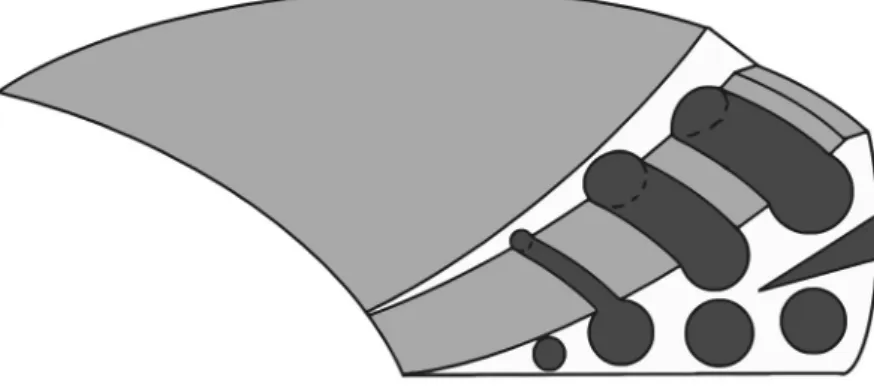
for meniscus regeneration. Ideally, such scaffolds could guide the infiltrating tissue into the preferable direction, for example peripheral collagen fibers into peripheral circumferential tunnels to better withstand hoop stresses during loading of the joint (Figure 6). Furthermore, these techniques may improve the biomechanical properties of the scaffolds and as such their initial performance in the knee joint before the actual tissue infiltration has completed.
An approach to avoid initial damage by high friction of the implant could be to coat the implant materials with substances to promote a smooth frictionless movement in the joint. Particularly hyaluronic acid (HA) and the other constituents
Figure 6. Schematic representation of scaffold design to enable the differentiation of tissue into the desired configuration.
1
of GAGs, such as the highly hydrated chondroitin sulphate, keratan-sulphate and/ or dermatan-sulphate could be used for this purpose. Particularly hyaluronic acid, a non-sulphated glycosaminoglycan, is present in high quantities in the synovial fluid and has a function in the lubrication of the joint. Currently, this substance is used as an injectable material to relieve complaints associated with severe osteoarthritis in patients. It also seems to have a protective effect on cartilage degeneration when used in animal models to prevent damage to the cartilage by surgical interventions98,99. HA
can also be used as a building block for drug delivery, tissue engineering, wound repair and visco-supplementation100.
OUTLINE OF THIS THESIS
This thesis shows the results of new potentials for meniscus tissue engineering. The first chapters are mainly focused on engineering a cell based meniscus construct. As stated in the paragraph “Scaffold materials for meniscus repair” the ideal scaffold material for meniscus tissue engineering should be biocompatible and biodegradable. The aim for CHAPTER 2 was to study a new scaffold material for meniscus tissue engineering. Besides biocompatibility and biodegradability, the scaffold should permit cell ingrowth, allow free diffusion of nutrients and should be strong enough to withstand the load in the knee-joint. The scaffold studied is a polyurethane (PU) based on poly D/L lactic acid and polycaprolactone with urethane linkers. Via solvent leaching a porous scaffold was obtained and subsequently tested for above mentioned properties for in vitro meniscus tissue engineering.
To increase the potential of the PU scaffold further, incorporation of bioactive macromolecules or growth factors are wanted (paragraph “Scaffold materials for meniscus repair”). These macromolecules should increase regeneration speed and quality of repair tissue. The macromolecule of interest is heparin, known for its growth factor adhering capacity and ability to re-differentiate meniscus cells. In
CHAPTER 3 a new surface treatment to coat this PU scaffold was studied.
In paragraph “New approaches”, current studies of meniscus tissue engineering have been discussed. An interesting option would be pre-culturing cells inside the scaffold. These cells would synthesize matrix and thereby increase mechanical stiffness and reduce incorporation time of the tissue engineered constructs. However, a big limitation of cell cultured constructs lies in the supplements added to the culture medium. Fetal bovine serum is a widely used and potent supplement of culture medium. It is known to induce proliferation and matrix synthesis of a wide variety of cells, including meniscus cells. But due to its bovine origin, application of cell cultured constructs with fetal bovine serum supplemented to the medium is limited due to potential bovine pathogens. Replacement for this fetal bovine serum is highly wanted for safety reasons and could facilitate future clinical applications of cell cultured tissue engineered constructs. In CHAPTER 4 we analyzed if platelet rich plasma could replace bovine serum in meniscus tissue engineering.
1
Scaffolds for musculoskeletal tissue engineering, e.g. meniscus and articular cartilage, should follow a number of requirements. A number of these requirements have been tested in chapter 2 and chapter 3. One property of scaffolds not mentioned before is the effect of scaffold architecture in tissue engineering. As proposed in paragraph “New approaches”, anisotropic scaffolds with a high porosity and aligned channels can guide extracellular matrix into similar organization as the native meniscus. In CHAPTER 5 we provided an overview of all current methods to produce different types of anisotropic scaffolds used for musculoskeletal tissue engineering. Subsequently, in CHAPTER 6 one of the described methods, modified Thermal Induced Phase Separation, was used to obtain anisotropic scaffolds from the studied PU scaffold material (chapter 2). Subsequently, these anisotropic scaffolds are tested for the ability to guide extra cellular matrix into similar organization as the native meniscus in a subcutaneous rat pocket model.
In addition to guidance for extracellular matrix, anisotropic scaffolds can also be used to guide cell migration. In articular cartilage repair, micro-fracturing/Pridy-drilling is used to create a channel between the cartilage and the subchondral bone. This enables mesenchymal stem cells from the underlying bone marrow to migrate into defect and regenerate the articular cartilage surface. However, in large defects regeneration is of an inferior fibrous cartilage type. In these large defects, scaffolds can be inserted into the defect to guide and stimulate cell migration, thereby increasing regeneration speed and quality. In CHAPTER 7 isotropic and anisotropic collagen scaffolds were inserted into articular cartilage defects of a rabbit model, and the effects of scaffold architecture on stem cell migration and articular cartilage regeneration were tested.
In CHAPTER 8 the results of the studies performed in this thesis were summarized and the implementation of this research into future perspectives of meniscus tissue engineering are discussed in CHAPTER 9.
1
REFERENCE LIST
1. Fithian DC, Kelly MA, Mow VC. Material properties and structure-function relationships in the menisci. Clin Orthop Relat Res 1990(252):19-31.
2. Walker PS, Erkman MJ. The role of the menisci in force transmission across the knee. Clin Orthop Relat Res 1975(109):184-92.
3. McDevitt CA, Webber RJ. The ultrastructure and biochemistry of meniscal cartilage. Clin Orthop Relat Res 1990(252):8-18.
4. Macnicol MF, Thomas NP. The knee after meniscectomy. J Bone Joint Surg Br 2000;82(2):157-9.
5. Setton LA, Guilak F, Hsu EW, Vail TP. Biomechanical factors in tissue engineered meniscal repair. Clin Orthop Relat Res 1999(367 Suppl):S254-72.
6. Ghosh P, Taylor TK. The knee joint meniscus. A fibrocartilage of some distinction. Clin Orthop Relat Res 1987(224):52-63.
7. Sun Y, Berger EJ, Zhao C, An KN, Amadio PC, Jay G. Mapping lubricin in canine musculoskeletal tissues. Connect Tissue Res 2006;47(4):215-21.
8. Brindle T, Nyland J, Johnson DL. The meniscus: review of basic principles with application to surgery and rehabilitation. J Athl Train 2001;36(2):160-9.
9. Ververidis AN, Verettas DA, Kazakos KJ, Tilkeridis CE, Chatzipapas CN. Meniscal bucket handle tears: a retrospective study of arthroscopy and the relation to MRI. Knee Surg Sports Traumatol Arthrosc 2006;14(4):343-9.
10. Petersen W, Tillmann B. Collagenous fibril texture of the human knee joint menisci. Anat Embryol (Berl) 1998;197(4):317-24.
11. Christoforakis J, Pradhan R, Sanchez-Ballester J, Hunt N, Strachan RK. Is there an association between articular cartilage changes and degenerative meniscus tears? Arthroscopy 2005;21(11):1366-9.
12. Englund M, Lohmander LS. Risk factors for symptomatic knee osteoarthritis fifteen to twenty-two years after meniscectomy. Arthritis Rheum 2004;50(9):2811-9.
13. Cox JS, Nye CE, Schaefer WW, Woodstein IJ. The degenerative effects of partial and total resection of the medial meniscus in dogs’ knees. Clin Orthop Relat Res 1975(109):178-83.
14. Jaureguito JW, Elliot JS, Lietner T, Dixon LB, Reider B. The effects of arthroscopic partial lateral meniscectomy in an otherwise normal knee: a retrospective review of functional, clinical, and radiographic results. Arthroscopy 1995;11(1):29-36.
15. Maletius W, Messner K. The effect of partial meniscectomy on the long-term prognosis of knees with localized, severe chondral damage. A twelve- to fifteen-year followup. Am J Sports Med 1996;24(3):258-62.
16. Hede A, Larsen E, Sandberg H. Partial versus total meniscectomy. A prospective, randomised study with long-term follow-up. J Bone Joint Surg Br 1992;74(1):118-21.
17. Hede A, Larsen E, Sandberg H. The long term outcome of open total and partial meniscectomy related to the quantity and site of the meniscus removed. Int Orthop 1992;16(2):122-5.
18. Ahmed AM. The load-bearing role of the knee meniscus. In: Mow VC, Arnoczky SP, Jackson DW, editors. Knee Meniscus: Basic and Clinical Foundations. New York: Raven Press, Ltd.; 1992. p 59-73.
19. Boyd KT, Myers PT. Meniscus preservation; rationale, repair techniques and results. Knee 2003;10(1):1-11. 20. Linke RD, Ulmer M, Imhoff AB. [Replacement of the meniscus with a collagen implant (CMI)]. Oper Orthop
Traumatol 2006;18(5-6):453-62.
21. Farng E, Sherman O. Meniscal repair devices: a clinical and biomechanical literature review. Arthroscopy 2004;20(3):273-86.
22. Miller MD, Kline AJ, Jepsen KG. “All-inside” meniscal repair devices: an experimental study in the goat model. Am J Sports Med 2004;32(4):858-62.
23. Laprell H, Stein V, Petersen W. Arthroscopic all-inside meniscus repair using a new refixation device: a prospective study. Arthroscopy 2002;18(4):387-93.
24. Kurzweil PR, Friedman MJ. Meniscus: Resection, repair, and replacement. Arthroscopy 2002;18(2 Suppl 1):33-9.
25. Lee GP, Diduch DR. Deteriorating outcomes after meniscal repair using the Meniscus Arrow in knees undergoing concurrent anterior cruciate ligament reconstruction: increased failure rate with long-term follow-up. Am J Sports Med 2005;33(8):1138-41.
26. Petersen W, Pufe T, Starke C, Fuchs T, Kopf S, Raschke M, Becker R, Tillmann B. Locally applied angiogenic factors--a new therapeutic tool for meniscal repair. Ann Anat 2005;187(5-6):509-19.
27. Kurz B, Lemke AK, Fay J, Pufe T, Grodzinsky AJ, Schunke M. Pathomechanisms of cartilage destruction by mechanical injury. Ann Anat 2005;187(5-6):473-85.
1
Differences in type II collagen degradation between peripheral and central cartilage of rat stifle joints after cranial cruciate ligament transection. Arthritis Rheum 2000;43(9):2121-31.
29. Stoop R, van der Kraan PM, Buma P, Hollander AP, Poole AR, van den Berg WB. Denaturation of type II collagen in articular cartilage in experimental murine arthritis. Evidence for collagen degradation in both reversible and irreversible cartilage damage. J Pathol 1999;188(3):329-37.
30. Arnoczky SP. Building a meniscus. Biologic considerations. Clin Orthop Relat Res 1999(367 Suppl):S244-53. 31. Gao JZ. [Experimental study on healing of old tear in the avascular portion of menisci in dogs]. Zhonghua Wai
Ke Za Zhi 1990;28(12):726-9, 782.
32. Behling CA, Spector M. Quantitative characterization of cells at the interface of long-term implants of selected polymers. J Biomed Mater Res 1986;20(5):653-66.
33. Veth RP, den Heeten GJ, Jansen HW, Nielsen HK. Repair of the meniscus. An experimental investigation in rabbits. Clin Orthop Relat Res 1983(175):258-62.
34. Veth RP, Jansen HW, Leenslag JW, Pennings AJ, Hartel RM, Nielsen HK. Experimental meniscal lesions reconstructed with a carbon fiber-polyurethane-poly(L-lactide) graft. Clin Orthop Relat Res 1986(202):286-93.
35. Buma P, van Tienen T, Veth R. The collagen meniscus implant. Expert Rev Med Devices 2007;4(4):507-16. 36. Klompmaker J, Jansen HW, Veth RP, Nielsen HK, de Groot JH, Pennings AJ, Kuijer R. Meniscal repair by
fibrocartilage? An experimental study in the dog. J Orthop Res 1992;10(3):359-70.
37. van Tienen TG, Heijkants RG, de Groot JH, Pennings AJ, Schouten AJ, Veth RP, Buma P. Replacement of the knee meniscus by a porous polymer implant: a study in dogs. Am J Sports Med 2006;34(1):64-71.
38. van Tienen TG, Heijkants RG, de Groot JH, Schouten AJ, Pennings AJ, Veth RP, Buma P. Meniscal replacement in dogs. Tissue regeneration in two different materials with similar properties. J Biomed Mater Res B Appl Biomater 2006;76(2):389-96.
39. van Tienen TG, Heijkants RG, Buma P, De Groot JH, Pennings AJ, Veth RP. A porous polymer scaffold for meniscal lesion repair--a study in dogs. Biomaterials 2003;24(14):2541-8.
40. Choi G, Vigorita VJ, DiCarlo EF. Second-look biopsy study of human collagen meniscal implants: A histological analysis of 81 cases.; 2008. p 128.
41. Reguzzoni M, Manelli A, Ronga M, Raspanti M, Grassi FA. Histology and ultrastructure of a tissue-engineered collagen meniscus before and after implantation. J Biomed Mater Res B Appl Biomater 2005;74(2):808-16. 42. Rodkey WG, Steadman JR, Li ST. A clinical study of collagen meniscus implants to restore the injured
meniscus. Clin Orthop Relat Res 1999(367 Suppl):S281-92.
43. Steadman JR, Rodkey WG. Tissue-engineered collagen meniscus implants: 5- to 6-year feasibility study results. Arthroscopy 2005;21(5):515-25.
44. Stone KR, Steadman JR, Rodkey WG, Li ST. Regeneration of meniscal cartilage with use of a collagen scaffold. Analysis of preliminary data. J Bone Joint Surg Am 1997;79(12):1770-7.
45. Bulgheroni P, Murena L, Ratti C, Bulgheroni E, Ronga M, Cherubino P. Follow-up of collagen meniscus implant patients: clinical, radiological, and magnetic resonance imaging results at 5 years. Knee 2010;17(3):224-9. 46. Brophy RH, Cottrell J, Rodeo SA, Wright TM, Warren RF, Maher SA. Implantation of a synthetic meniscal
scaffold improves joint contact mechanics in a partial meniscectomy cadaver model. J Biomed Mater Res A 2010;92(3):1154-61.
47. Verdonk P, Beaufils P, Bellemans J, Djian P, Heinrichs EL, Huysse W, Laprell H, Siebold R, Verdonk R, Actifit Study G. Successful treatment of painful irreparable partial meniscal defects with a polyurethane scaffold: two-year safety and clinical outcomes. Am J Sports Med 2012;40(4):844-53.
48. Bruns J, Kahrs J, Kampen J, Behrens P, Plitz W. Autologous perichondral tissue for meniscal replacement. J Bone Joint Surg Br 1998;80(5):918-23.
49. Kohn D, Wirth CJ, Reiss G, Plitz W, Maschek H, Erhardt W, Wulker N. Medial meniscus replacement by a tendon autograft. Experiments in sheep. J Bone Joint Surg Br 1992;74(6):910-7.
50. Veth RP, den Heeten GJ, Jansen HW, Nielsen HK. An experimental study of reconstructive procedures in lesions of the meniscus. Use of synovial flaps and carbon fiber implants for artificially made lesions in the meniscus of the rabbit. Clin Orthop Relat Res 1983(181):250-4.
51. Cummins JF, Mansour JN, Howe Z, Allan DG. Meniscal transplantation and degenerative articular change: an experimental study in the rabbit. Arthroscopy 1997;13(4):485-91.
52. Kohn D, Verdonk R, Aagaard H, Seil R, Dienst M. Meniscal substitutes--animal experience. Scand J Med Sci Sports 1999;9(3):141-5.
53. Messner K, Kohn D, Verdonk R. Future research in meniscal replacement. Scand J Med Sci Sports 1999;9(3):181-3.
54. Rijk PC. Meniscal allograft transplantation--part II: alternative treatments, effects on articular cartilage, and future directions. Arthroscopy 2004;20(8):851-9.
1
surgical considerations. Arthroscopy 2004;20(7):728-43.
56. Rijk PC, Tigchelaar-Gutter W, Bernoski FP, Van Noorden CJ. Functional changes in articular cartilage after meniscal allograft transplantation: a quantitative histochemical evaluation in rabbits. Arthroscopy 2006;22(2):152-8.
57. Stone KR. Meniscus replacement. Clin Sports Med 1996;15(3):557-71.
58. van Arkel ER, de Boer HH. Survival analysis of human meniscal transplantations. J Bone Joint Surg Br 2002;84(2):227-31.
59. Verdonk R, Kohn D. Harvest and conservation of meniscal allografts. Scand J Med Sci Sports 1999;9(3):158-9.
60. King D. The healing of semilunar cartilages. 1936. Clin Orthop Relat Res 1990(252):4-7.
61. Kobayashi M, Toguchida J, Oka M. Preliminary study of polyvinyl alcohol-hydrogel (PVA-H) artificial meniscus. Biomaterials 2003;24(4):639-47.
62. Messner K, Lohmander LS, Gillquist J. Cartilage mechanics and morphology, synovitis and proteoglycan fragments in rabbit joint fluid after prosthetic meniscal substitution. Biomaterials 1993;14(3):163-8. 63. Wood DJ, Minns RJ, Strover A. Replacement of the rabbit medial meniscus with a polyester-carbon fibre
bioprosthesis. Biomaterials 1990;11(1):13-6.
64. Stone KR, Rodkey WG, Webber R, McKinney L, Steadman JR. Meniscal regeneration with copolymeric collagen scaffolds. In vitro and in vivo studies evaluated clinically, histologically, and biochemically. Am J Sports Med 1992;20(2):104-11.
65. Alhalki MM, Hull ML, Howell SM. Contact mechanics of the medial tibial plateau after implantation of a medial meniscal allograft. A human cadaveric study. Am J Sports Med 2000;28(3):370-6.
66. Aagaard H, Jorgensen U, Bojsen-Moller F. Reduced degenerative articular cartilage changes after meniscal allograft transplantation in sheep. Knee Surg Sports Traumatol Arthrosc 1999;7(3):184-91.
67. Rijk PC, de Rooy TP, Coerkamp EG, Bernoski FP, van Noorden CJ. Radiographic evaluation of the knee joint after meniscal allograft transplantation. An experimental study in rabbits. Knee Surg Sports Traumatol Arthrosc 2002;10(4):241-6.
68. Rijk PC, Van Eck-Smit BL, Van Noorden CJ. Scintigraphic assessment of rabbit knee joints after meniscal allograft transplantation. Arthroscopy 2003;19(5):506-10.
69. Noyes FR, Barber-Westin SD. Irradiated meniscus allografts in the human knee. A two to five year follow-up study. Orthop Trans 1995;19:417.
70. van Arkel ER, de Boer HH. Human meniscal transplantation. Preliminary results at 2 to 5-year follow-up. J Bone Joint Surg Br 1995;77(4):589-95.
71. van Arkel ER, Goei R, de Ploeg I, de Boer HH. Meniscal allografts: evaluation with magnetic resonance imaging and correlation with arthroscopy. Arthroscopy 2000;16(5):517-21.
72. Yahia LH, Drouin G, Zukor D. The irradiation effect on the initial mechanical properties of meniscal grafts. Biomed Mater Eng 1993;3(4):211-21.
73. Garret JC. Free meniscal transplantation: a prospective study of 44 cases. Arthroscopy 1993;9:368-369. 74. Milachowski KA, Weismeier K, Wirth CJ. Homologous meniscus transplantation. Experimental and clinical
results. Int Orthop 1989;13(1):1-11.
75. Rath E, Richmond JC. The menisci: basic science and advances in treatment. Br J Sports Med 2000;34(4):252-7.
76. Wills NJ, LaPrade RF. Clinical outcomes of meniscal allografts.; 2008. p 127.
77. Stapleton TW, Ingram J, Katta J, Knight R, Korossis S, Fisher J, Ingham E. Development and characterization of an acellular porcine medial meniscus for use in tissue engineering. Tissue Eng Part A 2008;14(4):505-18. 78. Toyonaga T, Uezaki N, Chikama H. Substitute meniscus of Teflon-net for the knee joint of dogs. Clin Orthop
Relat Res 1983(179):291-7.
79. Messner K, Fahlgren A, Persliden J, Andersson BM. Radiographic joint space narrowing and histologic changes in a rabbit meniscectomy model of early knee osteoarthrosis. Am J Sports Med 2001;29(2):151-60. 80. Messner K, Gillquist J. Prosthetic replacement of the rabbit medial meniscus. J Biomed Mater Res
1993;27(9):1165-73.
81. Sommerlath K, Gillquist J. The effect of a meniscal prosthesis on knee biomechanics and cartilage. An experimental study in rabbits. Am J Sports Med 1992;20(1):73-81.
82. Kobayashi M, Chang YS, Oka M. A two year in vivo study of polyvinyl alcohol-hydrogel (PVA-H) artificial meniscus. Biomaterials 2005;26(16):3243-8.
83. Smillie IS. Observations on the regeneration of the semilunar cartilages in man. Br J Surg 1944;31:398-401. 84. Moon MS, Kim JM, Ok IY. The normal and regenerated meniscus in rabbits. Morphologic and histologic
studies. Clin Orthop Relat Res 1984(182):264-9.
85. Klompmaker J, Veth RP, Jansen HW, Nielsen HK, de Groot JH, Pennings AJ. Meniscal replacement using a porous polymer prosthesis: a preliminary study in the dog. Biomaterials 1996;17(12):1169-75.
1
86. Heijkants RG, van Calck RV, van Tienen TG, de Groot JH, Buma P, Pennings AJ, Veth RP, Schouten AJ. Uncatalyzed synthesis, thermal and mechanical properties of polyurethanes based on poly(epsilon-caprolactone) and 1,4-butane diisocyanate with uniform hard segment. Biomaterials 2005;26(20):4219-28. 87. Klompmaker J, Jansen HW, Veth RP, Nielsen HK, de Groot JH, Pennings AJ. Porous implants for knee joint
meniscus reconstruction: a preliminary study on the role of pore sizes in ingrowth and differentiation of fibrocartilage. Clin Mater 1993;14(1):1-11.
88. Moroni L, Poort G, Van Keulen F, de Wijn JR, van Blitterswijk CA. Dynamic mechanical properties of 3D fiber-deposited PEOT/PBT scaffolds: an experimental and numerical analysis. J Biomed Mater Res A 2006;78(3):605-14.
89. van Tienen TG, Heijkants RG, Buma P, de Groot JH, Pennings AJ, Veth RP. Tissue ingrowth and degradation of two biodegradable porous polymers with different porosities and pore sizes. Biomaterials 2002;23(8):1731-8.
90. Ibarra C, Ellisseff J, Vacanti JP, Cao Y, Kim TH, Upton J, Langer R, Vacanti CA. Meniscal tissue engineered by subcutaneous implantation of fibrochondrocytes on polymer develops biomechanical compressive properties similar to normal meniscus. . 1997. p 549.
91. Kang SW, Son SM, Lee JS, Lee ES, Lee KY, Park SG, Park JH, Kim BS. Regeneration of whole meniscus using meniscal cells and polymer scaffolds in a rabbit total meniscectomy model. J Biomed Mater Res A 2006;78(3):659-71.
92. Kon E, Chiari C, Marcacci M, Delcogliano M, Salter DM, Martin I, Ambrosio L, Fini M, Tschon M, Tognana E and others. Tissue engineering for total meniscal substitution: animal study in sheep model. Tissue Eng Part A 2008;14(6):1067-80.
93. Aufderheide AC, Athanasiou KA. Assessment of a bovine co-culture, scaffold-free method for growing meniscus-shaped constructs. Tissue Eng 2007;13(9):2195-205.
94. Ishida K, Kuroda R, Miwa M, Tabata Y, Hokugo A, Kawamoto T, Sasaki K, Doita M, Kurosaka M. The regenerative effects of platelet-rich plasma on meniscal cells in vitro and its in vivo application with biodegradable gelatin hydrogel. Tissue Eng 2007;13(5):1103-12.
95. Murphy JM, Fink DJ, Hunziker EB, Barry FP. Stem cell therapy in a caprine model of osteoarthritis. Arthritis Rheum 2003;48(12):3464-74.
96. Chiari C, Koller U, Dorotka R, Eder C, Plasenzotti R, Lang S, Ambrosio L, Tognana E, Kon E, Salter D and others. A tissue engineering approach to meniscus regeneration in a sheep model. Osteoarthritis Cartilage 2006;14(10):1056-65.
97. de Groot JH, Zijlstra FM, Kuipers HW, Pennings AJ, Klompmaker J, Veth RP, Jansen HW. Meniscal tissue regeneration in porous 50/50 copoly(L-lactide/epsilon-caprolactone) implants. Biomaterials 1997;18(8):613-22.
98. Han F, Ishiguro N, Ito T, Sakai T, Iwata H. Effects of sodium hyaluronate on experimental osteoarthritis in rabbit knee joints. Nagoya J Med Sci 1999;62(3-4):115-26.
99. Kobayashi K, Amiel M, Harwood FL, Healey RM, Sonoda M, Moriya H, Amiel D. The long-term effects of hyaluronan during development of osteoarthritis following partial meniscectomy in a rabbit model. Osteoarthritis Cartilage 2000;8(5):359-65.
100. Luo Y, Prestwich GD. Hyaluronic acid-N-hydroxysuccinimide: a useful intermediate for bioconjugation. Bioconjug Chem 2001;12(6):1085-8.
Chapter 2
Proliferation of meniscal
fibrochondrocytes cultured on a new
polyurethane scaffold is stimulated
by TGF-beta
J. Biomaterials Applications, 2013Eric de Mulder
Gerjon Hannink
Maud Giele
Nico Verdonschot
Pieter Buma
2
ABSTRACT
The aim of this study was to investigate if newly developed polyurethane (PU) scaffolds are suitable as scaffold for cell seeded meniscus tissue engineered constructs. Scaffolds were seeded with goat meniscal fibrochondrocytes and cultured to assess changes in biological and mechanical properties. Furthermore, the effect of TGF-β on these properties were investigated. For this PU scaffolds were made from poly D/L lactide and caprolactone as soft segments and 1,4-butanediisocyanate for the urethane hard segments. The porosity of the scaffolds was 95%. Isolated goat meniscal fibrochondrocytes were seeded on the scaffolds and cultured with or without the addition of 10 ng/ml TGF-β in standard culture medium. After 2, 4 and 6 weeks of culture, scaffolds were analyzed for cell proliferation, matrix synthesis and mechanical properties.
Results of scanning electron microscopy and histology showed that the scaffolds had an interconnected isotropic pore structure. Without addition of TGF-β cells did not proliferate during the culture period and isolated meniscus fibrochondrocytes were more frequently located in the peripheral parts of the scaffold. Fibrochondrocytes supplemented with TGF-β were distributed throughout the construct. Clustered cells were surrounded by matrix which stained slightly positive for GAGs. Also collagen production was increased significantly after 4 and 6 weeks of culture compared to cultures without TGF-β and also more GAG staining was found after 4 and 6 weeks in the sections of the TGF-β stimulated cultures. Despite the increase in matrix production, the compressive stiffness of the constructs was not increased during the culture period.
In conclusion, meniscal fibrochondrocytes were able to adhere to the polyurethane scaffold. However, the scaffold itself does not stimulate proliferation and matrix production. The addition of TGF-β resulted in a strong induction of both proliferation and extracellular matrix production.
2
INTRODUCTION
Menisci are semilunar shaped structures located in pairs in each knee joint. They fill the gap between the round femoral condyle and the rather flat (medial) or even concave (lateral) tibia plateau. Although the meniscus has quite a number of functions in the knee joint, the main function of the meniscus is to distribute stresses in a homogenous way over the articular surfaces of the tibia and femur. To withstand the high forces in the knee, the fibro-cartilage of the meniscus has a highly circumferentially oriented type I collagen fiber organization. Moreover, it contains also type II collagen and some glycosaminoglycans (GAG’s), particularly in the inner highly loaded part of the meniscus1. The composition of the tissue enables
the meniscus to withstand compressive and tensile shear forces and to maintain its position under load. Thereby it transfers axial loads into circumferential directed forces2,3.
Meniscus injury will generate pain and cause mechanical locking of the knee joint. The most frequent occurring meniscus trauma’s are tears. The peripheral part of the meniscus is the so-called red zone which is vascularized. Since vascularization is a key player in regeneration after trauma4,5, tears in this location normally can
heal4-6. When tears occur in the inner 2/3 of the meniscus, also called the white
zone of the meniscus, no or very limited regeneration is possible due to a lack of vascularization.
The flap tear and bucket handle tear are the most common meniscus injuries. In the past, the resulting loose fragments were arthroscopically removed. Although this treatment is an enormous improvement compared to a total meniscectomy, even a partial meniscectomy will result in osteoarthritic changes in the articular cartilage7. Also the amount of meniscal tissue removed has a direct relationship with
the induction of late osteoarthritis8,9. Upon removal of meniscal tissue more load is
directly transferred from the condyles to the tibial plateau resulting in large peak stresses which causes an early onset of osteoarthritis3,7. Therefore, in order to preserve
the contact area of the meniscus, the general practice these days is fixation of the loose fragment to the main body of the meniscus by suturing, anchors or screws10.
Unfortunately the used fixation devices are still inferior to cope with the forces applied on the meniscus, resulting in high rates of ruptures or even osteochondral defects11. When fixation is not possible or insufficient, a partial meniscectomy is
inevitable.
Regeneration of the lost part of the meniscus would be the most optimal treatment. A scaffold that replaces the meniscus tissue could guide new tissue ingrowth into the lost meniscus and by that meniscus regeneration could be established. Two implants based on scaffolds are already in clinical use, namely a scaffold based on collagen (Menaflex™) and one based on a synthetic polymer(Actifit®)12,13. The
Menaflex™ implant has already been implanted for a number of years and supports ingrowth of new meniscus like tissue. However, the scaffold shrinks in size after 5 years13-15. The Actifit® implant, based on a synthetic caprolactone-urethane polymer,
2
is only recently available in Europe. Preclinical animal experiments showed that implantation of this scaffold in a partial defect will enable equal contact pressures as a normal meniscus, whereas without implantation contact pressures increase16.
After implantation the scaffold had rapid ingrowth of tissue and no cartilage defects were present after one year17. These experiments were conducted in partial meniscus
defects, but when implanted as a total meniscus replacement, the scaffold was not strong enough and failed to protect the cartilage from damage18,19. Recently, the first
patient study with Actifit® as a partial replacement has been published. After one year follow up, implant was integrated with native meniscus tissue, and biopsies showed fully vital tissue20.
In contrast to partial meniscal replacement, the only clinical used treatment for total meniscus replacement is allograft transplantation21. Even though clinical results
are satisfactory, there exists a minor risk of disease transfer, the availability is limited due size matching and the allograft meniscus can re-tear, shrink and dislocate22.
Tissue engineering could be a promising alternative approach to replace the lost tissue after (partial) menisectomy. Pre-cultured cell seeded scaffolds showed better integration with the surrounding meniscus tissue in an ex vivo setup23. Also in in
vivo experiments cell seeded constructs showed better integration with adjacent tissue after implantation in defects24,25. Pre-seeding cells on a scaffold also strongly
improved the mechanical properties compared to the initial scaffold26. Three
cell types are most often used so far, namely mesenchymal stem cells (MSC)23,
chondrocytes27, meniscal fibrochondrocytes (MFCs)28, or a combination of these
cell types27. Which cell type is best suited for these cell seeded constructs remains
unknown. To stimulate these cell based meniscal constructs, a number of growth factors (GF) have been tested. Most commonly used are TGF-β, IGF, bFGF and PDGF28-32. These GFs stimulate cell proliferation, matrix production and migration,
thereby reducing the time needed to engineer new meniscus tissue.
Which type of scaffold is best suited for meniscus TE is still pending. Scaffolds of many different composites, e.g., based on hydrogels, biologic polymer (collagen / silk) or synthetic polymer (PLA / PGA / PCL), have been described in literature18,33,34.
A synthetic polymer scaffold with very promising characteristics has already been described in detail by Spaans et al. and van Minnen et al.35,36. They synthesized a
polyurethane (PU) co-polymer porous scaffold with poly D/L lactic acid and poly-ε-caprolactone as soft block segments and 1,4-butanediisocyanate as hard block urethane segments36. These highly porous scaffolds were tested both in vitro and
in vivo for cytotoxicity and degradation. The in vitro experiments showed that after 12 weeks only 3.4% mass was degraded, thereby categorized as a slow degrading scaffold. Cytotoxicity tests showed that scaffolds did not change the proliferation speed and metabolic activity of fibroblast after direct contact37. The long term
test showed some cytotoxicity, however, this was ascribed to the accumulation of degradation products in their in vitro test system37. The in vivo experiments of short
2
a gradual resorption pattern over 3 years. First encapsulation and infiltration with connective tissue was observed. Subsequently, degradation and resorption by macrophages and giant cells was found. Scaffolds were not completely resorbed after 3 years but they expected it to be totally resorbed after 3.5 – 4 years38.
The PU described above has a number of novel aspects compared to other synthetic polymer scaffolds previously used for meniscus tissue engineering purposes or in clinical practice. The scaffold described in this study has a 95% porosity compared to approximately 80% porosity of most other scaffolds19,39. This high porosity is
required to increase surface area for cell attachment, tissue ingrowth and moreover, facilitating adequate transport of nutrients and cellular waste products40. With
decreasing porosity, nutrient and waste exchange of cells in the deeper regions is limited, leading to necrotic sites in the center of the scaffold19. Another advantage of
a high porosity is that it, in general, will lead to a faster speed of resorption as less polymer material has to be degraded. Reducing the time foreign materials remain present in the body has been shown to lower the risk on immunological responses. In addition, with smaller amounts of polymer material to be degraded, lower acidic levels will be reached. Higher acidity levels are known to reduce tissue regeneration speed37. The high porosity of the material used in our study also aims to ensure a
faster resorption rate, low immunological risk, and high tissue regeneration speed. Furthermore, the use of a co-polymer from caprolactone and both isomers of lactic acid exclude crystalline regions in this material. It has been described that increasing crystallinity results in highly stable remnants and increase inflammatory risk in a late degradation stage41.
The aim of this study was to improve the properties of the PU scaffolds as described by van Minnen et al.36 by a tissue engineering approach for total
meniscus replacement. Pre-seeding cells on scaffolds could have beneficial effects on biological and mechanical properties. In this study biological properties (i.e. proliferation, distribution and matrix production) were tested after culture MFCs on this PU scaffold. Furthermore, we tested the effect of TGF-β on these biological properties. Finally the mechanical properties of this tissue engineered construct was determined to assess whether it is comparable to native meniscus.
MATERIAL AND METHODS
Scaffold preparation.
Scaffolds were composed of 50% DL(50/50)-lactide (PDLLA, Purac) and 50% ε-caprolactone (PCL, Acros Organics) by ring opening polymerization with 1,4-butanediol (BDO) as initiator. The final macrodiol prepolymer had a molecular weight (MN) of 2000 g/mole. Subsequently this was end-capped with 1,4-butanediisocyanate (BDI). Chain extension was performed with a BDO-BDI-BDO urethane block which resulted in a hard block urethane segment with a uniform length of five urethane moieties. Final PU consisted of 22.5 w/w % hard urethane
2
segments and 77.5 w/w % amorphous polyester soft segments. Polymers were solvent casted, frozen and lyophilized. The obtained scaffolds had a 95% porosity36.
Finally, scaffolds were frozen in liquid N2, punched with a 6 mm internal diameter dermal punch and cut into 3 mm high cylinders. After cutting the scaffolds, SEM images were made to evaluate the internal pore structure of the scaffolds (JEOL JSM 6310, JEOL Ltd, Herts, UK).
Cell isolation
Both lateral and medial menisci from four skeletal mature female Dutch milk goats (Capra Hircus Sana) were isolated immediately after sacrifice under aseptic conditions. Tissue collection was approved by the Animal Ethics Committee of the Radboud University, Nijmegen. Isolation of meniscal fibrochondrocytes was performed according to earlier reported protocols28. In short, menisci were washed
3 times with PBS/1x PSF (Penicillin/streptomycin/fungizone, Gibco, Carlsbad, CA USA). Subsequently, the outer rim was removed and inner part was cut into small pieces. These pieces were digested in DMEM/F12 + 1x PSF with 3 mg/ml collagenase II (Worthington, Lakewood, NJ USA) for 48 hour in a 5% CO2, 37OC
controlled culture incubator. Digest was filtered over 70 µm tissue filter bags and filtrate was centrifuged for 10 min at 1100 rpm to pellet the cells. Cells were washed 2 times with PBS and plated in culture flasks. Cells were cultured in standard medium (DMEM/F12 + 10% FBS + 1x PSF) in a culture incubator. At 70% confluence cells were passed once by trypsinization using standard procedures. Passage 1 cells were harvested, frozen in cryo-vials and preserved in liquid nitrogen until use.
Cell culture
Before start of the experiment cells were defrosted and expanded twice. Finally, cells were isolated, counted and used for cell culture experiments. MFCs isolated had a >95% cell viability determined by trypaan blue exclusion. Scaffolds were ethanol desinfected in series till 96% EtOH, washed three times with sterile PBS and incubated o/n in standard culture medium. Scaffolds were squeezed to remove excess medium and passage 4 MFCs, a total of 5x10⁵ in 50 µl, were placed on top of
each scaffold and incubated for 60 minutes in an incubator. After incubation standard medium was added with or without 10 ng/ml TGF-β (R&D Systems, Minneapolis, MN USA). Total medium was refreshed three times a week.
Pilot tests were performed with different concentrations of TGF-β (1 ng, 10 ng and 100 ng/ml). If 10 ng/ml or 100 ng/ml was added, proliferation was shown to increase. However, no differences were observed between 10 ng and 100 ng. Therefore 10 ng/ml was used in the current experiments. Scaffolds were cultured for 2, 4 and 6 weeks without or with the addition of 10 ng/ml TGF-β in standard medium and performed in triplo.
2
Picogreen analyses
After 2, 4 and 6 weeks of culture samples were washed with PBS and frozen in Milli Q. After repeated freeze/thaw cycles samples were measured using PicoGreen dsDNA assay kit as per manufacturer’s protocol (Invitrogen, Carlsbad, CA USA).
Collagen analyses
After 2, 4 and 6 weeks of culture samples were washed and frozen in PBS. Samples were analyzed to quantify the amount of collagen using a colorimetric hydroxyproline assay42. In short, samples were hydrolyzed in 6N HCl by autoclaving for 20 hours
at 110OC. The hydrolyzate was washed twice with Milli Q. Chloramin-T was added
and incubated for 20 minutes at room temperature. Subsequently, Ehrlich’s reagent was added and chromophore was developed during 20 minutes at 60OC. Finally,
absorbance was read at 570 nm using a spectrophotometer.
Routine histology
Scaffolds were paraffin embedded and sections of 7 µm in thickness were cut. Sections were deparaffinized, and rehydrated. Oil Red O with hematoxylin (ORO/H) for cell staining, and alcian blue for GAG staining, were used for routine histology. For ORO/H staining, sections were shortly incubated in oil red O. Hereafter cell nuclei were counterstained with hematoxylin and mounted with aquamount. For alcian blue, sections were washed in 3% acetic acid, incubated for 45 min in alcian blue. After washing cell nuclei were counterstained with nucleus red and mounted with DPX. All samples were analyzed under light microscopy and pictures were recorded with an Olympus digital camera and a photo program software (analySIS Soft Imaging System, GmbH, Münster, Germany).
Mechanical analyses
At day 0 and after 2, 4, and 6 weeks of culture samples were tested for mechanical stiffness. Samples were placed in a Bose ElectroForce BioDynamic bioreactor mounted with a 22N load cell housed in a 37OC incubator (BOSE, MN USA). The
specimen chamber was filled with PBS and scaffolds were compressed at 1 mm/min till 20% compression. With the obtained stress/strain curve the Young’s Modulus was calculated as the steepness of this curve.
Statistics
A two way ANOVA was used on all datasets with treatment (TGF yes/no) and time (0, 2, 4 and 6 weeks) as factors. All data were expressed as mean ± SEM. For all experiments, differences were considered statistically significant for P values <0.05. Statistical analysis was performed using SPSS 16.0 software (SPSS Inc., Chicago, IL USA).