LARGE-SCALE GENERATION OF SYNTHETIC DNA LIBRARIES: SEQUENCE-SPECIFIC PRIMING OF REVERSE TRANSCRIPTION
by
Yusuf Esmail Murgha
A dissertation submitted in partial fulfillment of the requirements for the degree of
Doctor of Philosophy (Biomedical Engineering) in The University of Michigan
2012
Doctoral Committee:
Professor Erdogan Gulari, Chair Professor Shuichi Takayama Associate Professor Michael Mayer Assistant Professor Peter Woolf
© Reserved Rights All Murgha Yusuf 2012
ii To my Mom and Dad
iii
ACKNOWLEDGMENTS
I would like to thank Prof. Erdogan Gulari for giving me the opportunity to pursue my career goals and for his advice and support during the doctoral work. I like to thank Dr. Jean-Marie Rouillard for being a great mentor and motivator. His calm, confident and professional demeanor under pressure is a quality I aspire to achieve.
I would like to thank Prof. Shuichi Takayama, Prof. Michael Mayer and Prof. Peter Woolf for their guidance and feedback towards the completion of my work. I thank past and current members of Gulari research group for their help and support. I like to thank all my friends and peers for their continuous motivation and making this an enjoyable journey. I like to thank the administrative staff in biomedical and chemical engineering department.
I dedicate this work to my family, my mom and dad, brother and sister-in-law, and all my relatives. I thank them for their continual support and encouragement. They are my rock.
iv
TABLE OF CONTENTS
DEDICATION ... ii
ACKNOWLEDGMENTS ... iii
LIST OF FIGURES ... viii
ABSTRACT ... xi
CHAPTER 1 INTRODUCTION ... 1
1.1 Applications of oligonucleotide libraries ... 2
1.1.1 Synthetic gene assembly ... 2
1.1.2 Cloned libraries ... 4
1.1.3 Baits . ... 7
1.1.4 Oligo-Selective Sequencing (OS-Seq) ... 9
1.1.5 Fluorescent In Situ Hybridization (FISH) ... 11
1.2 Source of oligonucleotide libraries ... 13
1.2.1 Conventional state-of-the art oligonucleotide synthesis ... 13
1.2.2 Microarray Technology ... 14
1.3 Current limitations ... 18
1.4 Motivation ... 20
1.5 Overview of the dissertation ... 21
CHAPTER 2 FROM DNA MICROARRAYS TO SEQUENCE-SPECIFIC SINGLE-STRANDED NUCLEIC ACIDS ... 23
2.1 Introduction ... 23
v
2.1.2 Emulsions background ... 27
2.1.3 W/O emulsions in biology ... 33
2.1.4 Methods to prepare single-strand DNA from double-stranded DNA ... 37
2.2 Experimental methods and protocols ... 43
2.2.1 Process outline ... 43
2.2.2 Yeast Knock-out (YKO) deletion collection ... 46
2.2.3 Enzyme based methods to prepare single-strand DNA from duplex DNA .. ... ... 48
2.2.4 Preparation of single-strand DNA with 5’-modified end ... 49
2.2.5 Optimize digestion time of Lambda exonuclease ... 49
2.2.6 In vitro transcription and reverse transcription ... 50
2.2.7 SYBR gold gel staining ... 50
2.2.8 Polyacrylamide gel (PAGE) purification ... 51
2.2.9 Phenol/ Chloroform/ Isoamyl Alcohol (PCIA) purification... 51
2.3 Experimental results and discussion... 52
2.3.1 Yeast knock-out collection (YKO) ... 52
2.3.2 Interpretation of gel images ... 52
2.3.3 Dependence of emulsion PCR on template size and concentration ... 52
2.3.4 Dependence of emulsion PCR on type of oil ... 55
2.3.5 Enzymatic hydrolysis methods to prepare single-stranded DNA from duplex DNA ... 57
2.3.6 Preparation of single-strand DNA with 5’-modified end ... 61
2.3.7 Optimal digestion time of Lambda exonuclease ... 64
2.3.8 Amplification of microarray-derived DNA library ... 65
2.3.9 In vitro transcription and reverse transcription (IVT-RT) ... 76
vi
CHAPTER 3 PRIMING REVERSE TRANSCRIPTION WITH SEQUENCE-SPECIFIC
PRIMER LIBRARIES ... 84
3.1 Introduction ... 84
3.1.1 Sequence-specific primer libraries for reverse transcription ... 86
3.1.2 Background ... 88
3.1.3 Insights from reverse transcription-polymerase chain reaction (RT-PCR) ... ... 90
3.2 Experimental methods and protocols ... 93
3.2.1 One round aRNA amplification with 5’-end dangling gene-specific primer ... 93
3.2.2 Removal of excess sequence-specific primers ... 93
3.2.3 Optimize RT conditions when using sequence-specific primers... 95
3.2.4 Detect differential expression of transcripts with sequence-specific primers ... 96
3.2.5 Activity of reverse transcriptase in Actinomycin D ... 97
3.2.6 Two step process to remove excess long primers ... 99
3.2.7 Reverse transcription with sequence-specific long primer library... 100
3.2.8 Saccharomyces cerevisiae growth conditions and total RNA extraction ... ... ... 101
3.2.9 Polymerase chain reaction (PCR) ... 101
3.2.10 Real-time PCR and Melt Curve ... 102
3.2.11 Microarray analysis ... 102
3.3 Experimental results and discussion... 103
3.3.1 Interpretation of real-time PCR plots... 103
3.3.2 One round aRNA amplification with long gene-specific primers 103 3.3.3 Removal of excess sequence-specific primers ... 106
vii
3.3.4 Optimize reverse transcription conditions ... 114
3.3.5 Detect differential expression of transcripts with sequence-specific primers ... 117
3.3.6 Activity of reverse transcriptase in Actinomycin D ... 122
3.3.7 Differential expression of yeast genes induced in glucose and galactose media ... 124
3.3.8 Two step process to remove excess long primers ... 131
3.3.9 Reverse transcription with sequence-specific long primer library ... 134
3.4 Conclusion ... 139
CHAPTER 4 CONCLUSION AND RECOMMENDATIONS ... 141
4.1 Conclusion ... 141
4.2 Recommendations for future work ... 143
4.2.1 Under generation of single-strand nucleic acid libraries ... 143
4.2.2 Under sequence-specific primer libraries ... 146
APPENDIX ... 148
viii
LIST OF FIGURES
Figure 1-1 Synthetic gene assembly ... 3
Figure 1-2 Cloned libraries – RNAi and peptide libraries ... 5
Figure 1-3 Solution hybrid selection – Baits ... 8
Figure 1-4 Padlock probes – MIPS and SGC ... 8
Figure 1-5 Oligonucleotide selective sequencing ... 10
Figure 1-6 Fluorescent In Situ Hybridization (FISH) ... 12
Figure 1-7 Oligonucleotide synthesis ... 13
Figure 1-8 Microarray synthesized oligonucleotide library ... 18
Figure 2-1 Process overview ... 25
Figure 2-2 Emulsion types ... 29
Figure 2-3 Emulsion breakdown processes ... 31
Figure 2-4 Emulsion PCR versus Conventional PCR ... 35
Figure 2-5 Exonucleolytic hydrolysis of duplex DNA to generate ssDNA ... 39
Figure 2-6 Formation of single-stranded DNA ... 41
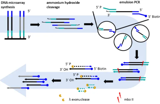
Figure 2-7 Emulsion PCR ... 44
Figure 2-8 Restriction digest of yeast genomic DNA ... 53
Figure 2-9 Effect of template size on emulsion PCR ... 54
Figure 2-10 Effect of oil type on emulsion PCR ... 56
Figure 2-11 T7 gene 6 exonuclease and phosphorothioate backbone ... 59
ix
Figure 2-13 Lambda exonuclease activity on modified 5’-ends ... 62
Figure 2-14 Optimal digestion time (lambda exonuclease) ... 64
Figure 2-15 Measurement of droplet size ... 66
Figure 2-16 Real-time PCR – Standard curve ... 69
Figure 2-17 Emulsion PCR versus Conventional PCR ... 71
Figure 2-18 Lambda exonuclease - Oligonucleotide library to ssDNA ... 74
Figure 2-19 Oligonucleotide library – IVT-RT method ... 78
Figure 3-1 cDNA synthesis with sequence-specific primers 1 ... 104
Figure 3-2 cDNA synthesis with sequence-specific primers 2 ... 105
Figure 3-3 Spin column removal of double-stranded primer ... 107
Figure 3-4 Effect of SSB protein on amount of ssDNA digested ... 109
Figure 3-5 Real-time PCR - Effect of SSB protein on amount of ssDNA digested... 110
Figure 3-6 Size dependent fractionation - PEG concentration (1.25 M NaCl) ... 112
Figure 3-7 Size dependent fractionation PEG and NaCl concentration ... 113
Figure 3-8 Effect of temperature on reverse transcription ... 115
Figure 3-9 Effect of primer concentration on revere transcription ... 116
Figure 3-10 Density Plot – cDNA synthesis with mouse sequence-specific primer 118 Figure 3-11 Differential gene expression – mouse sequence-specific primers ... 118
Figure 3-12 Exonuclease I digestion and reverse transcription of ssDNA ... 120
Figure 3-13 ‘no RNA’ reverse transcription with sequence-specific primers ... 121
Figure 3-14 Activity of reverse transcriptase in presence of Actinomycin D ... 123
Figure 3-15 Melt curve of GAL1 gene - optimal sample processing method ... 125
Figure 3-16 Number of gene detected across sample processing methods ... 126
Figure 3-17 List of yeast genes expressed in galactose - glucose ... 128
x
Figure 3-19 Exonuclease I digestion in presence of actinomycin D... 132
Figure 3-20 AMPure bead - Effect of DNA concentration ... 133
Figure 3-21 AMPure bead - Effect of PEG8000 concentration ... 133
Figure 3-22 Gene expression – exonuclease I and 11 % PEG 8000 ... 135
xi ABSTRACT
Custom-designed DNA and RNA oligonucleotide collections are used as building blocks in synthetic biology, complementary probes for targeted sequencing, and nucleic acid aptamers. They encode information for technologies like RNA interference, protein engineering and DNA-encoded chemical libraries. These applications require an economical source of diverse libraries.
High-throughput microarray technology produces hundreds of thousands of diverse sequences on a single planar substrate at low cost (<$200 for 20,000 oligonucleotides). However, the quantity and quality of individual oligonucleotides obtained from this technology is low (~ 5 fmol). In order to produce feasible amounts of high quality oligonucleotides (10 – 100 pmol), it is necessary and more economical to go through a molecular amplification procedure. Existing methods of amplification lead to the formation of chimeric products. We developed a 2-step amplification process which gives an overall ~450 fold increase in the amount of single-stranded oligonucleotides. The first-step, emulsion polymerase chain reaction (PCR), provides the initial amplification while preserving the library complexity (~25 μM product). However, the overall product yield from a single reaction after removal of primer binding sequences and single-strand DNA formation is limited (~15 pmol) as a result of restrictions imposed on the input reactant quantity. Therefore, this step alone is labor intensive to produce large amounts of products due to lack of scalability. This limitation is addressed via transcription-reverse
xii
transcription of the emulsion PCR amplicons. This two stage process allows scalability of microarray synthesized oligonucleotides for preparation of large quantities of ssDNA libraries (~6500 pmol).
As an application of the amplification technology we developed an integrated method for cDNA library construction with sequence-specific primers incorporating a unique tag and universal primer sequence. The method suffers from the formation of three types of false positives that need to be sufficiently removed to reduce contribution of false-positives signals. A 3 step process is implemented to reduce the false positives contributors and still detect differential expression of yeast genes in galactose and glucose conditions. The sequence-specific primers libraries can be used for applications not limited to detection of low abundance and rare RNA and identification of aberrant splicing variants and gene-fusions.
1 CHAPTER 1
INTRODUCTION
The last decade has seen the emergence of a broad range of applications for DNA and RNA oligonucleotide libraries. DNA oligonucleotides are the synthetic biology building blocks for the assembly of single genes [1-3] to whole genomes [4, 5]. Targeted next-generation sequencing relies heavily on oligonucleotide libraries as a source of baits to capture, either in the form of DNA padlock probes for the circularization of targeted sequences [2, 6] or in the form of RNA baits for the direct capture of sequencing genomic DNA library fragments [7]. Recently Millykangas et al. pushed the application of oligonucleotide libraries for targeted sequencing even further by integrating the target capture into the sequencing device, using a DNA oligonucleotide library to customize the primer lawn on a sequencing flowcell [8]. Oligonucleotides libraries are widely used to encode active RNA such as shRNA [9], or peptides [10, 11] after cloning in appropriate vectors. Labeled libraries can be also used for molecular detection probes in fluorescent in situ hybridization (FISH) techniques such as OligoPaint [12].
2 1.1 Applications of oligonucleotide libraries
1.1.1Synthetic gene assembly
Gene-synthesis is the process of creating a gene in vitro without a precursor template [Figure 1-1]. The process of gene-synthesis begins with hybridization of melting temperature-normalized oligonucleotides to each other to make the gene backbone. The oligonucleotides overlap at their ends to simultaneous act as templates and primers for the synthesis of other strand. These are subsequently joined by various PCR techniques (ligation PCR, assembly PCR and fusion PCR) to form a new gene. Here, the primary requirement is for long oligonucleotides so that fewer numbers are required to make longer gene segments or genes. Also, a large amount of oligonucleotides are required to make many genes and it variants in a single reaction.
3
4 In the field of synthetic biology, oligonucleotide libraries have accelerated the optimization of gene-expression and protein activity [13] , enabled the assembly of difficult to make antibodies and antigens [14, 15], engineering of metabolic pathways [16] and construction of mitochondrial, viral and bacterial genomes [4, 17, 18]. Compared to conventional methods of gene-generation, which pick elements from existing elements by PCR and DNA cloning, they provide greater flexibility to gene-engineering to ultimately create organisms with new properties (bioremediation, biosafety, biofuels, drugs and vaccines). Another variant of assembly of synthetic oligonucleotides is in the field of DNA computing and storage [19, 20].
1.1.2Cloned libraries
Here, the oligonucleotide libraries are used to encode information for downstream applications. This can be either for a short hairpin RNA (shRNA) or a gene coding for a peptide [Figure 1-2]. Small interfering RNAs (siRNAs) are used for specific silencing of many genes. The process has been used to study cell behavior in many mammalian cells [21]. For this application the oligonucleotide libraries has self-complementary ends separated by common loop, which then folds to form short-hairpin DNAs (shDNA) constructs. The individual shDNA are cloned into vectors to produce shRNAs (or siRNA) that get incorporated into RNA-induced silencing complex (RISC) to guide mRNA cleavage and degradation [22, 23]. Unlike other methods to make siRNA, namely i) chemical in situ synthesis of RNA, which is cost prohibitive, ii) biological sources that give variable amounts of siRNA depending on mRNA expression levels and abundance in the tissue and iii) enzymatic digestion (RNase III, Dicer) of long dsRNA molecules into randomly chopped molecules, complex oligonucleotide libraries give more defined and
5 uniform representation of siRNA without any of the non-specifics effect such as binding to genes with homologous sequences or to splice isoforms [Figure 1-2] [24, 25].
6 Phage display is a high-throughput technique to screen, select and optimize for protein interactions with other molecules such as ligands, peptides, other proteins and DNA [26]. Here the phenotype (individual peptide) is linked to its genotype (coding sequence) which enables the isolation of binding clones from large populations. Historically, random nucleic acid libraries used to encode for peptides or proteins expressed on the bacteriophage cell surface is a great method to identify good candidates. However, they are not able to cover the entire diversity of peptide library (e.g. for a 12 mer peptide, there are possible 1220 = 4x10¹⁵ individual sequences of which less than 0.001% are screened in a typical experiment) [27]. Also, there is a decrease in the diversity of the library during the panning and selection rounds, further reducing the number of binding clones identified during the screening process [28]. Thus once good candidate sequences are found, they can be mutated in a controlled manner by using oligonucleotide libraries to access the best candidates with the highest binding affinities.
Another application of oligonucleotide libraries is to produce peptide or short protein libraries [Figure 1-2]. Here systematic combinations of DNA fragments are cloned into vectors to express peptides and short proteins to identify biologically active peptides (e.g. anti-microbial), discover ligand-binding proteins, study and optimize enzyme functionalities, epitope mapping and generate vaccines [29]. Although, solid-phase peptide synthesis competes with the flexibility provided by DNA libraries, it is limited by the peptide length (<18 amino acids), and high costs of synthesizing large libraries [30]. For both peptide libraries and phage display, oligonucleotide libraries provide flexibility in choosing codons to optimize for protein expression.
7 1.1.3Baits
The past few years has witnessed rapid development in ‘next-generation’ or massively parallel sequencing technology leading to sequencing whole genomes [31-33]. Whole genome sequencing of humans is resource-intensive as multiple runs are required to get sufficient depth and coverage. In addition, most disease-causing mutations (85%) are estimated to be located within the exome (protein coding regions), which constitute only 1 % - 1.5 % of the human genome (3 Gb) [34, 35]. Hence, methods are required to selectively enrich and amplify gene-subsets for targeted sequencing. The underlying principle of the methods namely, molecular inversion probes (MIP) [36], selective genomic circularization (SGC) [37] and solution hybrid selection (SHS) [38] is solution-based hybridization of targets with complementary sequences. For these applications a large number and quantities of diverse oligonucleotides are required to increase genome coverage and sensitivity and reproducibility of the assays. In case of MIPs, the use of longer oligonucleotides enables the incorporation of barcodes of sample multiplexing [39].
8
Figure 1-3 Solution hybrid selection – Baits
9 In SHS method, biotinylated target-specific RNA probes (or baits) bind to fragmented genomic DNA libraries and separated by capture on magnetic beads. As single-stranded RNA display a different spectrum of sensitivity to chemical agents and enzymes they can be easily removed from DNA prior to sequencing [Figure 1-3]. On the hand, DNA baits (MIPs or padlock probes - SGC) are used for copying target sequences between two primer binding sites [Figure 1-4]. The DNA probes have common linker flanked by two target-specific sequences. In MIPs method the baits hybridize to both end of the intended target, followed by primer extension (DNA polymerase) and ligation (DNA ligase) to form circular probes. Linear species molecules are removed by exonucleases to enrich for the circularized probes. In SGC method, the biotinylated probes anneal to either end of the target DNA fragments and are ligated to form circles. Unlike, MIPs method where the circularized probe is a complement of the DNA, in SGC method the DNA is directly incorporated into the circular probe [40].
1.1.4Oligo-Selective Sequencing (OS-Seq)
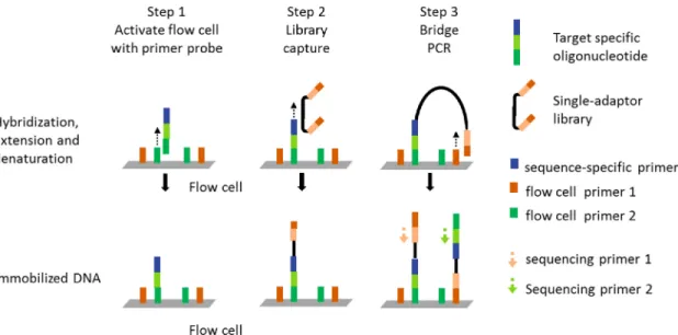
Recently, a new approach to targeted sequencing of single nucleotide variants (SNV) in cancer has been published that uses oligonucleotides libraries as a tool to re-engineer the Illumina GaxII flow cell [8].
10
11 The surface functionality of flow cell is changed using oligonucleotides that serve as probes to capture specific DNA library fragment and as sequencing primers [Figure 1-5]. The probe binds to one of the two immobilized short PCR primers on the flow cell and is extended by DNA polymerase to build a highly customized flow cell (Step 1). In step 2, the probes hybridize to their target fragments. The sheared DNA library is prepped such that either end is flanked by primer sequences to flow cell primer. The library is amplified on the flow cell by bridge PCR. This results in both strands being immobilized on the flow cell for bio-directional sequencing (Step 3).
1.1.5Fluorescent In Situ Hybridization (FISH)
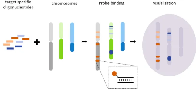
Fluorescent in situ hybridization (FISH) is a method to visualize and map an individual’s genetic material. The method uses fluorescent probes to tag chromosomal regions within cells to detect chromosomal rearrangements, translocations, deletions and copy number variations. It is used for clinical diagnosis of many diseases such as Down syndrome (trisomy 21) and Angelman syndrome (deletion of genes on maternal chromosome 15). This techniques has been used to detect localized mRNA in bacteria [41] and yeast [42]. Currently, dye-labeled oligonucleotide libraries are being investigated to increase the resolution and dynamic range of existing FISH technologies [12]. This application of FISH probes as Oligopaints in particular requires microgram amounts of diverse oligonucleotides for significant coverage of chromosomal regions [Figure 1-6].
12
13 1.2 Source of oligonucleotide libraries
1.2.1Conventional state-of-the art oligonucleotide synthesis
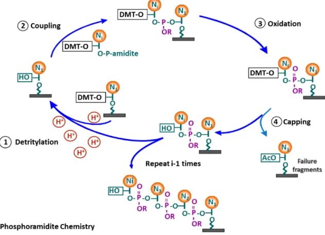
14 Oligonucleotides are synthesized using standard phosphoramidite chemistry starting from 4 tiny bottles of DNA monomers namely adenine, guanine, cytosine and thymidine [Figure 1-7]. Acid-cleavable phosphoramidite monomers are added in cyclical process to grow the nucleotide polymer chain from 3’-end to the 5’-end. In the first synthesis cycle, the desired monomer is linked to a solid support (e.g. controlled pore glass) pre-functionalized with a 3’ end tethered monomer. This is followed by step-wise addition of deoxynucleoside monomer in 4 step-process, which are i) deprotection (detritylation) – weak acid such as tricholoroacetic acid (TCA) is used to remove the dimethoxytrityl (DMT) group from the 5’ end of monomer, ii) coupling – 5’-hydroxyl end of monomer is covalently linked to the desired monomer in the presence of a activator agent (e.g. 1-H tetrazole), iii) capping – unreacted 5’-hydroxyl monomers are capped from further reactions by acetyl group to minimize deletion sequences and vi) oxidation – the two monomers on the oligonucleotide chain linked by unstable phosphite triester is converted to phosphate (e.g. iodine, pyridine solution). This process is repeated several times to grow DNA polymer, which is then cleaved from the surface using a base-catalyzed reaction (ammonium hydroxide), which also removes the remaining protecting groups [43].
This process is the principle method used by most commercial DNA synthesis companies such as Integrated DNA technologies. Individual oligonucleotides (up to 200 bases) are synthesized in large reactors (column-based synthesis) resulting in yields from 10 - 100 nmol (10-9) at low costs ($0.1 per base) [44].
1.2.2Microarray Technology
DNA microarrays are an abundant source of inexpensive complex DNA libraries. Currently, commercially available, in situ synthesized oligonucleotide
15 microarray come under two categories i) photo-labile 5’-O protecting group (Affymetrix, Nimblegen) and ii) acid-labile 5’-O protecting group (Agilent, CustomArray, LC Sciences, Mycroarray). The protection group serves the same function as DMT group used for column synthesis, i.e. prevents monomers to form homopolymers.
Photo-labile protecting group
Affymetrix is the dominant player in the field of microarrays. They leverage semiconductor-based photolithographic fabrication techniques and use photo-labile groups, which enables them to synthesize a large variety of defined probes (1 million) on a large surface with feature size ranging from 1 – 5 µm. Their arrays are made using photolithographic masks, which require up to 100 masks to make a 25-mer oligonucleotide. The high cost of making pre-fabricated masks significantly adds up when used to make custom oligonucleotide libraries required for the applications above [45, 46].
Nimblegen like Affymetix uses photo-labile protection groups but differs in two aspects, i) they use mask-less projection technology for deprotection – Digital Micromirror Device (DMD) and ii) they use a different protecting group. The DMD consists of electromechanically controlled mirrors that give them the flexibility to target any area on the chip. Also, their protecting group (5’-ortho-nitrophenyl-type [NPPOC]) enables them to achieve higher step-wise efficiency (~96%) than Affymetrix (ortho-nitro-benzyl-type [MeNPOC]; <94%) and they have been able to make approximately million probes of 80 bases length [47, 48]. As both methods are dependent on photolytic cleavage of protecting groups, and as extent of cleavage is dependent on the exposure time (order of 1-2 min to get 99% deprotection), they
16 are unable to make long oligonucleotide libraries (100 – 150 bases). For 100-mer oligonucleotide, 96% step-wise yield gives 0.96100 = ~1.69% full length sequences). Acid-labile protecting group
Agilent microarrays are based on inkjet printer technology that enables the synthesis of high density arrays (244k spots) with up to 150-mer long oligonucleotides [49]. An inkjet printer head controlled by motion controllers delivers the synthesis reagents to desired spots on the array. Like, column-based synthesis, they use standard phosphoramidite chemistry which allows them to achieve close to 99.5 % step-wise efficiency [50].
CustomArray uses electrochemical reactions for DNA synthesis on silicon substrate. The acid produced then catalyzes the removal of the protecting group to enable coupling with incoming monomer. Their arrays are fabricated using CMOS technology to produce a matrix of individually addressable microelectrodes. The application of current, leads to oxidation of electrolyte at the anode to generate acid that diffuses to the spot for oligonucleotide synthesis and in a defined region, which is then neutralized at adjacent cathodes. They can make up to 12k spots on the array with 50 base oligonucleotides [51].
LC sciences and Mycoarray have adapted the standard phosphoramidite chemistry to digital photolithography for synthesis of DNA microarrays. This is achieved by replacing the acid with a precursor molecule which generates acid upon UV illumination for synthesis at desired spots. Both technologies achieve > 99% step-wise yield and can synthesize oligonucleotides up to 120 – 150-mer [52, 53]. However, because the acid generated is in the liquid phase, microfluidic devices with individual microchambers are fabricated from silicon to prevent diffusion. This limits the density of features to ~3,698 for PicoArray chip marketed by LC Sciences.
17 On the other hand Mycoarray have further optimized the technology to perform synthesis on open planar substrates. This has enabled them to achieve 20 k spot density.
18 1.3 Current limitations
The common theme to all the above applications is the requirement of complex oligonucleotide libraries. Commercial DNA synthesizers make individual oligonucleotides in large reactors (column-based synthesis) and have been scaled up to produce 96 or 384 oligonucleotides in a single batch. They give really high yields (nanomoles) of individual oligonucleotides at relatively low cost ($0.1 per base). Nevertheless, they are still too expensive to make libraries for applications such routine assembly of denovo genetic circuits or organisms. For instance, Craig Venter Institute used this option to synthesize bacterial genomes of M. genitalium (580 kb) and M. mycoides (1.08 Mb) at costs of several hundred thousand dollars (1.08Mb x $0.1/base * 2 strands = $216,000) [4, 5].
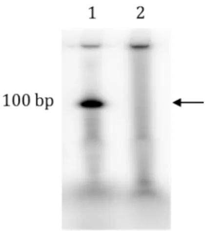
Figure 1-8 Microarray synthesized oligonucleotide library 1. Control oligonucleotide : 100 bp is run as marker (1 pmol)
19 Diverse oligonucleotide synthesis prices can be reduced by using high throughput microarray technology. They produce hundreds of thousands of oligonucleotide however their Achilles heel is the limited spot size where the synthesis occurs, which is usually well below 100 microns in diameters [2, 48, 51]. This results in very small synthesis scale. The overall yield of individual oligonucleotides per chip is in the range of 10 - 15 fmol, which is 106 times less than column-synthesis. This amount of oligonucleotides is further reduced as chemical reactions are not 100 % efficient; there are errors most often deletions. This leads to the formation of truncated DNA molecules as depicted in Figure 1-8. Thus, a 100 base oligonucleotide synthesized using state-of-art column synthesis (>99.5% step-wise efficiency) gives about 0.995100 = 60.5 percent full length oligonucleotides. For array based synthesis the yield is about 36.6 percent (99% step-wise efficiency) resulting in about ~3.6 – 5.5 fmol of individual oligonucleotide. This dictates a molecular amplification step; polymerase chain reaction (PCR) being the optimal choice to high amplification (>106-fold) amount. Conventional PCR has two major limitations related to amplification of multiple templates:
• Preferential amplification of some templates • Formation of chimeric products.
• Truncated products within the oligonucleotide library can act as primers to make non-specific products.
Also, PCR leads to formation of double-stranded DNA (dsDNA) and incorporates primer binding sequences (PBS). The addition of PBS increases the length of oligonucleotide synthesized, raising the percentage of undesired
20 sequences. After PCR amplification, the PBS is removed by type II restriction enzymes. For applications like MIPs or gene-assembly, the enzyme used must cut outside their recognition site [36]. The former i.e. formation of dsDNA requires additional processing steps to make them single-stranded DNA (ssDNA). Most methods to prepare single-stranded DNA (ssDNA) (e.g. exonuclease digestion) protect the desired strand from exonuclease digestion. For these methods:
• The amount of single-strand DNA produced depends on the starting amount of PCR products
Finally, the single-strand DNA reaction products consists of 5 different DNA species; desired ssDNA, dsDNA that is not digested by the restriction enzyme and that is not converted to single-stranded form, as well as any leftover PCR primers and cut primer binding DNA fragments. High quality ssDNA is obtained by purification on polyacrylamide gels.
1.4 Motivation
Many current (targeted sequencing, synthetic biology) and upcoming applications (FISH, RNA interference and bioactive peptides) use oligonucleotide libraries pools as a starting point. These applications require anywhere from 50 ng (1.6 pmol) – 1 µg (30 pmol) of single-strand nucleic acid (size 100bp) per reaction [39]. Technologies like targeted-sequencing, RNAi and fluorescent in situ hybridization (FISH) are being developed for clinical applications. Just during the development stages they require 10 - 100 reactions (even more for pilot studies) for robust data analysis. The current method to amplify microarray derived libraries i.e. conventional PCR falls short as it amplifies non-specific products. Also,
21 exonucleolytic hydrolysis of unprotected strand combined with polyacrylamide purification to remove PCR artifacts and primer binding sequences leads to > 50 % loss of single-strand DNA. Typical yields obtained per reaction ranges from 5 – 15 pmol.
This dissertation aims to develop a methodology to make large quantities of diverse oligonucleotides by integrating solid-phase oligonucleotide synthesis technology with molecular biology methods. In the second part, it develops the method to use oligonucleotide libraries as primers for first strand cDNA synthesis. Finally, the oligonucleotide libraries will help accelerate progress in the fields of synthetic biology, protein engineering, biotechnology and clinical diagnostics and therapeutics.
1.5 Overview of the dissertation
Chapter 2 presents an overall process outline starting from getting diverse oligonucleotide pools to generation of single-stranded DNA (ssDNA) and describes in detail two key components, i) molecular amplification method and ii) methods to make ssDNA. First emulsion PCR amplification technique is adapted to increase the yield of chip-derived oligonucleotides. Here, we investigated parameters to get amplification with the objective of preserving sequence fidelity and library. Second, many molecular biology methods are tested to obtain large amount of single-stranded DNA. Two of these methods are compared on the basis on yield and depending on the method different strategies are used to remove the PCR primer sequences.
Chapter 3 describes in a step-wise manner the problems encountered when using a library of sequence-specific primers to make complementary DNA (cDNA)
22 from RNA templates. In this case many enzymatic and non-enzymatic methods are tested in varying combinations to effectively reduce any false positives. The success of the methods is determined by measuring the level of genes induced in the yeast galactose versus glucose growth media. Gene-expression is studied initially using microarrays and later by polymerase chain reaction.
23 CHAPTER 2
FROM DNA MICROARRAYS TO SEQUENCE-SPECIFIC SINGLE-STRANDED NUCLEIC ACIDS
2.1 Introduction
DNA microarrays combine the benefits of solid phase synthesis and combinatorial chemistry [54]. They have ten to hundreds of thousands of polynucleotides fabricated with high resolution at precise locations. This makes them making them an inexpensive source of diverse DNA libraries [55]. Traditionally, microarrays have been used for hybridization based studies to i) measure changes in gene expression levels [56], ii) identify genomic gains or loss (microarray comparative genomic hybridization - CGH) [57], and iii) identify DNA mutations (e.g. single nucleotide polymorphisms – SNP) [58, 59]. Applications of microarrays have been described for almost every field of biological science.
Recently, there have been reports that use microarray synthesized DNA (oligonucleotide) libraries to assemble short DNA fragments into synthetic genes [2, 60-62]. Complex oligonucleotide libraries have been used in a variety of ‘front-end’ capture methods to selectively enrich and amplify regions of the genome for targeted sequencing [36, 37, 63, 64] as well as generation of mutant DNA libraries for peptide evolution [10]. For all these applications the bound oligonucleotides has to be released from the surface. This is done by either breaking the covalent bond between the first nucleoside and array surface or an on-chip second strand DNA
24 synthesis followed by DNA melting to release single-strand DNA. The success of latter method (i.e. on-chip PCR) depends on many factors such as steric hindrance, surface passivity and evaporation and is reviewed here [65].
Using the former method, the oligonucleotides are released from the surface in one big pool. Two major drawbacks limit their direct use namely, i) low yield of synthesized oligonucleotides released per chip and ii) less than 100% step-wise monomer coupling efficiency. A step-wise efficiency of 99%, will give only 0.9925 = 77.7 % full length DNA compared to 36.6 % for a 100-mer. Also at each synthesis reaction cycle there may be a shift in the acid diffusion boundary may by ± a few nanometers. This leads to formation of undesired oligonucleotide products whose size and sequence are highly variable.
The methods to select and purify correct size oligonucleotides such as polyacrylamide gel (PAGE) purification and high performance liquid chromatography (HPLC) gives less than 50% recovery yields further reducing the amount of oligonucleotides. To ensure enough probes are available for downstream applications, the template library is amplified by PCR, which necessitates extra steps to get single-stranded DNA (ssDNA). Here, both the amplification and ssDNA formation method is important to get high quality nucleic acid probes.
This chapter details the issues encountered and process to make large quantities of oligonucleotide libraries starting from microarray-derived DNA pool
2.1.1 Process outline
An overall outline of the process to get functional ssDNA from oligonucleotide probes is shown in Figure 2-1. Two methods integral to the overall process, i) Emulsion PCR and ii) conversion of double-strand DNA (dsDNA) to single-strand DNA (ssDNA) are described in more detail in separate sections.
25
Figure 2-1 Process overview
(1) ... DNA microarray synthesis of custom oligonucleotide library.
(2) ... Overnight ammonium hydroxide release of surface bound oligonucleotides. (3) ... Emulsion PCR – amplification step
(4) ... Removal of primer binding sequence
26 Purification
The cleaved DNA library is purified from short DNA fragments (<10 nt), linkers and other contaminants that inhibit the yield of polymerase chain reaction. In addition they give inaccurate spectrophotometric quantification of full length oligonucleotides. Some methods to purify oligonucleotides are i) size exclusion chromatography, ii) silica-membrane spin-column purification, iii) magnetic bead selection, and iv) gel-size selection. Size exclusion chromatography is based on gel matrix capture of low molecular weight contaminants (salts, NTP’s and other LMW compounds) while large molecules are exchanged with the buffer of choice and eluted. Purification with size exclusion columns does not completely remove the unwanted molecules. In contrast, there is loss of full length oligonucleotides when purified with commercially available silica-membrane based spin columns. This is because of their high cutoff threshold of 200 bp for ssDNA (cut off threshold for single-strand DNA = 2 * cut off threshold for double strand DNA). Magnetic selection for isolation of single-strand DNA can be done either by binding biotinylated probe to streptavidin coated beads [66] or using beads with probes complementary to a common sequence [67]. The former method requires an additional on-chip coupling of biotin phosphoramidite to 5’ prime terminus of oligonucleotide which labels probes irrespective of their size and consequently truncated and full length probes are purified upon magnetic bead selection. The latter method requires custom manufacturing of magnetic beads with common sequence that anneals to 3’ prime end of DNA probe to achieve good purification yields [68]. As probes are synthesized from 3’-5’ prime end, majority of truncated probes have the common binding sequence making this method infeasible. Thus, the only method that to some degree removes unwanted molecules prior to amplification is size exclusion spin-columns.
27 Amplification
The oligonucleotides have common primer binding sequence (PBS) at either ends for one-tube PCR amplification of all templates. To avoid loss of specific oligonucleotides, or introduce bias in template to product ratio as well as prevent any recombination events, the templates are amplified individually by compartmentalization in tiny droplet reactors formed upon emulsification of PCR mix.
Restriction digest
The primer binding sequences (PBS) flanking the user-sequence, especially the one at the 3’ end of the user-sequence is removed with restriction enzymes that cut outside their recognition. This make the gene-specific sequence available for polymerase mediated extension and creates 5’ phosphoryl terminus on the opposing strand.
Single-strand generation
After the primer removal step, one strand of dsDNA has 5’-phosphoryl terminus, while the complementary strand is not phosphorylated. Only the strand with 5’-phosphoryl group is hydrolyzed with lambda exonuclease followed by purification of products on native acrylamide gel to get high quality single-stranded DNA.
2.1.2Emulsions background General considerations
Emulsion is a mixture of two or more completely or partially immiscible liquids (e.g. oil and water) where one liquid (dispersed phase) is distributed in the
28 form of droplets (e.g. oil droplets) in the other (continuous phase; e.g. water). As oil and water do not mix spontaneously, the formation of dispersion phase requires the input of external energy, such as mechanical energy (e.g. stirring) or sound energy (i.e. ultrasound). Here, continuous energy input is required to stabilize the large surface energy (interfacial tension) associated with an increase in surface area/ volume ratio. Upon cessation of external energy, the oil droplets coalesce, forming a single blob of droplet that floats to the surface (generally, oil is less dense than water). Thus emulsions are thermodynamically unstable [69, 70].
Emulsifying agents such as surfactants (surface active agents) are added to stabilize emulsions. Surfactants are amphiphilic molecules i.e. their molecular structure has both a hydrophilic and a hydrophobic part that form a protective coating (steric or electrical barrier) between the phases. Surfactants reduce surface tension, provide repulsive interaction potential between droplet interfaces and inhibit coalescence of emulsion system [71]. Surfactants are characterized by an empirical value known as Hydrophile – Lipophile Balance (HLB) on the basis of their solubility in water or oil (higher the HLB value - more water-soluble). Also, known as Bancroft’s rule, it qualitatively determines the type and stability of emulsions [72]. On a scale of 1 to 20, emulsifiers with HLB value < 10, are good to stabilize w/o emulsion i.e. water droplets dispersed in oil phase and those with HLB value >10 stabilize o/w emulsion in which oil is dispersed in the water phase. The principle of w/o and o/w emulsion is shown in Figure 2-2 [73, 74].
29
30 Stability and breakdown mechanisms
The stability of an emulsion is measured by the rate of droplet coalescence. Factors that contribute to emulsion stability are i) Surfactant characteristics – they must diffuse rapidly to the new created interface, be soluble in both the internal and external phases and form relatively elastic films at the interface. Blends of two more surfactants (high and low HLB) make more robust emulsions (Gibbs-Marangoni effect) and improve stability against temperature changes. ii) Nature of electrical or steric interfacial barrier – interplay of repulsive effects (electrostatic, elastic, entropic, or mixing energy) with van der Waals force of attraction. iii) Viscosity of continuous phase – increase in viscosity reduces the diffusion coefficient (Einstein-Stokes Equation), thereby reducing the frequency of collisions and lowers coalescence. iv) Droplet size and distribution – monodisperse droplets form more stable dispersion against aggregation [75], v) Phase volume ratio – increase in volume of dispersed phase, decrease stability of emulsion. At high temperatures, o/w emulsions invert to form w/o emulsions. vi) Electrolyte concentration – w/o emulsions with >74% dispersed phase volume become more stable on addition of electrolytes. Here, addition of electrolytes helps to lower the refractive index difference between two phases [76]. vii) Density difference between phases and viii) Temperature – temperature effects factors such as strength of interfacial film, interfacial tension, solubility of surfactant, kinetic energy of droplets, and viscosity of liquid.
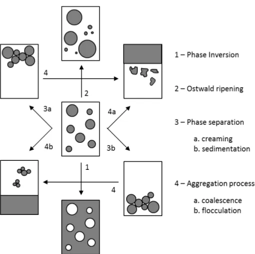
Emulsions are prone to various breakdown processes that may occur simultaneously as shown in Figure 2-3 [77, 78].
31
32 Phase Inversion – There is a sudden reversal of phases whereby an o/w emulsion becomes w/o type or vice versa. It is due to either increase in phase volume ratio above a critical value or change in solubility of surfactant towards the two phases (Bankcroft’s rule). Factors that affect phase inversion are temperature, polarity of the oil phase, kind of electrolyte and its concentration, other additives (organic, water-soluble solvents increasing oil solubility in water), and oil volume fraction.
Ostwald Ripening – Is a direct consequence of the polydispersity in the droplet size distribution. It is characterized by time dependent increase is droplet size. The difference in solubility (chemical potential) between droplets with different diameters (solubility increases with decreasing droplet size – effect of surface area/volume ratio), leads to the diffusion of molecules from small droplets to large ones. For w/o emulsions, the addition of electrolytes to dispersed phase decreases ripening rate [79]. Other approaches to reduce Ostwald ripening are i) increase surfactant concentration (below critical micelles concentration, it hinders rate of new molecule incorporation), ii) add components that reduce rate of diffusion of molecules within dispersed phase. Finally, Ostwald ripening does not take place in monodisperse solutions.
Phase separation – Gravitational forces may cause large droplet (diameter > 1 µm) to settle preferentially to the top (creaming – o/w emulsion) or bottom (sedimentation – w/o emulsion) of the container. This is observed due to density difference between the phases. Shaking the container reverses the process back to its initial state. Increase in viscosity of continuous phase or lowering density difference between phases reduces probability of phase separation.
Aggregation processes – During flocculation individual dispersed droplets cluster together without losing their identity, while coalescence is the process
33 whereby two or more droplets fuse together to form a larger droplet. Fusion occurs as a consequence of the rupturing of the thin liquid that separates droplets at flocculation stage under the action of attractive forces or due to thermodynamic instabilities. Flocculation is the initial step towards phase separation. It can be prevented by using polymeric surfactants which provide good steric stabilization. One method to enhance emulsion stability (i.e. prevent flocculation and coalescence) is addition of small amount of ionic surfactant to high concentrations of nonionic surfactant.
2.1.3W/O emulsions in biology Emulsion Polymerase Chain Reaction
During replication, cells produce multiple copies of genes (DNA), transcripts (RNA) and encoded peptides and proteins. The content of an individual cell is separated from others by a phospholipid barrier (cell wall), which helps to link genotype and phenotype. Two in vitro methods that mimic this replicating function of cells have formed the basis of a major number of significant advances in the field of biotechnology. The first technique employs bacterial cells (yeast, mammalian cells) to clone and express multiple genes whereby populations arising from single molecules are spatially separated in colonies for easy identification and differentiation[80]. The second technique uses a cell free method known as polymerase chain reaction (PCR) to multiply molecules [81, 82].
During PCR, some templates are preferentially amplified over others attributed to factors such as primer binding energy, amplicon G+C content, amplicon length, secondary structure of gene and its accessibility within genome, gene copy number and amplification efficiency[83-87]. The co-amplification of genes or organisms (viruses, bacteria) with high sequence similarity or repetitive sequence
34 stretches leads to formation of chimeric DNA molecules [88, 89]. Also, DNA artifacts are generated during PCR by cross-hybridization of heterologous sequences [90, 91]. Techniques to lessen PCR bias include optimizing template concentration with number of PCR cycles [66, 84], mixing multiple replicate reactions [84, 86], optimize annealing temperature and elongation times [85, 92], and increasing temperature ramp rates during PCR cycling[93].
Other techniques involve modifying the basic PCR protocol. For example, touchdown PCR (gradual decrease of annealing temperature during cycles) and hot-start PCR (addition of polymerase at denaturation temperature) increase the specificity of DNA amplification enabling detection of low template numbers. The former prevents mispriming during gene-amplification, while the latter prevents pre-PCR mispriming and primer-dimer formation. A two-step PCR (Nested PCR) where amplification first proceeds with external primer set, followed by an internal primer set that binds within the target has been shown to improve the specificity (closely related sequences) and sensitivity (very small amount of starting material; rare DNA) over single-step conventional PCR. Finally, commonly used TAQ DNA (T. aquatics) polymerase introduces base incorporation errors during replication process. Although, these random inserted incorrect bases are not detected during total amplicon DNA sequencing, they may prove problematic for subsequent functional analysis (for example, large scale production of proteins, development of nucleic or peptide assays) and therefore require additional selection steps. The discovery of thermostable polymerases with 3’ – 5’ exonuclease activity has made the fidelity of in vitro DNA replication comparable to cell-based cloning methods [94].
35
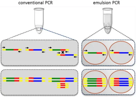
Figure 2-4 Emulsion PCR versus Conventional PCR
conventional PCR: Recombination of homologous regions leads to formation of chimeric products.
emulsion PCR: Isolated amplification in an emulsion results in equal representation of each fragment.
36 The use of w/o emulsions to carry out polymerase chain reaction (Emulsion PCR), whereby individual templates are segregated and amplified in millions of aqueous droplet micro-sized reactors eliminates biases associated with multi-template PCR [Figure 2-4] [95]. The knowledge of average droplet size and aqueous phase volume enable scientists to tune the template concentration such that there is only 1 template per reactor droplet. This ensures that the abundance and diversity of different templates is unaltered. The detection of single molecules requires optimization of reaction components, cycling conditions and rigorous primer design. Additionally, isolation of individual templates increases probability of primer binding to template and prevents dominance of non-specific product amplification within the reaction. Emulsion reactions have been described for detecting low copy number DNA and RNA molecules [96, 97].
Other applications
Directed evolution - Is the process of evolving proteins within the laboratory to generate protein with novel functionalities. The complexity of protein structure makes it impossible to predict the effect of change in structure on function. Many mutants of wild-type nucleotide sequence have to be analyzed to generate a protein molecule with desired traits. In vitro compartmentalization of genes and their products in emulsion droplets have been described for the evolution of many enzymes [98-103]. High throughput screens are possible as >1010 reactors are formed within 1 ml of emulsion with reaction volumes in the order of femtoliters (10-15 L) [104]. The resultant increase in effective concentration of reactants and reduction in diffusional distance between them may cause increase in rates of reaction [105].
37 BEAMing (beads, emulsions, amplification and magnetics) - Is a process of solid-phase immobilization of amplicons during emulsion PCR [106]. Besides preserving clonal information upon emulsion breaking, it lends to ease in downstream manipulation of desired products, for example fluorescence activated cell-sorting [107]. Methods to immobilize amplicons to solid support either bind the amplicon directly to the bead surface via streptavidin-biotin interaction or the amplicons anneal to a covalently surface bound complementary DNA fragment. Currently, BEAMing methods are used to prepare samples for next generation sequencing with 454 life science [108] and ABI SOLiD platforms [109]. The number of templates within droplets is modeled as a Poisson distribution translating to the possibility that a large number of emulsion droplets have beads but no templates [96]. To enrich for amplicons carrying beads and increase the number of sequencing reads, modifications have been introduced during sample preparation steps [110].
2.1.4Methods to prepare single-strand DNA from double-stranded DNA
The preparation of single stranded DNA is an important component of many procedures in molecular biology such as strand-specific microarray hybridization [111], next-generation sequencing [112], solution-based target enrichment [113, 114] and Systematic Evolution of Ligands by EXponential Enrichment (SELEX) [115].
Existing methods to convert library of double-strand DNA (dsDNA), specifically PCR amplicons to get single-strand DNA (ssDNA) can be divided into two categories, i) enzyme based or ii) affinity capture based. Enzymatic methods either selectively degrade or preferentially amplify one strand over the other. Exonuclease III and T7 gene 6 exonuclease digest both strands of dsDNA at equal rates to form
38 two shorter DNA fragments of half the dsDNA template length [116, 117]. Both methods require modification of the dsDNA to prepare full length ssDNA.
39
40 T7 exonuclease attacks double-strand DNA substrates from 5’ end. The use of phosphorothioate bonds in the DNA backbone of the first few bases at 5’ end of DNA strand makes it resistant to exonucleolytic hydrolysis. PCR primers with the above modification have been used to prepare single-strand DNA for PCR based Elisa-type single nucleotide polymorphisms (SNP) detection assays [118], and Sanger DNA sequencing [Figure 2-5.a].
Exonuclease III digests duplex DNA from 3’ end. The incorporation of 2-phosphorothioate bonds at 3’ end of DNA strand makes them resistant to exonucleolytic hydrolysis. As Exonuclease III is inactive on ssDNA, duplex DNA digested with restriction enzymes that generate four-base or longer 3'-protrusions to generate single-strand DNA have been successfully used for sequencing [Figure 2-5.b] [119].
Lambda exonuclease is a highly processive enzyme that preferentially digests duplex DNA strands with 5’ terminal phosphate [Figure 2-5.c] [120]. The enzyme is well characterized and has been shown to have greatly reduced activity for 5’ terminal hydroxyl strand of native and denatured DNA Article [121, 122]. Lambda exonuclease has been used to prepare single-stranded DNA for wide variety of applications. For example, probes for in situ hybridization [123], capture probes for next-generation DNA sequencing [124], dye-labeled targets that improve microarray hybridization signal-to-noise ratios [125], and DNA-based sensors [126].
Asymmetric PCR (AsymPCR; thermostable DNA polymerase) is used for preferential amplification of one strand over the other by using unequal primer concentrations to get single-strand DNA targets [Figure 2-6.a]. The technique requires rigorous primer design dependent on thermodynamic properties of both the product and primers and is limited by the size of PCR product [127]. Although, difficult to optimize and requiring 50+ cycles to produce high single-strand to
41 double-strand DNA ratio, variations of asymmetric PCR have been the used in diverse applications such as profiling of yeast knockout strain collection [128], in multiplexed detection of drug resistant genes [129], and single cell genetic diagnostic [130].
42 Magnetic isolation - An alternative non-enzyme method to get single-stranded targets from double-strand DNA is strand separation using biotinylated primers and streptavidin-coated magnetic beads [Figure 2-6.b]. Depending on downstream application, they can be captured either directly onto the bead surface Article [131, 132] indirectly captured via hybridization on pre-immobilized probes [133]. Next-generation-sequencing technologies use both capture techniques during library preparation and sequencing steps, for example enrichment of starting sample (poly (A) tail RNA, known DNA or RNA sub-populations), capture of biotinylated DNA for emulsion PCR, and selection of beads that have emulsion PCR products [134].
In vitro transcription and reverse transcription - Reverse transcriptase in
the presence of a suitable primer synthesizes a complementary DNA copy of an RNA template [135, 136]. This coupled with an upstream RNA amplification has the potential to generate large quantities of ssDNA probes [137].
43 2.2 Experimental methods and protocols
2.2.1Process outline
Processing chip-derived oligonucleotide libraries
The synthesized oligonucleotide library is released from microarray in large volume of alkaline solution (30% ammonium hydroxide). The volume is reduced to 30 – 40 μl by either evaporating the excess solution under vacuum or ethanol precipitation. The DNA library is purified on size-exclusion spin columns using recommended protocol (CS-20, Princeton Separations) and quantified by Nanodrop spectrophotometer (Thermo Scientific).
Standard curve – Real time PCR
115-mer gel-purified oligonucleotide is obtained from Integrated DNA Technologies. 10 x serial dilutions of the oligonucleotide ranging from 100 femtomole (~60 billion molecules) to 100 yoctomole (~6 molecules) are amplified by real time PCR (Chromo4-PTC 200 thermocycler, MJ Research). The PCR master (devoid template) consists of 0.5 μM forward and reverse primers, 2.5 mM MgCl2, 1 X GoTaq-flexi buffer, 0.2mM dNTP, 2 μM SYTO13 dye and 2.5 U GoTaq-flexi polymerase (Promega Inc). The final reaction volume at each template concentration is 50 μl. The amplification program is comprised of 35 cycles with the following steps: initial denaturation at 94°C for 2 min, 35 cycles of denaturation at 94°C for 15 s; primer annealing at Tm-5°C for 20 s; primer extension at 72°C for 20; and final elongation at 72°C for 5 min. The signal intensity of SYTO13 dyes is measured immediately after the annealing and extension temperature [138] . A melt curve is generated by slowly increasing the temperature in steps of 0.5°C from 60°C
44 to 95°C. The tubes are held for 5 s at every temperature increment, followed by measurement of dye intensity. The data is analyzed using Opticon software available with the thermocycler.
Emulsion PCR
80 μl of ABIL EM90 (4%) and 1 μl of Triton X-100 (0.5%) surfactants are added to 2 ml of light mineral oil and rigorously mixed for 1 min and out on ice. The PCR (aqueous) mix contains 1 – 5 μl of template, 0.5 µM each of forward and reverse primer, 1 X GC buffer (proprietary solution), 1 mM MgCl2, 0.5 µg/µl BSA and 2 U/50 µl hot-start high-fidelity phusion DNA polymerase.
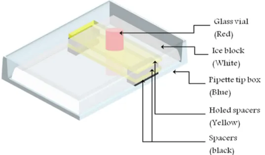
45 The oil phase (4% ABIL EM90; 0.05% Triton X-100) is transferred to 2 ml glass vial containing magnetic stirrer [Figure 2-7]. The emulsion is prepared by drop-wise addition of PCR solution to oil phase (stirred at 1000 rpm), followed by additional stirring for 15 minutes. The ratio of aqueous: oil phase is maintained at either 1:4 or 1:6 and the final volume of emulsion is kept between 200 – 600 µl. The emulsified product is transferred to 0.2 ml PCR tubes or PCR plate in aliquots of 50 µl. PCR is performed in an Eppendorf thermocycler. The amplification program is comprised of 35 cycles with the following steps: initial denaturation at 98°C for 1 min, 30 cycles of denaturation at 98°C for 15 s; primer annealing at Tm-5°C for 20 s; primer extension at 72°C for 20 s; and final elongation at 72°C for 5 min. After PCR amplification, the products equivalent to 100 μl of aqueous phase volume (i.e. 400 – 600 μl emulsion volume) are pooled together in 1.6 ml centrifuge tube containing 5 µl of loading buffer (indicator – orange G/xylene cyanol, to help identify water droplet in organic solvents), followed by addition of mineral oil to 1.4 ml final volume. The contents of tubes are mixed by vortex for 1 min. The emulsion is separated into oil and aqueous-surfactant phase by centrifugation at 13,000 g for 10 min. The upper (oil) phase is removed and the aqueous-surfactant phase is broken by successive washes with 1 ml water saturated solutions of diethyl ether and ethyl acetate. In each case, the contents of the tubes are mixed by vortex (10 s) and collected by centrifugation (5 s), followed by removal of upper solvent phase. The bottom phase contains the PCR products. After the final diethyl ether wash step the tubes are placed on 37°C heat block to evaporate trace amounts of leftover diethyl ether.
46 Restriction digest
1 μg of PCR amplicons are digested with 20 units of Mbo II restriction enzyme (New England Biolabs Inc.) in 50 μl reaction for minimum 2 h at 37°C. The buffer formulation is 50 mM NaCl, 10 mM Tris-HCl, 10 mM MgCl2, 1 mM Dithiothreitol pH 7.9 @ 25°C (1 X NEB buffer 2). For DNA size >100 bp, the enzyme is heat inactivated at 65°C for 20 min. To avoid possible strand separation and incorrect strand re-annealing, the enzyme is not heat activated for duplex DNA < 100 bp. dsDNA is purified with MinElute PCR purification kit (Qiagen Inc.) following manufacturer’s protocol with following modifications, i) nucleic binding step is performed at 6,000 g for 1 minute, ii) a second elution step is performed with 10 μl of EB buffer and centrifuged for 3 minutes. The concentration of DNA (2 μl) is measured with Nanodrop spectrophotometer and saved for gel analysis.
Preparation of single-strand DNA
The 5’-phosphoryl strand of restriction digest product is hydrolyzed in 1 X lambda exonuclease buffer (67 mM Glycine-KOH, 2.5 mM MgCl2, 50 µg/ml BSA, pH 9.4 @ 25°C) at 37°C for 30 minutes (unless stated). 1 unit of enzyme is used for every 5 pm of double strand DNA. The concentration of dsDNA in final reaction volume is kept between 0.5 pm/µ to 2 pm/µl. Finally, the enzyme is inactivated by heating at 75°C for 15 min and the products are run on acrylamide gel for purification.
2.2.2Yeast Knock-out (YKO) deletion collection
Frozen (-80°C) samples of pooled mat-alpha mating YKO deletion strains is thawed on ice and diluted in 2% glucose rich media. The cells are spread on rich media supplemented with 200 µg/ml geneticin (G-418; Gibco) and grown at 28°C (2
47 days). They are recovered in ringer’s solution (0.4% NaCl) and cells are counted using hematocytometer.
Yeast genomic DNA (20kb – 40kb) is extracted from 10 OD600 of cells (1 OD600 = 2.7E+07 cells/ml)using ‘smash and grab’ method [139]. Acid washed glass beads (400 – 500 μm) and phenol chloroform solution (pH 8.0) are used to break open the yeast cell wall under rigorous mixing followed by multiple steps of phenol/chloroform extraction. The protocol is modified to include a protein digestion (Proteinase K) and additional phenol/chloroform and chloroform extraction steps after RNase A treatment to yield high quality genomic DNA (OD260/OD280 > 1.8).
Touchdown PCR
PCR amplicons corresponding to the either UPTAG or DNTAG modules are amplified using universal primer sequences. The PCR reaction mix (50 μl) contains 200 ng yeast genomic DNA, 0.5 μM forward and reverse of UPTAG or DNTAG primers, 2.5 mM MgCl2, 1 X GoTAQ flexi buffer, 0.2mM dNTP and 2.5 U GoTAQ polymerase (Promega Inc). The cycling parameters are as follows: initial denaturation at 94°C for 2 min; 4 cycles of dsDNA denaturation at 94°C for 20 s, primer annealing at initially Tm+6°C for 30 s with 2°C decrease per cycle; primer extension at 72°C for 30 s; 30 cycles of denaturation, primer annealing and extension at constant annealing temperature Tm-4°C; and final elongation at 72°C for 5 min.
Restriction digest of yeast genomic DNA
1 µg of genomic DNA is separately digested with 20 U DraI (recognition site: 5’-TTT/AAA-3’) and 20 U NdeI (recognition site: 5’-CA/TATG-3’) for 37°C for 60
48 minutes. The buffer components are 50 mM potassium acetate, 20 mM Tris-acetate, 10 mM magnesium acetate and 1 mM dithiothreitol (DTT) buffer (1 X NEB 4 buffer). In a third tube, 20 µg of genomic DNA is then digested overnight with 60 units each of DraI and NdeI in 1 X NEB buffer at 37°C. In all cases, the enzymes are inactivated after digestion by incubation at 65°C for 20 min.
2.2.3Enzyme based methods to prepare single-strand DNA from duplex DNA PCR amplification and restriction digest
300 base-pair (bp) amplicons with UPTAG module is amplified with modified U1 universal primer and 5’-hydroxyl KanBby touchdown PCR. In case of T7 gene 6, the first 5 bases at 5’ end of U1 primer are linked by phosphorothioate bonds. In case of lambda exonuclease, the U1 primer has a 5’-phosphoryl group. The amplicons are purified on QiaQuick PCR purification columns and eluted with 50 µl of Tris-HCl pH 8.0. 1 µg of purified amplicons are digested with 20 units of Bgl II restriction enzyme (20 µl, 1x NEB buffer 4) for 30 min at 37°C, followed by heat inactivation of the enzyme for 20 min at 65°C.
T7 gene 6 exonuclease and 5’-phosphorothioate backbone strand
0.5 µg of amplicon (phosphorothioate backbone) is digested with 50 U of T7 gene 6 exonuclease (Affymetrix Inc.) for 45 min at 37°C in 1x T7 gene 6 exonuclease reaction buffer (20 μl). The enzyme is inactivated by heating at 75°C for 10 min. Lambda exonuclease and 5’-phosphoryl terminus strand
0.5 µg of PCR amplicons with 5’ phosphorylated ends are digested with 10U of lambda exonuclease (New England Biolabs) for 60 min at 37°C in 1x lambda exonuclease buffer (20 μl). The enzyme is inactivated by heating at 75°C for 10 min.
49 Exonuclease I hydrolysis of ssDNA
An aliquot of ssDNA product is hydrolyzed with 1 U/μl exonuclease I (20 μl; New England biolabs) for 60 min at 37°C to detect for presence of ssDNA. Equal concentrations of unpurified PCR, BglII digest, single-strand formation reaction and exonuclease digestion products are electrophoresed on native polyacrylamide gels.
2.2.4Preparation of single-strand DNA with 5’-modified end
300 bp UPTAG module region of YKO collection is amplified in six separate PCR reactions with forward U1 primer modified by 3 different 5’-terminus groups (Cy3, Cy5 and hydroxyl) and reverse KanB primer with 5’-hydroxyl or 5’-phosphoryl group. 5 pmol of PCR product is digested with lambda exonuclease (1 U enzyme / 5 pm DNA) at 37°C for 45 min (10 µl). The enzyme is heat inactivated at 75°C for 15 minutes. 2.5 pmol of PCR and lambda exonuclease products are digested with exonuclease I for 30 min (5 μl). Equal concentrations of the 4 products are run on 6% native acrylamide gel.
2.2.5Optimize digestion time of Lambda exonuclease
146 bp region of the YKO genomic DNA is amplified using 5’-phosphoryl forward primer and 5’-hydroxyl reverse primer by PCR. 400 ng (~ 4 pm) of PCR amplicons are digested with lambda exonuclease (1 U enzyme / 5 pmol dsDNA) in 5 µl reaction at 37°C at varying time points ( 1, 2, 5, 10, 15, 30 and 60 min) in separate 0.2 ml PCR tubes. Lambda exonuclease samples at 15, 30 and 60 minutes as well as PCR sample are hydrolyzed with exonuclease I enzyme at 37°C for 30 min (10 µl). The products are run on 8% native acrylamide gel.
50 2.2.6In vitro transcription and reverse transcription
4 pmol of emulsion PCR products is transcribed in 40 μl reaction with AmpliScribe™ T7-Flash™ Transcription Kit (Epicentre Biotechnologies Inc.) at 42°C for 4 h. 1 µl of TURBO DNase is added to the reaction and incubated at 37°C for 15 min, followed by inactivation of DNase with 2 µl of inactivation reagent following TURBO DNA-free kit (Ambion Inc.). 36 µl of the DNase inactivated solution is suspended in 144 µl nuclease free water and 20 µl 3 M sodium acetate (pH 5.2) and purified on two RNeasy columns (Qiagen Inc.). The RNA is eluted with 50 µl of nuclease free (total 150 µl). The concentration is measured with Nandrop spectrophotometer. RNA is reverse transcribed with superscript II (Invitrogen Inc.) following manufacture’s protocol with a few modifications; i) 100 pmol of RNA is used per 20 µl reaction, ii) use of 1.5 x sequence-specific primer (150 pmol), iii) the hybridization reaction is heated at 65°C for 5 min and slowly cooled (-0.1°C/s) to 37°C, iv) the reaction after addition of enzyme is incubated at 37°C for 3h and v) the reaction is scaled to process 1200 pmol (240 µl) of RNA. RNA is hydrolyzed with 0.43 volume (103.2 µl) of 1 M NaOH at 65°C for 15 min, followed by inactivation with equal volume (103.2 µl) of 1 M HCl. The cDNA is ethanol precipitated with 0.3 M sodium acetate (pH 5.2, 50 µl). Post precipitation the DNA pellet is suspended in 75 µl nuclease free water and purified with three centri-spin20 column (25 µl each).
2.2.7SYBR gold gel staining
Polyacrylamide gels are stained using SYBR gold dye [80]. The top plate is removed with the gel on the bottom plate. Excess gel not containing any nucleic acid products is cut out, followed by washing the gel with deionized water. 5 µl of 10,000x SYBR gold (Invitrogen Inc.) is suspended in 10 ml of tris-HCl pH 8.0 (10x) and poured over the gel. The nucleic acids are stained in the dark for at least 20 min.