Vol. 43, No. 1, 2005 pp. 3-10
Rafts - the current picture
Michał Grzybek
1, Agnieszka Kozubek
2, Patrycja Dubielecka
1and Aleksander F. Sikorski
1,2 1Institute of Biochemistry and Molecular Biology, University of Wrocław and 2
Academic Centre for Biotechnology of Lipid Aggregates, Wrocław, Poland
Abstract: Although evidences that cell membrane contains microdomains are accumulating, the exact properties, diversity
and levels of organization of small lipid patches built mainly of cholesterol and sphingomyelin, termed rafts, remain to be elucidated. Our understanding of the cell membrane is increasing with each new raft feature discovered. Nowadays rafts are suggested to act as sites of cell signaling events, to be a part of protein sorting machinery but also they are used by several pathogens as gates into the cells. It is still unclear how rafts are connected to the membrane skeleton and cytoskeleton and with how many different types of rafts are we actually dealing with. This review summarizes some of the most recent discoveries trying to make a view of the complex raft properties.
Key words: Rafts - Detergent-resistant membranes - Membrane skeleton - Immunological synapse
Introduction
Plasma membrane is no longer seen as a lipid sea with embedded protein islands. The discovery of mem-brane domains both, caveolae-containing and flat rafts, prompted new research efforts towards cell membrane biology. Huge resources are engaged into identification of the components and the role of lipid rafts in various cell types [23, 26, 32, 38, 57]. After several years of studies it is generally assumed that eukaryotic cell plasma membrane is subdivided into lipid raft and non-raft regions. However, the term "raft" is not unique. One can find terms as: DRMs -detergent resistant membranes [48, 62]; DIGs - deter-gent insoluble glycolipid rich complexes [46]; GEMs - glycolipid enriched membranes [42]; LDMs - low density membranes [24] etc. Each of these to some extent characterizes the features of lipid domains i.e. rafts are membrane parts that are resistant to cold detergent extraction (usually Triton X-100 but also other detergents are used - see below) [48, 55], which are enriched in cholesterol, glycosphingolipids and phospholipids with saturated acyl chains and are iso-lated by sucrose gradient centrifugation with the low density fraction. Apart from lipids that are
charac-teristic to rafts, there is also a specific subset of proteins that localize preferentially to the rafts. These are the GPI-anchored proteins [11] and proteins attached to the membrane via its other lipid components [4, 20].
An enormous number of sophisticated techniques such as single particle tracking [21], single dye trac-ing [49], fluorescence microscopy [27], FRET [59], ESR [56], have been involved in studies aiming at the explanation of the function of raft components but also at the observation of the behavior of rafts
per se. The picture that is emerging from all these
studies shows rafts as membrane platforms that are spatio-temporally organizing the signaling events in the cell [22, 29]. The lipid domains are, however, also sites of various pathogen entries into the cell and some viruses are known to preferentially bud from the raft regions [4, 34].
In this paper we try to draw a picture of lipid rafts, present some evidence for their role in the cell, and show some problems that urgently need to be solved before one can clearly demonstrate a complete view on the raft concept.
Correspondence: A.F. Sikorski, Institute of Biochemistry and
Molecular Biology, University of Wrocław, Przybyszewskiego 63/77, 51-148 Wrocław, Poland; e-mail: [email protected]
Abbreviations used in the text: GPI - glycosylphosphatidylinositol,
FRET - fluorescence resonance energy transfer, ESR - electron spin resonance, PC - phosphatidylcholine, DOPC - dioleoylphosphati-dylcholine, SM - sphingomyelin, Chol - cholesterol, ld - liquid
disordered state, lo - liquid ordered state, DRM - detergent resistant
membrane, IS - immunological synapse, TCR - T cell receptor, BCR - B cell receptor
Rafts - isolation artifact or natural necessity?
It is generally known that hydrated pure lipids may exist in different phases. The solid-like state (or gel phase) describes lipids that posses tightly packed lipid acyl chains, parallel to one another (all trans conformation), whereas the liquid disordered (ld) state characterizes acyl chains (in trans-gauche conformation) with high mo-bility, tumbling around the axis perpendicular to the membrane surface. Even in artificial membrane both phases can coexist [19]. Between those two there is a middle phase. The so called liquid ordered (lo) state describes lipids that are tightly packed but show rather high mobility [15]. Many experiments demonstrate that the lo state is crucial for raft existence [13, 47]. Certain lipids have the propensity to associate with one another, therefore abolishing the ld state in model membranes, including liposomes [31]. Sphingolipids, cholesterol and saturated glycerophospholipids can easily associate forming domains [47]. Sphingolipids contain mostly long saturated acyl chains, which allow them to pack tightly together - a property that makes their Tm (melting
temperature) higher compared to glycerophospholipids, which contain more unsaturated acyl chains. Sphingoli-pids would normally exist in a gel-phase, but the presence of cholesterol prevents them from entering the gel-phase, changing it instead into the lo state. The ld phase is usually abolished in favor to the lo phase if the content of cholesterol reaches ~30 mol% [15]. And so, vesicles composed of DOPC:SM:Chol undergo phase separations, exhibiting the coexistence of lo and ld phases at varying compositions [3]. It is then likely that the lo phases exist in a cell membrane with a sufficient content of cholesterol and sphingolipids [6]. The lo state is thought to form discrete microdomains (rafts) inter-spersed in the continuous ld phase.
Rafts are thought to be thicker than the rest of the membrane. This is due to the presence of sphingomyelin. The molecule contains long sphingosine moiety and a long saturated fatty acid chain. Therefore SM:Chol pat-ches are thought to be thicker than the surrounding lipid matrix that contains more unsaturated phospholipids. The X-ray diffraction shows that DRMs are ~0.9 nm (~30%) thicker than the rest of the bilayer [31].
Another relevant feature of SM:Chol bilayers is that they have a much larger area compressibility modulus than do unsaturated phosphatidylcholine bilayers [33]. Since the bilayer bending modulus is proportional to the compressibility modulus times the square of the bilayer thickness [12], the bending modulus of SM:Chol bilayer would also be larger than that of unsaturated PC mem-brane. Therefore, to accommodate proteins or peptides of a given hydrophobic length it should take more energy to separate or deform adjacent lipid molecules in raft bilayers compared to non-raft regions with lower com-pressibility and bending modulus. This implies that for
a given extent of hydrophobic mismatch, the lo SM:Chol
bilayer would provide a more energetically unfavorable enviroment for a protein than would a ld phospholipid
bilayer [31].
Some facts, however, put doubts about the raft theory - the presence of Triton X-100 can induce or even promote domain formation even in an initially homogen-ous, fluid PC:SM:Chol membrane and it is also possible to isolate raft domains from raft-free membranes [13]. One immediately asks a question whether rafts really exists in living cell membranes, or whether they are just an artifact created during extraction.
It has been suggested [47] that DRMs isolated from living cell membranes arise from native raft regions. Therefore many groups either use detergents other than Triton [48] or extract raft domains without detergents [10] in identifying the composition of natural rafts. These studies showed that DRMs vary depending on the way of extraction not only in the quantity of extracted proteins but also in the quality. Detergent is said to disrupt most of the lipid-lipid and lipid-protein interac-tions, solubilizing most of the membrane proteins. How-ever, the lo state is said to be sufficient to avoid detergent
extraction [39]. The GPI-anchored proteins that are mostly thought to reside in natural raft domains were shown to resist the Triton X-100 extraction, due to the strong associations of their lipid tail with SM:Chol pat-ches [47]. The difference in raft composition isolated with various detergents is suggested to be due to differ-ent association strength between protein and lipids. The ability to isolate a peptide with the DRM fraction means only that its interactions with the surrounding lipids were strong enough to resist the solubilization. A protein that is not present with the raft fraction may still be present in native raft regions, but its interactions are too weak to resist the solubilization process [48]. The distribution of cholesterol and peptides containing a single hydro-phobic α-helix has been reported to be different at 37˚C and at 4˚C, a temperature at which most detergent solu-bilization experiments are performed on biological membranes [31]. It has been shown, however, that DRM solubilization proceeds upon heating and the membrane solubilization is expected to proceed upon cooling [13]. This indicates that detergent solubility experiments per-formed on biological membranes at low temperatures may not give a completely accurate insight into the concentrations of specific proteins and lipids in raft membranes at physiological temperatures [31]. It does not mean that rafts do not exist, but that some common assumptions are likely to be wrong.
When a raft becomes a platform
Rafts in biological bilayers containing a large variety of molecules [22, 32, 62] should not be considered as stable areas, the size and number of which depends only on the
SM and Cholesterol content and the temperature. In-stead, they form and disappear, grow or shrink and cluster or break up triggered by small amounts of other compounds or other effect such as structural changes of proteins interfering with lipid packaging [13].
In T cells, TCR and negative regulators of signal transduction like CD43 and CD45 reside outside lipid microdomains [7, 16, 42], whereas the positive signal transducers like LAT, Fyn, Lck and CD48, all localize preferentially to raft regions in the membrane [7, 65] (Fig. 1). Such localization of proteins is normally ob-served in resting cells and prevents unnecessary signal-ing. Upon stimulation, the small dynamic rafts aggregate into bigger platforms sustaining signal transduction [17, 38, 61]. As the small dynamic microdomains coalesce into bigger ones, new signal propagator proteins are recruited to raft regions, forming the so-called Immuno-logical Synapse (IS) (reviewed in [29]), a domain that may be observable even under light microscope. The redistribution of several receptors into IS is correlated with their gained resistance to solubility in Triton solu-tions [57]. All those changes lead to the creation of a signalling center which "informs" the cell about the contact it has been engaged into. The cell response depends on which costimulators (apart from the TCR) have been cross-linked, e.g. CD28/TCR or CD48/TCR costimulation leads to raft coalescence [38], while CD45/TCR cross-linking strongly downregulates the signaling [17]. Protein-protein and protein lipid interac-tions which are strongly enhanced upon raft aggregation act as an impediment to the mobility of raft localized molecules [57].
The recruitment of signal transduction regulators to rafts is not only restricted to T cells, although it is most widely studied. The critical signal-competent Lyn (an essential protein tyrosine kinase in T lymphocytes) is
constitutively expressed and resides in raft region, but the BCR relocates to lipid rafts only upon antigen cross-linking [20]. Other signal propagators accompany the same domain after they are phosphorylated [8]. Activa-tion of the IGF-I also leads to translocaActiva-tion in the lipid rafts [26] and the Fas-induced apoptosis leads to exten-sive raft reconstruction [51].
The presence of some proteins inside and others outside rafts has its implications on the cell itself. Rafts are said to constitute about 4% of the total membrane proteins [44]. The close proximity of proteins residing in rafts allows their interactions. Membranes contain both receptors for ligands and the regulators for signal transduction. It seems very useful for the cell to contain both receptors and regulators in the membrane in close proximity in order to quickly respond to the incoming signal. But the true masterpiece is to separate one from another or at least to put the receptor in the vicinity of negative regulators only as long as no outside signal reaches the cell membrane. Then those proteins that were normally excluded from raft regions would be recruited into rafts in order to transduce a signal into the cell. The small size of rafts in resting cells may suggest that each contains only a few proteins randomly dis-tributed between different rafts. This would explain the reasons for the formation of IS in T cells [17]. Although this hypothesis is very attractive, further studies are needed to confirm it. Also, the molecular mechanism responsible for the rearrangement of small domains into big IS needs further studies.
Rafts - gates into the cell
The presence of so many different molecules that par-ticipate in signaling processes highlights the great im-portance of rafts for the cell. However, different
Fig. 1. Model of protein
organisa-tion in the cell membrane of resting T cell. TCR is localized outside lipid microdomains in the vicinity of ne-gative regulators of signal transduc-tion (CD43/CD45 etc.), whereas the signal propagators (LAT, Lck, CD28 etc.) are located in the raft region of the cell membrane. In this way rafts act as sites for the spatio-temporal organization of molecules engaged in signaling. The raft region is probably thicker than the rest of the membrane, containing most of the GPI-anchored proteins. The pic-ture is highly schematic, therefore the relation of size of lipids and pro-teins are neglected.
receptors that are present in raft regions also play a role in some pathogen entries. Both CD4 and CCR5 were found to associate with the raft region [28, 63]. For example, HIV-1 enters the cell by binding to CD4 [25], whereas CCR5 strongly promotes the virus entry [9]. A similar situation concerns Coxackievirus A9. The MHC class I, which is required for virus internalization [60], and the integrin αvβ3, which is a coreceptor, both reside in DRMs [59]. But it is not only viruses that use rafts for their entry. The raft disruption by methyl-β-cyclodex-trin, a potent cholesterol binding molecule both on ery-throcyte membrane and parasite vacuolar membrane,
inhibits Plasmodium falciparum infection of the ery-throcytes [44]. Some of the GPI-anchored proteins (al-kaline phosphatase, carboxypeptidase M) associated with DRMs have been identified, and it has been sug-gested that rafts function by concentrating parasite li-gands for interaction with erythrocyte receptors, thereby speeding up the binding process and thus increasing the binding avidity [51].
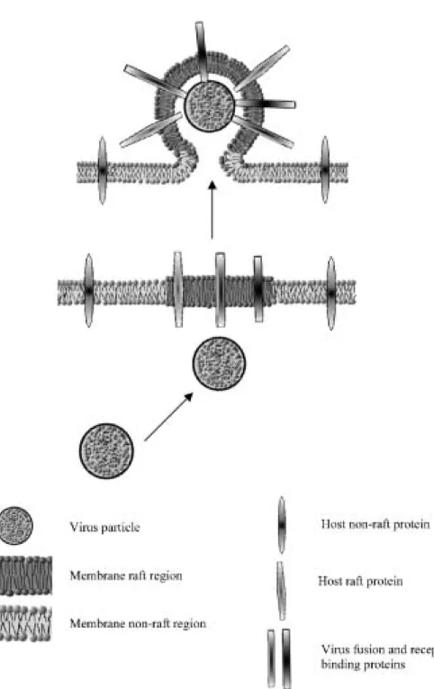
The doors that lead into the cell, lead also out of it. The HIV proteins co-localize with the DRM’s markers and HIV-1 virions, although possessing many proteins and lipids of host cells, lack CD45, a protein that covers
Fig. 2. The viral proteins are
pref-erentially localized in the raft region of the host cell. The complete virus buds from this region of plasma membrane. Due to the very small area of the raft and the proximity of viral proteins in it, the chances for budding a complete virus increase. During the budding process the vi-rion is encapsulated by plasma membrane with incorporated host cell proteins, which may act as a sort of camouflage against immune sys-tem. The picture is highly sche-matic, therefore the relations of size of lipids and proteins are neglected.
from 10% to 25% of lymphocyte surface [2]. It has been proved that this characteristic feature is due to the fact that HIV-1 proteins are preferentially targeted to lipid rafts and that the virus assembly and budding proceed in raft regions [34]. HIV-1 is not the only virus to use rafts for those processes. Filoviruses, Ebola and Marburg are two of the most deadly viruses. It has been demonstrated that their envelopes incorporate raft-associated GM1 and exclude transferrin receptor, a protein that is com-monly used as non-raft marker [4]. Also the influenza virus hemagglutinin preferentially localizes to lipid rafts [45]. It does not mean, however, that all viruses use lipid rafts for their assembly and budding processes. The VSV (vesicular stomatitis virus) and SFV (Semliki Forest virus) do not posses high amounts of detergent-insoluble complexes in their envelopes [46].
The process of virion assembly and budding from the plasma membrane is quite complicated and requires that viral nucleocapsid, matrix and glycoprotein envelope are put together in an orchestrated manner. Thus, the compartmentalization of the process in a special mem-brane microdomain may provide the required coordina-tion and may increase the virus budding efficiency and decrease the release of defective non-infectious particles [4]. The presence of several receptors, normally occur-ring in lipid rafts, in the viral envelopes may act as a camouflage for the host immunity system, but also may efficiently increase the ability to infect new cells because of the enrichment of virus envelope with certain adhe-sion molecules [59].
Sorting and trafficking - how does a protein
know where to go?
The association of raft proteins with raft regions usually takes place in the Golgi apparatus [55]. The GPI anchor as well as palmitoylation of proteins seems to be a hallmark of raft-associated proteins. The acyl chains of GPI anchor of proteins are largely saturated [30]. It is proposed, then, that such GPI anchors are sufficient for lo domains localization [47]. Also the studies on some of the Ebola virus glycoproteins that were mutated at specific cysteine residues (the putative palmitoylation sites) showed that the proteins that normally partici-pate in DRMs failed to localize to raft regions [4]. N-terminal mirystoylation is required for Lyn kinase anchoring to the plasma membrane and raft partition-ing [20].
Another feature that has been proposed as a regulator of protein sorting between detergent-soluble and deter-gent-insoluble membrane regions is the bilayer thick-ness [53]. The DRMs are supposed to be thicker than the non-raft regions, therefore they would incorporate pro-teins with relatively longer transmembrane domains. Some experiments with synthetic transbilayer peptides supported this hypothesis [31].
Rafts play also important role in apical trafficking of proteins in polarized cells. The transport of proteins to the apical membrane may occur directly from the TGN (Trans Golgi Network) or indirectly, by sending the protein first to the basolateral membrane and then via transcytosis to the apical compartment. The cholesterol depletion in HepG2 cells has been shown to strongly affect trafficking, causing mislocalization of newly syn-thesized MDR1 protein to the basolateral surface [55]. Caveolae are flask-shaped rafts that are known to operate as transcytotic carriers [35]. The cholesterol depletion has been shown to affect caveolae causing gradual disappearance of these structures and diffusion of caveolae-associated proteins [58].
Cytoskeleton - raft’s anchor
Even at the time the Singer’s and Nicolson’s model was published [54], the problem of non-uniform lateral or-ganisation of the biological membrane was known; e.g. large membrane domains such as mitochondrial cytoch-rome oxidase or purple membrane of Halobacterium
halobium. Restriction of integral membrane protein
mo-bility by membrane skeletal or cytoskeletal elements suggested by the authors of the model and many other researchers [18, 36, 37] led Sheetz to propose "corrals" (membrane skeleton) and "fence posts" (transmembrane proteins associated with skeletal elements) as interpre-tation of membrane lateral organization [52]. This model was further developed by others (reviewed in [40, 41]). The "fence posts" which are the immobilized by mem-brane skeleton proteins contain lipid of restricted mo-bility as well, therefore they could function as the organizing centres of at least one kind of protein-lipid rafts. Indeed, Nebl et al. [32] isolated from neutrophil plasma membrane so-called DRM-H which is rich in membrane skeleton components.
Membrane rafts which are involved in so many dif-ferent cellular functions and processes such as signaling, protein transport, cell adhesion and movement need to be connected with the cytoskeleton. Raft proteomic ana-lysis reveals that many proteins of membrane cytoskele-ton co-isolate with DRM fraction. Raft-associated cytoskeletal proteins from Jurkat cells and neutrophils show many similarities. Fodrin (non-erythroid spectrin), actin, myosin IIa, supervillin, flotillin are just a subset of those [32, 62]. Also, the DRMs isolated from erythro-cytes are enriched in those proteins [43].
The formation of immunological synapse seems cru-cial for the signal sustaining and propagation [17, 38]. Although the molecular basis of synapse formation re-mains to be elucidated, the role of cytoskeleton cannot be omitted when discussing dynamic changes in rafts (reviewed in [50]). The coalescence of small patches and the formation of immunological synapse require dy-namic membrane changes and therefore involvement of
the membrane skeleton [61]. DRMs in Jurkat cells have been shown to include many cytoskeleton-associated proteins [62] which may be involved in the IS formation and preservation of its integrity. The proposed cytos-keletal reorganization coordinated with raft clustering could be responsible for the mobility restriction of mole-cules localized to aggregated rafts [57]. Some evidence is brought by the fact that EBP50, an ERM family protein that actively binds to actin cytoskeleton, is also a binding partner for the PAG protein (raft-associated protein). This interaction seems to be crucial in connect-ing membrane rafts to actin cytoskeleton, which in turn may be essential for re-distribution of rafts during im-munoreceptor signaling [5].
The transmembrane CD99 molecule is abundantly present on plasma membrane of T cells. CD99 appeared to be incorporated into lipid rafts in a regulated manner. It has been shown that upon engagement of CD99, the molecule becomes associated with the cytoskeleton as well as lipid rafts. After the engagement, CD99 elicits export of several transmembrane proteins and GM1 and the association of CD99 with the cytoskeletal compart-ment occurs in a lipid-dependent manner [64]. In neur-onal cells, all major NCAMs promote incorporation of spectrin into DRMs. It has been demonstrated that spec-trin-mediated coordination between NCAM and PKCβ2
is required to trigger NCAM-mediated neurite out-growth [23]. It has also been demonstrated that lipid rafts are intrinsically linked to cytoskeleton and it may be hypothesized whether cytoskeleton molecules are re-quired for the stabilization and/or localization of rafts within the membrane [32]. However, some of the lipid-binding proteins (annexins, proteins containing pleck-strin homology domain) have been suggested to act as a linker between the rafts and the cytoskeleton and con-tribute for the raft formation and stabilization [1].
Questions to be answered
The interest in rafts has put new energy into a broad range of research fields such as cell biology, membrane biophysics or signal transduction. And as the raft hypo-thesis is enriched with new exciting data, each answer results in the appearance of several new problems. Those that are recently under investigation include:
• The three-dimensional structure of lipid rafts. The specific lipid-lipid and lipid-protein interactions are still one of the most urgent problems when discussing rafts. The problem seems even more complicated in view of the recently demonstrated lack of specific interactions between cholesterol and sphingomyelin [14]. It would be also of great interest to obtain a clear picture of the cytoplasmic face of the rafts.
• The molecular mechanism determining the stability and size of lipid rafts is still unknown. What is the role
of membrane skeleton in localization of raft domains within the biological membranes?
• What are the exact relationships between lipid rafts, caveolae and smooth invaginations free of caveolin and clathrin? Are there different subclasses of rafts?
Although much has already been said, and a lot has been discovered, no one can assure that rafts really exist. Anyway, they are still an exciting phenomenon to inves-tigate.
References
[1] Bandorowicz-Pikuła J (2000) Lipid-binding proteins as stabi-lizers of membrane microdomains - possible physiological sig-nificance. Acta Biochim Pol 47: 553-564
[2] Barclay AN, Beyers AD, Birkeland MR, Brown MH, Davis SJ, Somoza C, Williams AF (1993) The leukocyte antigen facts book. Academic Press, San Diego, California
[3] Baumgart T, Hess ST, Webb WW (2003) Imaging coexisting fluid domains in biomembrane models coupling curvature and line tension. Nature 425: 821-824
[4] Bavari S, Bosio CM, Wiegand E, Ruthel G, Will AB, Geisbert TW, Hevey M, Schmaljohn C, Schmaljohn A, Aman MJ (2002) Lipid raft microdomains: a gateway for compartmentalized trafficking of Ebola and Marburg viruses. J Exp Med 195: 593-602,
[5] Brdickova N, Brdicka T, Andera L, Spicka J, Angelisova P, Milgram SL, Horejsi V (2001) Interaction between two adapter proteins PAG and EBP50: a possible link between membrane rafts and actin cytoskleton. FEBS Lett 507: 133-136
[6] Brown DA, London E (1998) Structure and origin of ordered lipid domains in biological membranes. J Membr Biol 164: 103-114
[7] Cerny J, Stockinger H, Horejsi V (1996) Noncovalent associ-ation of T lymphocyte surface proteins. Eur J Immunol 26: 2335-2343
[8] Cherukuri A, Cheng PC, Pierce SK (2001) The role of CD19/CD21 complex in B cell processing and presentation of complement-tagged antigen. J Immunol 167: 163-172 [9] Dimitrov D (1997) How do viruses enter cells? The HIV
coreceptors teach us a lesson of complexity. Cell 91: 721-730 [10] Eckert GP, Igbavboa U, Mller WE, Wood WG (2003) Lipid rafts
of purified brain synaptosomes prepared with or without deter-gent reveal different lipid and protein domains. Brain Res 962: 144-150
[11] Elortza F, Nuhse TS, Foster LJ, Stensballe A, Peck SC Jensen ON (2003) Proteomic analysis of glycosylphosphatidylinositol-anchored membrane proteins. Mol Cell Proteomics 12: 1261-1270
[12] Evans E, Rawicz W (1990) Entropy-driven tension and bending elasticity in condensed-fluid membranes. Phys Rev Lett 64: 2094-2097
[13] Heerklotz H (2002) Triton promotes domain formation in lipid raft mixtures. Biophys J 83: 2693-2701
[14] Holopainen JM, Metso AJ, Mattila J, Jutila A, Kinnunen PKJ (2004) Evidence for the lack of a specific interaction between cholesterol and sphingomyelin. Biophys J 86: 1510-1520 [15] Ipsen JH, Karlstrom G, Mouritsen OG, Wennerstrom H,
Zuc-kermann MJ (1987) Phase equilibria in the phosphati-dylcholine-cholesterol systems. Biochim Biophys Acta 905: 162-172
[16] Janes PW, Ley SC, Magee AI (1999) Aggregation of lipid rafts accompanies signaling via T cell antigen receptor. J Cell Biol 147: 447-461
[17] Janes PW, Ley SC, Magee AI, Kabouridis PS (2000) The role of lipid rafts in T cell receptor (TCR) signalling. Sem Immunol 12: 23-34
[18] Ji TH, Nicolson GL (1974) Lectin binding and perturbation of the outer surface of the cell membrane induces a transmembrane organizational alteration at the inner surface. Proc Natl Acad Sci USA 71: 2212-2216
[19] Korlach J, Schwille P, Webb WW, Feigenson GW (1999) Characterization of lipid bilayer phases by confocal microscopy and fluorescence correlation spectroscopy. Proc Natl Acad Sci USA 96: 8461-8466
[20] Kovárová M, Tolar P, Arudchandran R, Dráberová L, Rivera J, Dráber P (2001) Structure-function analysis of Lyn kinase as-sociation with lipid rafts and initiation of early signaling events after Fcε receptor I aggregation. Mol Cell Biol 21: 8318-8328, [21] Kusumi A, Sako Y (1996) Cell surface organization by the
membrane skeleton. Curr Opin Cell Biol 8: 566-574
[22] Kwiatkowska K, Frey J, Sobota A (2003) Phosphorylation of FcγRIIA is required for the receptor induced actin rearrange-ment and capping: the role of membrane rafts. J Cell Sci 116: 537-550
[23] Leshchyns’ka I, Sytnyk V, Morrow JS, Schachner M (2003) Neural cell adhesion molecule (NCAM) association with PKCβ2 via βI spectrin is implicated in NCAM-mediated neurite outgrowth. J Cell Biol 161: 625-639
[24] Li H, Ayer LM, Polyak MJ, Mutch CM, Petrie RJ Gauthier L, Shariat N, Hendzel MJ, Shaw AR, Patel KD, Deans JP (2004) The CD20 calcium channel is localized to microvilli and con-stitutively associated with membrane rafts; antibody binding increases the affinity of the association through an epitope-de-pendent crosslinking-indeepitope-de-pendent mechanism. J Biol Chem 279: 19893-19901
[25] Maddon P, Dalgleish A, McDougal J, Clapham P, Weis R, Axel R (1986) The T4 gene encodes the AIDS virus receptor and is expressed in the immune system and the brain. Cell 47: 333-348 [26] Maggi D, Biedi C, Segat D, Barbero D, Panetta D, Cordera R (2002) IGF-I induces caveolin 1 tyrosine phosphorylation and translocation in the lipid rafts. Bioch Biophys Res Commun 295: 1085-1089
[27] Manes S, del Real G, Lacalle RA, Lucas P, Gómez-Moutón C, Sánchez-Palomino S, Delgado R, Alcami J, Mira E, Matinez-A C (2000) Membrane raft microdomains mediate lateral assem-blies required for HIV-1 infection. EMBO Rep 21: 190-196 [28] Manes S, Mira E, Gómez-Moutón C, Lacalle R, Keller P,
Labrador J, Matinez AC (1999) Membrane raft microdomains mediate front-rear polarity in migrating cells. EMBO J 18: 6211-6220
[29] Matkó J, Szöllosi J (2002) Landing of immune receptors on lipid rafts: a safe way to be spatio-temporally coordinated? Immunol Lett 82: 3-15
[30] McConville MJ, Ferguson MAJ (1993) The structure, biosyn-thesis and function of glycosylated phosphatidylinositols in the parasitic protozoa and higher eukaryotes. Biochem J 294: 305-324
[31] McIntosh TJ, Vidal A, Simon SA (2003) Sorting of lipids and transmembrane peptides between detergent-soluble bilayers and detergent-resistant rafts. Biophys J 85: 1656-1666 [32] Nebl T, Pestonjamasp KN, Leszyk JD, Crowley JD, Oh SW,
Luna EJ (2002) Proteomic analysis of a detergent-resistant membrane skeleton from neutrophil plasma membranes. J Biol Chem 277: 43399-43409
[33] Needham D, Nunn RS (1990) Elastic deformation and failure of lipid bilayer membranes containing cholesterol. Biophys J 58: 997-1009
[34] Nguyen DH, Hildreth JEK (2000) Evidence for budding of Human Immunodeficiency Virus type 1 selectively from gly-colipid-enriched membrane lipid rafts. J Virol 74: 3264-3272
[35] Nichols B, Lippincott-Schwartz J (2001) Endocythosis without clathrin coats. Trends Cell Biol 11: 406-412
[36] Nicolson GL, Painter RG (1973) Anionic sites of human ery-throcyte membranes. II. Antispectrin-induced transmembrane aggregation of the binding sites for positively charged colloidal particles. J Cell Biol 59: 395-406
[37] Nicolson GL, Singer SJ (1974) The distribution and asymmetry of mammalian cell surface saccharides utilizing ferritin-con-jugated plant agglutinins as specific saccharide stains. J Cell Biol 60: 236-248
[38] Patel VP, Moran M, Low TA, Micelli MC (2001) A molecular framework for two-step T cell signaling: Lck Src homology 3 mutations discriminate distinctly regulated lipid raft reorganiz-ation events. J Immunol 166: 754-764
[39] Rietveld A, Simons K (1998) The differential miscibility of lipids as the basis for the formation of functional membrane rafts. Biochim Biophys Acta 1376: 467-479
[40] Ritchie K, Iino R, Fujiwara T, Murase K, Kusumi A (2003) The fence and picket structure of the plasma membrane of live cells as revealed by single molecule techniques (Review). Mol Membr Biol 20: 13-18
[41] Ritchie K, Kusumi A (2004) Role of the membrane skeleton in creation of microdomains. Subcell Biochem 37: 233-245 [42] Rodgers W, Rose JK (1996) Exclusion of CD45 inhibits activity
of p56lck associated with glycolipid-enriched membrane
do-mains. J Cell Biol 135: 1515-1523
[43] Salzer U, Prohaska R (2001) Stomatin, flotilin-1 and flotilin-2 are major integral proteins of erythrocyte lipid rafts. Blood 97: 1141-1143
[44] Samuel BU, Mohandas N, Harrison T, McManus H, Rosse W, Reid M, Haldar K (2001) The role of cholesterol and glycosyl-phosphatidylinositol-anchored proteins of erythrocyte rafts in regulating raft protein content and malaria infection. J Biol Chem 276: 29319-29329
[45] Scheiffele P, Rietveld A, Wilk T, Simons K (1999) Influenza viruses select ordered lipid domains during budding from the plasma membrane. J Biol Chem 274: 2038-2044
[46] Scheiffele P, Roth MG, Simons K (1997) Interaction of influen-za virus hemagglutinin with sphingolipid-cholesterol mem-brane domains via its transmemmem-brane domain. EMBO J 16: 5501-5508
[47] Schroeder RJ, Ahmed SN, Zhu Y, London E, Brown DA (1998) Cholesterol and sphingolipid enhance the Triton X-100 insolu-bility of glycosylphosphatidylinositol-anchored proteins by promoting the formation of detergent-insoluble ordered mem-brane domains. J Biol Chem 273: 1150-1157
[48] Schuck S, Honsho M, Ekroos K, Shevchenko A, Simons K (2003) Resitance of cell membranes to different detergents. Proc Natl Acad Sci USA 100: 5795-5800
[49] Schütz GJ, Kada G, Pastushenko VP, Schindler H (2000) Properties of lipid microdomains in a muscle cell membrane visualized by single molecule microscopy. EMBO J 19: 892-901
[50] Sechi AS, Wehland J (2004) Interplay between TCR signaling and actin cytoskeleton dynamics. Trends Immunol 25: 257-265 [51] Sheel-Toelner D, Wang K, Singh R, Majeed S, Raza K, Curnow SJ, Salmon M, Lord JM (2002) The death-inducing complex is recruited to lipid rafts in Fas-induced apoptosis. Biochem Bio-phys Res Commun 297: 876-879
[52] Sheetz MP (1983) Membrane skeletal dynamics: role in modu-lation of red cell deformability, mobility of transmembrane proteins, and shape. Semin Hematol 20: 175-188
[53] Simons K, Ikonen E (1997) Functional rafts in cell membranes. Nature 387: 569-572
[54] Singer SJ, Nicolson GL (1972) The fluid mosaic model of the structure of cell membranes. Science 175: 720-731
[55] Slimane TA, Trugnan G, van Ijzendoorn SCD, Hoekstra D (2003) Raft mediated trafficking of apical resident proteins
occurs in both direct and transcytotic pathways in polarized hepatic cells: role of distinct lipid microdomains. Mol Biol Cell 14: 611-624
[56] Subczyn´ski WK, Kusumi A (2003) Dynamics of raft molecules in the cell and artificial membranes: approaches by pulse EPR spin labeling and single molecule optical microscopy. Biochim Biophys Acta 1610: 231-243
[57] Tanimura N, Nagafuku M, Minaki Y, Umeda Y, Hayashi F, Sakakura J, Kato A, Liddicoat DR, Ogata M, Hamaoka T, Kosugi A (2003) Dynamic changes in the mobility of LAT in aggregated lipid rafts upon T cell activation. J Cell Biol 160: 125-135
[58] Thomsen P, Roepstorff K, Stahlhut M, van Deurs B (2002) Caveolae are highly immobile plasma membrane microdo-mains, which are not involved in constitutive endocytic traffick-ing. Mol Biol Cell 13: 238-250
[59] Triantafilou K, Fradelizi D, Wilson KM, Triantafilou M (2002) GRP78 a co-receptor for Coxackievirus A9, interacts with MHC-class-I molecules which mediate virus internalization. J Virol 76: 633-643
[60] Triantafilou K, Triantafilou M (2003) Lipid raft microdomains: key sites for Coxackievirus A9 infectious cycle. Virology 317: 128-135
[61] Van der Merwe, Davis SJ, Shaw AS, Dustin ML (2000) Cytos-keletal polarization and redistribution of cell surface molecules during T cell antigen recognition. Semin Immunol 12: 5-21 [62] Von Haller PD, Donohoe S, Goodlett DR, Aebersold R, Watts
JD (2001) Mass spectrometric characterization of proteins ex-tracted from Jurkat T cell detergent-resistant membrane do-mains. Proteomics 1: 1010-1021
[63] Xavier R, Brennan T, Li Q, McCormack C, Seed B (1998) Membrane compartmentalization is required for efficient T cell activation. Immunity 8: 723-732
[64] Yoon SS, Jung KI, Choi Y, Choi EY, Lee I, Park SH, Kim TJ (2003) Engagement of CD99 triggers the exocytic transport of ganglioside GM1 and the reorganization of actin cytoskeleton. FEBS Lett 540: 217-222
[65] Zhang W, Trible RP, Samelson LE (1998) LAT palmitoylation: its essential role in membrane microdomain targeting and tyro-sine phosphorylation during T cell activation. Immunity 9: 239-246
Received: September 28, 2004 Accepted after revision: November 25, 2004