Original Researches
Modern medicine tends to incorporate elements of globalized approach into the systematization of pathological conditions, while the synthetic processes are reserved for diagnostics and treatment. The above mentioned tendency gave rise to the notions of chronic venous disease (CVD), chronic obstructive pulmonary disease (COPD) etc. [1, 2]. Articular diseases make no exception. There is an opinion that hip arthritis should be divided into central and superolateral forms accord-ing to the displacement vector of the femoral head [3]. Each form may have its own causes. However, their end result is similar for both, having the same nature and clinical manifestations.
The principal cause of superolateral form is the dys-plastic change of hip joint. It induces the dysdys-plastic hip arthritis, the degenerative-dystrophic condition, char-acterized by the developing articular tip deformity taking the pattern of acetabular wall defects or CCD (centrum-collum-diaphyseal) angle modification and/or femoral anteversion [4, 5]. At the moment, there are several clas-sifications of dysplastic hip arthritis [6-9], which might be applied in hip arthroplasty. Crowe’s classification is preferable while determining the extent of cranial dis-placement, as it gives a notion as to the arthroplastic difficulties and necessary manipulations to bring down the center of artificial joint rotation. Hartofilakidis’ and © «Біль. Суглоби. Хребет» / «Боль. Суставы. Позвоночник» / «Pain. Joints. Spine» (« »), 2019
© Видавець Заславський О.Ю. / Издатель Заславский А.Ю. / Publisher Zaslavsky O.Yu., 2019
Для корреспонденции: Зуб Татьяна Александровна, кандидат медицинских наук, ассистент кафедры медико-социальной экспертизы и реабилитации факультета последипломно-го образования, Государственное учреждение «Днепропетровская медицинская академия Министерства здравоохранения Украины», ул. В. Вернадскопоследипломно-го, 9, г. Днепр, 49044; факс +38 (056) 766-48-10; e-mail: [email protected], контактный телефон +38 (095) 88-15-146.
For correspondence: Tetiana Zub, PhD, Assistant at the Department of medical and social expertise and rehabilitation of the Faculty of postgraduate education, State Institution "Dnipropetrovsk Medical Academy of Health Ministry of Ukraine", Vernadsky st., 9, Dnipro, 49044, Ukraine; fax +38 (056) 766-48-10; e-mail: [email protected], phone +38 (095) 88-15-146.
Full list of author information is available at the end of the article.
УДК 616.728.2–018.3–007.24–036.1:616.718.16–073.7 DOI: 10.22141/2224-1507.9.3.2019.178645
O.Ye. Oleynik , T.O. Zub
State Institution “Dnipropetrovsk Medical Academy of the Ministry of Health of Ukraine”, Dnipro, Ukraine
Methods of acetabular defect visualization
in dysplastic hip arthritis
For citation: Bol', sustavy, pozvonočnik. 2019;9(3):205-211. doi: 10.22141/2224-1507.9.3.2019.178645
Abstract. Background. There is an opinion that according to the displacement of the femoral head, hip arthritis may be divided into central and superolateral forms; the latter is usually caused by the dysplastic changes of the hip joint. The extant classifications of the developmental dysplasia of the hip mostly describe either an acetabular contour or the cranial distance at which the femoral head becomes displaced. However, none of them takes into account dysplastic defects of the acetabulum or their influence on the results of the surgery. The purpose of the study was to evaluate the roentgenanthropometric parameters of the dysplastic acetabulum and visualize the de fects, affecting the outcomes of the total hip arthroplasty. Materials and methods. The roentgenanthropometric analysis of 201 dysplastic hips and 70 normal hips acetabula was performed. We’ve studied the acetabular width, depth, thickness of the acetabular bottom, acetabular index and inclination and vo lume of the acetabular dysplas tic defect. Statistical analysis included mean, relative values, median, quartiles, nonparametric MannWhitney and KruskalWallis criterion with mediantest, nonparametric Kendall correlation. The difference between values was considered significant if p < 0.05. Results. On performing statistical analysis, we found out that in types AC by Eft ekhar classification, the acetabular parameters were changing linearly. Some of them were combined in pairs. For example, the acetabular width increase was accompanied by an inclination increase; however, inclination increased more significantly. The bottom thickness and acetabular depth had an inverse correlation. The diagram for visual ization of the changing parameters was drawn, and a pairing table was built. The latter allows creating a formula for any dysplastic acetabulum. A concept of the dysplastic defect was considered separately. Its prevalence rate and linear sizes represent the difference between the location of the rotation center of the femoral head in case of dysplastic hip arthritis and rotation center of the normal hip joint. Conclusions. Deformations of the acetabulum in the developmental dysplasia of the hip reflect the difficulties the surgeon faces during the preoperative planning and implantation. However, understanding of the pathomorphology of this process helps to solve some problems associated with implantation technique, e.g. bottom resection or required bone grafting of the dysplastic defect, and need for an additional preoperative examination in the patients.
Оригінальні дослідження /
Original Researches
Eftekhar’s classifications inform about the acetabularmorphological changes. The latter classification, for instance, includes 4 stages and describes acetabular de-formations in a somewhat more detailed manner [10].
While there exist various approaches to the dysplas-tic hip arthritis classification, no one quite covers the initial parameters of acetabular modifications in light of the further arthroplasty, despite their describing the morphological changes attending dysplastic hip arthri-tis (DHA) [4, 11, 12]. This fact explains the importance of integral methodology of acetabular evaluation in or-der to compare the obtained results and interpret the preceding acetabular defect modifications, extrapolat-ing them from the arthroplastic outcomes and chances of long-term acetabular component and prosthetic sta-bility maintenance.
The purpose of the study was to evaluate the X-ray parameters of the dysplastic acetabulum and visualize the defects defining the results of the total hip arthro-plasty.
Materials and methods
We’ve studied the roentgen-anthropometric param-eters of 201 dysplastic hips (88 % of them female). For the control group, 70 normal hip acetabula of patients with a unilateral hip arthritis of non-dysplastic genesis were chosen (86 % of them female). All of the patients belonging to the main group had an arthroplasty of the damaged joints. In order to classify the patients from the main group, we’ve used N. S. Eftekhar’s classifica-tion [7]: Type А – 68 joints, Type В – 58, Type С – 63 and Type D – 12 joints.
Acetabular parameters were analyzed according to the plan radiographs of the patients’ pelvises with en-largement markers. By their nature, principal roentgen-anthropometric parameters whose modifications define acetabulum’s dysplastic deformation reflect the final stage of superolateral hip arthritis. It is widely known that with dysplastic hip arthritis acetabulum flattens out, its bottom thickens, and upper tip is skewed due to its contact with a decentered femoral head [4, 13]. This is why we’ve singled out several roentgen-anthro-pometric parameters describing the above mentioned modification.
Acetabular width is a line connecting the most cra-nial and caudal acetabular points, without taking into account ossificate filling its lower sections in case when femoral head has a subluxated posistion. It stands to reason that the acetabular width measured at the plan radiographs does not correspond to the acetabular im-plant’s size in case of dysplastic hip arthritis. However, from the superolateral hip arthritis’ morphological per-spective, this size this very important.
Acetabular inclination is an angle between the ac-etabular width and a horizontal line drawn across the lower edge of pelvic teardrop on both sides (horizontal teardrop line).
Acetabular depth is a perpendicular section, drawn to the center of acetabular width, between its initial point
and acetabular bottom. When a regular hemispheri-cal implant is used during arthroplasty this parameter would increase by 1 mm if the cup’s diameter increases by 2 mm (corresponding to one size of implant), ac-cording to the geometrical principles.
Acetabular bottom thickness is the same perpendic-ular section between the acetabperpendic-ular bottom and inner cortical plate of the innominal bone. Thickness includes the ossificate filling the acetabular bottom in response to the hemispherical femoral head’s absence.
Acetabular index is an integral parameter reflecting the acetabular spherical nature, calculated as a ratio of acetabular depth to width and described in lobes start-ing from 1.
Positioning of the acetabular component with res-toration of femoral head’ rotation center during the pre-op planning leads to appearance of a dysplastic ac-etabular defect. That one is a section delineated by an upper surface of the cup on the bottom and a sclerosed upper edge of the deformed acetabulum on the medial part. The defect doesn’t have a lateral wall. It should be noted that its volume and presence is determined by the implanted cup’s position [14]. There are sources mentioning that a certain size of acetabular defect rules out the initial cup’s stabilization or promotes the sta-bility’s loss in a long-term perspective [15, 16]. Thus, the acetabular defect should be filled with bone grafts. The defect’s volume determining the amount of bone grafts is calculated according to the existing formula during the pre-op planning of the optimal cup inser-tion; however, the necessary parameters are measured in advance [17].
Statistical analysis was performed with biometric analysis of the licensed Microsoft Excel-2003® and Statistica v 6.1 (Statsoft Inc., США) (serial number - AGAR 909 E415822FA). We’ve measured mean (М), standard deviation (SD) for the regular distribution da-ta, median (Ме), 25th and 75th quartiles – for the non- regular distribution data. Depending on the type, distri-bution principle, pair-wise or multiple comparison for the unrelated groups, Student’s t-test and nonparamet-ric Mann-Whitney, Kruskal-Wallis tests with median-test, nonparametric Kendall correlation were used. The difference between values was considered significant if p<0.05.
Results and discussion
Results of the statistically processed findings are pre-sented in Table 1.
A healthy acetabulum has a nearly hemispherical shape (acetabular index 0,47±0,04), providing ana-tomical ground for implanting press-fit hemispherical cups in case of a primary hip arthritis. It should be also noted that the mean linear dimensions of a healthy ac-etabulum are relatively small, explaining why the cups of 50-56 mm in diameter are used most often [18]. The smallest applicable unit size allows preserving the great-er part of bone tissue and its trabecular structure at the implantation site [15, 19].
In case of dysplastic hip arthritis acetabular shape changes. Its width grows from Type A to Type C (there is a significant direct correlation, р≤0,05), which might be attributed to a cranial displacement of femoral head. During childhood and adolescence before the growth plates close, acetabular ‘roof’ becomes sloped due to an eccentric femoral head pressure. In the adulthood, this excessive pressure may even destroy the acetabular ‘roof’. Type D belongs to a separate category, being a complete femoral head dislocation where both lum and head are formed separately. In Type D acetabu-lar width is very small.
Due to the acetabular ‘roof’ destruction and the up-permost acetabular tip’s displacement upwards, its me-dial inclination angle growth. Furthermore, healthy and dysplastic acetabula have a significantly different incli-nation even for Type A.
Acetabular depth diminishes with dysplastic hip ar-thritis’ degree by N. S. Eftekhar, while the bottom’s thickening increases (there are significant direct and inverse correlations for Types A – C, р≤0,05; respec-tively).
Acetabular index also diminishes from Type A to Type C. However with Type D it starts to grow. Thus the acetabulum which has never contained the femoral head has a more spherical shape than the acetabulum affected by dysplastic hip arthritis of Type C.
Based on the mean width, depth, inclination and bottom thickness values, we’ve created two-dimension-al models of the acetabula affected by dysplastic hip ar-thritis (Fig 1, 2) for all the types by N. S. Eftekhar.
However, the Figures don’t represent differences be-tween the acetabular deformation types in a noticeable way. This is why we’ve decided to calculate a percent-age ratio between the normal and modified parameters. The healthy acetabular parameters were set at 1, while the dysplastic hip arthritis parameters described in lobes starting with 1. To visualize the obtained results, we’ve made a radar chart where each of the rays represents one of the parameters (Fig. 3).
Discussion
As it was mentioned above, the increase of width and inclination is caused by the same process of skewing and destruction of acetabular ‘roof’. It may be expected that
these parameters are changing in the same way. Howev-er, in Type A the increase of width to 1,08 of the healthy acetabulum is associated with the increase of inclina-tion of up to 1,33 times at once. In Types B and C, the difference between the parameters in relation to the healthy ones remains almost the same. In Type D, there is an inverse correlation. Underdeveloped acetabular walls lead to a dramatic increase of inclination paired with extremely narrow acetabular width.
As to the depth and bottom’s thickness, these two pa-rameters are also interrelated, as they are adjacent sec-tions of the same line. Furthermore, for the healthy ace-tabulum and Types A, B and C the sum of their absolute values is practically similar and makes from 34,3 mm to 35,9 mm. In this way, with one parameter diminished, another one will increase. For the healthy ones, the bot-tom thickness is more representative, and in Type A it makes 1,27 of the healthy acetabulum. The maximum bottom thickness is observed in Type C, amounting to 1,5 of the healthy acetabulum. The depth is not so sig-nificantly modified, which might be attributed to the fact that the absolute value of the normal parameter is more than twice the value of bottom thickness. In Type A, the depth is 0,88 of the healthy acetabulum, while in Type C - 0,70. In Type D, the sum of absolute values is only 26,4 mm, while the bottom thickness is 1,4 times the healthy acetabulum’s value. The bottom’s thicken-ing associated with dysplastic hip arthritis occurs due to the ossificate’s filling the bottom up and deepening of bottom to the level of a round ligament’s of the femoral head bed is permissible. This phenomenon gave rise to a method of successful acetabular component arthro-plasty. Its extreme variety, cotiloplasty, which is consid-ered to be less physiologically grounded, as it results in an excessive medicalization of implant’s rotation center and diminishing of hip abductors’ strength [20, 21].
In Type A, acetabular index is almost 20 % smaller than the healthy one. Its minimum is observed in Type C, with only 0,6 of the healthy one, while in Type D it reaches 0,7 of the healthy one.
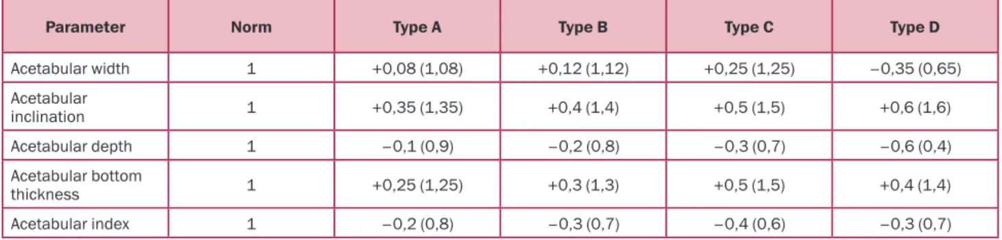
Taking into account the X-ray data, we may suggest the following parameters for the dysplastic acetabular deformation by N. S. Eftekhar (Table 2).
Using this Table, one may describe any dysplastic acetabulum. For instance, the formula describing Type Table 1. Linear, angular and relative parameters of the healthy
and dysplastic acetabulum
Acetabular width, mm Acetabular inclination, degrees Acetabular depth, mm Acetabular bottom thickness, mm Acetabular index Dysplastic acetabular defect, cm3 Healthy (n=70) 50,5 ± 3,8 43,4 ± 7,4 24,2 ± 2,6 11,6 ± 1,6 0,47 ± 0,04 Отсутствует Type А (n=68) 54,6 [50,0–58,2] 58,2 ± 5,9 21,2 ± 3,2 14,7 ± 3,1 0,39 ± 0,06 0,23 [0,00–0,96] Type В (n=58) 56,6 ± 7,0 61,1 ± 7,8 19,1 ± 2,9 15,2 ± 2,8 0,34 ± 0,06 1,39 ± 1,11 Type С (n=63) 62,9 ± 8,9 64,6 ± 6,9 17,1 ± 4,2 17,3 [15,3–20,0] 0,28 ± 0,07 3,22 ± 2,07 Type D (n=12) 32,0 ± 2,5 68,7 ± 9,8 10,1 ± 6,2 16,3 ± 5,4 0,33 ± 0,21 4,90 ± 2,07 Note: parameters are presented accordingly: М ± SD for the regular distribution data and Ме [25th quartile – 75th quartile] for the
Оригінальні дослідження /
Original Researches
B dysplastic hip arthritis would be as follows: widthin-creased by 10%, inclination by 40 %, bottom thick-ness by 30 %, depth and index diminished by 20 %. A minor increase of width does not affect the location of vertical rotation center for an artificial joint; how-ever, the increased inclination requires a careful cup positioning in order to avoid verticalization [14, 22]. Acetabular sphericity may be recovered by its bottom’s resection.
By contrast, the formula describing Type D dysplas-tic hip arthritis is calculated in the following way: ac-etabular width reduced by 35 %, inclination increased by 60 %, depth reduced by 60 %, bottom thickness increased by 40 %, acetabular index reduced by 30 %. It is evident that drilling-out is required as even the smallest acetabular element has extensive geometrical parameters. Drilling-out may be performed by width, as well as by depth (the thickened bottom permitting). Underdeveloped acetabular walls inevitably require the bone grafting of the loaded site (more often at the im-plant cup’s superior pole). One should be careful about the inclination and cup’s anteversion because all the acetabular walls are underdeveloped.
It is impossible to determine the ratio of dysplas-tic defect’s volume to the healthy acetabulum, as the defect is absent in the healthy acetabulum (Table 1, Fig. 4).
In Type A, the defect is revealed in 57,8% of cases, in Type B – in 81,8% of cases, in Type C – in 93,6% of cases, in Type D – in 100 % of cases. The findings reflect the difference between the femoral head’s ro-tation center with dysplastic hip arthritis and roro-tation
center of the healthy hip joint. One may find references to the “recovery of true center of rotation” [19, 23, 24], as even despite the dysplasia, normal rotation center is optimal from the biomechanical perspective. This ‘re-covery’ presupposes bringing down and optimal medi-calization of artificial joint’s center of rotation during cup’s insertion [22, 25].
Conclusions
1. With progressing acetabular dysplasia, its de-formation parameters are changing unevenly even though they are interconnected. Acetabular inclination increases more noticeably than acetabular width, even though the extent of deformation remains the same. The association between bottom thickness and depth al-lows the bottom’s deepening-out to correct the rotation center of artificial joint.
2. Acetabular index with a complete subluxation is closer to the norm than with dysplastic hip arthritis Type C.
3. Dysplastic defect occurs during the recovery of true center of rotation. Its extent depends on the cranial position of the femoral head before the surgery.
4. The suggested analysis of acetabular evaluation according to the X-ray data allows describing the defect and choosing the optimal implanting technique.
5. Defect evaluation method provides the clear screening parameters of visualization requiring speci-fication by precise diagnostic tools. However, without these parameters it’s impossible to evaluate the prin-cipal vectors and trends of diagnostic precision proce-dures.
Fig.1. Shape of a healthy acetabulum and the one affected by dysplastic hip arthritis: healthy –––, Type А ••••,
Type В -•-•-, Type С
---Fig.2. Acetabular shape for dysplastic hip arthritis of Type D. The arrow points at the ‘false’ acetabulum in contact with a
Conflicts of interests. Authors declare the absence of any conflicts of interests and their own financial in-terest that might be construed to influence the results or interpretation of their manuscript. Authors received no financial compensation and report no sponsorship in the study ‘Methods of acetabular defect visualization in dysplastic hip arthritis’.
Information on the authors’ individual contribu-tions: O.Ye. Oleynik – concept and study design, writ-ing the article; T.O. Zub – study design, collection and processing of data, writing the article
References
1. Wittens C, Davies AH, Bækgaard N, et al. Edi -tor's Choice - Management of Chronic Venous Dis-ease: Clinical Practice Guidelines of the European So-ciety for Vascular Surgery (ESVS). Eur J Vasc Endovasc Surg. 2015 Jun;49(6):678-737. https://doi.org/10.1016/j. ejvs.2015.02.007.
2. Vestbo J, Hurd SS, Agustí AG, et al. Global strategy
for the diagnosis, management, and prevention of chronic
obstructive pulmonary disease: GOLD executive summary. Am J Respir Crit Care Med. 2013 Feb 15;187(4):347-65.
https://doi.org/10.1164/rccm.201204-0596PP.
3. Ledingham J, Dawson S, Preston B, Milligan G, Doherty M. Radiographic patterns and associations of osteo-arthritis of the hip. Ann Rheum Dis. 1992 Oct;51(10):1111-6. https://doi.org/10.1136/ard.51.10.1111.
4. Anisimova EA, Yusupov KS, Anisimov DI.
Mor-phology of bone structures of hip joint in normal state
and in dysplastic coxarthrosis (review). Saratov Journal of
Medical Scientific Research. 2014;10(3):373-377. (in Rus -sian).
5. Postel M. Anatomical abnormalities of the hip.
Rev Chir Orthop Reparatrice Appar Mot. 1976 Jul-Aug;62(5):515-8. (in French).
6. Crowe JF, Mani VJ, Ranawat CS. Total hip replace-ment in congenital dislocation and dysplasia of the hip. J Bone Joint Surg Am. 1979 Jan;61(1):15-23.
7. Eftekhar NS. Total hip arthroplasty. 7th ed. St Louis:
Mosby; 1993.
8. Hartofilakidis G, Karachalios T. Total hip arthro -plasty for congenital hip disease. J Bone Joint Surg Am.
2004 Feb;86(2):242-50.
https://doi.org/10.2106/00004623-200402000-00005.
9. Hartofilakidis G, Stamos K, Ioannidis TT. Low fric -tion arthroplasty for old untreated congenital disloca-tion of the hip. J Bone and Joint Surg. 1988;70B(2):182-186. https://doi.org/10.1302/0301-620X.70B2.3346284.
10. Zub TO, Loskutov OYe, Loskutov OA. On the clas
-sification of dysplastic coxarthrosis in adults. Orthopaedics,
Traumatology and Prosthetics. 2010;(2):83-87. https://doi. org/10.15674/0030-59872010283-87. (in Russian).
11. Sharp IK. Acetabular dysplasia: the acetabular an -gle. J Bone Joint Surg. 1961;43B(2):268-272. https://doi. org/10.1302/0301-620X.43B2.268.
12. Oleynik OYe, Zub TO. An integral analysis of the
acetabular parameters for the pathomorphological evalua
-Fig. 3. Morphological changes of healthy acetabulum compared to the one affected by dysplastic hip arthritis
Table 2. Parameters of dysplastic acetabulum in light of defect by N.S. Eftekar’s classification (norm is set at 1)
Parameter Norm Type А Type В Type С Type D
Acetabular width 1 +0,08 (1,08) +0,12 (1,12) +0,25 (1,25) –0,35 (0,65) Acetabular inclination 1 +0,35 (1,35) +0,4 (1,4) +0,5 (1,5) +0,6 (1,6) Acetabular depth 1 –0,1 (0,9) –0,2 (0,8) –0,3 (0,7) –0,6 (0,4) Acetabular bottom thickness 1 +0,25 (1,25) +0,3 (1,3) +0,5 (1,5) +0,4 (1,4) Acetabular index 1 –0,2 (0,8) –0,3 (0,7) –0,4 (0,6) –0,3 (0,7)
Fig.4. Change of dysplastic defect’s volume with dysplastic hip arthritis
Оригінальні дослідження /
Original Researches
Information about authors
O.Ye. Oleynik, MD, PhD, Professor at the Department of traumatology, State Institution "Dnipropetrovsk Medical Academy of Health Ministry of Ukraine", Dnipro, Ukraine; ORCID iD: https: //orcid.org/0000-0002-0382-2590.
Tetiana Zub, PhD, Assistant at the Department of medical and social expertise and rehabilitation of the Faculty of postgraduate education, State Institution "Dnipropetrovsk Medical Academy of Health Ministry of Ukraine", Dnipro, Ukraine; ORCID iD: https://orcid.org/0000-0001-8404-0437.
tion of dysplastic hip arthritis. Morphologia. 2018;12(2):55-61. https://doi.org/10.26641/1997- 9665.2018.2.55-2018;12(2):55-61. (in Russian).
13. Korzh AA, Tikhonenkov ES, Andrianov VL, et al.
Displasticheskii koksartroz: khirurgicheskaia profilaktika
i lechenie [Dysplastic coxarthrosis: surgical prevention and treatment]. Moscow: Meditsina; 1986. 208 p. (in Rus-sian).
14. Loskutov OYe, Oleynik OYe, Zub TO. The features
of deformation in dysplastic hip arthritis from the position of hip replacement. Orthopaedics, Traumatology and Pros-thetics. 2011;(2):23-28. (in Russian).
15. Inao S, Matsuno T. Cemented total hip arthroplasty
with autogenous acetabular bone grafting for hips with de -velopmental dysplasia in adults: the results at a minimum of ten years. J Bone Joint Surg Br. 2000 Apr;82(3):375-7.
16. Tikhilov RM, Shubnyakov II, Mazurenko AV, et al.
Experimental substantiation of acetabular component im
-paction with uncoverage in arthroplasty of patients with severe hip dysplasia. Traumatology and Orthopedics of Russia. 2013;(4):42-51. https://doi.org/10.21823/2311-2905-2013--4-42-51. (in Russian).
17. Loskutov OYe, Oleynik OYe, Zub TO. The method
of estimation of the acetabular dysplastic defect. Litopys
of traumatology and orthopedics. 2012;(1-2):70-72. (in Ukrainian).
18. Zeng Y, Wang Y, Zhu Z, Tang T, Dai K, Qiu S.
Dif-ferences in acetabular morphology related to side and sex
in a Chinese population. J Anat. 2012 Mar;220(3):256-62. https://doi.org/10.1111/j.1469-7580.2011.01471.x.
19. Della Valle AG, Padgett DE, Salvati EA. Preopera-tive planning for primary total hip arthroplasty. J Am Acad Orthop Surg. 2005 Nov;13(7):455-62.
20. Hartofilakidis G, Stamos K, Karachalios T, Ioan -nidis TT, Zacharakis N. Congenital hip disease in adults.
Classification of acetabular deficiencies and operative treatment with acetabuloplasty combined with total hip ar -throplasty. J Bone Joint Surg Am. 1996 May;78(5):683-92. https://doi.org/10.2106/00004623-199605000-00007.
21. Dorr LD, Tawakkol S, Moorthy M, Long W, Wan Z.
Medial protrusio technique for placement of a
porous-coat-ed, hemispherical acetabular component without cement in a total hip arthroplasty in patients who have acetabular
dysplasia. J Bone Joint Surg Am. 1999 Jan;81(1):83-92. https://doi.org/10.2106/00004623-199901000-00012.
22. Daines BK, Dennis DA. The importance of
ac-etabular component position in total hip arthroplasty. Or -thop Clin North Am. 2012 Nov;43(5):e23-34. https://doi. org/10.1016/j.ocl.2012.08.002.
23. Bonnin MP, Archbold PH, Basiglini L, Fessy MH,
Beverland DE. Do we medialise the hip centre of rotation
in total hip arthroplasty? Influence of acetabular offset and
surgical technique. Hip Int. 2012 Jul-Aug;22(4):371-8. https://doi:10.5301/HIP.2012.9350.
24. Schofer MD, Pressel T, Heyse TJ, Schmitt J, Bou-driot U. Radiological determination of the anatomic hip centre from pelvic landmarks. Acta Orthop Belg. 2010 Aug;76(4):479-85.
25. Fukui K, Kaneuji A, Sugimori T, Ichiseki T,
Mat-sumoto T. How far above the true anatomic position can the acetabular cup be placed in total hip arthroplasty? Hip
Int. 2013 Mar-Apr;23(2):129-34. https://doi.org/10.5301/ hipint.5000010. Received 02.06.2019 Revised 23.06.2019 Accepted 15.07.2019 Олійник О.Є., Зуб Т.О. Державний заклад «Дніпропетровська медична академія Міністерства охорони здоров’я України», м. Дніпро, Україна
Методи візуалізації дефектів кульшової западини при диспластичному коксартрозі
Резюме. Актуальність. Залежно від напрямку зміщення го-ловки стегнової кістки коксартроз можна поділити на цен-тральну та суперлатеральну форми; у другому випадку осно-вною причиною є диспластичні зміни суглобових кінців кіс-ток, що формують кульшовий суглоб. Наявні класифікації диспластичного коксартрозу мають описовий характер і оці-нюють вибірково або форму кульшової западини, або від-стань, на яку головка стегнової кістки зміщується в краніаль-ному напрямку, але жодна з них не враховує дефекти куль-шової западини, викликані дисплазію, або їх вплив на резуль-тати оперативного лікування.Мета дослідження — оцінити рентгенантропометричні показники кульшової западини при диспластичному коксартрозі з наступною візуалізацією де-фектів, що визначають результати ендопротезування кульшо-вого суглоба.Матеріали та методи. Виконано аналіз осно-вних рентгенантропометричних показників кульшової за-падини 201 кульшового суглоба з ознаками диспластично-го коксартрозу та 70 здорових кульшових суглобів без ознак дисплазії. Вивчено показники ширини, глибини, товщини дна кульшової западини, індекс кульшової западини (відно-шення глибини до ширини), її інклінацію й обсяг дисплас-тичного дефекту. Статистичний аналіз містив розрахунки се-редніх, відносних величин, медіани, квартилів, непараме-тричних критеріїв Манна — Уїтні, Крускала — Уолліса з меді-анним тестом, непараметричної кореляції Кендалла. Різницю між порівнюваними величинами вважали статистично значу-щою за умови р < 0,05.Результати. Після статистичної об-робки визначено, що при дисплазії кульшової западини(ти-пи А–С за N.S. Eftekhar) показники змінюються лінійно. Бу-ли виділені пари показників, що пов’язані між собою. Так, збільшення ширини кульшової западини відбувається одно-часно зі збільшенням її інклінації, але збільшення останньої є більш істотним. Товщина дна та глибина кульшової запади-ни, навпаки, мають зворотню кореляцію. Побудовано діагра-му, що візуалізує зміни кульшової западини при диспластич-ному коксартрозі, а також таблицю сполучення, що дозволяє скласти формулу кульшової западини для будь-якого типу диспластичної деформації. Окремо розглянуте поняття дис-пластичного дефекту кульшової западини. Частота його по-ширеності та розміри залежать від різниці між положенням центру ротації головки стегнової кістки при диспластичному коксартрозі та центру обертання кульшового суглоба в нормі. Висновки. Деформації кульшового суглоба при диспластич-ному коксартрозі відображають труднощі, що виникають пе-ред хірургом у процесі пепе-редопераційного планування й ви-конання ендопротезування кульшового суглоба. Проте розу-міння патоморфології цього захворювання дозволяє виріши-ти низку проблем, що пов’язані з хірургічною технікою імп-лантації, наприклад, поглиблення дна кульшової западини за рахунок дозованої резекції чи визначення потреби у кістко-вій пластиці диспластичного дефекту, необхідність виконан-ня додаткових методів обстеженвиконан-ня при підготовці пацієнтів цієї категорії до операції. Ключові слова: диспластичний коксартроз; кульшова за-падина; площинна рентгенантропометрія; ендопротезування кульшового суглоба Олейник А.Е., Зуб Т.А. Государственное учреждение «Днепропетровская медицинская академия Министерства здравоохранения Украины», г. Днепр, Украина