Received: December 25, 2012; Revised: March 15, 2013; Accepted: March 26, 2013
*
Corresponding Author: Ming-Cheng Tai, Department of Ophthalmology, Tri-Service General Hospital, National Defense Medical Center, No. 325, Sec. 2, Cheng-gong Road, Taipei 114, Taiwan, Republic of China. Tel: +886-2-87927165; Fax: +886-2-87927164; E-mail: mingtai [email protected]
Copyright © 2013 JMS
Accuracy of LASIK Flap Thickness Obtained Using 2 Types of Moria M2
Single-use Head Microkeratome Measured Postoperatively by Optical
Coherence Tomography
Yen-Shou Chou1,2,4, Chang-Min Liang1,2, Jiann-Torng Chen1,2, Da-Wen Lu1,2, Yu-Ching Chou3, and Ming-Cheng Tai1,2*
1
Department of Ophthalmology, Tri-Service General Hospital, National Defense Medical Center, Taipei, Taiwan;
2
Department of Ophthalmology; 3
School of Public Health, National Defense Medical Center, Taipei, Taiwan;
4
Department of Ophthalmology, Tri-Service General Hospital Penghu Branch, Penghu, Republic of China
Background: To investigate the accuracy of corneal fl ap thickness (FT) and to examine the potential factors that can infl uence fl ap thickness obtained using Moria M2 microkeratomes with two kinds of single-use heads. Methods: A ret-rospective review of the records of 90 patients who had undergone LASIK between Jun 2011 and Jan 2012 at Tri-Service General Hospital was performed. Creation of corneal fl ap was achieved by automated Moria M2 microkeratome with two different types of single-use head (SU90 and SU130). The right eye was operated before the left eye in each patient, using the same blade. Additionally, the corneal fl ap thickness was measured in all cases using the Fourier-domain anterior seg-ment optical coherence tomography (AS-OCT) one week later. Results: The mean central thickness of corneal fl ap was 114.26±4.49μm (Range, 106~127μm) in the SU 90 head and 146.98±3.62μm (Range, 140~157μm) in the SU130 head. The difference between the fi rst and second eye operated was not signifi cant. There were no free fl aps, incomplete fl aps or fl aps with buttonholes in any of our cases. Conclusions: Moria M2 SU 90 and SU 130 produce accurate, repro-ducible, safe and cost-effective corneal fl ap. Thin fl aps achieved by both single-use heads did not increase the rate of fl ap-related complications. Furthermore, AS-OCT is a noncontact, rapid, and repeatable tool for measuring corneal fl ap thick-ness and making morphological observations.
Key words: laser in situ keratomileusis (LASIK), corneal fl ap thickness, keratometry reading, mechanical microkeratome, optical coherence tomography
INTRODUCTION
Creation of an accurate corneal fl ap is the step most crucial to the success of laser in situ keratomileusis (LASIK) surgery owing to the importance of stromal preservation and surgical planning. In previous studies1-3, the flap thickness achieved was different from the at-tempted flap thickness using the mechanical microker-atome. Since the introduction of the femtosecond laser as an alternative to the mechanical microkeratome, thinner and more accurate fl aps can be achieved with the advan-tage of preserving more stroma and potentially reducing the incidence of corneal ectasis.
The recently developed, automated microkeratomes with single-use head have advantages over the traditional metallic head, such as no need for assembly, enhanced transparency and decreased risk of infection. This current study was performed to evaluate the accuracy of the cor-neal fl ap produced using automated Moria M2 microker-atomes with two kinds of single-use head and measured by OCT, and to investigate the potential factors that may influence flap thickness, such as preoperative corneal thickness, preoperative spherical equivalent, patient’s age and keratometry readings.
MATERIAL AND METHODS
We performed a retrospective review of the records of 180 eyes of the 90 enrolled patients who had undergone LASIK between Jun 2011 and Jan 2012 at Tri-Service General Hospital. The study protocol complied with the requirements of the the Institutional Review Boards of the Tri-Service General Hospital, Taipei, Taiwan. The study followed the Good Clinical Practice (GCP) guide-lines of Taiwan and in accordance with the Declaration of
Helsinki, 1964, and later revisions. The inclusion criteria were no history or slit-lamp evidence of ocular trauma, no ocular surgery or corneal abnormality, no history of systemic or ocular disease that contraindicated LASIK, no use of systemic antimetabolites or immunosuppres-sants. Besides, if the predicted value of the postoperative residual stromal bed (with the 2 kinds of microkeratomes for calculating) was less than 250 micrometers, the sub-jects were excluded. Before the operation, use of soft and hard contact lens was suspended for at least 2 and 4 weeks, respectively. All patients underwent a complete preoperative ophthalmological examination including biomicroscopy, measurement of corneal topography and thickness (Obscan), determination of refraction, mea-surement of intraocular pressure (Topcon Computerized Tonometer CT-60; Topcon Corp, Tokyo, Japan), mea-surement of uncorrected (UCVA) and best spectacle-corrected visual acuity (BSCVA).
Surgical technique
The LASIK procedures were performed at our center by the same surgeon (M-C Tai). The automated Moria M2 microkeratome with the control unit ME-LSK Evolu-tion 2 was used for the creaEvolu-tion of corneal fl aps. There are two kinds of plastic single-use head 90 and 130 mi-crokeratomes, designed to create a 120- and 150-μm fl ap, respectively. The randomly selected the both eyes of each patient were operated with either plastic single-use head 90 microkeratome or plastic single-use head 130 microkeratome. Prior to the operation, a notepaper repre-senting either Moria M2 SU 90 or Moria M2 SU 130 was randomly picked by the surgeon’s assistant. The right eye was operated before the left eye in each patient, using the same blade. The suction ring was chosen according to the manufacturer’s recommendations, a nomogram based on the keratometric value. All fl aps had a superior hinge. Stromal ablations were performed with a broad beam excimer laser (Visx S4; AMO). Flap morphology was evaluated using an RTVue Fourier-domain anterior segment optical coherence tomography (Optovue, Inc.), which has a depth resolution of 5μm and a speed of 26000 axial scans per second. The scan was centered on the vertex reflection, and the horizontal meridian OCT images with 6 mm in diameter were acquired and ana-lyzed in a blind fashion by the same skilled technician at 1 week postoperatively. Due to poor contrast between the fl ap and stroma in the central zone, the central corneal fl ap thickness was interpolated from the paracentral mea-surements3 (±2 mm from the corneal vertex) by semi-automated software (Fig 1). Flap-related complications
and post-operative visual acuity were also recorded till 2 weeks after operation. In addition, AS-OCT scanning was performed 3 times with the patients repositioned af-ter each scan.
Statistical analysis
The eyes were grouped for statistical analysis accord-ing to the type of head used. The mean values and SDs of ages, corneal thickness, flap thickness, preoperative refraction, and keratometric values of 180 eyes were cal-culated. Statistical analyses were performed using SPSS version 19.0 for windows (SPSS Inc, Chicago, Illinois). Unpaired Student’s t-test was used to analyze differences between the two groups (SU 90 and SU 130). Single-variable correlation of fl ap thickness between preopera-tive refraction, corneal thickness, keratometric values and age was performed using a Pearson correlation coef-fi cient analysis. A p value of less than 0.05 was consid-ered statistically signifi cant.
RESULTS
A total of 180 consecutive eyes of 90 patients who had undergone LASIK (44 patients treated with Moria M2 SU 90 and 46 patients with Moria M2 SU 130) between Jun 2011 and Jan 2012 at Tri-Service General Hospi-tal were identifi ed. The preoperative values for the two groups are presented in Table 1.
The flap was identified from the stromal bed by its increased internal refl ectivity and interface signal peak. This contrast was best seen in the paracentral region (±2 mm from the vertex) and worse seen near the corneal vertex. The interface peak was clearly visible at 1 week after the operation. In Moria M2 SU 90-treated eyes, the intended corneal flap thickness was 120 μm, and the mean fl ap thickness was 114.26±4.49 μm (range, 106-127 μm). In Moria M2 SU 130-treated eyes, the intend-ed corneal fl ap thickness was 150 μm, and the mean fl ap thickness was 146.98±3.62 μm (range, 140-157 μm).
Fig. 1 High-resolution corneal anterior segment optical co-herence tomography image showing the fl ap
thick-ness at measurement±2mm from the vertex at 1
week postoperatively.
The Moria M2 SU 130 group revealed a much smaller deviation from the target thickness (mean difference: SU 90: -5.74, SU 130: -3.02) (Table 2) but both groups
showed good precision (Coeffi cient of Variation: SU 90: 3.93%, SU 130: 2.46%). The second flap cut with the same blade w a s n o t s i g n i f i c a n t l y thinner for fl aps cut with either the Moria M2 SU 90 microkeratome (P = 0.267), showing an aver-age thickness of 114.80
± 4.54 μm (fi rst) versus 113.73 ± 4.42 μm (sec-ond), or the Moria M2 SU 130 microkeratome (P = 0.186), s h o w i n g a n a v e r a g e t h i c k n e s s o f 147.48 ± 3.69 μm (f i r s t) v e r s u s 146.48 ± 3.51 μm (s e c o n d). Moreover, there were no free or incomplete flaps, or fl aps with buttonholes in any of the cases in this study.
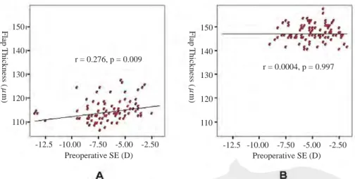
A w e a k c o r r e l a t i o n was found between cor-neal flap thickness and corneal thickness in the Moria M2 SU 90 group (r = -0.335, P = 0.001) but no significant correlation in the M2 SU 130 group (r = -0.025, P = 0.812) (F i g. 2). T h e r e w a s a weak correlation between corneal fl ap thickness and preoperative spherical equivalent in the M2 SU 90 group (r = 0.276, P = 0.009) but no significant correlation in the M2 SU 130 group (r = 0.0004, P = 0.997) (Fig. 3). Moreover, corneal flap thickness showed no correlation with age (r = 0.061, P = 0.572 in M2 SU 90 group; r = 0.134, P = 0.202 in M2 SU 130 group) (Fig. 4) or preoperative keratometry K1 (r = 0.04, P = 0.709 in M2 SU 90 group; r = 0.087, P = 0.074
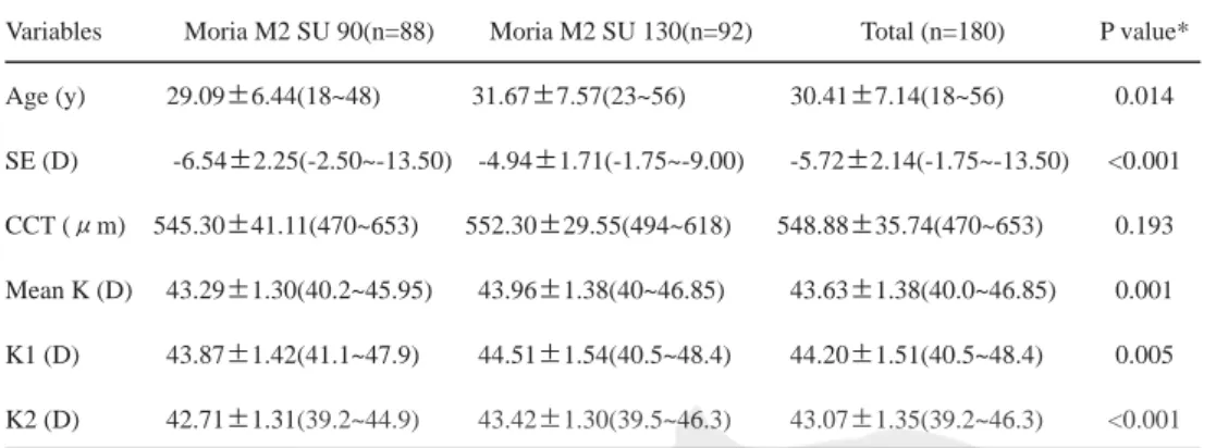
Table 1 Preoperative characteristics of patients undergoing LASIK fl ap creation with Moria M2 SU 90 and SU 130 microkeratomes
Variables Moria M2 SU 90(n=88) Moria M2 SU 130(n=92) Total (n=180) P value* Age (y) 29.09±6.44(18~48) 31.67±7.57(23~56) 30.41±7.14(18~56) 0.014 SE (D) -6.54±2.25(-2.50~-13.50) -4.94±1.71(-1.75~-9.00) -5.72±2.14(-1.75~-13.50) <0.001 CCT (μm) 545.30±41.11(470~653) 552.30±29.55(494~618) 548.88±35.74(470~653) 0.193 Mean K (D) 43.29±1.30(40.2~45.95) 43.96±1.38(40~46.85) 43.63±1.38(40.0~46.85) 0.001 K1 (D) 43.87±1.42(41.1~47.9) 44.51±1.54(40.5~48.4) 44.20±1.51(40.5~48.4) 0.005 K2 (D) 42.71±1.31(39.2~44.9) 43.42±1.30(39.5~46.3) 43.07±1.35(39.2~46.3) <0.001 CCT = Central corneal thickness, SE = Spherical equivalent, K =keratometry
*The P values of Moria M2 SU 90 and 130 groups were calculated using t test
Table 2 Thickness of corneal flap cut using Moria M2 SU 90 and the Moria M2 SU 130 microkeratomes
Microkeratome group Flap thickness (μm)
Mean ± SD Range Mean difference P value Moria M2 SU 90 (n=88) 114.26 ± 4.49 106~127 -5.74 <0.001 Moria M2 SU 130 (n=92) 146.98 ± 3.62 140~157 -3.02 <0.001
Fig. 2 Graph showing a weakly negative linear relationship between corneal fl ap thickness and corneal thickness in the SU 90 (A), but no signifi cant correlation in the SU 130 group (B).
150 140 130 120 110 Flap Thickness ( μ m) 500.00 550.00 600.00 650.00 Comeal Thickness (μm) r = 0.343, p = 0.001 150 140 130 120 110 Flap Thickness ( μ m) 500.00 550.00 600.00 650.00 Comeal Thickness (μm) r = 0.039, p = 0.712 TAO
in M2 SU 130 group) (Fig. 5) in the two groups. DISCUSSION
The incidence of microkeratome-related fl ap compli-cations has been reported to range from 0.3% to 1.9%.4-6 In addition, iatrogenic keratectasia is one of the long-term major complications after LASIK. Therefore, creat-ing an accurate and thin corneal fl ap to preserve as thick as possible the residual corneal bed is critical in LASIK surgery. In the current study, the mean corneal fl ap thick-ness in the Moria M2 SU 90 group was 114.26 μm. The standard deviation of 4.49 μm was clinically insignifi -cant and the same conclusion can be made for the Moria
M2 SU130 group. Moreover, the precision in fl ap thickness in terms of Coeffi cient of Variation (SU 90: 3.93%, SU 130: 2.46%) and the accuracy in flap thick-ness in terms of mean difference (SU 90: -5.74, SU 130: -3.02) are great in both groups. Accord-ing to the findAccord-ings above, both groups appeared to produce ac-curate and reproducible corneal fl aps. Besides, there are plenty of microkeratomes in clinical use and their most common charac-teristic is thinner flap thickness achieved than intended7-9, agree-ment with our current study. Pre-vious studies1,2,10 showed that the standard deviation of fl ap thick-ness achieved by mechanical mi-crokeratomes was in the range of
±20 to ±40. The improvement in standard deviation of our study may be attributed to the learning curve of the procedure or better design of microkeratome. Fur-thermore, thin fl aps are prone to buttonholes and necessitate more complicated handling during surgery. However, in our study, incomplete fl aps, buttonholes and other microkeratome-related fl ap complications were not observed.
To determine the preoperative factors associated with corneal flap thickness, we investigated the correlation between corneal flap thickness and the preoperative SE, age, keratometry K1 power and central corneal thickness, respectively. We found a weak cor-relation between fl ap thickness and central corneal thick-ness and a weak correlation between fl ap thickthick-ness and preoperative spherical equivalent only in the M2 SU 90 group. However, in previous studies7,8, a positive correla-tion between fl ap thickness and central corneal thickness was found. Furthermore, Pietila et al.7 found a tendency towards thinner fl aps created by Moria M2 SU 130 with higher keratometric K1 value in myopic eyes, but the dif-ference was not signifi cant. Huhtala et al.8 reported that increasing thickness of fl aps produced by Moria M2 SU 90 was associated with fl atter keratometric power K1 in
Fig. 3 Graph showing a weakly direct linear relationship between corneal fl ap thick-ness and preoperative spherical equivalent in the M2 SU 90 group (A), but no signifi cant correlation in the SU 130 group (B).
150 140 130 120 110 Flap Thickness ( μ m) -12.5 -10.00 -7.50 -5.00 -2.50 Preoperative SE (D) r = 0.276, p = 0.009 150 140 130 120 110 Flap Thickness ( μ m) -12.5 -10.00 -7.50 -5.00 -2.50 Preoperative SE (D) r = 0.0004, p = 0.997
Fig. 4 Graph showing no correlation between flap thickness and age in the SU 90 group (A) or the SU 130 group (B).
150 140 130 120 110 Flap Thickness ( μ m) 20 30 40 50 Age r = 0.061, p = 0.572 Flap Thickness ( μ m) r = 0.134, p = 0.202 150 140 130 120 110 20 30 40 50 Age TAO
hyperopic eyes but not in myopic eyes. In addition, a negative correlation between patient’s age and corneal fl ap thickness was also noted in that study. The discrep-ancies between our results and previous fi ndings7,8 may be attributed to the dissimilar methods adopted (OCT vs. ultrasonic pachymetry) or the different time points (1 week after operation vs. immediately after fl ap creation) of flap measurement. The anticipated errors11 with the measurement technique using ultrasound pachymetry and the subtraction method of calculating fl ap thickness have led investigators to use the OCT as a tool for measuring postoperative fl ap thickness.12,13 Fourier-domain anterior segment OCT provides noncontact high-resolution cor-neal cross-sectional images. We have recently reported that AS-OCT provided excellent reproducibility and quantitative parameters in the anterior chamber angle.14 In this study, we also confirmed previous results15 that routine measurement of flap thickness with OCT was recommended at 1 week postoperatively, when the mea-surement was most precise and edema has resolved. Moreover, the mean corneal fl ap thickness in both groups of our study was less than that in previous studies.7,8 Such difference may be due to too dry residual stromal bed after fl ap creation or compression of residual stromal bed when making measurement, thus increasing the pachym-etry-derived fl ap thickness.
Another common issue of microkeratomes concerning the discrepancy in flap thickness between the first and second eye also occurred when the same blade was used for both eyes. David et al.16 showed that the reuse of mi-crokeratome blades created signifi cantly thinner fl aps on the second cut. Our study demonstrated a slightly
thin-ner fl ap in the second cut, which was consistent with previous findings.7,8 Some authors postu-lated that the reason for thinner flap obtained in the second cut was the increased dullness of the blade following the fi rst cut.17,18
The newly developed femto-second lasers were designed to produce thinner fl aps, with closer range of thickness around the mean. Salomao et al.19 obtained a standard deviation of fl aps to be
±14.5 μm using the IntraLase femtosecond laser. The standard deviation was comparable with that obtained using the two kinds of mechanical microkeratomes in our study (SU90: ±4.49 and SU130: ±3.62), mean-ing that Moria M2 SU 90 and SU 130 microkeratomes could also effectively produce thinner flaps with a smaller standard deviation in more eyes with relatively thinner corneas. In contrast to the high-priced IntraLase femtosecond laser device and the extra charges to the patients using femtosecond laser, either Moria M2 SU 90 or SU 130 produce accurate, reproducible, safe and cost-effective corneal fl ap. Moreover, Moria M2 SU 130 could be more precise and accurate than Moria M2 SU 90 although the difference was not signifi cant. However, with the advantages of preserving more stromal tissue and potentially reducing the incidence of corneal ectasis, M2 SU 90 could create thinner and accurate flaps (but not superior to M2 SU 130) and thus more suitable for the patients with thinner central corneal thickness.
There were some drawbacks that need improvement in our present study. First, because our study was a ret-rospective study with relative small patients, when they were divided into groups, analysis of the data was lim-ited to one factor at a time. Therefore, we were unable to benefit from an interaction analysis of factors. Such as the result of factors correlated the fl ap thickness was not totally compatible with the previous studies7,8 and the rate of LASIK fl ap complication might be underesti-mated, as the incidence of actual microkeratome-related fl ap complications had been reported to range from 0.3% to 1.9% in previous literature4-6 but none in our study. A large prospective study is required to establish more sta-tistically powerful results. Second, because poor contrast between the fl ap and stroma in the central zone but best in the paracentral region as an intrinsic limitation by the
Fig. 5 Graph showing no correlation between fl ap thickness and preoperative keratom-etry K1 in the SU 90 group (A) or the SU 130 group (B).
Flap Thickness ( μ m) r = 0.187, p = 0.074 150 140 130 120 110 42.0 44.0 46.0 48.0 Keratometry K1 (D) 42.0 44.0 46.0 48.0 Keratometry K1 (D) Flap Thickness ( μ m) r = 0.040, p = 0.709 150 140 130 120 110 TAO
corneal geometry, the central corneal fl ap thickness was interpolated from the paracentral measurements to lower the effect of fl ap thickness variation3 and was only along the horizontal meridian of the cornea. As the previously study20 state, in order to avoid false measurement and misinterpretation of the fl ap data that might be caused by interference from the upper eyelid or by superior location of the hinge-pocket complex, the horizontal meridian was concentrated. However, the horizontal profi le only is not enough to determine the homogeneity of the whole fl ap; fl ap profi le shown on vertical and oblique scans should be included in future studies.
CONCLUSION
This study revealed that automated microkeratome Moria M2 with SU 90 and SU 130 heads could produce accurate, reproducible, safe and cost-effective corneal fl ap. Furthermore, they did not increase the rate of fl ap-related complications, showing results comparable with those created using femtosecond laser. Moreover, ante-rior segment OCT is a noncontact, rapid, and repeatable tool for measuring corneal flap thickness and making morphological observations.
ACKNOWLEDGEMENTS
A. This study was supported by funding from Tri Service General Hospital (TSGH-C100-1-SO4).
B. The authors contributed to the manuscript and study as follows: CM Liang, JT Chen and DW Lu collected data, reviewed the literature and writing part of the manuscript. YC Chou was responsible for analysis and interpretation of data and writing part of the man-uscript. YS Chou was the chief editor and MC Tai was the surgeon and corresponding author.
DISCLOSURE
A. The authors have no additional fi nancial disclosures. B. The article has not been presented in a meeting. C. The authors have no fi nancial or proprietary interest in
a product, method, or material described herein.
REFERENCES
1. Arbelaez MC. Nidek MK 2000 microkeratome clini-cal evaluation J Refract Surg 2002;18:S357-S360. 2. Ucakhan OO. Corneal flap thickness in laser in
situ keratomileusis using the Summit
Krumeich-Barraquer microkeratome. J Cataract Refract Surg 2002;28:798-804.
3. Li Y, Netto MV, Shekhar R, Krueger RR, Huang D. A longitudinal study of LASIK fl ap and stromal thick-ness with high-speed optical coherence tomography. Ophthalmology 2007;114:1124-1132, doi: 10.1016/ j.ophtha.2006.09.031.
4. Gimbel HV, Penno EE, van Westenbrugge JA, Fe-rensowicz M and Furlong MT. Incidence and man-agement of intraoperative and early postoperative complication in 1000 consecutive laser in situ ker-atomileusis cases. Ophthalmology 1998;105:1839-1847.
5. Jacobs JM and Taravella MJ. Incidence of intraopera-tive fl ap complications in laser in situ keratomileusis. J Cataract Refract Surg 2002;28:23-28.
6. Tham VM and Maloney RK. Microkeratome compli-cations of laser in situ keratomileusis. Ophthalmol-ogy 2000;107:920-924.
7. Pietila J, Makinen P, Suominen S Huhtala A, and Uusitalo H. Bilateral comparison of corneal flap dimensions with the Moria M2 reusable head and single use head microkeratomes. J Refract Surg 2006;22:354-357.
8. Huhtala A, Pietila J, Makinen P, Suominen S, Sep-panen M and Uusitalo H. Corneal fl ap thickness with the Moria M2 single-use head 90 microkeratome. Acta Ophthalmol Scand 2007;85:401-406.
9. Hsu SY, Liu YL, Chang MS and Lin CP. Accuracy of corneal fl ap thickness achieved by two different age MK-2000 microkeratomes. Eye 2009; 23:2200-2205, doi: 10.1038/eye.2008.435.
10. Shemesh G, Dotan G, Lipshitz I. Predictability of corneal fl ap thickness in laser in situ keratomileusis using three different microkeratomes. J Refract Surg 2002;18:S347-S351.
11. Foulkes RB. LASIK flap thickness is trickier than you think. Ocular Surgery News May 1, 2002:10-11. 12. Nam SM, Im CY, Lee HK, Kim EK, Kim T-I, and
Seo KY. Accuracy of RTVue optical coherence to-mography, Pentacam, and ultrasonic pachymetry for the measurement of central corneal thickness. Ophthalmology 2010;117:2096-2103, doi: 10.1016/ j.ophtha.2010.03.002.
13. Li Y, Shekhar R, and Huang D. Corneal pachymetry mapping with high speed optical coherence tomogra-phy. Ophthalmology 2006;113:792-799.
14. Ming-Cheng Tai, Ke-Hung Chien, Da-Wen Lu, Jiann-Torng Chen. Angle changes before and after cataract surgery obtained with Fourier- Domain anterior
ment optical coherence tomography. Journal of Cata-ract & RefCata-ractive Surgery 2010;36:1758-1762, doi: 10.1016/j.jcrs.2010.05.011.
15. Thompson RW, Choi DM, Price MO, Potrezbowski L and Price FW. Noncontact optical coherence tomog-raphy for measurement of corneal fl ap and residual stromal bed thickness after laser in situ keratomileu-sis. J Refract Surg 2003;19:507-515.
16. David W, Lin W, and Douglas D. Accuracy and pre-cision of the Amadeus microkeratome in producing LASIK fl aps. Cornea 2003;22:504-507.
17. Schultze RL. Microkeratome update. Int Ophthalmol Clin 2002;42:55-65.
18. Seiler T, Koufala K, and Richter G. Iatrogenic kerate-ctasia after laser in situ keratomileusis. J Refract Surg 1998;14:312-317.
19. Salomao MQ, Ambrosio R Jr and Wilson SE. Dry eye associated with laser in situ keratomileusis: mechani-cal microkeratome versus femtosecond laser. J Cata-ract RefCata-ract Surg 2009;35:1756-1760, doi: 10.1016/ j.jcrs.2009.05.032.
20. Kucumen RB, Yenerel NM, Gorgun E, Oral D, Al-tunsoy M, Utine CA and Ciftci F. AS-OCT as a tool for fl ap thickness measurement after femtosecond-as-sisted LASIK. Journal of Ophthalmic Surgery, Lasers & Image 2011;42:31-36, doi: 10.3928/15428877-20101124-03.