T-cell prolymphocytic leukemia Amit Sud1, Claire Dearden*1
1Department of Haemato-Oncology, The Royal Marsden Biomedical Research Centre,
London, UK
*Corresponding author:
Dr Claire Dearden BSc MBBS MD FRCP FRCPath
Department of Haemato-Oncology, The Royal Marsden NHS Foundation Trust, Downs Road, Sutton, Surrey SM2 5PT, UK
Tel: + 208 661 3116 Fax: + 208 642 9634
Email: [email protected] Dr Amit Sud MBChB (Hons) MRes MRCP
Department of Haemato-Oncology, The Royal Marsden NHS Foundation Trust, Downs Road, Sutton, Surrey SM2 5PT, UK
Tel: + 20 8661 3071 Email: [email protected]
Keywords: T-cell prolymphocytic leukemia, T-cell, leukemia, alemtuzumab
Disclosure statement:
A.S. has no conflict of interest to declare. C.D. undertakes consultancy for Genzyme.
Synopsis
molecular features. The current therapeutic approach relies upon immunotherapy followed by a hematopoietic stem cell transplant (HSCT) in selected cases. Clinical outcomes are generally poor, although insights from genomic and molecular studies may increase our understanding of this disease, with the promise of additional effective therapeutic options.
Key Points:
T-cell prolymphocytic leukemia (T-PLL) is a rare and aggressive leukemia.
The current therapeutic approach utilizes immunotherapy followed by a hematopoietic stem cell transplant (HSCT) in eligible cases.
INTRODUCTION
T-cell prolymphocytic leukemia (T-PLL) is a rare and aggressive T-cell malignancy first described by Catovsky et al over 40 years ago.1 Although termed ‘prolymphocytic’, the
disease is characterized by the proliferation of post-thymic T-lymphocytes. T-PLL can be distinguished from other lymphoid diseases by the evaluation and integration of clinical features, morphology, immunophenotyping, cytogenetics and molecular features. The current therapeutic approach relies upon immunotherapy followed by a hematopoietic stem cell transplant (HSCT) in selected cases. Clinical outcomes are generally poor, although insights from genomic and molecular studies may increase our understanding of this disease, with the promise of additional effective therapeutic options.
EPIDEMIOLOGY
T-PLL accounts for 2% of mature lymphocytic leukemia in adults.2 The median age at
presentation is 61 years of age and there is a male predominance.3 Three cases of
children with T-PLL have been reported, although incomplete diagnostics were reported in one.4-6 Patients with ataxia telangiectasia are at increased risk of developing
T-PLL (as well as other lymphoid malignancies) with a younger median age at presentation of approximately 31 years of age.7 An individual with Nijmegen breakage
syndrome developing T-PLL has been reported.8 Aside from these findings, no other
genetic or environmental risk factor has been robustly identified thus far.
CLINICAL FEATURES
(typically >100x109/l).3 Lymphadenopathy, although present in a majority of patients,
is rarely bulky. Anemia and thrombocytopenia are seen in up to half of patients.3
Erythematous or nodular skin rashes involving the trunk or limbs, peripheral edema and pleuro-peritoneal effusions may be seen in up to a quarter of patients with T-PLL.9
T-PLL may also involve the face, where it manifests as purpura and edema, often in a periorbital distribution.10,11 Central nervous system (CNS) involvement is rare. A
minority of patients have no symptoms at diagnosis. This ‘indolent’ phase can persist for a variable length of time, and can be as long as years. Disease progression may be rapid when it occurs.
LABORATORY DIAGNOSIS
The diagnosis of T-PLL relies on an integrated evaluation of clinical features, peripheral blood, morphology, immunophenotyping, bone marrow, cytogenetics and molecular tests.
Morphology
The ‘typical’ morphology observed in 75 percent of cases consists of medium sized lymphoid cells with partial chromatin condensation, a visible nucleolus and a round or oval nucleus (Figure 1).2,9 A slight basophilic cytoplasm is present, often with
biopsy of affected areas demonstrates a wide cyto-morphological spectrum similar to that observed in the peripheral blood with a perivascular or diffuse dermal infiltrate, sometimes with accompanying haemorrhage.10,11 These findings are distinct from MF in
which atypical small to medium sized T-cells (with characteristic ‘cerebriform’ nuclei), infiltrate the epidermis and upper dermis, form Pautrier microabscesses with Langerhans cells, and accumulate along the basal layer of the epidermis (termed ‘string of pearls’).12 The spleen demonstrates an atrophied white pulp with dense lymphoid
infiltrates in the red pulp that invade the capsule. Lymph node involvement is diffuse, often with prominent high-endothelial venules.
Immunophenotype
Flow cytometry confirms a post-thymic T-cell population (TDT-, CD1A-, CD5+, CD2+, CD7+).2 The majority of cases are CD4+/CD8-. Dual CD4+/CD8+ cells occur in
approximately 25% of cases (this is unique to the post-thymic T-cell malignancies) and only a minority of cases express a CD4-/CD8+ phenotype. CD52 is expressed strongly. Cytoplasmic CD3 is always present, but membrane expression may be weak or negative. NK and cytoplasmic granule markers are consistently negative. Typically CD7 expression is strong, whilst CD25 may be negative, thus helping to distinguish T-PLL from adult cell leukemia (ATL) and SS. PLL patients are also negative for human T-cell leukemia virus type 1 (HTLV-1).
Molecular genetics
identification of abnormalities can aid diagnosis as well as provide insight into the pathogenesis of T-PLL.
The most frequently observed group of cytogenetic abnormalities involve chromosome 14 (90%). These may take the form of inv(14), t(14;14)(q11;q32) which involve the TCL1A and TCL1B locus and t(X;14)(q28;q11) involving a homolog of TCL1, MTCP1 (mature T cell proliferation 1 gene), which is located on the X-chromosome.13
Transgenic mouse models have confirmed the oncogenic roles of TCL1 and MTCP1,14,15
and functional work identifies TCL1 as an Akt kinase coactivator, promoting cell survival and proliferation.16 Cytogenetic abnormalities involving chromosome 8 are the
next most frequently observed (idic(8p11), t(8;8) and trisomy 8q).17,18 Other recurrent
abnormalities seen with conventional techniques include loss of 11q23 (ATM inactivation) together with additional losses (22q, 13q, 6q, 9p, 12p, 17p) and gains (22q and 6p).17,18 12p13 deletion, which probably occurs in up to half of T-PLL cases, is
thought to contribute to the pathogenesis of T-PLL by causing haploinsufficiency of CDKN1B.19 With the advent of high-throughput sequencing, additional mutations in
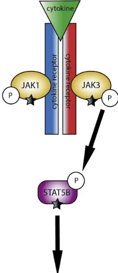
T-PLL have been identified. These include highly recurrent, largely exclusive, gain-of-function mutations involving IL2RG, JAK1/3, and STAT5B, which lead to constitutive STAT5 signaling (Figure 2)20-22. Deleterious mutations in EZH2, FBXW10 and CHEK2
may further contribute to the pathogenesis of T-PLL through their roles in DNA repair, epigenetic transcriptional regulation and proteasome degradation pathways.20 Further
TREATMENT
Due to the rarity of T-PLL, little published data exists regarding treatment. No randomized clinical trials have been conducted. The following recommendations are based on best available evidence and personal experience.
Watch and wait
Not all patients diagnosed with T-PLL require treatment immediately. Chemo-immunotherapy can be associated with significant toxicity and, aside from HSCT, current chemo-immunotherapy regimens in T-PLL are not curative. Furthermore, some patients present with an ‘indolent phase’ of the disease. Although disease progression eventually occurs, patients can be monitored for years before requiring intervention. Close monitoring (for example, blood count and clinical examination at regular intervals) is required as disease progression can be rapid and fatal. A pre-treatment lymphocyte doubling time (LDT) of less than 8.5 months has been shown to be associated with a worse outcome, 23 although an absolute lymphocyte count with LDT
should be taken into consideration when deciding upon treatment initiation. Indications for treatment include B symptoms, symptomatic anemia or thrombocytopenia, disease infiltration in the skin, lungs or CNS, and progressive disease demonstrated by an increasing lymphocytosis or rapidly enlarging spleen, liver or lymph nodes.
First line therapy
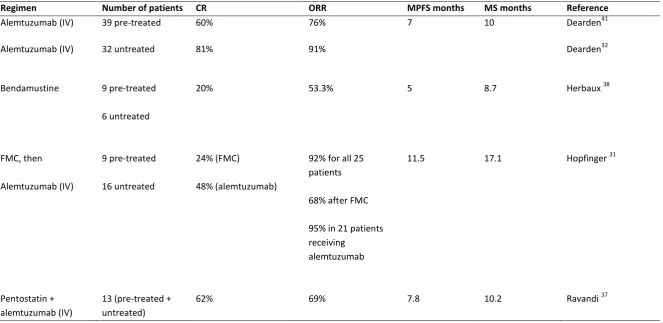
trial, patients should be offered an alemtuzumab (anti-CD52) – regimen. This was initially employed over 2 decades ago and was first used due to the strong CD52 expression on treatment-naïve T-PLL cells. Studies suggest an overall response rate (ORR) of >80% in the first-line setting and in 50-76% of relapsed-refractory cases (Table 1).24-27 Although progression-free survival (PFS) is longer when compared to
other therapies (over a year in responders), relapse invariably occurs and there are few long-term survivors, with a median overall survival (OS) from treatment of less than 2 years. For this reason, eligible patients should be considered for consolidation therapies such as HSCT. The results of alemtuzumab therapy compare favorably with outcomes reported with the use of purine analogues in which ORR are <50% and remission durations are less than one year.28-30 Single-agent pentostatin has shown the greatest
efficacy of all purine analogues in T-PLL,28 although no randomized controlled trials
have directly compared single agent pentostatin and alemtuzumab. The use of pentostatin is discussed further in relapsed/refractory disease. Small prospective studies have evaluated the use of alemtuzumab in combination with chemotherapy agents. For example, Hopfinger et al reported a prospective multi-center phase II trial investigating the use of fludarabine, mitoxantrone and cyclophosphamide (FMC) induction followed by alemtuzumab in 16 treatment-naïve patients and 9 previously-treated patients.31 The ORR to FMC was 68% increasing to 92% following the addition
be monitored for the early detection and management of CMV reactivation. Due to the risk of infertility with chemotherapy and HSCT, men and women should receive appropriate counselling and options for fertility preservation prior to commencing any treatment. Intravenous administration of alemtuzumab is more effective than subcutaneous administration.32 Infusion reactions are common with alemtuzumab and
measures should be employed to reduce the severity and occurrence of infusional reactions. One month following completion of therapy response to treatment should be measured by history, physical examination, full blood count, bone marrow aspirate and biopsy and computed tomography of the chest, abdomen and pelvis. Response is defined using the criteria created for disease assessment in chronic lymphocytic leukemia (CLL).
Post remission therapy
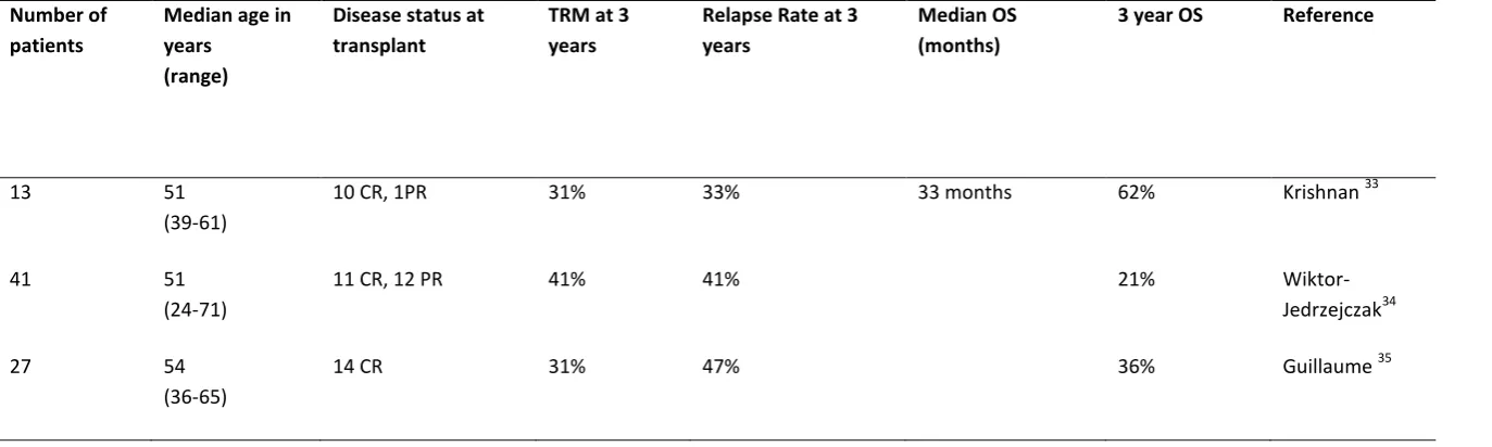
Approximately 80% of patients achieve a CR following alemtuzumab treatment. However, without additional therapy, a majority of patients will relapse within two years. HSCT is used to consolidate responses in eligible patients. A number of studies have investigated the use of HSCT in T-PLL and suggest that OS can be improved and in a minority of cases can achieve a cure (Table 2).33-36 The main challenges to contend
with are the treatment-related mortality (TRM) and risk of relapse. A retrospective study by Guillaume et al reported 27 patients undergoing allogeneic HSCT (14 of who were in CR at time of HSCT).35 The relapse rate at 3 years was 47%, with a TRM of 31%
and an OS of 36%. The European Group for Blood and Marrow Transplantation (EBMT) registry had 41 patients with T-PLL who had received an allogeneic HCT.34 3 year OS
year relapse rate from a smaller cohort of patients although a larger proportion of patients in our study were in CR, highlighting the importance of disease-status at time of HSCT.33 Although the number of patients is small, we also demonstrated similar OS
in patients who received an autologous-SCT compared to those who received an allogenic-SCT.33 Although not offering a cure, given lower risk of treatment-related
toxicity, autologous-SCT may be an option for less fit patients. Relapse following HSCT is usually within 2 years but can occur late. Given the increasing use of reduced-intensity conditioning and matched unrelated donors, as well as improvements in supportive care, more patients are eligible for HSCT, and the data which exists currently regarding HSCT may not be applicable to prospective cohorts of T-PLL patients.
Therapy for relapsed and refractory disease
Little data exists regarding the treatment of relapsed or refractory disease. Approximately half of the patients who relapse following a previous response to alemtuzumab can achieve a second disease remission with further alemtuzumab therapy, although this is usually of shorter duration. Flow cytometry should be repeated as T-PLL cells can lose CD52 expression.
Patients who fail to achieve a remission with single-agent alemtuzumab should have pentostatin added to the treatment regimen. Although no randomized trials have compared single-agent therapy with combination therapy, pentostatin has demonstrated efficacy as a single-agent in a small retrospective study. The ORR was 45% independent of previous treatment with a median PFS and OS of 6 months and 9 months respectively.28 A phase II study evaluated combination alemtuzumab with
ORR was 69% with a median OS and PFS of 10.2 and 7.8 months, respectively.37 Despite
adequate prophylaxis, common side effects included infection (including CMV reactivation) as well as neutropenia, thrombocytopenia, anemia and nausea.
Other treatment options include nelarabine or bendamustine, although durable remissions with these therapies are uncommon.30 Herbaux et al report 15 patients with
T-PLL treated with bendamustine, 7 of whom had failed front-line therapy with alemtuzumab. The ORR was 53% (20% CR), median PFS of 5 months and OS of 8.7 months, independent of prior exposure to alemtuzumab.38 Treatment of patients with
relapsed or refractory disease is currently suboptimal. Effective novel therapies are needed to improve the outcome for these patients.
Novel therapies
New approaches aim to utilize our expanding knowledge of T-PLL in order to target pathways involved in disease pathogenesis and resistance. Given the high frequency of mutations observed, and the perturbed signaling pathways, small molecule inhibitors targeting JAK-STAT pathway represents a therapeutic strategy available for patients. Pimozide, a STAT5 inhibitor, has been shown to induce apoptosis in primary T-PLL cells.20 Histone-deacetlyase inhibitors (HDACi) in combination with hypomethylating
agents aim to act synergistically to increase expression of silenced tumor suppressor genes. The combination of cladribine and alemtuzumab with or without an HDACi can overcome alemtuzumab resistance and induce expression of other molecules liable to targeting with additional agents.39 Cells with inactive ATM demonstrate impaired DNA
therefore selectively sensitize ATM-deficient tumor cells to killing.40 Finally, chimeric
antigen receptor natural killer cells targeting CD7 may represent a novel therapeutic avenue not yet explored.
SUMMARY
REFERENCES
1. Catovsky D, Galetto J, Okos A, Galton DA, Wiltshaw E, Stathopoulos G. Prolymphocytic leukaemia of B and T cell type. Lancet (London, England). 1973;2(7823):232-234.
2. Swerdlow S, Campo E, Harris N. World Health Organization Classification of Tumours of Haematopoietic and Lymphoid Tissues. Lyon: IARC Press 2008.
3. Matutes E, Brito-Babapulle V, Swansbury J, et al. Clinical and laboratory features of 78 cases of T-prolymphocytic leukemia. Blood. 1991;78(12):3269-3274.
4. Bellone M, Svensson AM, Zaslav AL, et al. Pediatric T-cell prolymphocytic leukemia with an isolated 12(p13) deletion and aberrant CD117 expression. Experimental hematology & oncology. 2012;1(1):7.
5. Mitton B, Coutre S, Willert J, et al. A pediatric case of T-cell prolymphocytic leukemia. Pediatric Blood & Cancer. 2015;62(6):1061-1062.
6. Moser AM, Quider AA, Groen JA, Shubinsky G, Kapelushnik J. A gamma/delta T-cell receptor prolymphocytic leukemia and CD4-/CD8- double-negative immunophenotype in a pediatric patient. Journal of pediatric hematology/oncology. 2015;37(4):e218-219.
7. Suarez F, Mahlaoui N, Canioni D, et al. Incidence, Presentation, and Prognosis of Malignancies in Ataxia-Telangiectasia: A Report From the French National Registry of Primary Immune Deficiencies. Journal of Clinical Oncology. 2015;33(2):202-208.
8. Michallet AS, Lesca G, Radford-Weiss I, Delarue R, Varet B, Buzyn A. T-cell prolymphocytic leukemia with autoimmune manifestations in Nijmegen breakage syndrome. Annals of hematology. 2003;82(8):515-517.
9. Ravandi F, O'Brien S. Chronic lymphoid leukemias other than chronic lymphocytic leukemia: diagnosis and treatment. Mayo Clinic proceedings. 2005;80(12):1660-1674.
10. Magro CM, Morrison CD, Heerema N, Porcu P, Sroa N, Deng AC. T-cell prolymphocytic leukemia: an aggressive T cell malignancy with frequent cutaneous tropism. Journal of the American Academy of Dermatology. 2006;55(3):467-477.
11. Herling M, Valbuena JR, Jones D, Medeiros LJ. Skin involvement in T-cell prolymphocytic leukemia. Journal of the American Academy of Dermatology. 2007;57(3):533-534.
12. Song SX, Willemze R, Swerdlow SH, Kinney MC, Said JW. Mycosis Fungoides. Report of the 2011 Society for Hematopathology/European Association for Haematopathology Workshop. 2013;139(4):466-490.
13. Maljaei SH, Brito-Babapulle V, Hiorns LR, Catovsky D. Abnormalities of chromosomes 8, 11, 14, and X in T-prolymphocytic leukemia studied by fluorescence in situ hybridization. Cancer genetics and cytogenetics. 1998;103(2):110-116.
14. Gritti C, Dastot H, Soulier J, et al. Transgenic Mice for MTCP1 Develop T-Cell Prolymphocytic Leukemia. Blood. 1998;92(2):368-373.
15. Virgilio L, Lazzeri C, Bichi R, et al. Deregulated expression of TCL1 causes T cell leukemia in mice. Proceedings of the National Academy of Sciences. 1998;95(7):3885-3889.
17. Costa D, Queralt R, Aymerich M, et al. High levels of chromosomal imbalances in typical and small-cell variants of T-cell prolymphocytic leukemia. Cancer Genetics. 2003;147(1):36-43.
18. Soulier J, Pierron G, Vecchione D, et al. A complex pattern of recurrent chromosomal losses and gains in T-cell prolymphocytic leukemia. Genes, chromosomes & cancer. 2001;31(3):248-254.
19. Le Toriellec E, Despouy G, Pierron G, et al. Haploinsufficiency of CDKN1B contributes to leukemogenesis in T-cell prolymphocytic leukemia. Blood. 2008;111(4):2321-2328.
20. Kiel MJ, Velusamy T, Rolland D, et al. Integrated genomic sequencing reveals mutational landscape of T-cell prolymphocytic leukemia. Blood. 2014;124(9):1460-1472.
21. Bellanger D, Jacquemin V, Chopin M, et al. Recurrent JAK1 and JAK3 somatic mutations in T-cell prolymphocytic leukemia. Leukemia. 2014;28(2):417-419. 22. Lopez C, Bergmann AK, Paul U, et al. Genes encoding members of the JAK-STAT
pathway or epigenetic regulators are recurrently mutated in T-cell prolymphocytic leukaemia. British journal of haematology. 2016;173(2):265-273. 23. Herling M, Patel KA, Teitell MA, et al. High TCL1 expression and intact T-cell receptor signaling define a hyperproliferative subset of T-cell prolymphocytic leukemia. Blood. 2008;111(1):328-337.
24. Pawson R, Dyer MJ, Barge R, et al. Treatment of T-cell prolymphocytic leukemia with human CD52 antibody. Journal of clinical oncology : official journal of the American Society of Clinical Oncology. 1997;15(7):2667-2672.
25. Ferrajoli A, O'Brien SM, Cortes JE, et al. Phase II study of alemtuzumab in chronic lymphoproliferative disorders. Cancer. 2003;98(4):773-778.
26. Keating MJ, Cazin B, Coutré S, et al. Campath-1H Treatment of T-Cell Prolymphocytic Leukemia in Patients for Whom at Least One Prior Chemotherapy Regimen Has Failed. Journal of Clinical Oncology. 2002;20(1):205-213.
27. Dearden CE, Matutes E, Cazin B, et al. High remission rate in T-cell prolymphocytic leukemia with CAMPATH-1H. Blood. 2001;98(6):1721-1726. 28. Mercieca J, Matutes E, Dearden C, MacLennan K, Catovsky D. The role of
pentostatin in the treatment of T-cell malignancies: analysis of response rate in 145 patients according to disease subtype. Journal of Clinical Oncology. 1994;12(12):2588-2593.
29. Kantarjian HM, Childs C, O'Brien S, et al. Efficacy of fludarabine, a new adenine nucleoside analogue, in patients with prolymphocytic leukemia and the prolymphocytoid variant of chronic lymphocytic leukemia. The American journal of medicine. 1991;90(2):223-228.
30. Gandhi V, Tam C, O'Brien S, et al. Phase I Trial of Nelarabine in Indolent Leukemias. Journal of Clinical Oncology. 2008;26(7):1098-1105.
31. Hopfinger G, Busch R, Pflug N, et al. Sequential chemoimmunotherapy of fludarabine, mitoxantrone, and cyclophosphamide induction followed by alemtuzumab consolidation is effective in T-cell prolymphocytic leukemia. Cancer. 2013;119(12):2258-2267.
33. Krishnan B, Else M, Tjonnfjord GE, et al. Stem cell transplantation after alemtuzumab in T-cell prolymphocytic leukaemia results in longer survival than after alemtuzumab alone: a multicentre retrospective study. British journal of haematology. 2010;149(6):907-910.
34. Wiktor-Jedrzejczak W, Dearden C, de Wreede L, et al. Hematopoietic stem cell transplantation in T-prolymphocytic leukemia: a retrospective study from the European Group for Blood and Marrow Transplantation and the Royal Marsden Consortium. Leukemia. 2012;26(5):972-976.
35. Guillaume T, Beguin Y, Tabrizi R, et al. Allogeneic hematopoietic stem cell transplantation for T-prolymphocytic leukemia: a report from the French society for stem cell transplantation (SFGM-TC). European journal of haematology. 2015;94(3):265-269.
36. Kalaycio ME, Kukreja M, Woolfrey AE, et al. Allogeneic Hematopoietic Cell Transplant for Prolymphocytic Leukemia. Biology of Blood and Marrow Transplant. 2010;16(4):543-547.
37. Ravandi F, Aribi A, O'Brien S, et al. Phase II Study of Alemtuzumab in Combination With Pentostatin in Patients With T-Cell Neoplasms. Journal of Clinical Oncology. 2009;27(32):5425-5430.
38. Herbaux C, Genet P, Bouabdallah K, et al. Bendamustine is effective in T-Cell prolymphocytic leukaemia. British journal of haematology. 2015;168(6):916-919. 39. Hasanali ZS, Saroya BS, Stuart A, et al. Epigenetic therapy overcomes treatment resistance in T-cell prolymphocytic leukemia. Science translational medicine. 2015;7(293):293ra102-293ra102.
40. Weston VJ, Oldreive CE, Skowronska A, et al. The PARP inhibitor olaparib induces significant killing of ATM-deficient lymphoid tumor cells in vitro and in vivo. Blood. 2010;116(22):4578-4587.
Table 1: Treatment Trials in T-PLL (> 10 patients)
CR, complete remission; ORR, overall response rate; MPFS, median progression-free survival; MS, median overall survival; IV, intravenous; FMC, fludarabine, mitoxantrone and cyclophosphamide.
Regimen Number of patients CR ORR MPFS months MS months Reference
Alemtuzumab (IV) 39 pre-treated 60% 76% 7 10 Dearden41
Alemtuzumab (IV) 32 untreated 81% 91% Dearden32
Bendamustine 9 pre-treated
6 untreated
20% 53.3% 5 8.7 Herbaux 38
FMC, then
Alemtuzumab (IV)
9 pre-treated
16 untreated
24% (FMC)
48% (alemtuzumab)
92% for all 25 patients
68% after FMC
95% in 21 patients receiving
alemtuzumab
11.5 17.1 Hopfinger 31
Pentostatin + alemtuzumab (IV)
13 (pre-treated + untreated)
Table 2: Allogeneic Stem Cell Transplant in T-PLL
TRM, transplant related mortality; OS, overall survival; CR, complete remission; PR, partial remission.
Number of patients
Median age in years
(range)
Disease status at transplant
TRM at 3 years
Relapse Rate at 3 years
Median OS (months)
3 year OS Reference
13 51
(39-61)
10 CR, 1PR 31% 33% 33 months 62% Krishnan 33
41 51
(24-71)
11 CR, 12 PR 41% 41% 21%
Wiktor-Jedrzejczak34
27 54
(36-65)
Figure 1: Peripheral blood smear from a patient with T-prolymphocytic leukemia demonstrating a ‘typical’ morphology.
The T-prolymphocytic leukemia cells are medium sized lymphoid cells with partial chromatin condensation and a visible nucleolus. The cytoplasm is basophilic with protrusions and an absence of granules.
Fig.1
Figure 2: A pathway diagram illustrates the interaction of JAK1, JAK3, and STAT5B during cytokine activation. Cytokine binding results in JAK autophosphorylaton, leading to STAT recruitment and activation through tyrosine phosphorylation. Activated STAT proteins then dimerize and translocate to the nucleus to regulate transcription of numerous genes involved in differentiation, proliferation, and survival. Mutated components of the JAK1-JAK3-STAT5B pathway are highlighted. Mutations have also been described in IL2RG, which is a cytokine receptor.