DOI 10.1007/s10086-004-0671-x
Y. Hosoo1 · M. Yoshida (*) · T. Okuyama
Laboratory of Bio-material Physics, Graduate School of
Bioagricultural Sciences, Nagoya University, Nagoya 464-8601, Japan Tel. ⫹81-52-789-4153; Fax ⫹81-52-789-4150
e-mail: [email protected] T. Imai
Laboratory of Biomass Resources Utilization, Graduate School of Bioagricultural Sciences, Nagoya University, Nagoya 464–8601, Japan
Present address:
1Graduate School of Science and Technology, Niigata University, Niigata 950-2181, Japan
Yoshihiro Hosoo · Masato Yoshida · Takanori Imai Takashi Okuyama
The effect of day length on diurnal differences in the innermost surface of
the S
2layer in differentiating tracheids
Received: February 4, 2004 / Accepted: August 20, 2004 ORIGINAL ARTICLE
Abstract This article describes the effect of day length
dur-ing the photoperiodic cycle on the diurnal differences in the innermost surface of developing secondary walls. Saplings of Cryptomeria japonica D. Don. were grown in growth chambers at constant temperature and relative humidity, but with different photoperiods. Samples of differentiating xylem were collected during the light and dark periods. The innermost surface of developing secondary walls in differ-entiating tracheids were observed using field emission scan-ning electron microscopy, and observations made during the light and dark periods were compared. In the saplings grown under long-day or short-day conditions, diurnal dif-ferences in aspects of the innermost surface of developing secondary walls were observed. Cellulose microfibrils were observed on the innermost surface of developing secondary walls during the light period when the volumes of differen-tiating cells were low, and amorphous material was observed during the dark period, when differentiating tracheids were turgid. The amorphous material was labeled with antiglucomannan antiserum. These results suggest that the range of day-length conditions set in this study does not affect the diurnal periodicity in the supply of cell wall com-ponents to the innermost surface of developing secondary walls.
Key words Cell wall formation · Cryptomeria japonica ·
Diurnal periodicity · Day-length · Tracheid
Introduction
Thin-walled cambial cells undergo dramatic transforma-tions including secondary wall deposition, bordered pit for-mation, lignification, and programmed cell death during secondary xylem differentiation.1
Secondary walls consist of cellulose microfibrils embedded in a lignin–hemicellulose matrix. Cellulose deposition increases cell wall thickness. In contrast, the deposition of hemicellulose and lignin increases cell wall density. In wood secondary walls, three main layers can be differentiated based on the orientation of the cellulose microfibrils: the outer, middle, and inner layers (S1, S2, and S3, respectively).
Differences in the innermost surface of developing sec-ondary walls of differentiating tracheids were seen between day and night using field emission scanning electron
micro-scopy (FE-SEM).2
Cellulose microfibrils were clearly evident during the day, and amorphous material was preva-lent at night. Using the immunogold labeling method, we demonstrated that the amorphous material was a matrix containing hemicellulose.3
Diurnal differences on the inner-most surface of developing secondary walls were also ob-served in saplings grown in climate chambers, in which all conditions were kept constant except the photoperiodic cycle. The diurnal differences were affected by light; cellu-lose microfibrils were clearly observed during the light pe-riod, and amorphous material was observed during the dark period, regardless of the time of sampling.4
Day length affects vegetative growth and reproductive differentiation. Many plants respond to day length as reli-able and predictreli-able indicators of seasonal progression. Because the diurnal differences on the innermost surface of developing secondary walls correspond to the light–dark cycle, the changes in day length during the photoperiodic cycle are likely to influence the diurnal differences.
The innermost surfaces of developing secondary walls in differentiating tracheids were observed using FE-SEM. The observations made during the light and dark periods were compared, and the relationship between the diurnal period-icity of cell wall formation and light was examined.
Materials and methods
Plant materials
Three-year-old cloned Cryptomeria japonica saplings (average height, 106 cm; average diameter 10 cm from the ground, 1.5 cm) were planted in pots (diameter, 20 cm; height, 30 cm) and grown in the field. At the beginning of June 2002, they were placed in two growth chambers (KG-50HLA; Koito, Yokohama, Japan) at a constant
tempera-ture (25° ⫾ 1°C) and constant relative humidity (70% ⫾
7%). In one growth chamber, the light cycle was set at 21 h of light (illuminance, 47 W/m2) and 3 h of darkness (21L3D; lights off at 1200 hours; lights on at 1500 hours). In the other chamber, the light cycle was set at 3 h of light and 21 h of darkness (3L21D; lights on at 1200 hours; lights off at 1500 hours). The saplings were irrigated with 200 ml of water at 1000 hours every day. After about 2 weeks, samples for FE-SEM observations were collected from both chambers just before lights-off (light period) and just before lights-on (dark period). The stems were cut into 5-cm-long segments and fixed in 3% glutaraldehyde/phosphate buffer (40 mM Na2HPO4 and 27 mM KH2PO4, adjusted to pH 7.0)
overnight at 4°C.
At the beginning of July 2002, more saplings were placed in two growth chambers. In one growth chamber, the light cycle was set at 18 h of light and 6 h of darkness (18L6D; lights off at 1200 hours; lights on at 1800 hours). In the other chamber, the light cycle was set at 6 h of light and 18 h of darkness (6L18D; lights on at 1200 hours; lights off at 1800 hours). The saplings were irrigated with 200 ml of water at 1000 hours every day. After about 2 weeks, samples were collected from both chambers just before lights-off (light period) and lights-on (dark period). The samples were pre-pared and fixed as above. We assumed that the environ-mental conditions in the growth chambers were uniform throughout the study.
Measurement of tangential strain on the inner bark
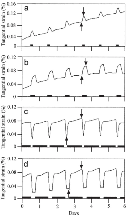
A knife was used to remove 15-mm squares of outer bark from the stem 10 cm above the ground. A 5-mm strain gauge (KFG-5–120-C-11; Kyowa, Tokyo, Japan) was glued tan-gentially to the surface of the inner bark using a cyanoacry-late adhesive (CC-33A; Kyowa). The strain gauge was connected to a strain meter (NEC-San-ei LoggerMate-DL 1200; NEC, Tokyo, Japan) by a three-wire connection. To prevent dehydration of the inner bark and to shield it from light, the strain gauge and inner bark were covered with layers of lanolin, vinyl, and aluminum foil. Measurements were made at 10-min intervals.
Scanning electron microscopy
Radial sections were studied because they were useful for detecting specific developmental stages of
differentiat-ing tracheids. Radial sections approximately 200µm thick
were cut from the fixed blocks using a freezing/sliding
microtome at ⫺20°C. The sections were washed with
dis-tilled water, treated with 50% sodium hypochlorite for 15 min to remove the protoplasm, and washed again (three times, 10 min each) with distilled water. Some sections were subjected to immunogold labeling before being prepared for FE-SEM.
Some sections were immersed in 50 mM glycine in phos-phate-buffered saline (PBS: 137 mM NaCl, 2.7 mM KCl, 8.1 mM Na2HPO4, and 1.5 mM KH2PO4, adjusted to pH 7.4)
for 15 min, incubated in blocking buffer (PBS containing 0.8% BSA) for 1 h, and then incubated in rabbit antiglu-comannan antiserum (diluted 1:20 in blocking buffer) for 2 h at 37°C. As a control, serial sections were incubated in preimmune serum or antiserum previously incubated with glucomannans at 0.5 mg/ml. The preparation and specificity of the antisera used in this study were described in detail previously.3
The sections were washed with PBS-T (PBS containing 0.5% Tween-20) six times for 10 min each and incubated for 2 h at 37°C with 15-nm-gold-labeled goat antirabbit IgG (Auro Probe EM GAR G15; Amersham, Little Chalfont, UK) diluted 1:50 in blocking buffer. The sections were washed six times for 10 min each in PBS-T, fixed with 2% glutaraldehyde/PBS for 5 min, and washed with distilled water six times for 5 min each.
All the sections were fixed in 2% osmium tetroxide for 2 h for conductive staining, dehydrated through a graded ethanol series, and processed using the t-butanol
freeze-drying method.5 The dried sections were coated with
ap-proximately 3.5-nm-thick platinum–palladium using an ion sputter coater (E-1030; Hitachi, Tokyo, Japan). Thereafter, the innermost surface of the developing secondary walls
in S2-forming tracheids was observed using a FE-SEM
(S-4500; Hitachi) at an accelerating voltage of 1.5 kV and working distance of 5 mm. Five to ten sections were pre-pared from each specimen and approximately ten different cells in the individual section were observed.
Results
Tangential strain
lights-off, continued to increase slowly until reaching a maximum just before lights-on, and decreased immediately after lights-on. All the sapling specimens were collected at the end of the light period, just before the tangential strain began to increase sharply, and at the end of the dark period, when the strain reached a maximum.
Scanning electron microscopy
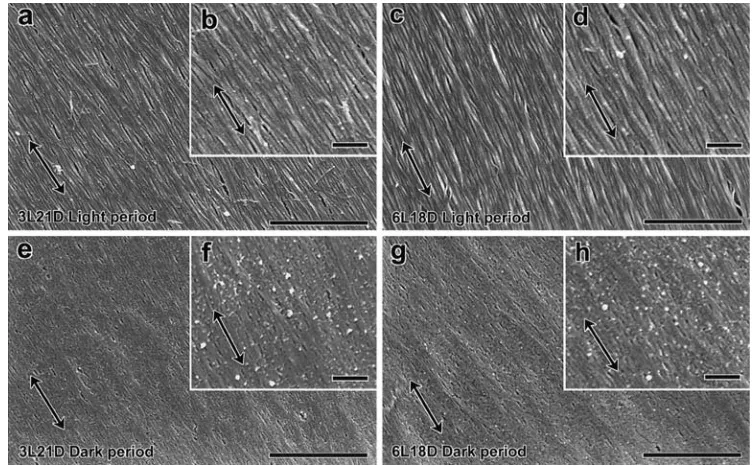
In the specimens collected from the long-day (21L3D and 18L6D) chambers during the light period, cellulose mi-crofibrils were clearly observed and amorphous material was rarely found on the innermost surface of developing secondary walls (Fig. 2a,c). Only a small amount of labeling was observed in the sections labeled with antiserum (Fig. 2b,d). In the specimens collected during the dark period, amorphous material was observed on the innermost sur-face of developing secondary walls (Fig. 2e,g), and a large
amount of immunogold labeling was observed as bright spherical particles in the amorphous material (Fig. 2f,h). In contrast, little labeling was observed in the sections labeled with preimmune serum or antiserum previously incubated with glucomannans.
The FE-SEM observations of the sapling specimens from the short-day (3L21D and 6L18D) chambers were the same as those for the long-day specimens (Fig. 3). Speci-mens collected during the light period clearly contained cellulose microfibrils (Fig. 3a,c) and were rarely labeled with immunogold (Fig. 3b,d). Specimens collected during the dark period, had amorphous material on the innermost surface of developing secondary walls (Fig. 3e,g) and exhib-ited strong antiglucomannan labeling (Fig. 3f,h). Few labels were observed in the sections labeled with preimmune se-rum or antisese-rum previously incubated with glucomannans. In each section, differences in aspects of the innermost sur-face of developing secondary walls were not seen between differentiating tracheids at early and late S2-forming stages.
Each photograph is representative of a large number of photographs of the innermost surface of developing secondary walls obtained at specific times from FE-SEM observations.
Discussion
We studied the effect of day length on the diurnal changes seen on the innermost surface of developing secondary walls using FE-SEM. The S2 layer composes the largest part
of the secondary cell wall in conifer tracheids, and most differentiating xylem cells are S2-forming tracheids.
There-fore, this study focused on the innermost surface of the developing secondary walls in S2-forming tracheids.
In the saplings grown under long-day conditions (21L3D and 18L6D) or short-day conditions (3L21D and 6L18D), cellulose microfibrils were clearly observed during the light period, and amorphous material was observed during the dark period. These results indicate that diurnal differences in aspects of the innermost surface of developing secondary walls occur in saplings grown under the range of day-length conditions used in this study.
SEM observations of immunogold-labeled cells revealed the topology of antigens on the cell surface. The resolution of SEM is adequate for detecting colloidal gold particles as small as 5–20 nm.6–10
We previously examined the specificity of the antiglucomannan antiserum used in this study and found that the antiserum is specific for glucomannan and
can be used for glucomannan labeling of the cell wall.3
Therefore, the immunogold labeling observed on the inner-most surface of developing secondary walls in this study reflects the localization of glucomannans.
Our results indicate that the amorphous material on the innermost surface of developing secondary walls during the dark period contains glucomannan. We propose that this amorphous material contains a hemicellulose matrix. The amorphous material was labeled with antiglucomannan an-tiserum regardless of the day length. It is thought that the
composition of the amorphous material rarely changes due to differences in the day length of the photoperiodic cycle.
It is possible that some of the glucomannans in the amor-phous material can be extracted with reagents, such as hypochlorite. All specimen sections were prepared and observed using exactly the same processes and conditions in this study. Even if some of the glucomannans were removed by hypochlorite, the amount of the supply of glucomannan to the innermost surface of developing secondary walls from the matrix during the dark period was greater than that during the light period.
The water status of a tree is reflected in changes in stem diameter.11,12
The diurnal fluctuation of stem diameter is caused mainly by changes in the water status of cells in the
cambium and developing cells in the xylem and phloem.13–16
During xylem cell development in woody plants, cell size is determined by the interaction between cell turgor and the cell wall properties of the developing xylem cells.17,18
The volumetric changes of differentiating cells can be estimated from changes in the tangential strain on the inner bark.19–21
The strain is proportional to the volume of differentiating cells. Our results indicate that the pattern of tangential strain corresponds to the day length of the photoperiodic
Fig. 2a–h. Innermost surfaces of the radial wall during secondary wall formation in specimens col-lected from the long-day cham-bers. Some sections were not labeled with any antibodies (a, c, e, g), and others were labeled with antiglucomannan antiserum (b, d, f, h). The longitudinal cell axes in the micrographs are verti-cal. The double-headed arrows show the orientation of cellulose microfibrils in each micrograph. a, b Specimens collected from the 21L3D chamber during the light period. c, d Specimens collected from the 18L6D chamber during the light period. e, f Specimens collected from the 21L3D cham-ber during the dark period. g, h Specimens collected from the 18L6D chamber during the dark period. Bars a, c, e, g 1µm; b, d, f, h 200 nm
cycle, such that the tangential strain is high during the dark period and low during the light period. This suggests that there is a diurnal periodicity of the volumetric changes in differentiating cells that corresponds to the 24-h light–dark cycle; that is in accord with the findings in our previous study.4
We assumed that the environmental conditions in the growth chambers were uniform throughout the study and that all saplings in the chambers grew under the same con-ditions. Therefore, the changes in tangential strain were applied to the analysis of all the saplings used for FE-SEM observations. When specimens were collected during the light period, the volumes of the differentiating cells were low; when specimens were collected during the dark period, the differentiating cells were fully turgid. In other words, cellulose microfibrils were observed on the innermost sur-face of developing secondary walls during the light period when the volumes of differentiating cells were low as a result of water loss by transpiration, and the matrix material was observed during the dark period when differentiating cells were turgid as a result of imbibition. Whether the turgor pressure controls the synthesis of cell wall compo-nents or their deposition to the cell wall has not been eluci-dated yet. This study clarified that the diurnal changes in aspects of the innermost surface of developing secondary walls corresponded to the day length of the photoperiodic cycle and volumetric changes of differentiating cells. Per-haps the diurnal differences in the innermost surface of developing secondary walls reflect changes in phenomena such as the turgor pressure or transpiration. Conversely, the diurnal differences may be affected by the circadian rhythm, which might change with day length.
The diurnal differences in the innermost surface of developing secondary walls were observed in the saplings grown under long-day conditions or short-day conditions. Cellulose microfibrils were observed during the light pe-riod, and amorphous material was observed during the dark period. The amorphous material contained abundant levels of glucomannans regardless of the day length, suggesting that the composition of the material rarely changed due to differences in the day length of the photoperiodic cycle. These findings indicate that the range of day-length condi-tions set in this study do not affect the diurnal periodicity in the supply of cell wall components to the innermost surface of developing secondary walls.
References
1. Savidge RA (1996) Xylogenesis. Int Assoc Wood Anat J 17:269– 310
2. Yoshida M, Hosoo Y, Okuyama T (2000) Periodicity as a factor in the generation of isotropic compressive growth stress between microfibrils in cell wall formation during a twenty-four hour pe-riod. Holzforschung 54:469–473
3. Hosoo Y, Yoshida M, Imai T, Okuyama T (2002) Diurnal differ-ences in the amount of immunogold-labeled glucomannans de-tected with field emission scanning electron microscopy at the innermost surface of developing secondary walls of differentiating conifer tracheids. Planta 215:1006–1012
4. Hosoo Y, Yoshida M, Imai T, Okuyama T (2003) Diurnal differ-ences in the innermost surface of the S2 layer in differentiating tracheids of Cryptomeria japonica corresponding to a light–dark cycle. Holzforschung 57:567–573
5. Inoue T, Osatake H (1988) A new drying method of biological specimens of scanning electron microscopy: the t-butyl alcohol freeze-drying method. Arch Histol Cytol 51:53–59
6. Osumi M, Yamada H, Kobori H, Yaguchi H (1992) Observation of colloidal gold particles on the surface of yeast protoplasts with UHR-LVSEM. J Electron Microsc 41:392–396
7. Osawa T, Yoshida Y, Tsuzuki F, Nozaka M, Takashiro M, Nozaka Y (1999) The advantage of osmium conductive metal coating for the detection of the colloidal gold-conjugated antibody by SEM. J Electron Microsc 48:665–669
8. Awano T, Takabe K, Fujita M, Daniel G (2000) Deposition of glucuronoxylans on the secondary cell wall of Japanese beech as observed by immuno-scanning electron microscopy. Protoplasma 212:72–79
9. Suzuki E (2002) High-resolution scanning electron microscopy of immunogold-labelled cells by the use of thin plasma coating of osmium. J Microsc 208:153–157
10. Elrandsen S, Chen Y, Frethem C, Detry J (2003) High-resolution backscatter electron imaging of colloidal gold in LVSEM. J Microsc 211:212–218
11. Sheriff DW, Whitehead D (1984) Photosynthesis and wood struc-ture in Pinus radiata D. Don during dehydration and immediately after rewatering. Plant Cell Environ 7:53–62
12. Horzog KM, Hasler R, Thum R (1995) Diurnal changes in the radius of a subalpine Norway spruce stem: their relation to the sap flow and their use to estimation transpiration. Trees 10:94–101 13. Klepper B, Browning VD, Taylor HM (1971) Stem diameter in
relation to plant water status. Ecology 45:149–155
14. Molz FJ, Klepper B (1973) On the mechanism of water-stress-induced stem deformation. Agron J 65:469–473
15. Zweifel R, Item H, Hasler R (2001) Link between diurnal stem radius changes and tree water relations. Tree Physiol 21:869–877 16. Perämäki M, Nikinmaa E, Sevanto S, Ilvesniemi H, Siivola E, Hari
P, Vesara T (2001) Tree stem diameter variations and transpiration in Scots pine: an analysis using a dynamic sap flow model. Tree Physiol 21:889–897
17. Abe H, Funada R, Ohtani J, Fukazawa K (1995) Changes in the arrangement of microtubules and microfibrils in differentiating conifer tracheids during the expansion of cells. Ann Bot 75:305– 310
18. Abe H, Funada R, Ohtani J, Fukazawa K (1997) Changes in the arrangement of cellulose microfibrils associated with the cessation of cell expansion in tracheids. Trees 11:328–332
19. Okuyama T, Yoshida M, Yamamoto H (1995) An estimation of the turgor pressure change as one of the factors of growth stress generation in cell walls (in Japanese). Mokuzai Gakkaishi 41:1070– 1078
20. Yoshida M, Yamamoto O, Okuyama T (2000) Strain change on the inner bark surface of an inclined coniferous sapling producing compression wood. Holzforschung 54:664–668