Research and Reports in Neonatology
Neonatal hypokalemia
Dilek Sarici1 S Umit Sarici2
1Kecioren Research and Education
Hospital, Kecioren, Ankara, 2Chief of
Division of Neonatology, Division of Neonatology, Department of Pediatrics, Gulhane Military Medical Academy, Ankara, Turkey
Correspondence: S Umit Sarici Division of Neonatology, Department of Pediatrics, Gulhane Military Medical Academy, Etlik-06018, Ankara, Turkey Tel +90 532 255 58 41
Fax +90 312 361 7074 Email [email protected]
Abstract: In this article, distribution of potassium (K+) in body fluids, pathophysiology,
causes, clinical signs and symptoms, and the evaluation and treatment of neonatal hypokalemia are reviewed. K+ is the most important intracellular cation and normal serum K+ is stabilized
between 3.5 and 5.5 mEq/L. Hypokalemia may be caused by increased renal losses, increased extrarenal (gastrointestinal) losses, redistribution or prolonged insufficient K+ intake. Clinical
signs and symptoms occur as the result of functional changes in striated muscle, smooth muscle, and the heart. Hypokalemia is usually asymptomatic when K+ levels are between
3.0 and 3.5 mEq/L; however, there may sometimes be slight muscle weakness. Moderate hypokalemia is observed when serum K+ is between 2.5 and 3.0 mEq/L. Proximal muscle
weakness is observed most commonly in lower extremities; cranial muscles are normal, but constipation and distention are prominent. Severe hypokalemia develops when serum K+ falls
below 2.5 mEq/L. Rhabdomyolysis, myoglobinuria, severe muscle weakness, paralysis, respi-ratory distress, and respirespi-ratory arrest are observed. The clinical signs and symptoms may be unremarkable in cases of chronically developing hypokalemia; however, appropriate treatment is essential when serum K+ level falls below 2.5 mEq/L as the most dangerous complication
of hypokalemia is fatal cardiac arrythmia, and changes visible with electrocardiography may not always correlate with the level of hypokalemia. Sodium (Na+), K+, chloride (Cl-),
bicar-bonate, creatinine, blood sugar, magnesium (Mg), plasma renin activity, aldosterone, and blood gases should be investigated by laboratory testing. Aspartate aminotransferase, alanine aminotransferase, creatinine kinase, and creatinine kinase isoenzyme MB should be studied if rhabdomyolysis is suspected. In urine sample density, pH, Na+, K+, Cl-, Mg, creatinine,
and myoglobinuria (blood reaction is positive in the absence of erythrocytes on microscopic examination of urine) should be investigated. The primary aim of therapy is to prevent and treat life-threatening cardiac and muscular complications. However, in the presence of severe symptomatic hypokalemia and gastrointestinal problems such as ileus, the intravenous route may be used in cases where serum K+ level is usually below 2.6 mEq/L. K+ given in intravenous
fluids should not exceed 40 mEq/L. In case of emergency, 0.3–1 mEq/kg of K+ may be given
intravenously over 1 hour. When higher concentrations (60–80 mEq/L) are needed, infusion through a central vein under electrocardiography monitoring may be used.
Keywords: neonatal, hypokalemia, newborn
Introduction
Potassium (K+) is the most important intracellular cation. The distribution of K+ inside
and outside of the cell is 98% and 2%, respectively. Normal serum K+ is stabilized
between 3.5 and 5.5 mEq/L. Changes in pH values of blood and other body components may lead to changes in serum K+ levels via changes in intracellular and extracellular
Dove
press
R E v i E w
open access to scientific and medical research
Open Access Full Text Article
Research and Reports in Neonatology downloaded from https://www.dovepress.com/ by 118.70.13.36 on 27-Aug-2020
For personal use only.
Number of times this article has been viewed
This article was published in the following Dove Press journal: Research and Reports in Neonatology
K+ concentrations. An increase of 0.1 U in blood pH (towards
alkalosis) causes a decrease of 0.3–1.3 mEq/L in serum K+
concentration, with the entry of K+ into the cell.1
K+ equilibrium and distribution provided by the Na+-K+
-ATP′ase pump between the intracellular and extracellular regions is the major determinant of membrane potential at rest. This is significant for the stabilization and excitability of cell membranes, especially those of neuromuscular tissues.2
The most dangerous complication of hypokalemia is fatal cardiac arrhythmias.
Total body K+ is provided by the equilibrium occurring
between K+ intake, and loss via urine and the gastrointestinal
tract.3 Almost all of K+ ingested through diet is absorbed. The
kidneys secrete more than 90% of daily intake, and are the organs primarily responsible for the elimination of K+. The
kidneys are only capable of secreting half of very high-dose K+ (over 4–6 hours), with the rest transiently distributed
into the cell. This intracellular distribution, which has only a very limited capacity, nevertheless has a very important role in the equilibrium of acute changes in serum K+. Even
the transfer of small amounts (1%–2%) of intracellular K+
into the extracellular region can easily increase serum K+ to
dangerous levels. Many factors affect the distribution of K+
between the intracellular and extracellular regions, and these factors are summarized in Table 1.
Under normal conditions, K+ excretion via the
gastro-intestinal route is negligible; however, colonic excretion increases in the presence of chronic renal failure, dominating the role of the kidneys. Any disorder in renal K+ equilibrium
may lead to excessive loss or accumulation.
Causes of neonatal hypokalemia
Neonatal hypokalemia may be caused by increased renal losses, increased extrarenal (gastrointestinal) losses, redistri-bution, or prolonged insufficient K+ intake. Laboratory data
for blood pressure, acid–base status, electrolytes, blood-urine osmolality, and renin-aldosterone axis should be collected before starting treatment in any patient with hypokalemia. Causes of neonatal hypokalemia are listed in Table 2.2–9
Table 1 Factors affecting the distribution of K+ between the
intracellular and extracellular regions
insulin increase of it causes hypokalemia, and decrease of it causes hyperkalemia
Catecholamines β-agonists cause hypokalemia, and β-antagonists cause hyperkalemia
Acid–base status Metabolic alkalosis causes hypokalemia and metabolic acidosis causes hyperkalemia Tissue injury Causes hyperkalemia
Table 2 Causes of neonatal hypokalemia
Increased renal losses
with hypertension Mineralocorticoid excess Primary aldosteronism
Congenital adrenal hyperplasia (17α-hydroxylase deficiency, 11β-hydroxylase deficiency)
Hyperreninemic hyperaldosteronism
Hyperaldosteronism that may be suppressed with glucocorticoid Exogen mineralocorticoids (9-alpha fluorocortisol, liquorice roots) Cushing’s syndrome
Liddle’s syndrome with normal blood pressure with acidosis
Renal tubular acidosis Diabetic ketoacidosis with alkalosis vomiting Diuretics
Congenital chloridorrhea
Bartter syndrome, pseudo-Bartter syndrome (cystic fibrosis, maternal eating disorder)
Gitelman syndrome
Nephrogenic diabetes insipidus Magnesium deficiency
Normotensive hyperaldosteronism with normal acid–base status
Healing stage of acute tubular necrosis Postobstructive diuresis
Drugs (amphotericin B, penicillins, foscarnet, aminoglycosides, vancomycin, B12 vitamin treatment, high-dose adrenalin, steroids)
Extrarenal losses
Diarrhea
Gastrointestinal fistulas, ileostomy villous adenoma
Malabsorption Excessive laxative use Excessive diaphoresis Use of dialysate with low K+
Redistribution
Alkalosis insulin
Calcium channel blockers Theophylline, caffeine Lithium
β-agonists Barbiturate coma Thyrotoxicosis Hypothermia Acute brain injury Barium intoxication
Frozen-packed red blood cell transfusion Familial hypokalemic periodic paralysis Prolonged insufficient K+ intake
Prolonged starvation
Insufficient K+ support in total parenteral nutrition
Dovepress Sarici and Sarici
Research and Reports in Neonatology downloaded from https://www.dovepress.com/ by 118.70.13.36 on 27-Aug-2020
Clinical signs and symptoms
of neonatal hypokalemia
Clinical signs and symptoms occur as the result of functional changes in striated muscle, smooth muscle, and the heart.10,11
Hypokalemia is usually asymptomatic when K+ levels are
between 3.0–3.5 mEq/L; however, there may sometimes be a slight muscle weakness.11 Moderate hypokalemia is observed
when serum K+ is between 2.5–3.0 mEq/L. Proximal muscle
weakness is observed most commonly in lower extremities; cranial muscles are normal, but constipation and distention are prominent. Severe hypokalemia develops when serum K+ falls
below 2.5 mEq/L. Rhabdomyolysis, myoglobinuria, severe muscle weakness, paralysis, respiratory distress, and respira-tory arrest are observed. Fasciculation and tetany are observed in muscles. Electrocardiography (ECG) changes such as an increase in the amplitude of P-waves, prolongation in PR and QT intervals, decrease in the amplitude of T-waves, inversion in T-waves, depression in ST segments, and the appearance of U-waves are observed as cardiac findings (Figure 1).11
However, ECG changes may not always correlate with the level of hypokalemia. Left ventricular hypertrophy and heart failure may be detected, and the risks of digoxin toxicity, dysrhythmia, and sudden death increase. It also weakens the effect of insulin. Paralytic ileus and gastric dilatation develop when the smooth muscles are affected.
The clinical signs and symptoms may be unremarkable in cases of chronically developing hypokalemia; however, appropriate treatment is essential when serum K+ levels fall
below 2.5 mEq/L, as the most dangerous complication of hypokalemia is fatal cardiac arrhythmias, and ECG changes
may not always correlate with the level of hypokalemia. It should also be noted that severe hypokalemia inversely affects growth and development, and its effects on the Na+-K+-ATP′ase
pump and all kinds of muscles must be considered.
Rhabdomyolysis may affect renal function, as renal con-centration capacity decreases with prolonged hypokalemia and causes polyuria. Prolonged hypokalemia increases urinary chloride (Cl-) loss, decreases bicarbonate and
citrate excretion, and increases ammonia synthesis. Consequently, persistent metabolic alkalosis develops with hypokalemia.11
Evaluation of neonatal hypokalemia
Vomiting, diarrhea, ileostomy, nasogastric drainage, and drugs (ie, use of diuretics in babies with bronchopulmonary dysplasia causes hypokalemia with alkalosis) should be questioned in the anamnesis. Na+, K+, Cl-, bicarbonate, creatinine, blood
sugar, magnesium (Mg), plasma renin activity, aldosterone, and blood gases should be investigated by laboratory testing. Aspartate aminotransferase, alanine aminotransferase, creatinine kinase, and creatinine kinase isoenzyme MB should be studied if rhabdomyolysis is suspected. In urine sample density, pH, Na+, K+, Cl-, Mg, creatinine, and myoglobinuria
(blood reaction is positive in the absence of erythrocytes on microscopic examination of urine) should be investigated. Gastric dilatation and ileus should be investigated by upright abdominal X-ray. An ECG should be ordered for cardiac findings and echocardiography may be necessary.
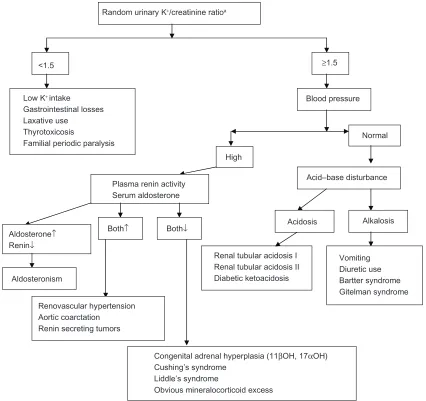
Measurement of blood pressure, blood gas analysis, and measurement of urinary Na+, K+, Cl-, Mg and creatinine
Figure 1 Electrocardiography changes in hypokalemia.
Dovepress Neonatal hypokalemia
Research and Reports in Neonatology downloaded from https://www.dovepress.com/ by 118.70.13.36 on 27-Aug-2020
are the most important steps in the etiologic evaluation of hypokalemia (Figure 2).12,13 In the evaluation of patients,
the first step is to determine whether the ratio of urinary K+
to creatinine (mmol:mmol) is below or above 1.5.14 If Mg is
high in urine, renal Mg loss should be considered. If uri-nary Cl- level is below 10 mEq/L, gastrointestinal losses
(vomiting, pyloric stenosis, drainage, fistulas, ileostomy, diarrhea, and chloridorrhea) should be considered.11,13 In
babies with a urinary Cl- level above 20 mEq/L, diuretic
use and Bartter and Gitelman syndromes should be considered.
In cases of increased blood pressure aldosteronism, Cushing’s syndrome, and congenital adrenal hyperplasia (17 alpha hydroxylase and 11 beta hydroxylase deficiencies) should be considered. If urinary (Na+ + K+) - Cl- is $–10,
gastrointestinal K+ loss with normal gap metabolic acidosis
should be considered. If urinary (Na++ K+) - Cl- is #–10,
renal K+ loss, renal tubular acidosis, drugs, and ureteral
diversion should be considered. Vomiting, nasogastric drainage and prolonged diuretic use should be considered if urinary Cl- is below 20 mEq/L, with hypokalemia and
meta-bolic alkalosis. Diuretic use and mineralocorticoid increase should be considered if urinary Cl- is above 20 mEq/L with
hypokalemia and metabolic alkalosis.11
Treatment of neonatal hypokalemia
The primary aim of therapy is to prevent and treat life-threatening cardiac and muscular complications. The secondary aim is to replenish the body’s K+ stores.
There is no absolute way of determining real K+ deficit,
Random urinary K+/creatinine ratioa
<1.5 ≥1.5
Low K+ intake Gastrointestinal losses Laxative use
Thyrotoxicosis
Familial periodic paralysis
Blood pressure
Normal
High
Acid–base disturbance
Acidosis Alkalosis
Renal tubular acidosis I Renal tubular acidosis II Diabetic ketoacidosis
Vomiting Diuretic use Bartter syndrome Gitelman syndrome Plasma renin activity
Serum aldosterone
Aldosterone↑
Renin↓
Aldosteronism
Both↑
Renovascular hypertension Aortic coarctation
Renin secreting tumors
Both↓
Congenital adrenal hyperplasia (11βOH, 17αOH) Cushing’s syndrome
Liddle’s syndrome
Obvious mineralocorticoid excess
Figure 2 Algorithma used in the evaluation of neonatal hypokalemia.
Note: ammol/L K+/mmol/L creatinine.
Dovepress Sarici and Sarici
Research and Reports in Neonatology downloaded from https://www.dovepress.com/ by 118.70.13.36 on 27-Aug-2020
Research and Reports in Neonatology
Publish your work in this journal
Submit your manuscript here: http://www.dovepress.com/research-and-reports-in-neonatology-journal
Research and Reports in Neonatology is an international, peer-reviewed, open access journal publishing original research, reports, editorials, reviews and commentaries on neonatal health. The manuscript manage-ment system is completely online and includes a very quick and fair
peer-review system. Visit http://www.dovepress.com/testimonials.php to read real quotes from published authors.
as there is no correlation between plasma K+ concentration
and the body’s K+ stores. A decrease of 1 mEq/L in serum
K+ concentration with K+ loss usually refers to a 10%–30%
decrease in body K+. In such conditions as acidosis and
hyperosmolarity, plasma K+ concentration may reflect a
lower value than actual K+ stores, and rapid correction of
acidosis with bicarbonate may lower serum K+
concentra-tion rapidly.
The safest treatment of K+ is via the oral/enteral route.
The normal daily required intake of K+ is 1–2 mEq/kg/day.
However, in the presence of severe symptomatic hypokalemia and gastrointestinal problems such as ileus, the intravenous route may be used in cases where serum K+ level is usually
below 2.6 mEq/L. K+ given in intravenous fluids should not
exceed 40 mEq/L. In case of emergency, 0.3–1 mEq/kg of K+ may be given intravenously over 1 hour.15 When higher
concentrations (60–80 mEq/L) are needed, infusion through a central vein under ECG monitoring may be used. Dextrose should not be used in initial fluids because increases in insulin secretion secondary to dextrose infusion may lower plasma K+ concentrations even further.
The choice of the type of K+ salt depends on the clinical
situation. KCl is usually appropriate if hypovolemia is present. In the presence of simultaneous metabolic acidosis, other K+
salts producing K+ bicarbonate, K+ citrate, and K+ acetate
may be given. In the presence of a phosphate-depleting situation such as diabetic ketoacidosis, K+ phosphate may
be used. It should be kept in mind that correction of total body K+ deficit may take days and even weeks. In cases of
hypokalemia resistant to treatment, hypomagnesemia should be considered. In these cases, K+ levels normalize following
magnesium treatment.
Disclosure
This article was published as a chapter in Turkish in the
Textbook of Pediatric Endocrinology edited by Selim
Kurtoglu and published by Nobel Tip in Istanbul, Turkey in 2011. The authors report no conflicts of interest in this work.
References
1. Simmons CF. Fluid and electrolyte management. In: Cloherty JP, Stark AR, editors. Manual of Neonatal Care. Philadelphia, PA: Lippincott Williams and Wilkins; 1998:87–100.
2. Chadha V, Alon US. Asid-baz ve elektrolit dengesizlikleri [Distur-bances of acid-base status and electrolyte imbalances]. In: Narli N, Balat A Bolum, editors. Pediatrik Tani ve Tedavide Pratik Yaklasimlar
[Practical approaches in pediatric diagnosis and treatment]. Istanbul, Turkey: Nobel Tip Kitabevleri; 2007:447–464. Turkish.
3. Ors R. Yenidoganda hiperpotasemi ve hipopotasemi [Hyperpotasemia and hypopotasemia in the newborn]. Turkiye Klinikleri J Pediatr Sci. 2008;4(2):113–118. Turkish.
4. Weiss-Guillet EM, Takala J, Jakob SM. Diagnosis and management of electrolyte emergencies. Best Pract Res Clin Endocrinol Metab. 2003;17(4):623–651.
5. Nanji AA. Drug-induced electrolyte disorders. Drug Intell Clin Pharm. 1983;17(3):175–185.
6. Fang W, Chen JY, Fang Y, Huang JL. Epinephrine overdose-associated hypokalemia and rhabdomyolysis in a newborn. Pharmacotherapy. 2005;25(9):1266–1270.
7. Rastergar A, Soleimani M. Hypokalemia and hyperkalemia. Postgrad Med J. 2001;77(914):759–764.
8. Le J, Adler-Shohet FC, Nguyen C, Lieberman JM. Nephrotoxicity associated with amphotericin B deoxycholate in neonates. Pediatr Infect Dis J. 2009;28(12):1061–1063.
9. Alfonzo AV, Isles C, Geddes C, Deighan C. Potassium disorders-clinical spectrum and emergency management. Resuscitation. 2006;70(1):10–25.
10. Mandal AK. Hypokalemia and hyperkalemia. Med Clin North Am. 1997;81(3):611–639.
11. Schaffer TJ, Wolford RW. Disorders of potassium. Emerg Med Clin North Am. 2005;23(3):723–747.
12. Assadi F. Diagnosis of hypokalemia: a problem-solving approach to clinical cases. Iran J Kidney Dis. 2008;2(3):115–122.
13. Lim S. Approach to hypokalemia. Acta Med Indones. 2007;39(1): 56–64.
14. Groeneveld JH, Sijpkens YW, Lin SH, Davids MR, Halperin ML. An approach to the patient with severe hypokalaemia: the potassium quiz.
QJM. 2005;98(4):305–316.
15. Galloway E, Doughty L. Electrolyte emergencies and acute renal failure in pediatric critical care. Clin Ped Emerg Med. 2007;8(3):176–189.
Dovepress
Dove
press
Neonatal hypokalemia
Research and Reports in Neonatology downloaded from https://www.dovepress.com/ by 118.70.13.36 on 27-Aug-2020