University of Pennsylvania
ScholarlyCommons
Publicly Accessible Penn Dissertations
1-1-2014
Epithelial Cell Shape Changes During Lung
Branching Morphogenesis: The Role of Wnt/Fzd2
signaling in directing new branch formation
Rachel S. Kadzik
University of Pennsylvania, [email protected]
Follow this and additional works at:
http://repository.upenn.edu/edissertations
Part of the
Cell Biology Commons
, and the
Developmental Biology Commons
This paper is posted at ScholarlyCommons.http://repository.upenn.edu/edissertations/1323
For more information, please [email protected].
Recommended Citation
Kadzik, Rachel S., "Epithelial Cell Shape Changes During Lung Branching Morphogenesis: The Role of Wnt/Fzd2 signaling in directing new branch formation" (2014).Publicly Accessible Penn Dissertations. 1323.
Epithelial Cell Shape Changes During Lung Branching Morphogenesis:
The Role of Wnt/Fzd2 signaling in directing new branch formation
Abstract
Formation of the intricately branched mammalian lung requires precise coordination between the epithelium and mesenchyme over the course of development. This coordination is mediated by molecular signaling between the two tissue compartments. How these signaling pathways coordinate changes in cellular and tissue morphology to give rise to the highly ramified branched network of the lung is not well understood. In this work, I show that signaling through Frizzled 2 (Fzd2) is required for promoting changes in epithelial cell shape that lead to tissue-level changes needed for branching morphogenesis in the lung. Through analysis of both fixed lungs and live imaging lung explants, I was able to identify the changes in individual cell
morphology and epithelial tissue organization that occur during new domain branch formation. Using this model, I characterized the defect in branching morphogenesis that arises due to loss of Fzd2 in the lung epithelium. I found that Fzd2 affects apical localization of phospho-myosin light chain 2, and through the RhoA pathway mediates changes in cell shape that lead to thickening of the lung epithelium and ultimately bending of the epithelium. These studies provide a mechanistic link between the Wnt signaling pathway and changes in cell morphology that lead to branching morphogenesis of the lung epithelium.
Degree Type
Dissertation
Degree Name
Doctor of Philosophy (PhD)
Graduate Group
Cell & Molecular Biology
First Advisor
Edward E. Morrisey
Keywords
Branching, Epithelial, Fzd2, Lung, Morphogenesis, Wnt
Subject Categories
Cell Biology | Developmental Biology
EPITHELIAL CELL SHAPE CHANGES
DURING LUNG BRANCHING MORPHOGENESIS:
THE ROLE OF WNT/FZD2 SIGNALING IN DIRECTING
NEW BRANCH FORMATION
Rachel S. Kadzik
A DISSERTATION
in
Cell and Molecular Biology
Presented to the Faculties of the University of Pennsylvania
in
Partial Fulfillment of the Requirements for the
Degree of Doctor of Philosophy
2014
Supervisor of Dissertation:
_____________________
Edward E. Morrisey, Ph.D., Professor of Medicine
_____________________
Daniel Kessler, Ph.D., Associate Professor of Cell and Developmental Biology
Graduate Group Chairperson
Dissertation Committee:
Peter Klein, MD, Ph.D., Professor of Medicine
Catherine Lee May, Ph.D., Adjunct Assistant Professor of Pathology and Lab Medicine Ken Zaret, Ph.D., Joseph Leidy Professor
EPITHELIAL CELL SHAPE CHANGES
DURING LUNG BRANCHING MORPHOGENESIS:
THE ROLE OF WNT/FZD2 SIGNALING IN DIRECTING
NEW BRANCH FORMATION
COPYRIGHT
2014
Rachel Sinclair Kadzik
This work is licensed under the Creative Commons Attribution- NonCommercial-ShareAlike 3.0 License
To view a copy of this license, visit
iii
ACKNOWLEDGMENTS
The work contained in this thesis could not have been completed without the help and
support of a number of people. I am grateful for the advice and guidance provided by my
advisor, Ed Morrisey. Additionally, there were many colleagues in the Morrisey lab who
provided helpful scientific discussions about overarching concepts as well as technical
assistance with experimental design and data analysis. The current and former lab
members who were particularly important include Dave Cohen, Tien Peng, and Mayumi
Miller. I am deeply indebted to Shelby Blythe, who provided unfailing emotional and
scientific support through the long days and nights that were required for finishing this
iv
ABSTRACT
EPITHELIAL CELL SHAPE CHANGES
DURING LUNG BRANCHING MORPHOGENESIS:
THE ROLE OF WNT/FZD2 SIGNALING IN DIRECTING
NEW BRANCH FORMATION
Rachel S. Kadzik
Edward Morrisey
Formation of the intricately branched mammalian lung requires precise coordination
between the epithelium and mesenchyme over the course of development. This
coordination is mediated by molecular signaling between the two tissue compartments.
How these signaling pathways coordinate changes in cellular and tissue morphology to
give rise to the highly ramified branched network of the lung is not well understood. In
this work, I show that signaling through Frizzled 2 (Fzd2) is required for promoting
changes in epithelial cell shape that lead to tissue-level changes needed for branching
morphogenesis in the lung. Through analysis of both fixed lungs and live imaging lung
explants, I was able to identify the changes in individual cell morphology and epithelial
tissue organization that occur during new domain branch formation. Using this model, I
characterized the defect in branching morphogenesis that arises due to loss of Fzd2 in
the lung epithelium. I found that Fzd2 affects apical localization of phospho-myosin
light chain 2, and through the RhoA pathway mediates changes in cell shape that lead to
thickening of the lung epithelium and ultimately bending of the epithelium. These
studies provide a mechanistic link between the Wnt signaling pathway and changes in
v
TABLE OF CONTENTS
ABSTRACT ... IV
LIST OF TABLES ... VIII
LIST OF FIGURES ... IX
CHAPTER 1: INTRODUCTION ... 1
Summary ... 1
Lung development: a brief overview ... 1
Molecular regulation of lung development ... 3
A map for the branched lung ... 5
Epithelial-Mesenchymal signaling interactions are required for branching morphogenesis ... 6
Fgf signaling ... 7
Evidence for the Fgf10-Fgfr2b-Sprouty branching circuit ... 8
An Fgf based signaling network for new branch induction ... 10
Wnt signaling pathways ... 12
Wnt signaling in lung development ... 14
Non-canonical Wnt signaling in the development of branched organs ... 17
Frizzled Receptors ... 20
CHAPTER 2: CHANGES IN EPITHELIAL BEHAVIOR UNDERLYING NEW DOMAIN BRANCH FORMATION ... 27
Summary ... 27
Introduction ... 27
Live imaging of the whole lung epithelium ... 28
vi
Apical localization of components of the actin-myosin contractile network ... 32
MATERIALS AND METHODS ... 33
CHAPTER 3: THE ROLE OF FZD2 IN LUNG EPITHELIAL DEVELOPMENT ... 46
Summary ... 46
Introduction ... 46
Loss of Fzd2 in the lung epithelium causes the formation of distal cysts in the lung ... 48
Fzd2 and Fzd1 show partial redundancy in maintenance of the cystic phenotype in the late pseudoglandular period ... 49
Fzd2 is required for formation of lateral domain branches during lung branching morphogenesis ... 50
Tertiary internal domain branching occurs in Fzd2 conditional knockout lungs .... 51
Proximal-distal patterning and cell differentiation are not significantly altered upon loss of Fzd2 in lung epithelium ... 53
The adult cystic phenotype is similar to a human disease, CCAM ... 53
Loss of Fzd2 does not significantly change Wnt/β-catenin signaling in lung epithelium ... 55
Fzd2 does not interact with Wnt5a ... 56
Loss of Fzd2 disrupts the paracrine signaling program for branching morphogenesis of the lung ... 57
Fzd2 is required for maintaining tube dimension and new branch formation through regulation of epithelial cell shape ... 59
Wnt/Fzd2 is required for sculpting new domain branch points by promoting apical constriction through the RhoA pathway ... 60
Discussion ... 64
MATERIALS AND METHODS ... 67
CHAPTER 4: CONCLUSIONS AND FUTURE DIRECTIONS ... 109
Summary ... 109
Preliminary Data on complete loss of Fzd2 in the embryo ... 111
vii
What Wnt ligand(s) interact with Fzd2 to promote branching? ... 117
What are the physical and mechanical forces acting in the lung during branching morphogenesis? ... 119
What constitutes the periodicity generator for stereotyped domain branch
formation? ... 122
viii
List of Tables
ix
List of Figures
Figure 1.1 Overview Of Lung Development………..23
Figure 1.2 Molecular Circuitry For Branching Of The Lung………25
Figure 1.3 Wnt Signaling Pathways………..26
Figure 2.1 Branching Morphogenesis In Lung Development……….35
Figure 2.2 Left Lobe Domain Branching………36
Figure 2.3 Live Imaging Of Branching Morphogenesis In The Lung Epithelium……37
Figure 2.4 Changes In Lung Epithelium During New Branch Formation………38
Figure 2.5 Analysis Of Epithelial Cell Types In The Bending, Stalk, And Developing Bud Regions Of The Epithelium………..39
Figure 2.6 Actin-Myosin Localization At The Luminal/Apical Surface Of The Lung Epithelium………41
Figure 2.7 Model For Epithelial Tissue Morphogenesis During New Branch Formation In The Lung………43
Figure 2.8 Diagram Of Lung Explant Culture System……….44
Figure 3.1 Diagram Of Fzd2-Floxed Allele………72
Figure 3.2 Efficient Loss Of Fzd2 In The Lung Epithelium By E12.5 With Shh Cre/+..73
Figure 3.3 Gross Phenotype Of Loss Of Fzd2 In The Lung Epithelium……….74
Figure 3.4 Fzd1 And Fzd2 Interaction In The Lung Epithelium………75
Figure 3.5 Defect In Domain Branching With Loss Of Fzd2………77
Figure 3.6 Interior Domain Branching In Fzd2-Null Epithelium……….79
Figure 3.7 Fzd2 Does Not Regulate Proximal-Distal Patterning Of Lung Epithelium During Development………..81
Figure 3.8 Fzd2 CKO Adult Lung Phenotype And CCAM……….83
Figure 3.9 Loss Of Fzd2 In Lung Epithelium Does Not Lead To Significant Alterations In Canonical Wnt Signaling………..85
Figure 3.10 Loss Of β-Catenin Does Not Affect The Fzd2-CKO Phenotype………..86
x
Figure 3.13 Loss Of Fzd2 In The Developing Lung Epithelium Does Not Lead To A
Significant Change In Cell Proliferation Or Orientation Of Cell Division
………90
Figure 3.14 Shhcre:Fzd2flox/Flox Mutant Lung Epithelium Responds To The
Chemoattractive Effect Of FGF10………91
Figure 3.15 Live Imaging Of Lung Explants………92
Figure 3.16 Increased Apical Cell Surface Area With Loss Of Fzd2………..94
Figure 3.17 Failure Of The Lung Epithelium To Thicken At Site Of New Branch
Formation With Loss Of Fzd2………..95
Figure 3.18 Defects In Cell Morphology At Sites Of New Branch Formation With Loss
Of Fzd2………..96
Figure 3.19 Defects In Cell-Cell Adhesion Molecules With Loss Of Fzd2……….98
Figure 3.20 Components Of The RhoA Pathway Are Decreased With Loss Of Fzd2.100
Figure 3.21 Inhibiting Or Activating Components Of The RhoA Pathway Affects Cell
Morphology And Branching Morphogenesis……….102
Figure 3.22 Quantification Of Calpeptin-Mediated Rescue Of Cystic Phenotype And
Branching Defect In Shhcre:Fzd2flox/Flox Mutant Explants………104
Figure 3.23 Model Of The Effect Of Loss Of Fzd2 On Epithelial Cell And Tissue
Biology In The Developing Lung………..105
Figure 4.1 Fzd2 Expression In The Early Embryo And Fzd2 Complete Null
Phenotype………..113
Figure 4.2 Wnt7b Mutant Branching Defect ………..…..125
Figure 4.3 Smooth Muscle Development And Basal Actin Organization And
1
CHAPTER 1: Introduction
A portion of this introduction and Figure 1.1 were published in a review in Cell Stem
Cell[1].
Summary
Significant effort from many labs has contributed to our understanding of the
signaling pathways that direct formation of the lung. Despite this insight into the
biochemical signals that underlie patterning and differentiation of the lung, very little is
understood regarding how these signals are translated into physical changes in cell
morphology and cell behavior to give rise to the structurally complex branched lung. As
the structure of the lung is intimately tied to its proper function, understanding the steps
in this morphogenetic process is important for addressing defects that arise during
embryogenesis. This introductory chapter will summarize the current understanding of
the major molecular pathways required for branching morphogenesis during lung
development. I will also highlight the outstanding questions in the field regarding this
molecular network as well as the morphogenetic movements that lead to formation of
the branched respiratory airways.
Lung development: a brief overview
The adult lung is a highly structured organ whose primary function is to promote
gas exchange between the surrounding environment and the circulating blood to allow
for terrestrial life. The lung is constructed such that the air from the external
environment is passed through a series of branched tubules prior to delivery to the
alveoli, where gas exchange with the vasculature occurs. The arborization of the airways
2
moistened and filtered for particulates. In addition, the highly branched network forms
a large terminal gas exchange surface in a spatially efficient manner. As such,
appropriate patterning of the branched lung epithelium along with the closely aligned
vascular network is vital for lung function.
The lung is composed of endoderm-derived epithelial cells that constitute the
luminal surface of the airways and alveolar spaces. Surrounding the epithelium are
mesenchymal derivatives including airway smooth muscle, pulmonary fibroblasts, and
vascular endothelium [2]. During development, the epithelium and mesenchyme are
involved in a complex circuit of paracrine and autocrine signals that act to drive
morphogenesis and patterning of the developing airway structure [3]. Initial
specification of the presumptive lung field is determined by signals from the surrounding
mesenchyme, which act to pattern the foregut endoderm. Within 24 hours of
specification of the lung the foregut endoderm undergoes a series of morphogenetic
movements to give rise to the trachea and the two main stem bronchi. These bronchi
ultimately form the 5 lobes of the murine lung: 1 lobe derived from the left bronchus and
4 lobes from the right. Through a series of stereotyped branching processes, these lobes
will eventually form the highly arborized respiratory tree [4]. By E16.5 in mice, the
primary branching of the lung is complete. Subsequent cell differentiation continues
through the early postnatal period to generate more than 40 different cell types in the
mature lung [2]. Throughout these processes, signals between the mesenchyme and
epithelium inform cell specification, determination, and differentiation as well as direct
3
Molecular Regulation of Lung Development
It is generally understood that development of the lung requires epithelial and
mesenchymal interactions induced by molecular signaling between the compartments.
While the requirement for a number of these molecular components has been
established through knockout analysis, how these signaling pathways interact and how
these molecules affect cell biology to shape the developing lung remain areas of active
investigation.
The lung initially arises from the anterior foregut endoderm region, which itself
arises from the definitive endoderm that develops soon after gastrulation. The definitive
endoderm folds to form the gut tube which is patterned along the anterior-posterior and
dorsal-ventral axes through paracrine signals from the surrounding mesoderm [5]. As
described above, one of the earliest steps in lung development is delineation of the initial
respiratory lineage from the foregut endoderm. Specification of the anterior foregut
occurs through the action of signaling pathways, including Wnt, Retinoic acid, Fgf, and
Bmp in the surrounding mesenchyme acting on the foregut. Integration of these signals
leads to specification of the presumptive lung region via expression of Nkx2.1 in the
ventral region of the foregut epithelium and restriction of Sox2 expression to the dorsal
region [6]. Following specification, the ventral foregut epithelium undergoes a
morphogenetic process that leads to a pinching of the foregut endodermal tube to give
rise to two new tubes: the dorsal esophagus that leads to the stomach, and the ventral
trachea, which leads to the lung. As the trachea and esophagus separate, the two initial
buds of the lung epithelium are visible arising from the tracheal region of the foregut
4
Examination of lungs shortly after the two initial buds form show that at this
early stage the lung is relatively simple, essentially two epithelial sacs, surrounding a
lumen that is continuous with the trachea. This lumen, surrounded by a single-cell
layered epithelium, is maintained throughout the branching morphogenesis of the
developing lung, and as such presents a different model of branching morphogenesis
from salivary gland [7] and mammary gland [8]. From these two simple epithelial sacs,
the 5 lobes of the mouse lung form, 4 lobes on the right: the cranial, medial, accessory
and caudal, and one lobe on the left. During the pseudoglandular stage, from E9.5 to
E16.5, these lobes undergo a series of morphogenetic changes coordinated between the
epithelium and the mesenchyme to give rise to the finely branched epithelial airways.
As the lung epithelium grows and branches during the pseudoglandular stage, it
is patterned along the proximal-distal axis. The epithelial cells lining the proximal
airways express the transcription factor Sox2, and the distal progenitor cells express
Sox9 (Fig 1.1). The Sox2-expressing proximal progenitor cells will later differentiate into
the ciliated and secretory cells that line the upper airways [9, 10]. Sox9, along with Id2,
mark distal multipotent progenitor cells that give rise to all the epithelial cells of the lung
[11]. Sox9 plays an important role both in branching morphogenesis and differentiation
of the distal cells into the alveolar lineages [12, 13]. The proximal to distal patterning of
the lung epithelium is directed and maintained by molecular signaling between the
mesenchyme and epithelium through a number of signaling pathways, including Fgf,
Wnt and Bmp [13-15].
Following the pseudoglandular stage, around E16.5 in the mouse, branching
morphogenesis slows and reaches completion. During the canicular (E17.5-18.5) and
5
differentiation into the cells that make up the mature, functional lung (Fig 1.1). As
described above, the proximal progenitor epithelial cells differentiate into the secretory
and ciliated cells of the upper airways that express CC10 and FoxJ1 respectively. SM22a
expressing smooth muscle cells, derived from the mesenchyme, surround and support
the main bronchioles. At the distal tips of the branched airways, the terminal buds
narrow, and undergo further division into small saccules. The cells of the distal
epithelium differentiate and go through a series of extensive morphogenetic changes to
give rise to the functional gas exchange unit of the lung, the alveoli. The distal epithelial
cells become either alveolar epithelial type I cells (AEC1) or alveolar epithelial type II
cells (AEC2) (Fig1.1). The AEC1 cells are flat and thin-walled and through close
association with the underlying vascular endothelial cells provide the interface that
allows for gas exchange. The AECII cells are cuboidal and secrete surfactant that reduces
surface tension required for maintenance of the alveolar unit. Molecular signaling
pathways including Fgf10, Retinoic acid, and Notch have emerged as critical molecular
components needed for maturation of the cell types that compose the alveolar unit [16].
While many of the molecular components of this process have been characterized, the
mechanical forces and cellular rearrangements contributing to alveolar development are
just beginning to be investigated [17].
A Map for the Branched Lung
Work from the Krasnow lab [4] established that the branching pattern during the
pseudoglandular period is highly reproducible and therefore presumably stereotyped
and genetically hard-wired. This work highlighted 3 basic types of branching
morphogenesis: domain branching, planar bifurcation and orthogonal bifurcation.
These modes of branch formation are deployed at different times during establishment
6
formation occurs earliest in development, and is characterized by daughter branches
budding laterally from the main bronchial tubule. Domain branching establishes the
underlying skeleton of the arborized respiratory tree that develops into the main
bronchioles of the lung. At the bud tips, the growing terminal bud expands, flattens and
undergoes bifurcation, either within the plane of the parent branch (planar bifurcation)
or perpendicular to the plane of the parent branch (orthogonal bifurcation). The three
modes of branch formation are used in specific sequence to give rise to the stereotyped
branching pattern of the lung. This careful analysis of the developmental history of the
lung resulted in a lineage diagram for the 5,000 branches of the bronchial tree that is
useful for analysis of defects in lung branch formation.
Epithelial-Mesenchymal signaling interactions are required for branching
morphogenesis
Initial evidence that signaling between the epithelium and mesenchyme drives
branch formation in the lung was derived from tissue transplant experiments in the
1960s. In these experiments, distal lung mesenchyme was placed adjacent to tracheal
epithelium, which normally does not undergo branching morphogenesis. The
transplanted distal mesenchyme was sufficient to promote branching morphogenesis in
the tracheal epithelium [3, 18, 19]. Later work established that isolated lung epithelium
could actively branch if cultured with mesenchyme on the opposite side of a filter,
suggesting that a diffusible biochemical agent produced by the mesenchyme promotes
epithelial branching [20]. Isolated respiratory epithelium is unable to survive in explant
culture without mesenchyme, but the denuded epithelium can survive if supplied with
Fgf10, Fgf7 or Fgf1. Furthermore, the isolated lung epithelium, cultured in matrigel
supplied with either Fgf10 or Fgf1, will undergo branching morphogenesis [21, 22].
7
large cystic structures. These results demonstrate that the different Fgf ligands
expressed in the lung can have distinct effects on epithelial behavior. These series of
experiments provide strong support that Fgf acts as the mesenchymal signal that
promotes survival and branching morphogenesis of the respiratory epithelium [22].
Fgf Signaling
Fgf ligands induce their downstream biological cascades by binding to and
activating Fgfrs, receptor tyrosine kinases. Fgf signaling can act in diverse
developmental processes including mesoderm induction and maintenance, cell
migration, somite development and proliferation [23]. There are a number of
downstream pathway cascades that can be activated with Fgf binding to its receptor
including the phospholipase C (PLC) pathway and the Ras/ERK pathway [23]. Because
of the diversity of downstream targets, unraveling the particular downstream effects in a
tissue-specific manner is an important, but incompletely understood, aspect of the role
of Fgfs in lung development. Formation of an active Fgf/Fgfr complex requires heparan
sulfate, and localization of heparan sulfates in the lung remains a relatively unexamined
aspect of control of Fgf signaling in respiratory development [24, 25]. Numerous
members of the Fgf ligand family are expressed in the lung during development, and it
has been proposed that each Fgf has a particular role during specific temporal points in
lung development. Fgf9 is expressed in the mesothelium and epithelium and has an
effect on mesenchymal proliferation and branching morphogenesis [26]. Other Fgf
ligands are expressed in the lung mesenchyme, including Fgf10 [21, 22], Fgf1 [27], and
Fgf7 [28] [21] [22, 29], and these ligands affect proliferation in both the epithelium and
mesenchyme. The Fgf receptor, Fgfr2b, is expressed in the lung epithelium and has a
role in branching morphogenesis of the lung [27, 30, 31]. Both Fgf10 and Fgf7 can bind
8
migration while an Fgf7-Fgfr2b complex promotes proliferation [32]. Although Fgf7 is
expressed in the lung mesenchyme, Fgf7 mutant mice do not have a reported lung
phenotype [33], which may indicate redundancy with other Fgfs expressed in the lung.
Despite the numerous Fgfs expressed in the lung, Fgf10 in the mesenchyme and its
cognate receptor Fgfr2b in the epithelium, have emerged as the Fgf ligand/receptor
interaction with the most profound effect on branching morphogenesis in the lung.
Evidence for the Fgf10-Fgfr2b-Sprouty Branching Circuit
Research over the last 40 years focusing on the molecular signals required for
development of a branched lung has established a central role for Fgf signaling in this
process. Analysis of the Fgfr2b knockout mouse revealed a requirement for Fgf
signaling through this receptor in early lung development. Loss of Fgfr2b causes
complete lung agenesis, although the trachea separates from the esophagus. The
tracheal tube forms a blind trachea without any additional bronchial tubes or budding of
the endoderm. Further insight into the requirement for FgrR2 in lung development was
evaluated by analysis of a dominant-negative Fgfr2 expressed in the lung epithelium [30,
34], or conditional deletion of Fgfr2b using an SPC-cre [35], a Cre line which is active
after initial lobation of the lung. Analysis of these mutants allows for evaluation of the
requirement for Fgf signaling after the two initial lung buds form. The two major
bronchi develop if Fgfr2b is lost after lung specification, and these bronchi extend the
length of the lung, but fail to undergo any additional branching. The conditional loss of
Fgfr2b in the epithelium further clarifies that Fgf signaling through this receptor is
required for the organized branching pattern of the lung. Epithelium with conditional
loss of Ffgr2b undergoes late stage branching, but branch formation is completely
9
branching circuitry that drives both the initial budding of the lung from the trachea and
new branch formation later in development.
Evidence that Fgf10 is the ligand that interacts with Fgfr2b in early lung
development comes from two independent analyses of Fgf10 knockout mice. Fgf10
mutant lungs recapitulate the Fgfr2b knockout, displaying complete lung agenesis and
formation of a blind trachea [36, 37]. Unfortunately, we currently do not have tools that
allow for analysis of the role of Fgf10 in branching morphogenesis after the initial lung
bud formation [35, 38]. But, analysis of an Fgf10 hypomorph [38] shows fewer and
shorter branches with loss of Fgf10, and complete loss of the accessory lobe. Conditional
loss of Fgf10 in the lung mesenchyme using the Dermo1-cre caused only partial loss of
Fgf10, but did result in secondary branching defects and complete loss of the cranial lobe
[35]. These results demonstrate a requirement for Fgf10 signaling in the initial budding
of the lung from the trachea, and strongly suggest a role for Fgf10 in later branching
processes.
Downstream of the Fgf10/Fgfr2 interaction, Fgf10 signaling activates the
Ras/ERK pathway, and phosphorylated ERK (pERK) is distinctly localized to the
growing distal tips of new epithelial branches [13]. During initial lobation of the lung, Fgf
signaling through the Ras pathway has been proposed to affect the orientation of cell
division and epithelial tubule diameter [39]. The role of Fgf signaling through the
Ras/ERK pathway in secondary and tertiary branching processes has yet to be
elucidated.
Another downstream target of Fgf signaling is Sprouty. In the lung, Sprouty acts
as both a downstream target of Fgf signaling and a negative regulator of Fgf signaling
10
affecting MAPK phosphorylation [43]. Focal regions of Sprouty2 expression are
increased in the epithelium prior to new bud formation [42]. This specific expression
pattern of an Fgf target gene provides additional support for a role for Fgf signaling in
new bud development. Furthermore, double knockouts of Spry1/2 [39] and Spry2/4 [44]
exhibit defects in branching morphogenesis and lung formation, similar to what is seen
with loss of Fgfr2b. These results suggest that both activation of Fgf10 signaling and
reciprocal down-regulation of Fgf signaling via Sprouty is required for proper branching
to occur.
An Fgf based signaling network for new branch induction
The knockout analyses detailed above lead to a proposed Fgf based signaling
circuit that directs the stereotyped branching program in the lung. In this signaling
network, Fgf10, expressed in the mesenchyme, (Fig1.2, A) signals to the epithelium
through the Fgfr2b receptor and activates Sprouty2 in epithelial cells. In addition to
activating the negative regulator Spry2, Fgf10 signaling promotes epithelial expression of
Shh and Bmp4, two signaling molecules that have been proposed to downregulate Fgf10
expression in the lung mesenchyme [15, 45-47]. As the epithelial buds grow and extend
into the surrounding mesenchyme, the mesenchyme immediately adjacent to the
growing branch does not express Fgf10, though the more distal mesenchyme continues
to express Fgf10. This halo of Fgf10-free mesenchyme is likely mediated through the
activity of Shh and Bmp4 from the epithelium. When the growing epithelial bud reaches
nearly to the edge of the mesenchyme, Fgf10 expression appears to be bifurcated due to
this region of inhibition surrounding the epithelium. It has been proposed that the new
focal regions of Fgf10 expression promote planar bifurcation of the growing bud tip,
further promoting branching morphogenesis. The expression pattern of Fgf10 has been
11
branching. But, careful evaluation of the timing of Fgf10 expression in the mesenchyme
reveals the distinct punctate pattern arises after new domain branch outgrowth, rather
than preceding new branch formation [22]. These results support a model for a dynamic
signaling network that drives branching morphogenesis rather than new branch
locations determined solely by Fgf10 patterning in the mesenchyme. This model
presents a complex signaling and negative feedback circuit that is required for proper
branching morphogenesis of the lung epithelium.
Although the described circuit is the generally accepted model for branch
formation based on years of experimental data and knockout analysis, this model has a
number of aspects that require further examination. As proposed, Fgf10 act as the sole
generator of this pattern, but recent data has suggested that correctly patterned
branching can occur even with broad, low level, expression of Fgf10 [48]. These results
are in agreement with experiments using isolated epithelium in Matrigel supplemented
with ubiquitous Fgf10, where individual branches form, but without the patterning
observed in vivo [22]. A remaining question in development of the respiratory tree is the
mechanism mediated by Fgf signaling that promotes branch outgrowth in the lung. In
the drosophila trachea, which also uses an Fgf (branchless/breathless) network for
branching morphogenesis, the epithelial cells extend filopodia and migrate through the
surrounding cells towards the Fgf10 source [49]. In the lung, no cellular extensions are
observed extending into the surrounding mesenchyme, and the epithelium is maintained
as a sheet of cells [50] (and our observations). This leaves open the question regarding
the cellular biology that underlies Fgf10 acting as a chemoattractant for the lung
epithelium. These issues highlight the many outstanding questions regarding the
mechanics of new bud formation as well as what factors confer specificity and control of
12
Wnt signaling pathways
As mentioned above, a number of molecular signaling pathways are important
for lung development. While there is a clear role for Fgf signaling in establishing the
branching pattern in lung development, the requirement for Wnt signaling in this
process remains obscure. Analysis of the phenotypic consequences of inactivation or
activation of the Wnt signaling pathway has primarily focused on the effect of Wnt
signaling on specification and cell fate determination.
The Wnt signaling pathway is activated when a Wnt ligand binds its receptor at
the cell surface. The primary Wnt receptors include Frizzleds, of which there are 10
identified in the mouse. Once the ligand-receptor interaction has occurred, a number of
different downstream signaling pathways are activated. These pathways are delineated
as the canonical, or β−catenin dependent pathway and the non-canonical, or β−catenin
independent pathways (Fig 1.3).
The best understood of Wnt pathways is the canonical or β−catenin dependent
Wnt pathway. Activation of the canonical Wnt pathway occurs when the Wnt ligand
binds to the Fzd/LRP co-receptor at the cell surface and recruits the Disheveled protein
(Dvl). Activation of Dvl causes the inactivation of a complex of proteins consisting of
serine-threonine kinase glycogen synthase kinase 3B (Gsk3B), casein kinase 1 alpha
(Ck1a), Axin2 and adenomatosis polyposis coli (APC). Without Wnt ligand binding, this
complex phosphorylates β−catenin, targeting it for proteosomal degradation. Upon Wnt
pathway activation, inhibition of this complex results in accumulation of β−catenin and
subsequent translocation to the nucleus. In the nucleus, β−catenin complexes with the
13
Canonical Wnt signaling frequently effects cell proliferation or cell fate
specification; in contrast, the non-canonical Wnt pathways control components of cell
migration and polarity that have effects on tissue morphogenesis. Non-canonical Wnt
signaling serves as an umbrella term for any effect of Wnt signaling that does not require
β−catenin. Wnt signaling through these non-canonical pathways can include the planar
cell polarity (PCP) pathway, which directs how cells align within the plane of a tissue.
Convergent extension (CE), another non-canonical pathway, drives the lengthening and
narrowing of a tissue as a result of cell intercalation. Both of these pathways involve
changes in cell shape and behavior controlled by alterations in the cellular cytoskeleton,
frequently mediated through RhoA activation [52-56]. Based on studies in Drosophila,
the core components of the PCP/CE pathway have been identified and include the
Frizzled receptor, Disheveled, Vangogh-like (vang), Celsr (Flamingo), and Ptk7(Prickle).
In addition to the PCP/CE pathway, a Wnt/calcium- dependent pathway has
been described. The Wnt/Ca2+ pathway signals through Fzd receptors to heterotrimeric
G-protein receptors activating phosphoinotisol signaling pathway to release intracellular
calcium stores [57-59]. Release of Ca2+ activates downstream calcium-sensitive proteins
such as PKC [60], CamKII [59] and calcineurin/NFAT [61]. It remains unclear what role
the calcium pathway plays in mammalian development, or if the Ca2+ exists in
mammalian systems.
A number of studies have demonstrated that several non-canonical pathways,
including the Wnt/NF-AT [61], Wnt/JNK [62],Wnt/Siah [63], and the Wnt/TAK1/NLK
pathway[64], can antagonize Wnt/β-catenin signaling, indicating possible cross-talk
14
focused on the role of non-canonical Wnt signaling in the developing lung, some analysis
has been completed evaluating the requirement for components of the PCP pathway.
Wnt signaling in lung development
There are 19 Wnts in vertebrates, and the expression patterns of 5 Wnts have
been characterized in the lung: Wnt2, Wnt2b; Wnt 7b; Wnt5a; and Wnt11. Loss of Wnt2
and Wnt2b, Wnt7b, and Wnt5a as well as loss of β−catenin all cause various defects in
lung development, establishing a role for Wnt signaling in promoting proper lung
development. Wnt11, is expressed in both the lung epithelium and mesenchyme [65],
but the Wnt11 knockout has no reported lung phenotype. Most of the analysis of Wnt
signaling in lung development has relied on analysis of knockout of single Wnts,
although recent work from our lab has highlighted the interaction of multiple Wnt
ligands during respiratory development [66, 67].
Analysis of loss of β−catenin at different time points in lung development using
conditional knockout mice has demonstrated that the Wnt/β−catenin pathway has a vital
and temporally specific role in respiratory organ development. Loss of β−catenin in the
foregut endoderm prior to lung specification causes complete lung agenesis [67, 68] due
to a failure to specify the Nkx2.1-positive respiratory epithelium. Later in development,
loss of epithelial β−catenin after lung specification leads to a loss of distal progenitor
cells and a concomitant expansion of proximal cell types [14]. This is accompanied by a
reported defect in branching morphogenesis, which may be due to the expanded
proximal progenitors being unable to respond to mesenchymal signals that promote
branching. Hyperactive Wnt/β-catenin signaling in embryonic lung endoderm causes a
switch in lineage commitment from lung to intestinal cell types [69]. These studies using
15
for Wnt/β−catenin in lung epithelium development is to direct lung specification,
differentiation and maintenance of distal progenitor cell types. As defects in
specification can affect cell behavior, parsing the interplay between specification and
downstream morphological defects remains a challenge in the field.
Wnt2 and Wnt2b are expressed in the splanchic mesoderm surrounding the
foregut endoderm at E7.5-E9, prior to lung specification. Complete loss of Wnt2/2b in
the mesoderm surrounding the anterior foregut endoderm results in complete lung
agenesis due to loss of ventral expression of Nkx2.1, expansion of the dorsal (esophageal)
marker Sox2, and a failure of the tracheolaryngeal groove to form. These results, along
with the β−catenin results above, demonstrate that Wnt2/2b acts through canonical Wnt
signaling to specify the foregut endoderm to lung fate early in development [67].
Analysis of Wnt2 null phenotype reveals a role for this ligand alone in later stages of lung
development. Wnt2 mutant lungs are hypoplastic with reduced smooth muscle
development surrounding the airways [70]. Wnt2 and Wnt2b expression is restricted to
the mesenchyme at all points in development, but their dual role in later aspects of lung
development remain relatively unexamined due to the early requirement for these
ligands in lung specification.
Wnt7b is expressed throughout the epithelium during lung development. Loss of
Wnt7b has effects on both the epithelium and mesenchyme, affecting proliferation in
both compartments [71, 72], but does not affect proximal to distal patterning of the lung.
Loss of Wnt7b causes decrease of Axin2 and Lef1 expression, downstream targets of
Wnt/β−catenin dependent signaling in both the epithelium and the mesenchyme.
Decrease in these Wnt target genes suggest that at Wnt7b may be acting through the
16
Wnt7b could also act through the non-canonical pathway. Loss of Wnt7b causes a
branching defect such that the right medial lobe arises from the cranial lobe rather than
the main bronchi [71] (and my observations). Characterization of a Wnt7b hypomorph
suggests that Wnt7b signaling from the epithelium to the mesenchyme affects
mesenchyme-derived vascular smooth muscle development [73]. Based on these
studies, the primary role for Wnt7b in lung development appears to be regulation of
proliferation and vascular smooth muscle development.
Previous work from our lab demonstrated Wnt2/Wnt7b act together in lung
development to promote lung development after specification [66]. Loss of both Wnt2
and Wnt7b causes loss of branching morphogenesis in the lung after formation of the
initial right and left lobes. Additionally, Wnt2/7b double knockouts have significantly
reduced smooth muscle development. Analysis of the double knockout focused on
canonical Wnt signaling in the mesenchyme, and defects in epithelial development were
not examined. Work remains to establish if the defect in branching morphogenesis with
loss of Wnt2/Wnt7b is due to the loss of smooth muscle or due to unreported defects in
the epithelium.
Wnt5a has traditionally been thought of as a non-canonical Wnt based on the
axis duplication test in Xenopus [74], but cell culture work has shown that Wnt5a can
activate β−catenin dependent Wnt signaling, depending on receptors expressed in the
receiving cells [75]. Wnt5a in the lung is highly expressed in the mesenchyme around
the developing trachea at E11.5, and at lower levels in the lung mesenchyme and
epithelium around the branching tips as development proceeds [76]. Wnt5a mutant
animals have a very distinct defect in lung development characterized by lung
17
any change in cell specification [76]. Loss of Wnt5a disrupts some signaling components
of the branching circuit, namely a slight increase in Shh expression and incongruously,
an increase in Fgf10 expression, as assessed by Northern blot. Overexpression of Wnt5a
in the lung epithelium causes decreased branching, shortened outgrowth of new buds
and complete loss of the accessory lobe [77]. In addition to defects in branching
morphogenesis there were disruptions in the molecular branching circuit. Specifically,
Fgf10 expression was increased and Shh expression in the epithelium was decreased.
These results lead to the conclusion that Wnt5a is required for down-regulation of Shh
expression in the lung epithelium late in development. Despite these findings, whether
Wnt5a has a direct role, or indirect role via regulation of Shh, in regulating branch
formation requires further investigation.
Non-canonical Wnt signaling in the development of branched organs
In addition to effects through the β−catenin dependent Wnt signaling pathway,
Wnt signaling can elicit β−catenin-independent effects on target cells. Rather than
affecting cell proliferation or cell fate specification, these alternative Wnt signaling
pathways control various aspects of cell migration and polarity which, in turn, can have
profound effects on tissue morphogenesis [78]. Molecular analyses of the alternative
Wnt pathways has been challenging because these pathways generally lack a robust
transcriptional readout. Instead, non-canonical Wnt signaling often leads to cytoskeletal
and migratory changes, which can be difficult to characterize and quantify. Despite these
caveats, some work has been completed evaluating the requirement for non-canonical
Wnt signaling proteins in branching morphogenesis in the mouse kidney and lung.
Some preliminary evaluation of the requirement for the Wnt/PCP/CE
18
due to the difficulty in determining an appropriate read-out of PCP/CE signaling in these
complex organs, there are limited conclusions to be drawn from the available studies. In
the kidney, defects in non-canonical Wnt signaling have been reported to cause
decreased branching of kidney tubules as well as cause cyst formation. The underlying
cellular biology driving these defects is still being examined, but loss of Wnt9b in the
kidney [79] causes a disruption in the orientation of cell division. Recent findings have
suggested canonical Wnt signaling can control orientation of cell division [80], so
assigning a non-canonical role to Wnt9b activity in the kidney may require re-evaluation.
Loss of Wnt11 (typically thought to act as a non-canonical Wnt) in the kidney causes
decreased branching. This defect in branching morphogenesis is mediated through Gdnf
expression, and it remains unclear if Gdnf is a canonical or non-canonical Wnt target
[81]. Loss of a core PCP component, Vangl2, in the kidney results in reduced branching
and dilated tubules, although this mutant does not develop cystic kidneys [82]. As these
results from the kidney make clear, the role and mechanism for non-canonical Wnt
signaling acting in branching morphogenesis requires significant additional
investigation.
Evaluation of the requirement for non-canonical Wnt signaling in the lung has
primarily focused on analysis of mutants of core PCP proteins. Loss of Vangl2 or Celsr1
causes reduced branching morphogenesis, defects in the shape of the major lobes of the
lungs, and collapsed lumens of epithelial branches [83]. These studies were completed
in the complete knockout, which may have defects in diaphragm development and
pleural compartment, potentially confounding interpretation of these results. Of
interest, lung explants not subject to the defects in diaphragm development, display
defects in branching morphogenesis and a subtle increase in tubule lumen size. Both
19
subcellular distribution within the apical plane has been completed, leaving open the
question if the lung epithelium exhibits planar polarity. Loss of another protein that can
act within the PCP pathway, Scribble, also results in fewer branches with a narrowed
epithelial lumen [84]. Although these studies have demonstrated that core components
of the PCP pathway are required for proper arborization of the respiratory epithelium,
the mechanism underlying these defects have not been characterized. Furthermore,
until it is established that the lung epithelium exhibits planar polarity, an alternative role
for these core components in tissue morphogenesis needs to be considered.
Elucidating the tissue specific effect of the Wnt signaling pathway in lung
development has remained a challenge, likely due to crosstalk between signaling
pathways, including the Fgf pathway. There is evidence for Fgf affecting Wnt expression
and canonical Wnt signaling can affect Fgfr2 expression. Fgf9, expressed in the lung
epithelium and mesenchyme, regulates Wnt2 expression in the mesenchyme as well as
having an effect on branching and mesenchymal growth[26, 85]. Additionally, a screen
for downstream effects of Fgf10 signaling in the lung epithelium during bud formation
identified Fgf10 as having a positive affect on β-catenin expression and a negative affect
on Fzd2 expression[86]. Evidence that Wnt/b-catenin signaling can affect Fgf signaling
comes from use of an inhibitor of canonical Wnt signaling, Dkk. Overexpression of Dkk,
or deletion of β-catenin, in the lung epithelium results in a decrease in both Fgfr2
expression and p-ERK staining[87]. Additionally, as described above, both loss and
overexpression of Wnt5a can cause an increase in Fgf10 expression in the lung. While it
is evident that there is crosstalk between the Wnt and Fgf signaling pathways, much
20
There are a few limitations to the approaches used to date to evaluate the
requirement for Wnt signaling in the lung. Analysis of the effect of Wnt signaling at the
level of the ligand makes it difficult to determine the requirement for Wnt signaling in
the epithelium verses effects on the mesenchyme. Additionally, the role for β-catenin in
the cell-cell adhesion complex in addition to its role in Wnt signaling complicates
assessment of β-catenin knockout phenotypes. In this dissertation I approached the
question of evaluating Wnt signaling in the lung from the perspective of the Wnt
receptor, Fzd2. Analysis of Wnt signaling at the level of the receptor allows for
evaluating tissue specific effects of Wnt signaling, which is not possible when evaluating
Wnt ligand mutants. Additionally, by evaluating Wnt signaling by knockout of the
receptor, I have not biased my analysis to the canonical Wnt pathway and potentially can
evaluate either the canonical and non-canonical pathway, depending which downstream
pathway is active in the epithelium.
Frizzled Receptors
Frizzleds are seven-pass transmembrane proteins that function as the principal
receptors for Wnt ligands. There are 10 Frizzled receptor genes expressed in the mouse,
and 5 are expressed in the lung: Fzd1, Fzd7, Fzd2, Fzd4 and Fzd10 [73]. Although a
number of targeted loss of function mutations of Fzd genes have been undertaken, no
effect on lung development has been reported. Of the Fzd genes expressed in the lung,
Fzd1, Fzd7 and Fzd2 form a distinct subfamily of mammalian Fzd genes and have highly
similar protein sequences [88]. Previous work in our lab found that Fzd2 is a
downstream target of Gata6 and may have an affect on lung development [89].
Furthermore, loss of Gata6 caused increased Wnt/β-catenin signaling as assessed by the
21
expression. These results suggested that Fzd2 might play a role in lung development
through activation of non-canonical Wnt signaling pathways.
Fzd2 is expressed in both the lung epithelium and the lung mesenchyme [89] (Fig
3.2). While Fzd2 was originally considered a non-canonical Wnt receptor, a number of
studies, both in vitro and in vivo, have demonstrated that Fzd2 can act in both pathways.
Results from in vitro work showed that selectivity of the branch of the Wnt pathway
activated is dependent on the co-receptors expressed and the Wnt ligands available [75].
Wnt5a signaling through Fzd2 and Ror1/2 can activate non-canonical signaling through
Rac and down-regulate canonical Wnt signaling [90]. In contrast, in the presence of
Wnt3a, Fzd2 can activate canonical Wnt signaling [90-92].
In vitro work on the role of Fzd2 in Wnt signaling has primarily identified
non-canonical Wnt pathways as downstream targets of signaling through Fzd2.
Overexpression of Fzd2 in Xenopus embryos elicits activation of the calcium pathway,
causing increased intercellular Ca2+ release [57, 58] and activating CAMKII [59].
Additionally, recent work on a LacZ knock-in into the Fzd2 locus demonstrates a role for
Fzd2 in palate closure and heart development, without activation of canonical
transcriptional targets [93]. Double knockout of Fzd2 and the closely related Fzd7 cause
defects in convergent extension during gastrulation, suggesting that Fzd2, in conjunction
with other Fzd receptors, may act through non-canonical Wnt signaling[94]. This group
did not report any effect of loss of Fzd2 on lung development. The conflicting data on
the downstream effects of Fzd2 in vivo and in vitro highlights the need for analysis of
Fzd2 function in a tissue-specific manner in vivo.
As I have highlighted throughout this introduction, while many aspects of the
22
signaling molecules are translated into the morphogenetic changes needed for lung
development has not been well examined. In this dissertation, I provide a detailed
description of the changes in tissue morphology and cellular biology that drive changes
in the lung epithelium during branching morphogenesis in the lung. I analyze a mutant
for a receptor in the Wnt signaling pathway and identify the defects in cellular biology
and Fgf signaling that underlie defects in branching morphogenesis. This analysis
provides a link between the Fgf and Wnt signaling pathways and changes in cellular
biology that drive tissue level deformations necessary for branching morphogenesis in
23
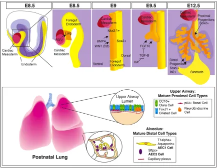
Figure 1.1 Signals Direct Lung Development into Specific Differentiated Cell
24
Figure 1.1 Signals Direct Lung Development into Specific Differentiated Cell
Types of the Adult Lung.
The foregut endoderm gives rise to the lung epithelium. Fgf signals from the cardiac
mesoderm pattern the foregut endoderm into organ specific fields. High levels of Fgf
signaling promote lung and thyroid specification, and lower levels promote liver
specification. Bmp and Wnt signals from the splanchnic mesoderm promote expression
of Nkx2.1 in the ventral endoderm. At E9.0 the trachea bifurcates from the foregut
endoderm and the primary lung buds form. Fgf10 signals from the mesenchyme drive
lung bud outgrowth and is in turn regulated by retinoic acid repression of Tgf-β
signaling. By E12.5 the 5 lobes of the mouse lung have formed, and stereotyped
branching morphogenesis has begun. Proximal progenitor cells express Sox2 and will
give rise to the cell types that populate the upper airways (club cells, ciliated cells,
neuroendocrine cells). Distal progenitor cells express Sox9 and Id2. Early in
development (prior to E13.5) these distal cell progenitors can give rise to all the epithelial
cells of the lung. Later in development (after E16.5), these distal progenitors will
25
Figure 1.2 Molecular Circuitry for Branching of the Lung
The proposed model for branching morphogenesis requires Fgf10 expression in the
mesenchyme to promote new branch formation. Fgf10 signals through Fgfr2 in the
epithelium to activate Fgf signaling. One of the downstream targets of Fgf signaling is
Sprouty2, which acts to repress Fgf signaling. Fgf10 also promotes expression of Shh
and Bmp4 in the epithelium. These molecular signals act to repress expression of Fgf10
adjacent to the epithelium. As the epithelium extends into the mesenchyme, the new
26
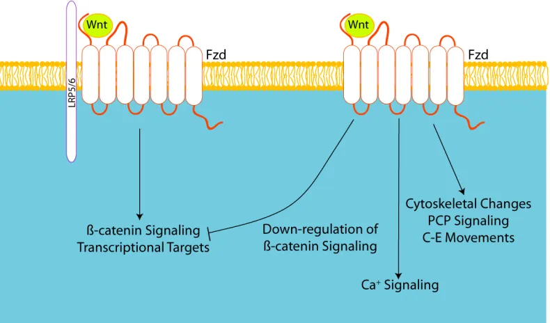
Figure 1.3 Wnt Signaling Pathways
Overview of the Wnt signaling pathways shows Wnt ligands can activate a number of
different downstream pathways. In Wnt/β-catenin dependent signaling, the Wnt ligand
binds to the Frizzled and Lrp5/6 receptors at the cell surface. The binding of the Wnt
ligand releases β-catenin from the destruction complex, and β-catenin translocates to
nucleus, where it activates transcriptional targets. The non-canonical Wnt signaling
pathway can cause downstream effects that include down-regulation of β−catenin
dependent signaling, calcium signaling and changes in cytoskeletal behavior and cell
27
CHAPTER 2: Changes in Epithelial Behavior Underlying
New Domain Branch Formation
Portions of this chapter were published in the Proceedings of the National Academy of
Sciences [95].
Summary
As reviewed in Chapter 1, the molecular component of branching morphogenesis
in the lung has been extensively studied, but how these signaling pathways are translated
into effects on cell biology has not been well examined. In this chapter I will describe the
changes in both cell morphology and tissue shape that the lung epithelium undergoes
during the initial development of the respiratory tree. From this analysis we have
developed a model for understanding and evaluating domain branch formation in the
lung and changes in cell and tissue morphology in the epithelium that lead to new
branch formation.
Introduction
Although a number of the biochemical pathways involved in establishing the
arborized airways have been elucidated, the physical forces and changes in cell biology
that drive these processes are not well understood. During the pseudoglandular stage,
the lung epithelium undergoes a stereotyped branching program to give rise to the
elaborately branched respiratory network of the adult lung (Fig 2.1). The branching
pattern has been delineated into a series of subroutines [4], all starting with domain
branching from the initial lung buds, formed by E10.5. Domain branching is
characterized by a new bud arising from the lateral aspect of the bronchiole tubule, and
28
epithelial tube. Domain branches are regularly spaced along the parent branch and
added in a proximal to distal manner as the parent branch extends and the lung matures
(Fig 2.2). Later in development, domain branches arise dorsally, medially and ventrally
to the initial sequence of domain branches, again in a proximal to distal direction. This
results in a bottlebrush appearance of branches along the main bronchioles and serves to
establish the main scaffolding of the respiratory tree. As these daughter domain
branches extend away from the parent tubule, they undergo a series of bifurcations to
establish the tertiary branches that fills in the established main arborized skeleton. With
this map in hand, we can start to evaluate the changes in cell biology that give rise to
morphological changes on the tissue level to form the elaborately branched lung.
Formation of the branches of the lung requires a bending, folding and molding of
the lung epithelial tubules. It is unclear at this point the respective force contributions
from the mesenchyme and the epithelium in to the process of new branch formation.
Explant studies of isolated epithelium in matrigel supplied with Fgf suggest that there
are sufficient forces within the lung epithelium to promote branch formation [22].
Despite these results, it is likely that, in vivo, the mesenchyme plays a role in branch
formation. Using newly available molecular tools, we were able to characterize the
epithelial cell behaviors that contribute to new domain branch formation and propose a
model for domain branching in the developing lung.
Live Imaging of the Whole Lung Epithelium
To begin to address the changes that the epithelium undergoes during branching
morphogenesis, we undertook live imaging of lung explants. We imaged up to the first
20 hours in culture, because the morphology of the developing lung is relatively well
29
With increased time in culture, the lung explant flattens onto the transwell and the
surrounding mesenchyme crawls away from the epithelium, potentially affecting the
epithelial-mesenchymal interactions that normally occur during branching
morphogenesis. Due to this restriction on the time course of live imaging experiments,
we primarily focused on the early processes in branching morphogenesis, domain branch
formation. We made use of the R26RmTmG line, which ubiquitously expresses
membrane-localized Tomato-Red under control of the Rosa locus, which is flanked by LoxP sites.
Upon cre-recombinase expression, the Tomato-Red cassette is removed, and membrane
localized GFP is expressed from the Rosa locus. The Shhcre line expresses
cre-recombinase downstream of the Shh promoter and is expressed throughout the foregut
endoderm at the time lung specification (approximately E9.5 in the mouse). We
generated Shhcre: R26RmTmG lungs to label the lung epithelium with GFP upon
recombination at E9.5, prior to commencement of branching morphogenesis. Over the
course of these experiments, we found that the lung was particularly sensitive to
phototoxic effects of laser exposure, as such we had to use very low laser power. The
level of resolution that we were able to obtain was relatively low, but did allow for live
imaging of the processes that the epithelium undergoes during establishment of the
respiratory tree. We started imaging at E11.5, when the two major airways of the lung
have established the five lobes of the lung. Over the course of the next 13 hours, new
branches grow and extend away from the main parent bronchi and the more proximal
region of the bud narrows and constricts (Fig. 2.3). Over this period of development,
formation of new domain branches in the right caudal lobe and left lobe are observed
(Fig. 2.3, arrows). These new domain branches arise not at the tip of the epithelial tube,
but rather in the middle of the tube, and as such represents domain branch formation.
30
bud will arise (Fig2.3, arrows). We can also observe planar bifurcation of the main
bronchiole of the right cranial lobe. As development proceeds, the flattened bud
bifurcates and two new buds grow away from the former bud tip (arrowheads in Fig2.3).
These live imaging studies provide an overview of the changes that the lung epithelium
undergoes during the early pseudoglandular period.
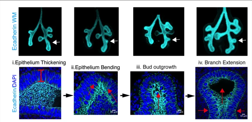
New domain branch formation requires apical constriction and epithelial
thickening
During our analysis of early branching, we observed that the epithelium
undergoes a number of stereotyped changes prior to new branch formation. At the site
of new bud formation, the epithelium thickens, and the epithelial tube deforms and
bends (Fig 2.4). Following this, a distinct kink in the epithelium is observed, and a newly
formed bud develops; then grows away from the main epithelial tube. As the new branch
extends, the region proximal to the bud tip constricts to promote further extension of the
branch into the surrounding mesenchyme. The lung epithelium maintains cell-cell
adhesion as a continuous sheet throughout these morphological changes, so the
tissue-level changes could requires change in cell shape, changes in proliferation and neighbor
exchange to accomplish these morphological changes at the tissue level. Proliferation
has long been proposed as a motive force behind new branch growth and formation in
the lung [96]. In fact, it has been shown that there is increased proliferation in the distal
tips of the developing lung epithelium [97]. Despite this proposed mechanism, there is
no evidence for a change in proliferation at regions in the epithelium preceding where
domain branches form [98, 99]. Rather, increased proliferation is observed after the
branches have budded from the epithelium, suggesting a requirement for proliferation in
31
morphology and behavior drive the bending of the epithelial tube to give rise to new
domain branches.
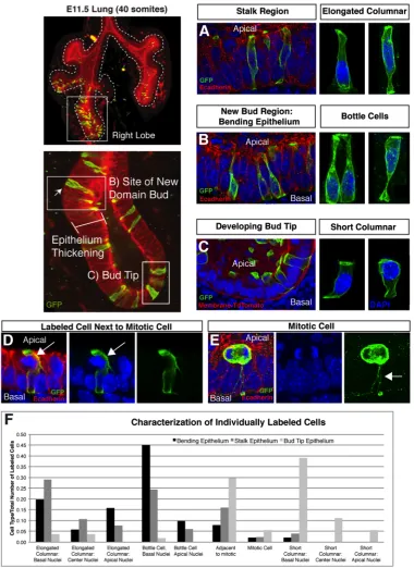
The thickening of the epithelium at sites of new branch formation suggested that
there were important changes in cell shape and size during this process. To better
visualize the individual changes in cell shape that occur prior to and during domain
branching, we generated ShhcreERT2:R26RmTmG mice which allow us to label individual
cells in the developing epithelium of the lung (Fig 2.5). The ShhcreERT2 is an inducible cre
recombinase driven by the Shh promoter. By injecting the pregnant dams with limiting
amounts of Tamoxifen to induce nuclear localization of the cre-recombinase in cells
expressing ShhcreERT2, we could induce excision of the Tomato-Red cassette and promote
expression of GFP in individual cells within the lung epithelium at E11.5, if the
Tamoxifen injections were completed at E10.5. Using this system, we observed a
number of spatially distinct cell morphologies in the developing epithelium at E11.5:
bottle-shaped cells located primarily at sites of new domain branch point formation,
elongated columnar cells in the stalk region, and short columnar cells located at the bud
tip (Fig. 2.5). In addition to these cell shapes, we also observed a distinct morphology of
cells adjacent to cells undergoing cell division (Fig2.5D). These cells were found
throughout the epithelium and they exhibited apical cellular extensions that were
deformed due to the rounded mitotic cells. The appearance of these cells that appear to
wrap the mitotic cells is notable as they represent a different model of mitotic and
neighboring cell behavior than what has recently been reported in the kidney branching
epithelium [100]. In the kidney, mitotic cells bud into the luminal space and the
daughter cell reinserts into the epithelium a few cells distant from the original cell. In
the lung, these cellular projections surrounding the mitotic cells maintain the dividing
32
of interest to examine if these neighboring cells had a role in where daughter cells
reinsert into the epithelial sheet, and without live imaging of individual cells it is
unknown if daughter cell dispersal occurs in the lung epithelium. Evaluation of
ShhcreERT2:R26RmTmG animals with a limiting dilution of tamoxifen suggests that there is
significant cell movement in the developing lung epithelium, as patches of clones are not
observed. Investigation as to whether this cell movement is mediated through mitotic
events or through neighbor exchange awaits advances in microscopy before this question
can be adequately addressed. Cells throughout the lung epithelium migrate to the apical
surface prior to mitosis. These mitotic cells have thin cellular projections that maintain
contact with the basal surface of the epithelium (Fig2.5E). While elongated columnar
cells and bottle cells are located both in the stalk and bending epithelium, quantification
of the appearance of labeled cells in distinct regions of the epithelium demonstrates an
increased frequency of bottle cells with basally localized nuclei at sites of new bud
formation (Figure 2.5F).
The presence of bottle cells and apical-basal cell lengthening are common
features of folding epithelial sheets as seen in gastrulation in Xenopus [101, 102], neural
tube bending [103, 104] }, optic vesicle formation [105], and Drosophilia salivary gland
development [106]. Consistent with these models, at the site of new bud formation the
lung epithelium thickens and adopts a pseudostratified morphology, suggesting the
epithelial thickening is due to epithelial cell lengthening and apical constriction.
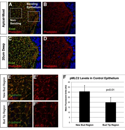
Apical localization of components of the actin-myosin contractile network
To further investigate evidence for apical constriction occurring in regions
undergoing apical-basal cell lengthening and bending, we imaged the luminal surface of