Extramammary Paget’s Disease in an Asian
Population: A Single Centre’s Experience
Lee Chee Meng; MBBS, MRCS, Cheong Kai Xiong, Kesavan S; MBBS, FRCPA, MBA, Melissa Teo Ching Ching; MBBS, MMed, FRCSEd, FAMS, MPH.
National Cancer Centre Singapore.
AbstrAct
Aims: Extramammary Paget’s Disease (EMPD) is a rare
intra-epithelial neoplasm affecting the skin of areas such as the scrotum, vulva and perineal region. This study examines the patient demographics, presenting symptoms, disease characteristics, and recurrence rates after surgical treatment, and overall prognosis of the condition.
Methods: This retrospective analysis examined all patients
(n=11) who were treated at the National Cancer Centre Singapore from 2003 to 2011. Information regarding the patients’ demographics, disease characteristics, treatment modalities and outcomes were collected.
Results: The median age was 68 years, and patients were
predominantly Chinese males who presented with skin rash. No concomitant malignancy was picked up amongst all the patients. All patients underwent wide excision, while groin dissection was performed in 2 patients with clinical lymphadenopathy. 5 patients had microscopically invasive disease while 6 had intraepidermal EMPD. Patients with intraepidermal EMPD had a significantly longer mean overall survival period of 35.5 months (range: 5-104) compared to 29 months (range: 3-59) for those with invasive disease. (p=0.0855). Mortality rate was 40% in those with micro-invasive disease, at time of follow up. The 2 patients who eventually passed away were noted to have lymphadenopathy at diagnosis.
Conclusions: Physicians should consider EMPD as
a differential in the evaluation of persistent skin rash, especially for that in the genital region. The best chance of cure remains surgical resection, with adequate margins. Computed Tomography should be used as an adjunct to look for concomitant malignancy and to assess nodal disease. Patients who presented with metastatic disease had poor outcomes even with surgery, and should be considered for palliative management.
INtrODUctION
Extramammary Paget’s Disease (EMPD) is a rare form of intra-epithelial neoplasm of apocrine glands of the skin, characterised by the presence of Paget cells on histology. the common sites of EMPD include the vulva, scrotum, perineum, and perianal region, with rarer manifestations in sites such as the axilla, thigh, umbilicus and nose. the first cases of penoscrotal, perianal, and vulva EMPD were reported by crocker,1 Darier and couillaud,2 and Dubreuilh3 respectively.
It predominantly affects patients between 50 years to 80 years of age, with a peak at 65 years. It has a higher prevalence in caucasians and in females.4 However, various Asian studies5-6 have reported more males being affected.
Major symptoms and signs of EMPD include rash, pruritus, plaque, pigmentary changes, erythema, ulceration, and erosion.7 As EMPD often presents very non-specifically, the diagnosis is often delayed, and is reached only after prolonged treatment failure. the median delay before the correct diagnosis is made is reported to be 2 years.8
EMPD can occasionally be associated with internal malignancies such as colorectal, gastric, renal, bladder, urethral, prostate, and endocervical carcinomas,9-14 and some authors have advocated screening for these cancers.
Localised EMPD has a good prognosis because the tumour cells tend to spread only intraepidermally. However, invasive EMPD is frequently associated with lymph node metastasis, with reported rates of close to 26% and thus has a poorer prognosis.8-9
surgery represents the mainstay of treatment, especially for localised EMPD. Non-surgical modalities, in the form of various forms of phototherapy, laser therapy, radiotherapy, and chemotherapy have also been developed to reduce
the morbidity and mortality associated with surgery.16-17 the recurrence rates post-surgical resection is also reported to be fairly high, i.e. from 8% to 44%,8,18 which is likely due to the tendency of EMPD to be multifocal and to have ill-defined margins.19 recurrences occurring up to 12 years after initial treatment have been reported.20
the rarity of this condition has caused difficulty in its characterisation. to date, there exist only a small number of retrospective studies which attempt to characterize EMPD.8,21-26
Hence, the aim of this paper is to describe our experience with EMPD at the National cancer centre singapore (Nccs) and suggest a management strategy for this rare disease.
MEtHODs
study Population
All patients who were evaluated and treated for EMPD at the singapore Ncc from January 2003 to December 2011 were included in this study. A retrospective cross-sectional study of the eligible patients was conducted. the study population was identified from a prospectively maintained database at the Ncc, and clinical charts were reviewed. Ethical approval was obtained from the centralised Institutional review board in singapore.
Data collection
Information regarding the patients’ demographics, age at diagnosis, presenting symptoms, disease sites and presence of synchronous lesions, evaluations, definitive treatments, post-operative adjuvant therapies, follow-up periods, survival periods, complications, recurrences, and deaths were collected.
Data Analysis
the data were reviewed and analysed using the computer software IbM sPss statistics for Windows, Version 19.0 (IbM, chicago, IL). Descriptive analysis was performed. the means, standard deviations, ranges, and medians were reported for categorical variables where appropriate, and the numbers and individual percentages were reported for categorical variables.
survival period was defined as the length of time from the date of surgery to the point of recurrence, or death, or time of analysis, whichever was the earliest, and was calculated for the patients who underwent resection for all known disease.
rEsULts
Demographics
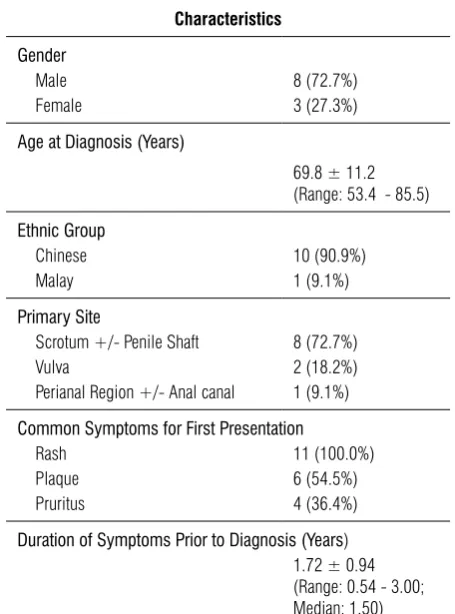
A total of 11 patients presented to the Ncc for EMPD from January 2003 to December 2011 and were included in this study. the patients were of 53 to 85 years of age at diagnosis, with the median age of 68 years. 8 patients were male, and the remaining 3 patients were female. All the patients in this study were of chinese in ethnicity, except for 1 patient who was Malay (table 1).
Presentation
the most common presenting symptoms were that of rash, plaque, followed by pruritus. rash was a common complaint amongst all of the patients. In addition, 6 patients presented with plaques, 4 with pruritus, and 3 with skin discolouration. 2 patients had palpable inguinal lymph nodes on examination.
Prior to referral to Ncc for management, patients had experienced symptoms for a mean duration of 1.65 years during which they either did not seek help or were conservatively managed by their primary healthcare provider.
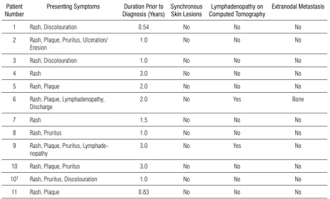
7 of the 8 male patients had scrotal EMPD. this was unilateral (left) in 5 of them, and bilateral in 2 others. 2 of the 3 female patients had vulva EMPD, with unilateral labial involvement in 1, and bilateral labial involvement in the other. the last male patient had perineal EMPD, similarly observed in the last female patient (table 2). Table 1. Summary of Demographics, Presentation,
Disease Characteristics, Evaluation, Management, and Outcomes
Characteristics
Gender
Male 8 (72.7%) Female 3 (27.3%)
Age at Diagnosis (Years)
69.8 ± 11.2 (Range: 53.4 - 85.5)
Ethnic Group
Chinese 10 (90.9%) Malay 1 (9.1%)
Primary Site
Scrotum +/- Penile Shaft 8 (72.7%) Vulva 2 (18.2%) Perianal Region +/- Anal canal 1 (9.1%)
Common Symptoms for First Presentation
Rash 11 (100.0%) Plaque 6 (54.5%) Pruritus 4 (36.4%)
Duration of Symptoms Prior to Diagnosis (Years) 1.72 ± 0.94 (Range: 0.54 - 3.00; Median: 1.50)
Surgico-Pathological Characteristics
Intraepidermal 6 (54.5%) Invasive Carcinoma 5 (45.5%)
Primary Treatment
Wide local excision 5 (45.5%) Wide local excision with skin grafts/
flaps
4 (36.3%) Wide local excision with lymph node
dissection
2 (18.2%)
Post-Operative Complications
Yes 5 (45.5%) No 6 (54.5%)
Recurrence
Yes 1 (9.1%) No 10 (90.9%)
Death
Yes 2 (18.2%) No 9 (81.8%)
Overall Survival Period (Months)
31.2
(Range: 3 - 104)
Disease-Free Survival Period (Months)
29.8 (Range: 3 - 89)
Investigation Modalities
All 11 patients were diagnosed with EMPD after incisional biopsies were performed. subsequently, computed tomography (ct) scans and a Fluorodeoxyglucose-Positron Emission tomography (FDG-PEt) with contrast-enhanced ct scan were performed in 10 and one patients respectively, to exclude the presence of nodal and distant metastases. It confirmed the presence of lymphadenopathy in 2 male patients in whom palpable lymph nodes were noted on physical examination. At diagnosis, one of these 2 patients already had extra-nodal metastasis to the bone, noted on the ct scan.
Underlying Malignancy
Only 1 patient had a background of malignancy. she was previously diagnosed with left breast ductal carcinoma in situ (DcIs), underwent a simple mastectomy with axillary clearance (in February 2007), and subsequently presented to us for EMPD in 2009. Her first malignancy was not thought to be associated with the diagnosis of
EMPD. 10 of the 11 patients had a ct Abdomen and Pelvis while 7 of the 11 patients underwent colonoscopies, which picked up benign colonic polyps in 3 patients. No one was found to have underlying malignancy after ct scans and colonoscopies.
Management
the main treatment modality was surgery. 5 patients underwent wide local excisions, while 4 patients underwent wide local excisions with skin grafts or regional flaps. the 2 patients with clinical lymphadenopathy, confirmed to be metastatic on fine needle aspiration cytology, underwent a concomitant modified inguinal lymphadenectomy, with dissection of the superficial and deep inguinal lymph node groups. the depth of the excision is down to scarpa’s fascia with a superior limit at the inguinal ligament, medial limit at the border of the adductor longus, lateral limit at the lateral border of the femoral artery and inferior limit at the apex of the femoral triangle.
Table 2. Presentation and Disease Characteristics at Diagnosis
Patient
Number Presenting Symptoms Diagnosis (Years)Duration Prior to Synchronous Skin Lesions Computed TomographyLymphadenopathy on Extranodal Metastasis
1 Rash, Discolouration 0.54 No No No 2 Rash, Plaque, Pruritus, Ulceration/
Erosion 1.0 No No No
3 Rash, Discolouration 1.0 No No No
4 Rash 3.0 No No No
5 Rash, Plaque 2.0 No No No
6 Rash, Plaque, Lymphadenopathy,
Discharge 2.0 No Yes Bone
7 Rash 1.5 No No No
8 Rash, Pruritus 1.0 No No No 9 Rash, Plaque, Pruritus,
Lymphade-nopathy 3.0 No Yes No
10 Rash, Plaque, Pruritus 3.0 No No No 10† Rash, Pruritus, Discolouration 1.0 No No No
11 Rash, Plaque 0.83 No No No
Intra-operative frozen section analyses of the margins were performed for all the 11 patients. Frozen section margins were clear for 9 patients while the remaining 2 patients with positive margins underwent wider excision. However, the wider margins remain persistently positive up to the perianal and peri-urethral region. Decision was made to withhold further surgery for these 2 patients as further resection would result in the involvement of the anal canal and urethra respectively. both patients were then counseled on wider resections that would entail an abdomino-perineal resection in one, and a urethral resection with an ileal conduit in the other. both patients declined the respective surgeries and the former patient underwent 10 cycles of radiotherapy (rt), whilst the latter had a further resection up to the level of urethra and closure with vertical rectus abdominis myocutaneous (VrAM) flap cover.
2 of the 9 patients who were thought to have clear resection margins after frozen section were subsequently found to have involved resection margins after the full histology was reviewed. these 2 patients underwent wider excision of involved margins with flap covers within a month of the initial surgery, attaining clear margins in the second surgery.
Post-operatively, both patients with nodal disease were offered chemotherapy. the patient who had nodal and bone metastases died before he could receive palliative chemotherapy, while the patient with isolated nodal involvement declined adjuvant chemotherapy.
Histological results
5 patients were found to have invasive carcinomas microscopically, and the remaining 6 patients were found to have intraepithelial lesions. the choice of immuno-histochemical markers for surgico-pathological evaluation was not standardised for all of the patients. Markers which were used included the cK5, cK6, cK7 and cK20. cK 7 was tested in 7 of the 11 patients and it was positive in all of them, while cK 20 was negative in all 6 patients who were tested for it.
Outcomes
A total of 3 patients suffered from wound infections post-operatively. 2 of these patients had undergone groin dissection, of whom 1 subsequently required vacuum assisted closure. All 3 patients were treated with a
course of oral antibiotics, and the infections resolved subsequently after 1 week. One of the patients who had undergone lymph node dissection for nodal involvement had persistent post-operative secondary lymphedema. No other post-operative complications were noted for other patients.
1 patient had metastatic disease on initial diagnosis. Of the remaining 10 patients, 1 had local recurrence, which occurred at 89 months. He had bilateral scrotal EMPD, for which he underwent wide local excision initially, and frozen sections revealed negative margins. Post-operatively, full histology revealed intraepidermal EMPD, but the surgical margins were involved, and he thus underwent a wider excision with a split skin graft cover, attaining clear margins. At the 7th year of follow up, he was noted to have recurrence of a scrotal rash at the previous operative site, and an incisional biopsy was performed, which confirmed the recurrence of Paget’s disease. A wider excision with split skin graft cover was subsequently performed. He has remained disease-free 14 months after the last surgery.
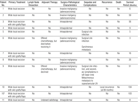
the mean overall survival period was 31.2 months (range: 3 - 104) while the mean disease-free survival period was 29.8 months (range: 3 - 89). both patients with nodal involvement at presentation were deceased at time of evaluation. the patient with bone metastases died 3 months after surgery. the other patient was noted to develop cervical lymph node metastasis at 29 months operatively and eventually died at 51 months post-operatively (table 3).
Differences in characteristics between
Intraepidermal and Invasive EMPD
Although patients with intra-epithelial disease only appeared to present at an earlier time than those who had invasive disease, with duration of symptoms experienced for a median of 1.00 and 1.75 years respectively, there was no significant difference (p=0.514; Mann-Whitney’s U test).
Table 3. Management and Outcomes Patient
No.
Primary Treatment Lymph Node Dissection
Adjuvant Therapy Surgico-Pathological Characteristics
Sequelae and Complications
Recurrence Death Survival Period (Months) 1 Wide local excision No No Invasive malignancy
(adenocarcinoma)
No No No 6
2 Wide local excision No No Invasive maligancy
(adenocarcinoma)
No No No 59
3 Wide local excision with skin grafts/flaps
No No Intraepidermal No No No 56
4 Wide local excision No No Intraepidermal No No No 9
5 Wide local excision No No Intraepidermal Surgical site
infection
No No 5
6 Wide local excision Yes Offered chemotherapy, but died before receiving it
Invasive malignancy (adenocarcinoma)
Surgical site infection
Synchronous metastasis
No Yes 3
7 Wide local excision
with skin grafts/flaps No No Intraepidermal No No No 16
8 Wide local excision No No Invasive malignancy
(adenocarcinoma) No No No 26
9 Wide local excision Yes Offered chemotherapy, but declined
Invasive malignancy
(adenocarcinoma) Surgical site infec-tion, and second-ary lymphedema in left lower limb Metachronous metastasis to supraclavicular LN
No Yes 51
10 Wide local excision
with skin grafts/flaps No No Intraepidermal No Local recurrence (at 89 months) No 104 10† Wide local excision
with skin grafts/flaps No No Intraepidermal No No No 103
11 Wide local excision No Underwent radiotherapy Intraepidermal No No No 8
†Patient experienced recurrence
DIscUssION
EMPD is a rare condition, and there is very little information available regarding the disease patterns and outcomes in an Asian population. to the best of our knowledge, this cohort of 11 patients over 8 years represents the largest number of cases treated at a single centre in singapore. Our aim was to analyze the epidemiology, presenting symptoms, disease characteristics, management strategies, and the long-term outcomes of patients who are treated at a large tertiary referral centre. In doing so, we hope to determine the prognostic factors and develop a better management strategy for patients with this rare disease.
Patients experienced symptoms of EMPD for a mean period of 1.65 years prior to presentation at our centre. As all of the patients complained of skin rash, the delay in diagnosis can be attributed to the fact that rash is a
common and non-specific skin condition. Many patients could have downplayed its significance, and not sought medical attention until further symptoms arose. Although EMPD is rare, clinicians should consider this differential diagnosis in a patient who presents with a persistent and suspicious-looking rash, especially that in the genitalia region.
Although chanda et al., in a review of 197 cases,10 had reported that EMPD may be associated with underlying malignancies, Asian studies had demonstrated lower incidences,25-26 particularly in men with penoscrotal EMPD. the results of our study are consistent with the Asian studies, i.e. none of the patients were found to have internal malignancies after colonoscopies and ct scans.
malignancy. ct revealed sizeable lymphadenopathy, both in the superficial and deep inguinal chains, in the 2 patients with clinically palpable lymph nodes, proving to be a useful adjunct to determine the extent of groin dissection. In one of the 2 patients with lymphadenopathy, ct also revealed the presence of synchronous bone metastases. PEt ct did not provide us with any additional information during staging and this concurred with a systematic review of 17 primary studies, where PEt-ct was found not to have any patient-relevant benefit27 over conventional ct in detecting lymphadenopathy during the staging of malignant
melanoma.
there is currently no internationally recognised consensus regarding the standard work up for the detection of concomitant internal malignancies after EMPD has been diagnosed. A thorough history and physical examination remain imperative in guiding the initial clinical evaluation, as with any other disease. specific investigations like gastroscopies, colonoscopies, PAP smears, colposcopies, cystoscopies, mammograms, abdomino-pelvic ultrasounds, serum cEA and PsA may then be carried out, especially if there are organ-specific symptoms and signs. the choice of specific investigations may also be guided by the site of the lesion, for instance, colonoscopy for perianal lesions, cystoscopy for genitourinary lesions, since chanda et al. has reported a strong association between the anatomical site of EMPD and that of internal malignancies. However, this was not noted in our study.
Patients with lymph node disease have poorer outcomes. As such, we propose a workflow for the management of such patients (Figure 1). Patients diagnosed with EMPD should be carefully examined for the presence of nodal involvement or distant metastases. In patients with clinically palpable nodes, a fine needle aspiration cytology should be performed. If FNAc is positive, a
is an issue, and the nodes are suspicious for metastasis by size criteria or are significantly FDG-avid on the PEt scan, the patient can be offered a nodal dissection. In cases where there is no lymphadenopathy detected on clinical examination and ct scan, a wide excision should be performed. It is our practice to perform a ct or PEt scan upon diagnosis, in order that surgical management can be confined to a single episode, although patients with intra-epidermal EMPD (non evidence of invasion) may possibly avoid the need for imaging. Post-operatively, clinicians should base their next management step on whether there is microscopically invasive disease on final histology.
As expected, patients with microscopically invasive disease fared poorly compared to those with intraepithelial disease. At time of follow up, there was a 40% mortality rate in the former group, and 2 of these 5 patients had lymph node metastases from the onset. studies28-29 have reported the high sensitivity of sentinel lymph node biopsy in cutaneous malignancies, with Hatta28 et al reporting a 100% detection rate in 23 patients with EMPD, and a 0% false negative rate. If there is micro-invasion and no clinical evidence of lmyphadenopathy, it might be worthwhile to consider offering an elective sentinel lymph node biopsy, keeping in view groin dissection in cases with positive nodes. Otherwise, regular follow up with careful clinical examination for lymphadenopathy, coupled with surveillance ct will be useful in this group of patients as it is difficult to predict if they will eventually develop nodal spread.
Frozen section analyses of resection margins were performed in all patients treated at our centre. In 9 patients, frozen sections reported clear margins, yet final histologies revealed false negative results in 2 of them (22.2%). this inherent risk was also reported in other studies with rates approaching 40%,19,30 but this had little effect on disease outcomes.17,31-32 In our series, outcomes were unchanged as the 2 mentioned patients were subjected to a second surgery allowing them to achieve negative margins.
studies on immuno-histochemical analysis33-34 have shown that certain markers have a relationship in the manifestation of EMPD. cK 7, cEA and GcDFP-15 are known to be positive for all forms of EMPD, while those patients with an underlying malignancy usually test positive for cK 20.35 Amongst the mucinous markers, MUc 1 and MUc 2 are occasionally expressed in EMPD but MUc 5 has the highest prognostic potential36 as they are expressed more frequently in microinvasive EMPD.
One of the limitations in our study was that immuno-histochemical analysis was not uniform. In our series, all patients who were tested with cK 7 all ended with positive results whereas cK 20 was negative amongst those tested, correlating with the absence of underlying malignancy. Apart from validating our results with known disease patterns associated with cK 7 and cK 20, we were unable to further derive any other meaningful results regarding immuno-histochemical evaluation of EMPD. We recommend that histological reports for future patients adopt a standardised set of markers for testing, namely cK 7, cK 20, cEA, GcDFP-15, MUc 1, MUc 2 and MUc 5 as this may allow researchers to find a link between the presence and absence of markers with prognosis.
besides chemotherapy, radiotherapy has been used as alternative form of treatment, particularly in patients who are at a high risk for surgery or in those with large lesions,37-38 where further wide excision may cause compromise to vital structures such as the anus or urethra. In such cases, radiotherapy may be more acceptable to patients than an abdominal perineal resection or the creation of an uretero-ileal conduit. Although radiotherapy is useful in some patients recurrences has also been reported in these studies. Furthermore, there are no studies that have recommended any fixed dose or duration of radiotherapy, and its use has to be carefully considered on a case-by-case basis as there are acute and chronic toxicity effects. the only patient who underwent radiotherapy for persistently positive margins at the perianal region remains disease-free to date, at 8 months of follow up.
EMPD is known to have high local recurrence rates despite adequate surgical resection.30 In our study, there was only 1 patient who had locoregional recurrence. this was the first EMPD patient managed by our center, with recurrence at 89 months after disease clearance. After the original surgery, full histology showed that margins were involved and he subsequently went for wider excision 1 month later. the margins were cleared after the second surgery and full histology revealed intraepithelial disease. Apart from him, there were 4 other patients who similarly did not have clear margins after the first operations and 2 of them underwent further surgeries, and the final margins were clear of EMPD. till date, these patients have not developed evidence of recurrence.
disease patterns in the local population. We recommend that patients have long term follow ups, and clinicians should have low thresholds for suspecting recurrence upon detecting new skin lesions on follow ups.
cONcLUsION
In summary, this study represents a retrospective analysis of EMPD. It has contributed to the small corpus of knowledge of EMPD, which has remained largely controversial. Our results reflected similarities with those in overseas centers apart from the findings of male predominance of the disease, low incidence of internal malignancy, and low recurrence rates.
Although EMPD is rare, it remains an important differential for non-resolving skin rash. With early diagnosis and adequate surgical resection, it remains treatable with a fairly good prognosis. Poor prognostic factors include those with nodal metastases on presentation and microscopically invasive disease on final histology; and the role of adjuvant chemotherapy in such patients should be further explored. Patients need to be placed on long term follow up to look for recurrence and scans should be considered, particularly in those with microscopically invasive disease.
We propose a workflow for the treatment of future patients in our center, and this can be validated with a prospective study.
AcKNOWLEDGEMENts
No funding has been sought in this research. The authors hereby thank all physicians who have been involved in the care of the patients described in this paper.
rEFErENcEs
1. crocker Hr: Paget’s disease affecting the scrotum and the penis. trans Pathol soc Lond 1889; 40:187–191.
2. Darier J, couillaud P: sur un cas de maladie de Paget de la region perinea-annale et scrotale. Ann Dermatol syphiligr 1893; 4:25–31. 3. Dubreuilh W: Paget’s disease of the vulva. br J Dermatol 1901;
13:403–13.
4. shepherd V, Davidson EJ, Davies-Humphreys J: Extramammary Paget’s disease. bJOG 2005; 112:273e9.
5. Kanitakis J: Mammary and extramammary Paget’s disease. J Eur Acad Dermatol Venereol 2007; 21(5): 581e90.
6. curtin JP, rubin sc, Jones Wb, et al.: Paget’s disease of the vulva. Gynecol Oncol 1990; 39:374e7.
7. chiba H, Kazama t, takenouchi t, et al.: two cases of vulval
pigmented extramammary Paget’s disease: Histochemical and immunohistochemical studies. br J Dermatol 2000; 142:1190e4. 8. Hatta N, Yamada M, Hirano t, et al.: Extramammary Paget’s
disease: treatment, prognostic factors and outcome in 76 patients. br J Dermatol. 2008 Feb; 158(2):313-8.
9. tsutsumida A, Yamamoto Y, Minakawa H, et al. Indications for lymph node dissection in the treatment of extramammary Paget’s disease. Dermatol surg. 2003;29:21–24.
10. chanda JJ: Extramammary Paget’s disease: Prognosis and relationship to internal malignancy. J Am Acad Dermatol 1985; 13:1009.
11. Ojeda VJ, Heenan PJ and Watson sH: Paget’s disease of the groin associated with adenocarcinoma of the urinary bladder. J cutan Pathol 1987; 14: 227.
12. Koh KbH and Nazarina Ar: Paget’s disease of the scrotum: report of a case with underlying carcinoma of the prostate. br J Dermatol 1995; 133:306.
13. Hoffman Ms, cavanagh D: Malignancies of the vulva. In: rock JA, thompson JD, (eds). the Linde’s operative gynecology. 8th ed. Philadelphia: Lippincott-raven; 1997. p.1370-3.
14. Allan sJr, McLaren K and Aldridge rD: Paget’s disease of the scrotum: A case exhibiting positive prostate-specific antigen staining and associated with prostatic adenocarcinoma. br J Dermatol 1998; 138: 689.
15. Im M, Kye Kc, Kim JM et al.: Extramammary Paget’s disease of the scrotum with adenocarcinoma of the stomach. J Am Acad Dermatol 2007; 57: s43.
16. Petkovic s, Jeremic K, Vidakovic s, et al.: Paget’s disease of the vulva review of our experience. Eur J Gynaecol Oncol 2006; 27(6):611e2.
17. Louis-sylvestre c, Haddad b, Paniel bJ: Paget’s disease of the vulva: results of different conservative treatments. Eur J Obstet Gynecol reprod biol 2001; 99:253e5.
18. Pierie JP, choudry U, Muzikansky A, et al.: Prognosis and management of extramammary Paget’s disease and the association with secondary malignancies. J Am coll surg 2003; 196:45e50. 19. Gunn rA, Gallager Hs: Vulvar Paget’s disease: A topographic
study. cancer 1980; 46:590e4.
20. Goldblum Jr, Hart Wr: Perianal Paget’s disease: A histologic and immunohistochemical study of 11 cases with and without associated rectal adenocarcinoma. Am J surg Pathol 1998; 22(2):170e9.
21. Hegarty PK, suh J, Fisher Mb, et al.: Penoscrotal extramammary Paget’s disease: the University of texas M. D. Anderson cancer center contemporary experience. J Urol. 2011 Jul; 186(1):97-102. 22. Zhang N, Gong K, Zhang X, et al.: Extramammary Paget’s disease of scrotum--report of 25 cases and literature review. Urol Oncol. 2010 Jan-Feb; 28(1):28-33.
23. Jones Is, crandon A, sanday K: Paget’s disease of the vulva: Diagnosis and follow-up key to management; a retrospective study of 50 cases from Queensland. Gynecol Oncol. 2011 Jul; 122(1):42-4.
24. Lee sJ, choe Ys, Jung HD, et al.: A multicenter study on extramammary Paget’s disease in Korea. Int J Dermatol. 2011 May; 50(5):508-15.
25. chiu tW, Wong PsY, Ahmed K, et al.: Extramammary Paget’s disease in chinese males: A 21-year experience. World J surg 2007; 31: 1941.
26. chang Yt, Liu HN, Wong cK: Extramammary Paget’s disease: A report of 22 cases in chinese males. J Dermatol. 1996 May; 23(5):320-4.
27. Morton DL, Wen Dr, Wong JH, et al.: technical details of intraoperative lymphatic mapping for early stage melanoma. Arch surg 1992; 127(4):392e9.
2004; 30(10):1329e34.
29. Longo MI, Lázaro P, bueno c, et al.: Fluorodeoxyglucose-positron emission tomography imaging versus sentinel node biopsy in the primary staging of melanoma patients. Dermatol surg. 2003 Mar;29(3):245-8.
30. Fishman DA, chambers sK, schwartz PE, et al.: Extramammary Paget’s disease of the vulva. Gynecol Oncol 1995; 56:266e70. 31. Zollo JD, Zeitouni Nc: the roswell Park cancer Institute
experience with extramammary Paget’s disease. br J Dermatol 2000 Jan; 142(1):59-65.
32. Preti M, Micheletti b, Ghiringhello b, et al.: La malattia di Paget vulvare. Minerva Ginecol 2000; 52:203e11.
33. Goldblum Jr, Hart Wr: Vulvar Paget’s disease: A clinicopathologic and immunohistochemical study of 19 cases. Am J surg Pathol. 1997 Oct; 21(10):1178-87.
34. Nowak MA, Guerriere-Kovach P, Pathan A, et al.: Perianal Paget’s disease – distinguishing primary and secondary lesions using
immunohistochemical studies including gross cystic disease fluid protein-15 and cytokeratin 20 expression. Arch Pathol Lab Med 1998; 12:1077–81.
35. Prieto VG, Ivan D: the use of immunohistochemistry in the differential diagnosis of primary cutaneous adnexal neoplasms and metastatic adenocarcinomas to the skin. Diagnostic Histopathology. 2010 sep; 16(9): 409-16.
36. Hata H, Abe r, Hoshina D, et al.: MUc5Ac expression correlates with invasiveness and progression of extramammary Paget’s disease. J Eur Acad Dermatol Venereol. doi: 10.1111/jdv.12156. 37. Luk NM, Yu KH, Yeung WK, et al.: Extramammary Paget’s disease:
outcome of radiotherapy with curative intent. clin Exp Dermatol. 2003 Jul; 28(4):36