© Universiti Tun Hussein Onn Malaysia Publisher’s Office
IJIE
Journal homepage:http://penerbit.uthm.edu.my/ojs/index.php/ijie
The International
Journal of
Integrated
Engineering
Remote Patient Identification based on ECG and Heart Beat
Pattern over Wireless Channel
Neeraj Garg
1*, Jagdeep S Lather
2, Sanjay K Dhurandher
21Maharaja Agrasen Institute of Technology, Delhi, INDIA 2National Institute of Technology, Kurukshetra, INDIA
DOI: https://doi.org/10.30880/ijie.2019.11.08.019
Received 6 September 2018; Accepted 18 November 2018; Available online 30 December 2019
Abstract:Wireless Body Area Network (WBAN) is an innovative solution for distant monitoring of patients. In WBAN sensors are placed on human body to monitor vital parameters, and these parameters send to physician using wireless network. As in wireless network data is aggregated before transmission therefore when data is de-aggregated on receiver side it is important to know which data belongs to particular intended patient. In this work, it is shown that electrocardiogram (ECG) and heart beat pattern itself can be used for the identification of patient as it carries distinctive features which are unique to each person. Moreover, ECG is an image thus when transferred over Orthogonal Frequency Division Multiplexing (OFDM) wireless channel adds noise, therefore a level of Signal-to-Noise Ratio (SNR) needs to be maintained for error free recovery. This paper proposes ECG and heart beat pattern based patient identification process over OFDM channel. Proposed methodology is tested on ECG database and it has been found that the accuracy of the method is 99.98%.
Keywords:ECG, OFDM, SNR
1. Introduction
In the present era, Body area sensor networks (BANs) acts as an effective technology due to its unbeatable features like simple usage, safe, and application in health sector [1]. These are used in number of ways such as in tracking the fitness trackers [2], in crucial following of emergency response teams [3], and in the medical implantable devices like heart pacemakers and insulin pumps. These kinds of medical and safety related applications of BAN require a decent class of controlling of access to them and proper and secured data [4–7]. For economical and practical reasons, the size of the nodes is not big considering the expense and practical application. These are resource -constrained and they give computation power and memory up to a particular limit. If we look in the recent past years, we will feel that the use of Wireless Sensor Network (WSN) technology has increased quickly. Among a number of applications, an important one is in wireless biomedical sensor network for collecting physiological signals. We can define the Wireless Body Area Network (WBAN) as a wireless network useful for making communication among operating sensor nodes on, in or around the body of an individual with the end purpose to evaluate the important body measurements and functions. The above-mentioned monitoring signals are after this collected by a personal device, such as Personal Digital Assistance (PDA) or smart phone that works as a data sink for the sensor nodes and transfers them to the specialist for further monitoring. In WBAN nodes are placed on the body, and communication protocol is to be developed for end to end secure information transfer [4]. In wireless body area network, the developed protocols are based on the distance among sensors and energy of the nodes [2-5].
can easily collect ECG with the help of the sensors that are connected with the body, as illustrated in [8–10]. The features of the recorded ECG can be used for the patient identification. We have organized complete paper in six sections. Section 2, of the paper discusses the related work and preliminaries of the work proposed. Section 3, of the paper discusses the proposed methodology. ECG transmission and reception using OFDM is discussed in section 4 of the paper, in section 5, simulation and results are presented. Finally, in section 6 major conclusions of the presented work are discussed.
2. Related Work and Preliminaries
In recent past ECG-based authentication and key-agreement protocols proposed in [11–13]. In these methods clinical ECG data is recorded and makes the processing of the data on PCs, and disregarding vulnerabilities.In WBAN security of data is a major issue and needs to be addressed properly. There are two security measures which need to be addressed:
1. Security of data from external attack.
2. As data are aggregated at the base station, therefore it is necessary at the receiving end data should be properly dissimilated and data belonging to each patient should be correctly identified.
From external attacks data can be protected using Reversible Data Hiding (RDH) techniques [14]. In Reversible Data Hiding first data is hidden using technique like difference expansion [15], histogram shifting [16], thereafter using encryption method data is encrypted, and at the receiver side data is decrypted and embedded data is recovered.
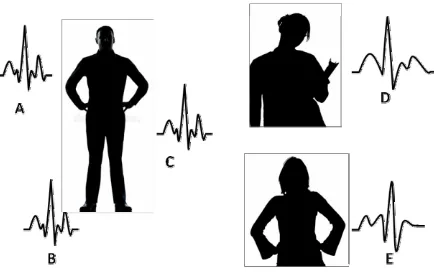
To distinguish data of individual patients EGC based authentication/verification is used [11-13]. In previous studies it has been found that ECG caries distinctive features and most importantly they are unique in each person [11-13]. Even the data is secured by security protocols and implementations on extremely constrained devices [17], still we have the problem how the devices of same body area can distinguish and trust each other. This is demonstrated by Fig. 1: Sensors connected with one person (Sensor A, B, and C) should identify and trust each other. On the other hand, sensors connected with other persons (E) or whole un-genuine data (D) should not be trusted. Certain options such as pre-deployed keys [18] or manual setups are awkward and have chances of error- specifically in conditions with numerous interfering BANs. It is also found that ECG pattern of a particular person remains unaltered irrespective of sensors location on the body.
Fig. 1 - Schematic of three persons ECG pattern
3. Proposed Methodology
In wireless body area networks, sensor nodes send data to base station and after aggregation and encryption data is send to physician using some transmission mechanism. In transmission mechanism wireless or mobile communica tion is preferred choices. In this work we have considered wireless communication as transmission media and for encrypted data transfer OFDM technology is considered.
this method works well in case of offline mode. In case of online transfer of data from one location to another, medium introduces impairments thus overall quality of the transmitted signal goes down. In such a situation it may not possible to detect Q-R-S complex, therefore it is useful to exploit other information of the ECG for patient identification and verification.
Fig. 2 - P, Q, R, S and T peaks in ECG signals
3.1 ECG Peak Detection and Analysis
In this work we use statistics of the ECG to identify patient. First of all, we use complete ECG pattern which is different of each patient, secondly detection of R-R peaks and calculation of heart beat, maximum and minimum heat beat, mean and variance of heart beat and its pattern is done. We found that these parameters are good enough to distinguish patients, finally encryption of this information is done using cover image, and ECG data can itself embedded on vacant space on ECG image [20].
Herat beat detection steps:
Step 1: Read ECG data from dat file. Defining ECG signal asx[n].
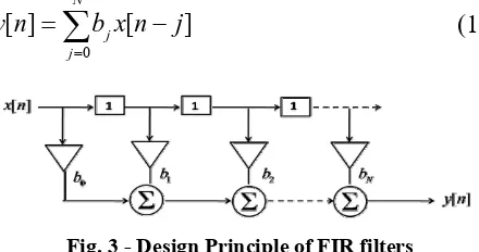
Step 2: To suppress noise and Q, R, S, and T waves ECG signal pass through a ‘high pass’ filter with impulse response ash[n], then the output of the filter isy[n] which is defined asy[n]=h[n]*x[n].
Step 3: The used filter is a Finite Impulse Response (FIR) filter, as shown below in Fig. 3. The filter equation is given by
N
y
[
n
]
b
jx
[
n
j
]
j0
(1)
Fig. 3 - Design Principle of FIR filters
Step 4: To further suppress the un-wanted terms we doz[n]=(y[n])m
Step 5: Finally, inter-arrival time between consecutive R-R peaks is evacuated, and heart rate is calculated.
It is also noticeable that, due to noise of ECG electrodes and additional noise due to the wireless channel some additional peaks can be observed in the ECG signal, to deal with such problem, initially first largest peak is detected. Considering,S(k) is kth sample amplitude value and ‘th’ is initially defined threshold. Than from the beginning find firstksuch that
Then for next time interval ‘t’ no peak will be detected to avoid false peaks. The time interval ‘t’ depends on sampling rates at which ECG is recorded. Considering that inter-pulse arrival (IPA) time between consecutive R-R peaks isT, and time between Q-R and Q-S pulses aretQR,tQSrespectively. Then ideally, t=T-tQRor roughly, t=T-tQS/2.
in noiseless systemtQR andtQS, can be find out using Pan–Tompkins real-time QRS detection algorithm, however, in noisy environment the detection of QRS peaks is a complex problem.
3.2. ECG Modeling as Stochastic Process
As discussed above, in case of noisy environment, detection of peaks and ECG feature is not easy. Therefore to tackle this issue we propose the use of other statistical measure for the ECG analysis. In ECG each sample follows i.i.d. process. Considering an ECG sample as
and represented as
x
(
n
)
, noise asw
(
n
)
and noise corrupted symbol can be defined asy
(
n
)
,y
(
n
)
x
(
n
)
w
(
n
)
(3)This will passes through the filter can be represented as
y
F(
n
)
x
(
n
)
w
(
n
)
h
(
n
)
y
F(
n
)
x
(
n
)
h
(
n
)
w
(
n
)
h
(
n
)
The time delayed version of the received signal is given by
y
(
n
n
0)
x
(
n
n
0)
w
(
n
n
0)
The autocorrelation function can be written as
R
YY(
n
0)
R
XX(
n
0)
E
[
x
(
n
)
w
(
n
n
0)]
E
[
x
(
n
n
0)
w
(
n
)]
R
ww(
n
0)
As ECG and noise is independent to each other, thereforeR
YY(
n
0)
R
XX(
n
0)
R
ww(
n
0)
(4)
(5)
(6)
(7)
(8)
If noise is suppressed using filtering process,
R
YY(
n
0)
R
XX(
n
0)
(9)If both original
x
(
n
)
and time delayed signaly
(
n
n
0)
obtain from filtering process are exactly same, then auto-correction is 1, however if they differ auto-auto-correction is less than 1.The auto and cross correlation of two discrete sequence is defined as
R
xx[
k
]
mx
[
m
]
x
[
m
k
]
and
R
yx[
k
]
R
xy[
k
]
mx
[
m
]
y
[
m
k
]
(10)As to measure similarity between two signals, cross co-relations co-efficient is defined as
c
xy
R
xy[0]
R
xx[0]
R
yy[0]
.
(11)
m
2
4. ECG Transmission and Reception Using OFDM
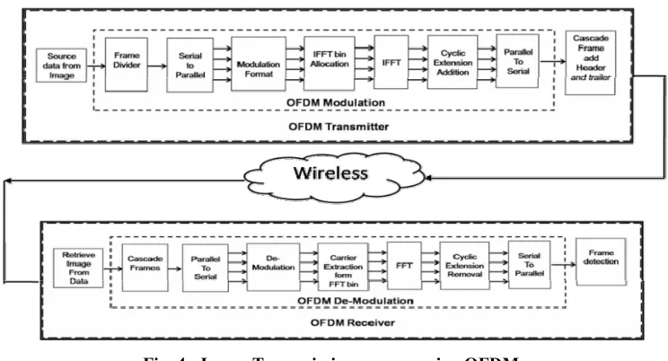
In OFDM system, at the transmitter side information is sent using IFFT symbols and on the receiver side FFT is performed to obtain received data. In OFDM in order to transmit information various sub-carriers are transmitted in parallel, which are orthogonal to each other and thus reduce ISI (Fig. 4). For transmission of information various modulation schemes (BPSK, QPSK etc.) can be used. Cyclic extensions are added for error free recovery. Finally, image data is transmitted in the form of frames which consist of header and trailer [21]. The information stored in the header is used for processing purpose only, like source address, destination address, frame number and time to live etc, while actual information is stored in trailer. At the receiver side, the steps at the transmitter are performed in reverse order.
Fig. 4 - Image Transmission process using OFDM
In OFDM information is transmitted using sub-carriers which are orthogonal to each other. Let the transmitted symbols are defined byxk, the channel response is representing ash(t) added while Gaussian noise asn(t), output asy(t) and received symbol as yk. The input symbols are taken from considered constellation. The digital-to-analog and analog-to-digital conversion processes consists of filter whose bandwidth is inverse of sampling period. The channel response is given by
M
h
(
t
)
a
m
(
t
m)
m1(11)
Where, M represents multi-path between transmitter and receiver. Each path is modeled as Gaussian process with mean zero and variance as2 with M
2
m m1
1.
The wireless channel can be modeled as [18]
y
k
x
k
d
k (12)Here, the parameter β is representing attenuating factor anddkis noise and distortion occur due to the transmission over wireless channel. Using elementary mathematics we get [22]
2
2
2
2SNR
x
1
d
x.
(13)2 2 2
x
In the above equation, the variance of additive white Gaussian variable, is denoted by 2, 2 is variance due to the d
distortion and 2is original signal transmitted power. Distortion in the OFDM signal arises due to the noise,
x
to reduce PAPR amplitude clipping is performed, and leads to some distortion which also contribute to distortion variance. Finally, the output signal for tonepof blockBis given by
y
[
B
,
p
]
H
[
B
,
p
]
x
[
B
,
p
]
w
[
B
,
p
]
. (14)Fig. 5 - Image Frame structure
In OFDM image is transmitted in the form of frames, which consists fixed number of bits in each frame (Fig. 5). So first of all, ECG image is converted into a grayscale image whose pixel values ranges from 0 to 255 and are represented by 8 bits. Depending on considered modulation scheme, image data will be converted into symbol size. Now at the OFDM transmitter, data is converted into frames. These frames are separated by guard bands, and thereafter after time alignment transmitted by transmitter. Due to noise of the channel some of bits get corrupted and thus bits are received in error. Therefore, demodulated frames also contain error and reconstructed image pixels values are different, finally pixels of original and reconstructed image are compared and error pixels are evaluated.
5. Simulation and Results
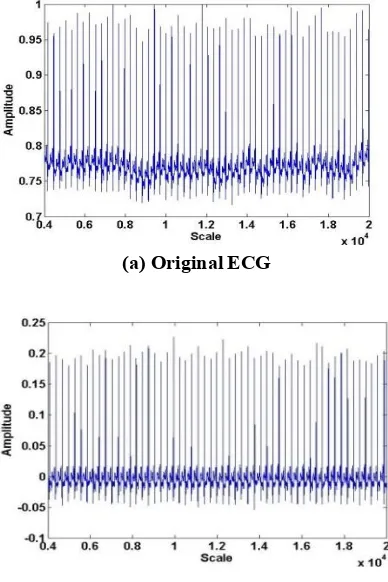
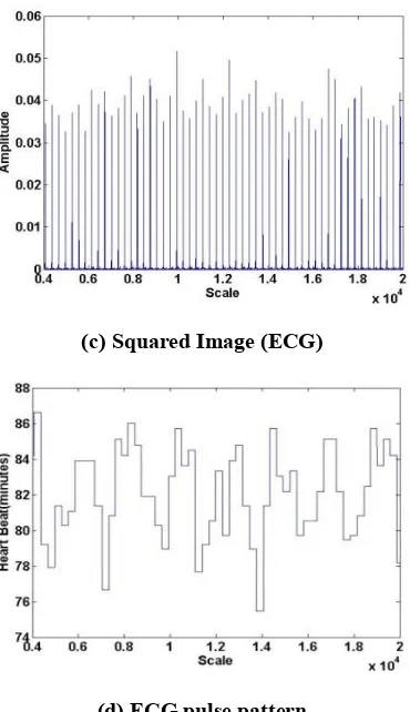
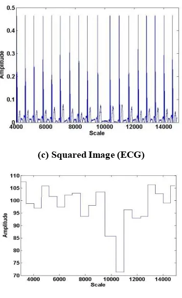
In Fig.6 (a), initial ECG is shown, where 20000 samples are taken, and 4000 samples are left to due to the edge effect of filtering process. So, processing of ECG is done on 16000 samples. In the first step baseline wander which arise due to the respiration, body movement etc. during ECG recording [23]. For baseline wander removal wavelet based filtering technique as proposed in [20] is used, and obtained result is shown in Fig. 6(b). it can be observed from the figure that base is flatter and detrimental effect of baseline wander is nearly removed. To further increase the difference between major and minor peaks, each sample value is squared (m = 2), as shown Fig.6(c). Finally, heat beat calculation is done, as shown in Fig. 6(d). Similarly, for other patient ECG pattern and associated graphs are shown Fig 7. The illustration of the above detailed method is detailed in Fig.7. In Fig. 7(a), original ECG-2 signal is shown, in 7(b) filtered signals is shown, while in Fig. 7(c) squaring of the filtered signal is shown and finally detected heart beats and its pattern in the specified window is shown 7(d).
(a) Original ECG
(c) Squared Image (ECG)
(d) ECG pulse pattern
Fig. 6 - ECG-1 heart beat detection process
(a) Original ECG
(c) Squared Image (ECG)
(d) ECG pulse pattern
Fig. 7 - ECG-2 heart beat detection process
For ECG-1, maximum heart beat is 86.46, minimum heart beat is 75.47, and average is 82.07 with standard deviation of 2.58. For ECG-2, maximum heart beat is 106.36, minimum heart beat is 71.34, and average is 96.67 with standard deviation of 8.6.
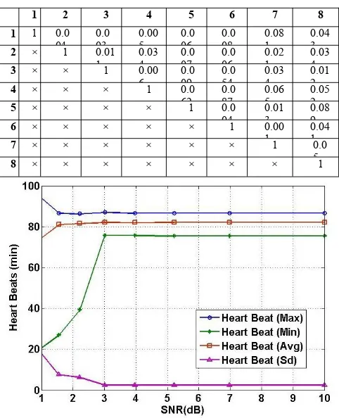
In the next experiment, we took auto and cross correlation of the original ECG signals (Fig. 8), and for 8 ECG signals it is shown in Table 1, it is clear that maximum value of cross-correlation is 0.089 which is much smaller to 1.
As it is evident from above details two ECG differs significantly in each statistical parameter we carried out experiments on a large dataset available at: www.physionet.org/physiobank/database/ptbdb, and it has been found that each ECG has unique distribution and characteristics.
Therefore, for authentication following procedure can be used:
A. ECG Encryption Process
Step 1:For ECG recording two sensors should be deployed in the body.
Step 2: One of the recorded ECG and its heart beat pattern should be transmitted as raw image, while other ECG should be embedded with patient other vital information like blood pressure, blood sugar, spO2, uric acid etc. using
reversible data hiding (RDH) process [24].
Step 3:Data embedding should be done on the upper part of the recorded image where no pulses are observed.
Step 4:After encryption ECG images can be transmitted over the wireless channel.
B. ECG Based Authentication Steps
Step 1: Decrypt ECG received image, and extract embedded data using RDH process.
Step 2: Obtain ECG and heart rate pattern for received ECGs.
Step 4:If obtained score for both the process is more than threshold, authenticate the patient otherwise reject it.
However, in case of noise-free environment, these parameters remain unaffected. In case of noisy channel, some part of the information can be disrupted therefore to detect information error free a minimum level of SNR needs to be maintained. In Fig. 8, various heart beat parameters for ECG -1, are shown, under different SNR while assuming random noise, and it is found that if SNR is kept above 5 dB, error free detection of important parameters are possible, while for ECG-2, the required SNR is 4 dB. Therefore, it is evident that a small amount of leftover noises after filtering process has no effect on ECG beat pattern as the time of transmission SNR is kept sufficiently high to tackle wireless channel attenuations and noises.
Fig. 8 - Cross-correlation between two ECGs
Table 1. Cross-Correlation among 8 Different ECGs
1 2 3 4 5 6 7 8
1 1 0.0
04 0.003 0.005 0.006 0.008 0.081 0.043 2 × 1 0.01
1 0.034 0.007 0.006 0.021 0.034 3 × × 1 0.00
6 0.009 0.054 0.034 0.012
4 × × × 1 0.0
62 0.0 87
0.06 5
0.05 2
5 × × × × 1 0.0
04
0.01 3
0.08 9
6 × × × × × 1 0.00
1 0.041
7 × × × × × × 1 0.0
5
8 × × × × × × × 1
Fig. 9 - Heart beat vs. SNR for ECG-1
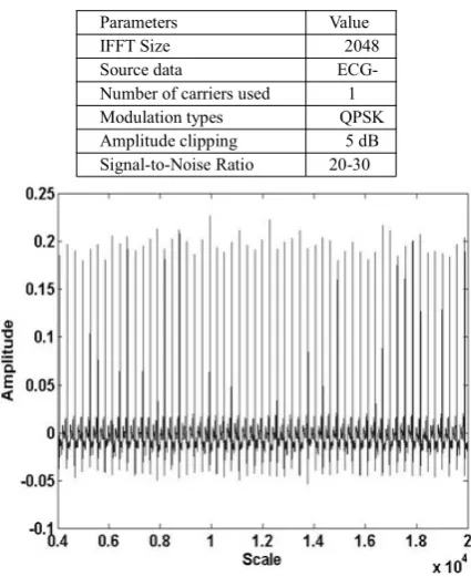
Fourier Transform (FFT) therefore their amplitude varies significantly, and due to variation in amplitudes peak and average values of power (square of amplitudes) varies significantly. Thus, OFDM suffers from PAPR (Peak to Average Power Ratio), and to suppress PAPR, amplitude clipping of 5 dB is considered This amplitude clipping is done for the subcarrier which has amplitude higher than average value, thus variation in amplitude reduces and in turn decrease PAPR. The used modulation is QPSK while SNR is varied from 5 to 25 dB. The transmitted ECG signal is shown in Fig. 11.
Fig. 10 - Heart beat vs. SNR for ECG-2
Table 2. OFDM SIMULATION PARAMETERS
Parameters Value
IFFT Size 2048
Source data
ECG-Number of carriers used 1 Modulation types QPSK Amplitude clipping 5 dB Signal-to-Noise Ratio 20-30
Fig. 12 - Received ECG (SNR=5 dB)
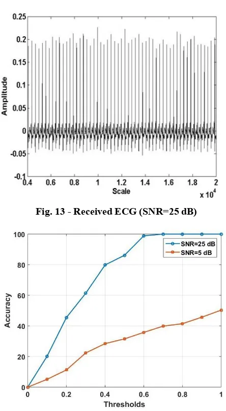
Fig. 13 - Received ECG (SNR=25 dB)
Fig. 14 - Accuracy vs. Thresholds for SNR =5 and 25 dB.
Finally, in Fig. 14, accuracy of correct patient identification vs. Thresholds is plotted while considering SNR of 5 and 25 dB. To obtain this curve 593 ECGs of different persons including both healthy and un-healthy persons are considered. It is clear from the figure that accuracy heavily dependent on SNR and for lower SNR the recovered ECG quality is poor and therefore accuracy is very low even at higher thresholds. However, in contrast to this at higher SNR accuracy is of excellent quality 99.98 % at sufficiently high thresholds > 0.7.
6. Conclusions
In WBAN authentication and verification of patient is an important problem without revealing their identity. In this paper, ECG based authentication and verification process is detailed, and on the basis of obtained results following conclusions can be made:
Each ECG has distinctive shape, features and statistics.
For identification and verification of patient ECG and its first and second order statistics i.e., mean and variance of heart rate can be used.
Correlation can be used for matching of ECGs, as each ECG is distinctive in nature.
Patient data can be hidden in ECG image itself, by using reversible data hiding technique.
For error free reception of ECG signal, a sufficiently high SNR (25 dB) is required.
References
[1] H B Li, K Y Yazdandoost, “Wireless body area network”, River publishers; 2010.
[2] H. Cao, V. Leung C. Chow H. Chan, “Enabling technologies for wireless body area networks: A survey and outlook. IEEE Communications Magazine, vol. 47, no. 12, 2009.
[3] U., Sana, H. Higgins, B. Braem, B. Latre, C. Blondia, I. Moerman, S. Saleem, Z.Rahman, and K. S. Kwak. "A comprehensive survey of wireless body area networks." Journal of medical systems, vol. 36, no. 3, 2012, pp.1065-1094..
[4] P. Kumar, H J. Lee, “Security issues in healthcare applications using wireless medical sensor networks: A survey”, Sensors, vol. 12, no. 1, 2011, pp.55-91.
[5] J. Sametinger, J. Rozenblit, R. Lysecky and P Ott, “Security challenges for medical devices”, Communications of the ACM, vol. 58, no. 4, 2015, pp.74-82.
[6] F. Momtaz, “Secure ECG-Based Biometric Authentication in Body Area Sensor Networks”, University of California, Irvine; 2016.
[7] M, Rushanan, AD Rubin, DF. Kune, CM Swanson, “SoK: Security and privacy in implantable medical devices and body area networks”, InSecurity and Privacy (SP), 2014 IEEE Symposium on 2014, pp.524-539.
[8] D. Da He, E S. Winokur and C G. Sodini, “A continuous, wearable, and wireless heart monitor using head ballistocardiogram (BCG) and head electrocardiogram (ECG)”, In Engineering in Medicine and Biology Society, EMBC, 2011 Annual International Conference of the IEEE 2011, 4729-4732.
[9] Al Taradeh N, Bastaki N, Saadat I, Al Ahmad M. “Non-invasive piezoelectric detection of heartbeat rate and blood pressure”, Electronics Letters, vol. 51, no. 6, 2011, pp.452-454.
[10] Y. Shu, C. Li, Z. Wang, W. Mi, Y. Li, TL. Ren, “A Pressure sensing system for heart rate monitoring with polymer-based pressure sensors and an anti-interference post processing circuit”, Sensors, vol.15, no.2, 2015, pp.
3224-3235.
[11] KK, Venkatasubramanian, A. Banerjee, and SK. Gupta, “PSKA: Usable and secure key agreement scheme for body area networks”, IEEE Transactions on Information Technology in Biomedicine, vol.14, no.1, 2010, pp.60-68.
[12] Z. Zhang, H. Wang, AV. Vasilakos, H. Fang, “ECG-cryptography and authentication in body area networks”, IEEE Transactions on Information Technology in Biomedicine, vol. 16, no. 6, 2012, pp.1070-1078.
[13] L. Yao, B. Liu, K. Yao, G. Wu, and J. Wang, “An ecg-based signal key establishment protocol in body area networks”, In Ubiquitous Intelligence & Computing and 7th International Conference on Autonomic & Trusted Computing (UIC/ATC), 2010, pp.233-238.
[14] X. Liao, C. Shu “Reversible data hiding in encrypted images based on absolute mean difference of multiple neighboring pixels,” Journal of Visual Communication and Image Representation, 2015, vol. 28, pp.21-27.
[15] Tian, Jun. "Reversible data embedding using a difference expansion."IEEE transactions on circuits and systems for video technology13.8 (2003): 890-896.
[16] Chen, Xianyi, et al. "Histogram shifting based reversible data hiding method using directed-prediction scheme."Multimedia Tools and Applications74.15 (2015): 5747-5765.
[17] C. Karlof, N. Sastry, D. Wagner, “TinySec: a link layer security architecture for wireless sensor networks”, In Proceedings of the 2nd international conference on Embedded networked sensor systems 2004, pp.162-175. [18] M. Toorani, “Cryptanalysis of Two PAKE Protocols for Body Area Networks and Smart Environments”,
International Journal of Network Security, vol. 17, no. 5, 2015, pp.629-636.
[19] Pan, Jiapu, and Willis J. Tompkins. "A real-time QRS detection algorithm."IEEE Trans. Biomed. Eng32.3 (1985): 230-236.
[21] US. Mohammed, HA Hamada, "Image transmission over OFDM channel with rate allocation scheme and minimum peak-to average power ratio", arXiv preprint arXiv:1006.0840. 2010.
[22] JHou,JGe, D. Zhai,JLi, "Peak-to-average power ratio reduction of OFDM signals with nonlinear companding scheme"' IEEE Transactions on Broadcasting. Vol.56, no.2, 2010, pp.258-62.
[23] G. Lenis, N. Pilia, A Loewe, WH, Schulze and 0. Dassel "Comparison of Baseline Wander Removal Techniques considering the Preservation of ST Changes in the Ischemic ECG: A Simulation Study". Computational and mathematical methods in medicine. 2017.
[24] D. Zhang, "Wavelet approach for ECG baseline wander correction and noise reduction", In Engineering in Medicine and Biology Society, 2005. IEEE-EMBS 2005. 27th Annual International Conference of the