Pharmacophore
ISSN-2229-5402
Journal home page: http://www.pharmacophorejournal.com
Corresponding Author: Mojtaba Ramezani, Assistant Professor, Department of Prosthodontics, School of Dentistry, Kermanshah University of medical science, Kermanshah, Iran.Email: dr.ramazani_ [email protected]
A COMPARATIVE ANALYSIS OF SHEAR BOND STRENGTH OF
COMPOSITE AND ACRYLIC TEETH TO HEAT-CURED ACRYLIC
RESIN BY DIFFERENCE PREPARATION METHODS
Mojtaba Ramezani
1*, Pezhman moradi
21. Assistant Professor, Department of Prosthodontics, School of Dentistry, Kermanshah
University of medical science, Kermanshah, Iran
2. Department of Prosthodontics, School of Dentistry, Kermanshah University of
medical science, Kermanshah, Iran
A R T I C L E I N F O A B S T R A C T
Received: 04th Jan 2017
Received in revised form: 09th Jun 2017
Accepted: 21th Jun 2017 Available online: 14th Aug 2017
Keywords: Shear bond strength, denture, heat-cured acrylic resin
Objectives: One of the basic problems in patients using removable denture is detachment of artificial teeth from the denture acrylic base. The bond strength of artificial teeth to acrylic base may affect this problem.
Materials & Methods: In this experimental study, five groups of artificial teeth (n=150), including 30 phoenix artificial teeth, 30 Emeral artificial teeth, 30 crystal artificial teeth, 30 ECL artificial teeth (Beta Dent Co, Iran), and 30 Ivoclar artificial teeth( Ivoclar Vivadent) were prepared by three different methods: 50 without surface preparation, 50 by methylmethacrylate monomer, and 50 by cavity preparation. Then, wax investment was performed, replacing the heat-cured acrylic resin. To determine the shear bond strength by Universal Testing Machine (UTM), a shear force with a speed of 1mm/min was delivered to samples and the force causing the fracture was recorded. The location and type of fracture were then investigated. The obtained data were analyzed by Kruskal-Wallis test. Results: The results showed a significant difference in shear bond strength between different artificial teeth in anterior teeth prepared by MM (p=0.004) and CV (p=0.048). Moreover, the findings indicated no significant difference in shear bond strength between different artificial teeth in posterior teeth prepared by MM (p=0.256) and CV (p=0.232).
Conclusion: The findings showed a significant difference between the shear bond strength of acrylic and composite denture artificial teeth to heat-cured acrylic resin.
Copyright © 2013 - All Rights Reserved - Pharmacophore
To Cite This Article: Mojtaba Ramezani*, Pezhman moradi, (2017), “A comparative analysis of shear bond strength of
composite and acrylic teeth to heat-cured acrylic resin by difference preparation methods”, Pharmacophore, 8(4), 27-34.
Introduction
Materials and Methods
After reviewing the studies conducted and consultation with the statistics advisor, a total of 150 artificial teeth, including 30 phoenix artificial teeth (Nano-hybrid composite, Beta Dent Co), 30 Emeral artificial teeth (Nano-composite, Beta Dent Co), 30 crystal artificial teeth (polymethyl methacrylate along with crystal, Beta Dent Co), 30 ECL artificial teeth (polymethyl methacrylate, Beta Dent Co), and 30 Ivoclar artificial teeth (Vivadent Co) (size: N3) were selected in this experimental study. In each group, 15 central incisors and 15 first molars were selected. Each group of artificial teeth was divided into three subgroups according to the preparation method for bonding to acrylic base.
Group 1. Without preparation: before packing acrylic resin, the teeth without any change in the bonding surface were prepared for flasking [21]
Group 2.Methylmethacrylate Monomer etching (MM) Preparation: before packing acrylic resin, methyl methacrylate monomer (SR Tripex hot, Italy Ivoclar, Vivadent) was used twice to moisturize the ridge lap base 1 mm above the cervical area of the artificial teeth [21].
Group 3. Cavity Preparation (CV): before packing acrylic resin, a cavity with 2 mm depth and 2.3 diameter was prepared by an 8 round bur (KG Sorenson) while the teeth were supported by polyvinyl siloxane putty (vinyl polysiloxane impression material heavy body/3M ESPE/USA)[21].
A line was drawn around the artificial teeth up to 1 mm above the ridge lap base using a caliper. This line indicated the finishing site of wax investment. The teeth were mounted on a wax cone (Poly wax/Desen/Turkiye) forming an angle of 45º between the wax surface and longitudinal axis of the tooth [21]. The height of prepared wax cone was 20 mm. Then a rectangular wax block with the height of 60 mm was prepared using a 15×20 mm aluminum block to which the wax bone was connected (Fig. 1).
Fig. 1. Preparation of wax cone with 45º angle
The waxes were molded around the cervical area into a crescent-shaped manner [21], and then flasking was done. Three samples were placed in each flask (Brass flask/USA) (Fig. 2).
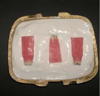
In the lower half of the flask, the dental plaster (Gypsom type II/Pars Dental/ Iran) was used [34]. After 20 minutes when the plaster was set, the first plaster was separated from the second one by Vaseline [34]. Then the upper half of the flask was mounted. The lower and upper parts of the flask were placed on vibrator to prevent bubbles, and dental plaster (Class IV die stone/Pars Dental/ IRAN) was then added [37]. Next, the flasks were placed under the 50 kg press (Hydraulic Press, Koosha Fan Pars/Iran) for 45 minutes [34]. After 45 minutes, the waxes were completely removed using hot water flow, soap and steam. Then the heat-cured acrylic resin (Italy/ Vivadent/ triplex hot Ivoclar) was mixed according to the manufacturer’s instructions (23.4g powder, 10ml liquid) and placed in a glass container [21]. According to the manufacturer’s instructions, after 10 minutes when acrylic resin turned into dough it was packed into the flask. The flasks were placed under 100 kg hydraulic press for 10 minutes and were then placed in clamp [34]. Next, the flasks were placed in cold water to increase their temperature to 100º and boil them for 45 minutes. After cooling down the water, the flasks were extracted and plaster was removed from the acrylic resin and artificial teeth (Fig. 3).
Fig. 3. Prepared samples
To rebuild the mouth, the artificial teeth were kept in distilled water at 37º in incubator (Incubator/Abzar Pezeshki Kavoosh MEGA/Iran) for 48-52 hours [34]. They were placed in a thermocycler (Iran/ tc-300/Vafaei Industrial) at 5000 rpm in a cold bath (4º) and a hot bath (55º). The samples were kept in each bath for 30 seconds with an interval of 20 seconds between the two baths [34].
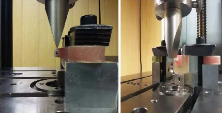
Having prepared the samples, they were numbered and delivered to the operator. To determine the shear bond strength, i.e. the bonding strength between acrylic resin and artificial teeth, a shear force with a speed of 1 mm/min was delivered to the samples by a universal testing machine (Germany/zwick/ roell z050) (Fig. 4). The forces in posterior teeth were delivered at 0.5 mm above marginal acrylic denture in the mesial region so that the blade had 4 mm contact with the teeth; 3 mm contact with polymethyl methacrylate and 1 mm contact with nano-composite [33] (Fig. 4). The forces in anterior teeth were delivered perpendicular to the longitudinal axis of the artificial teeth; 3 mm being in contact with the teeth [38] (Fig. 4). The fracture modes were assessed with a magnifying glass (×5 magnification), according to the following classification: 1) Adhesive - when the separation occurred at the tooth/denture base interface; 2) Cohesive in PMMA denture base - when the denture base material remained bonded to the dislodged teeth, covering it completely; 3) Cohesive in the tooth - when total tooth base remained bonded to the denture base; 4) Cohesive in the tooth associated with cohesive in the denture base; and 5) Mixed - when significant areas of adhesive and cohesive failures occurred simultaneously[21].
Results
The results showed no significant difference between different artificial teeth, including phoenix, Emeral, crystal, ECL, and Ivoclar in anterior teeth without surface preparation in terms of bonding strength (p=0.451) (Fig. 5). The maximum bond strength was reported for phoenix artificial teeth and the minimum strength was found for ECL artificial teeth. However, the findings indicated a significant difference between different artificial teeth, including phoenix, Emeral, crystal, ECL, and Ivoclar in anterior teeth after preparation with methylmethacrylate (p=0.004) (Fig. 6).
The maximum shear bond strength was reported for Ivoclar, crystal, phoenix, Emeral, and ECL, respectively. Further, the results revealed a significant difference between different artificial teeth regarding shear bond strength after cavity preparation (p=0.048) (Fig. 7). The maximum shear bond strength was reported for Ivoclar, phoenix, crystal, Emeral, and ECL, respectively. Moreover, the findings showed a significant difference between different artificial teeth with regard to bond strength in posterior teeth without surface preparation (p=0.42) (Fig. 8). The maximum shear bond strength was reported for Ivoclar, crystal, Emeral, ECL, and phoenix, respectively.
In addition, the findings showed no significant difference in the bond strength of posterior artificial teeth after preparation with methylmethacrylate (p=0.256) (Fig. 9). The maximum and minimum bond strength were reported for Ivoclar and ECL artificial teeth, respectively. Furthermore, no significant difference was observed for bond strength after cavity preparation in posterior teeth (p=0.232) (Fig. 10). The maximum and minimum bond strength were reported for phoenix and ECL artificial teeth, respectively.
Discussion
The results of study for different preparation methods showed that in all samples, both in anterior and posterior teeth, the maximum levels of shear bond strength was reported for cavity preparation, methylmethacrylate preparation, and artificial teeth without preparation. The increased bond strength in MM technique could be due to the fact that when there are two solid surfaces, or one solid and one liquid surface that soak the surface well, the surface tension value is greater than the surface active one and increases the bond strength [40]. Also, after some time the speed conversion of monomer to polymer is reduced due to two reasons: first, the number of reactive monomers is reduced and second, the propagation velocity of monomers in a substance with high viscosity is highly reduced toward the reaction sites [41]. Therefore, putting the monomer in the ridge lap area increases the number of monomers as well as polymerization in this area, thereby increasing the bond strength. The increased bond strength in artificial teeth prepared with diatoric cavity than teeth prepared with methylmethacrylate and teeth without preparation can be associated with the micro- and macro-mechanical attachment generated by diatoric cavities [40].
The results of the study regarding the bond strength of anterior teeth without preparation indicated no significant difference among different artificial teeth, including phoenix, Emeral, crystal, ECL, and Ivoclar artificial teeth (p=0.451). The maximum and minimum levels of bond strength were found for nano-hybrid composite phoenix and ECL artificial teeth, respectively. The equal bond strength of the artificial teeth could be possibly due to this fact that in all samples polymethyl methacrylate resin was used in the ridge lap area and the ridge lap surface of anterior teeth was less than that of posterior teeth.
Adhesive fracture was the most frequent type of fracture in Ivoclar denture possibly because the teeth were monolithic, indicating their high structural strength. Also, cohesive fracture within the tooth and acrylic resin was the most frequent type of fracture in crystal artificial teeth, and cohesive fracture within the tooth was the most frequent type of fracture in ECL artificial teeth. These findings for crystal and ECL artificial teeth indicate their lower structural strength than Ivoclar teeth. The maximum frequency of fracture in phoenix teeth was reported for cohesive fracture within the tooth probably because these teeth were multilithic, and bonding strength of composite to acrylic resin was low. Despite the multilithic nature of Emeral denture, the most frequent type of fracture in these teeth was adhesive fracture, which might be due to low polymerization of polymethyl methacrylate at the ridge lap area.
The findings of anterior teeth prepared by methylmethacrylate showed a significant difference between different artificial teeth, including phoenix, Emeral, crystal, ECL, and Ivoclar (p=0.004). Ivoclar, crystal, phoenix, Emeral, and ECL were found to have the maximum strength, respectively. The increased bond strength in acrylic teeth compared with composite artificial teeth might be due to increased polymerization at the ridge lap area while using methylmethacrylate. Although polymethyl methacrylate was used at the ridge lap region in all samples, the reduced bond strength in ECL teeth than other teeth could be due to the use of cross linking monomer in ECL structure, which caused higher cross linking at the ridge lap region and reduced the bond strength. The type of fracture in the teeth prepared with methylmethacrylate was the same as that of the teeth with no preparation; the only difference, however, was that the most frequent type of fracture in Ivoclar and Emeral teeth was mixed fracture. Mixed fracture is a kind of adhesive fracture with this difference that some acrylic resin remains in the cervical region possibly due to the wax investment method.
The results of anterior teeth prepared by diatoric cavity revealed a significant difference between various anterior artificial teeth after cavity preparation with regard to bonding strength (p=0.048). The maximum levels of bond strength were found for Ivoclar, phoenix, crystal, Emeral, and ECL artificial teeth, respectively. The possible reason for the increase of bond strength in phoenix than in Emeral denture is higher content of filler in phoenix denture. The presence of inorganic fillers
(particle or fi brous) has a remarkable impact on the properties of polymers, which in turn increases the The modulus of
of anterior teeth because the ridge lap surface of anterior teeth was lower than that of posterior teeth and forces were delivered at different locations.
The results of posterior artificial teeth without preparation showed a significant difference between different artificial teeth in terms of the bonding strength (p=0.042). The highest level of bond strength was reported for Ivoclar, crystal, Emeral, ECL, and phoenix artificial teeth, respectively. The enhanced bond strength in Ivoclar and crystal artificial teeth is possibly because they are monolithic and acrylic, but the reduced bond strength in phoenix denture may be due to the delivery of force. Because the posterior teeth receive more forces along the longitudinal axis, the string fillers should be oriented in such a manner to be resistant against these forces. On the other hand, in phoenix artificial teeth some degree of anisotropy occurs (delivery of force in different directions changes the bond strength). The most frequent type of fracture in Ivoclar and crystal artificial teeth was found to be mixed fracture, which is indicative of the higher structural strength of these artificial teeth than ECL in which the most frequent type of fracture was cohesive fracture within the tooth and acrylic resin. As for phoenix and Emeral artificial teeth, cohesive fracture within the tooth and acrylic resin was the most frequent type of fracture, which could be because of the multilithic nature of these artificial teeth and location of forces applied.
The findings related to the posterior teeth revealed no significant difference between the bond strength of different artificial teeth following preparation with methylmethacrylate (p=0.256). The maximum and minimum levels of bond strength were reported for Ivoclar and ECL artificial teeth .The improved bond strength of phoenix artificial teeth might be due to increased polymerization at the ridge lap region of the teeth. The most frequent type of fracture in Ivoclar and crystal artificial teeth was found to be cohesive fracture within acrylic resin possibly because of increased polymerization at the ridge lap region as a result of application of methylmethacrylate. In the case of ECL artificial teeth, cohesive fracture within the tooth and acrylic resin was reported to be the most frequent fracture, which is indicative of low bond strength in these teeth. In phoenix and Emeral artificial teeth, cohesive fracture within the tooth and acrylic resin was the most frequent type of fracture, which could be because of the multilithic nature of these teeth and location of forces delivered.
The results of posterior teeth prepared by diatoric cavity indicated no significant difference between the bond strength of different artificial teeth after cavity preparation (p=0.232). The maximum and minimum levels of bond strength were reported for phoenix and ECL artificial teeth. The increased bond strength in phoenix and Emeral artificial teeth might be due to the mechanical attachment in the structure of the nano-composite, which reduced the effect of forces delivered to the nano-composite. Cohesive in the acrylic fracture within Ivoclar and crystal artificial teeth was found to be the most frequent type of fracture possibly because the acrylic resin was stuck within diatoric cavity. As for ECL artificial teeth, cohesive fracture within the tooth and acrylic resin was the most frequent fracture, indicating their low structural strength. With regard to phoenix and Emeral artificial teeth, cohesive fracture within the tooth and acrylic resin was observed to be the most frequent type of fracture, which might be due to the multilithic nature of these artificial teeth and location of forced delivered.
Similar to the present study, the studies of Farid Bahrani [25], Lauro Egídio Bragaglia [21], and Adelina Stoia [31] showed that preparation with methylmethacrylate increased the bond strength. Farid Bahrani reported preparation by diatoric cavity enhanced the bond strength but the research by Lauro Egídio Bragaglia showed a reduction in bond strength in preparation by diatoric cavity. This difference might be due to the fact that in the present study acrylic resin was packed at diatoric cavity while adding acrylic resin by condenser, which was intended to make sure air entrapment did not prevent chemical bonding at this region.
Ghahramani [34] reported that the bond strength of composite artificial teeth was lower than that of acrylic artificial teeth in posterior teeth, which is in line with the results of present study considering less bond strength of composite teeth than acrylic teeth. Using bending strength test, Kawara [17] reported that the bond strength of acrylic monolithic teeth was significantly higher than that of resin-based multilithic teeth. Compared with the results of the current study, this difference may be associated with different types of artificial teeth used as well as the structural difference of multilithic artificial teeth manufactured by Beta Dent Company compared with the artificial teeth tested in Kawara’s study.
In their study analyzing the . Effect of surface treatments on the bond strength ofdifferent resin teeth to complete artificial teeth base material, Carolina et al. [20] studied four types of polymethyl methacrylate artificial teeth manufactured by various companies. The results showed no significant difference considering the effect of surface preparation on intragroup bond strength of commercial brands. In this study, a different acrylic resin was used, so the results cannot be compared with the results of the current research.
Rostamkhani et al. [27] evaluated the tensile strength of four types of acrylic artificial teeth (Italian Ivoclar, Yaghoot, Herasit plus and Acradent) in vitro in the presence of artificial saliva. Their findings showed the highest tensile strength for Ivoclar, Herasit plus, Yaghoot, and Acradent artificial teeth, respectively. In the present study, Ivoclar denture was reported to have the maximum bond strength but due to different types of artificial teeth used, type of preparation, and measurement of tensile strength, this study is not comparable with the current study.
types of Iranian resin-based artificial teeth (Marjan, Super Newclar, and Diamond) made by Ideal Makoo Company and compared them with a German denture. Their results revealed no significant difference between the Iranian and the foreign artificial teeth. This study is also not comparable with the present study because of the different types of artificial teeth evaluated.
Conclusion
1. In all samples, the maximum level of shear bond strength in both anterior and posterior artificial teeth was reported for teeth with CV preparation, teeth with MM preparation, and teeth without preparation, respectively.
2. In posterior teeth, there was only a significant difference in Ivoclar artificial teeth regarding shear bond strength from among different preparation techniques. No significant different was found for other artificial teeth.
3. In posterior artificial teeth, no significant difference was found for the shear bond strength of Ivoclar and crystal artificial teeth from among various preparation methods. There was a significant difference between the shear bond strength of other artificial teeth.
4. In all anterior artificial teeth, no significant difference was reported for the shear bond strength of the artificial teeth without preparation. Methylmethacrylate and cavity preparation methods were reported to show a significant difference. 5. In all posterior artificial teeth, a significant difference was found for the shear bond strength of the artificial teeth with no preparation. However, no significant difference was found for methylmethacrylate and cavity preparation methods.
References
1. Barbosa DB, Ricardo Barao VA, Monteiro DR, Compagnoni MA, Marra J. Bond strength of denture teeth to acrylic resin: effect of thermocycling and polymerization methods. Gerodontology .2008, No.25, P: 237-44. 2. Craig RG, Powers JM, wataha JC. Dental material properties and manipulation. Saint louis: Mosby; 2000, No.1, P:
257-81.
3. Cunningham JL. Shear bond strength of resin teeth to heat cured and light-cured denture base resin. J Oral Rehabil. 2000, No.27, P: 312-6.
4. Cunningham JL. Bond strength of denture teeth to acrylic bases. J Dent 1993, No.21, P: 274-80.
5. Cunningham JL, Benington IC. Effect of an experimental cement on denture tooth bond. J Dent Res. 1994, No.2, P; 74:949.
6. Darbar UR, Hugget R, Harrison A. Denture fracture: A survey. Br Dent J 1994, No.176, P: 342-5.
7. Cunningham JL, Benington IC. An investigation of the variables which may affect the bond between plastic teeth and denture base resin. J Dent. 1999, No.27, P: 129-35.
8. Shahabi S, Fadavi H. Comparison of bond strength of three denture teeth made in Iran with resin bases and Ivoclar denture teeth. JIDA. 2005, No.16, P: 44-52.
9. Mosharraf R, Pooya E, Maleky V. The evaluation of an Iranian denture tooth (Marjan) bond strength with denture base resins in four different preparing methods. JIDA. 2001, No.; 372, P: 49-59.
10. Mosharraf R, Feiz A, Barani B. Comparison of bond strength of three denture teeth made in Iran with resin bases and Ivoclar denture teeth. J Res Med Sci. 2002, NO.73, P: 243-5.
11. Naser Khaki M, Ehsani S. Comparing the bond strength of four kinds of Ideal-Makoo artificial teeth (Iran) and two Leichtenstein & Italy Ivoclar teeth with prosthetic acrylic base. J Dent Sch. 2007, No.; 25(3), P: 310-5.
12. Moshref p. Comparison of the bond strength of denture teeth (Coral) to denture base resins by the four treatments. Jour I. S Dentists.2000, No. 2, p: 49-59.
13. Zarb j. prosthetic treatment of edentulous patients. 1st ed., Royan pazhouh, Tehran, 2011, No.3, p: 221-2.
14. Ejlali-M. Patients without dental treatment. Third Edition. Tehran: Jihad Publishing Institute, 1999: Chapters 16, 24 and 25, p. 588-589 and 570.
15. Clancy JM, Boyer DB: Comparative bond strengths of light cured, heat cured and atuopolymerizing denture resins to denture teeth. J Prosthet Dent. 1989, No.61, P: 457-462.
16. Clancy JM, Hawkins LF,Bond strength and failure analysis of light cured denture resins bonded to denture teeth. J Prosthet Dent. 1991, No.65, P: 315-324.
17. Kawara M, Carter JM, Ogle RE, Johnson RR: Bonding of plastic teeth to denture base resins. J Prosthet Dent. 1991, No.66, P: 566-571.
18. Takahashi Y, Chai J, Takahashi T, Habu T: Bond Strength of denture teeth to denture base resins. Int J Prosthodont.2000, No.13, P: 59-65.
19. Rostamkhani, F, compared to the tensile strength of four denture acrylic in vitro in the presence of saliva. Journal of Mashhad Dental School.2011, No. 36, P: 2-14.
21. Lauro Egídio bragaglia1؛ luiz henrique maykot prates2؛ maria cristina marino calvo3. The role of surface treatments on the bond between acrylic denture base and teeth. braz dent j.2009, No. 20(2), P: 156-161.
22. Chittaranjan B, Taruna M, Sudheer N, Nagesh S Patil. Evaluation of shear bond strength of three different types of artificial teeth to heat cure denture base resin: An in vitro study. Kamineni Institute of Dental Sciences. 2012 No.24, P: 321-325.
23. Buyukyilmaz S, Ruyter IE: The effects of polymerization temperature on the acrylic resin denture base tooth bond. Int J Prosthodont. 1997, No.10, P: 49-54.
24. Mohammad Reza Hafeziahmadi, Mojgan Javedani, Bahare Ghiasi, Samiramis Ghavam, (2017). Investigation Of The Relationship Between Phase Angle And Micro-Albuminuria In Type 2 Diabetic Patients With A History Of More Than 5 Years Of The Disease In Ilam Province, Iran, Acta Medica Mediterranea, 2017, 33: 357.
25. Bahrani F, Khaledi AA. Effect of surface treatments on shear bond strength of denture teeth to denture base resins. Dent Res J (Isfahan). 2014, No.111, P: 114–118.
26. Nematollahi F, Azizi N, Shahabi S, Ghahremani L, Asgari Z, Bagheri H. Comparison effect of artificial tooth type and cyclic loading on the bond strength to auto-polymerized acrylic denture base resins, Journal of Dental Medicine-Tehran University of Medical Sciences. 2014, No.26(2), P:81-90۰
27. Rosthamkhani F, Gharehchahi J, Asadollahzadeh M, Zebarjad S, Gharehchahi M. Comparison of Tensile Strength of Four Kind of Acrylic Artificial Teeth to Acrylic Denture Base In Vitro. J Mash Dent Sch. 2012, No 36(2), P: 105-12.
28. Ghasemi A, Mosherf R, Eadi aghfiabadi A. survey of Strength of a few denture of Acrylic Artificial Teeth. Juor of jameslami, 2011, No.4, P: 240-247.
29. Naserkhaki M, Ehsani S.Comparison of four denture adhesive power plant Ivoclar ideal Maku Iran, two countries with acrylic denture base Aykhtn Stein and Italy. Martyr Beheshti University Dental Journal. 2007, No.3, P: 310-315.
30. Thean HP, Chew CL, GohK: Shear bond strength of denture teeth to base. Quintessence Int. 1996, No.27, P: 425-428.
31. Stoia AE, Cosmin S, Meda N, Marius E, Roxana R, Mircea P,Anca T, Mihai R. Tensile Bond Strength of Acrylic Resin Teeth to Denture Base Repair Resin. Advances in Communications, Computers, Systems, Circuits and Devices.32 Conference Paper.
32. Ghasemi E, Mosharaf R, Eidi A. A study of several types of bonding strength of artificial teeth to acrylic resin-based composite removable prostheses. J Dent Sch. 2012, No. 22(4): 240-247.
33. Mian H, Sucena Pita M, Do Nascimento C, Henrique Carriço Nogueira Fernandes F, Linares Calefi P, Manço de Oliveira-Neto J, Pedrazzi V.shear bond strength of acrylic teeth to heat –curing denture base resin under different disinfectant methods. int.j odontosiomat. 2013, No.71, P: 99-105.
34. Ghahramani L, Shahabi S, Amirjan A, Fazel A. Comparison of bond strength of composite and acrylic teeth to heat-cured and auto-polymerized acrylic denture base. Journal of Dental Medicine-Tehran University of Medical Sciences. 2010, No. 4, P; 215-226.
35. Azad A, Siddiqui A, Jawad A, Zia M, Tahir A. effect of mechanical modification of acrylic resin denture teeth bonded to acrylic denture base. Pakistan oral & dental journal. 2012, No.1, P: 22-40. 36. Adelina E, Cosmin S, Meda N, Marius E, Roxana R, Mircea P, Anca T, Mihai R. Tensile Bond Strength of Acrylic
Resin Teeth to Denture Base Repair Resin. Advances in Communications, Computers, Systems, Circuits and Devices, 2st ed, March 2012, No.2, P:111-122:
37. Tony J, Duncan J. Wood. Techniques in Complete Denture Technology. 1st ed, March 2012, No.1, P: 91-100: 38. Naveen S , Surabhi S, Sunil K, Puja H, Rajkiran Ch, Shilpi K . Evaluation of Bond Strength of Acrylic Teeth to
Denture Base using Different Polymerization Techniques: A Comparative Study. Journal of International Oral Health. 2015, No. 1, P: 54-56.
39. Organization Participating In The Edight Edition Of The Glossary Of Prosthodontic.Terms J Prosthet.Dent.2005, No. 94(1), P:1-104
40. Ronald L. John M. Craig’s RESTORATIVE DENTAL MATERIALS. Philadelphia, 3TH. United States. 2012, No.1, p: 52-53.
41. Ronald L. John M. Craig’s restorative dental materials. Philadelphia. 2012, No.1, p: 280-327