Pharmacophore
ISSN-2229-5402
Journal home page: http://www.pharmacophorejournal.com
Corresponding Author: K.V. Nuzhnaya; Laboratory of 3D technologies, Stavropol State Medical University, Stavropol, Russian Federation, 355000Russian Federation
COMPUTER SIMULATION AND NAVIGATION IN SURGICAL
OPERATIONS
K.V. Nuzhnaya
1*, A. E. Mishvelov
1, S.S Osadchiy
1, M. V.Tsoma
2, R. H. Slanova
2, A. M.
Kurbanova
2, KA Guzheva
2, I.A. Rodin
3, A.A. Nagdalian
4, I. V. Rzhepakovskiy
4, S.I.
Piskov
4, S.N. Povetkin
4, V. V. Mikhailenko
51.
Laboratory of 3D technologies, Stavropol State Medical University, Russian Federation,
2.
Stavropol regional clinical and consulting diagnostic center (ANMO
“
SKKKDC
”),
Russian
Federation
3.
Department of anatomy, veterinary obstetrics and surgery Federal State Budgetary
Educational Institution of Higher Education
“
Kuban State Agrarian University named
after I.T. Trubilin
”,
Russian Federation.
4.
Institute of Life systems, North Caucasus Federal University, Russian Federation
5.
Department of Parasitology and veterinary-sanitary expertise, anatomy and pathologic
anatomy of Professor S. N. Nikolsky, Stavropol State Agrarian University, Russian
Federation
To Cite This Article: K.V. Nuzhnaya, A. E. Mishvelov, S.S Osadchiy, M. V.Tsoma, R. H. Slanova, A. M. Kurbanova and et al .
(2019), “Computer Simulation and Navigation in Surgical Operations ”, Pharmacophore, 10(4), 43-48.
Introduction
Currently, the number of patients requiring high-tech surgical interventions in the provision of medical care in urology, oncology, cardiovascular surgery is growing. Patients with the purpose of diagnosis of complex pathologies are prescribed examinations, which are based on digital technologies. To date, these methods of examination include computer and magnetic resonance imaging, ultrasound with three-dimensional image reconstruction, etc. [1, 2].
Computed tomography (CT) is a promising method in the diagnosis of diseases of the internal organs, as well as in the use of intraoperative navigation surgery, which is confirmed by its extensive use in the preoperative use of various diseases [3, 4]. Nowadays, multispiral computed tomography is being actively implemented to obtain isotropic voxels, the combination of which with post-processing allows to obtain accurate data on the localization of the tumor, invasion of vessels and neighboring organs [5, 6].
The location of tumors in relation to surgically significant vessels and adjacent organs has a determining effect in the planning of the operation. No less important factor is the ability to assess the volume of the tumor in relation to the tissu e. Previously, we obtained a three-dimensional visualization in the interactive planning of complex liver resection operations A R T I C L E I N F O A B S T R A C T
Received:
26th Mar 2019
Received in revised form: 03th Aug 2019
Accepted:
10th Aug 2019 Available online: 28th Aug 2019
Keywords: heart, 3D graphics,
simulator, simulation surgical system, cardiology, augmented and mixed reality.
A prototype of the software package module for planning surgical interventions in real time using clinical trials has been developed. Tested software module HoloSurgeri designed for simulations of cuts in the surface of the 3D model in real-time. 3D models obtained by computed tomography are used as objects. Thus, developed system allows the user to perform operations on the object model in real-time.
Pharmacophore, 10(4) 2019, Pages 43-48
[1]. Grenacher and co-authors have shown that three-dimensional pancreatic reconstruction with semi-automatic segmentation can be created similarly to hepatic imaging [7]. Despite this, there are practically no medical programs based on the use of holographic images superimposed on real objects (HoloLens mixed reality technology) for both diagnostic doctors and surgeons.
For surgeons performing abdominal surgery, it is important to the accuracy of positioning and depth of penetration of tools. Often, urologist surgeons removing the tumor, located in the brain substance of the kidney, act almost blindly. Specialists use ultrasound machines and intraoperative CT for updating the location of the tumor. But this method of navigation is inconvenient, cumbersome and harms the health of the surgeon [4, 8]. Often the cardiology surgery has a problem of reliable assessment of the severity of the problem of complex diseases, such as a tumor in the heart (myxoma) without invasive intervention.
The solution of these problems is associated with the creation of a new type of simulator for the planning and navigation of surgical interventions. This work is already being carried out on the basis of auto-program modules and equipment developed by our team earlier, combined into software systems that use mixed and augmented reality technologies with the use of certain sensors for navigating surgical intervention [9].
Materials and Methods
At the first stage of creating a simulation surgical system for the development of a surgical intervention simulator module, a selection of diagnostic studies of patients by methods of computed tomography (CT), magnetic resonance imaging (MRI), ultrasound diagnostics (ultrasound) in the format of DICOM files for processing using the DoctorCT-Slicer software and a simulation surgical system was made. The patients were selected according to the principle of selection of complex clinical cases in Stavropol Diagnosis (40 cases in 2018).
Simulation software was developed in the framework of the program "UMNIK-2015", under the State contract 9492GU/2015 "Development of software DoctorCT, allowing you to create three-dimensional models of internal organs based on images of computer and magnetic resonance imaging".
The developed application is designed to stimulate cuts on the surface of the 3D model in real time. 3D models obtained by computed tomography are used as objects.
The software allows to create a visual cut on the surface, eliminate it, and cut a closed contour of the object of the user's choice. The basis of the cutting system is a special Shader model that allows you to translate the polygons of the object in a transparent state. Stitching works as a reverse operation, returning the normal Shader of the model and turning its parts int o visible mode. A similar algorithm was chosen to reduce the load on the system - because most of the currently used computers do not have enough resources to emulate a physical cut based on the material, which causes long processing of each click.
At the second stage, in order to simulate the work with tumors and simulate the work of the surgeon, we have prepared and optimized the HoloSurgeri system (within the framework of the research on the state program), which allows to make a surgical incision, and its stitching, the possibility of choosing surgical instruments (scalpel, retractor, laser scalpel), choose the thickness and depth of the incision on the 3D model of the organ.
Model cuts are based on pre-breaking algorithm “chunks”. This mechanism facilitates the operation of the system: real-time counting of millions of polygons of high-quality model is almost impossible, especially with limited computing power. Using pre-preparing method each part of a model is processed independently, reducing the recalculation time in proportion to the size and number of “chunks” [1, 10].
To make a cut, the user draws a closed contour, and then there is an assessment - to which part or several parts of the model belongs to this area. Next, the algorithm “cleans” all polygons that are outside the contour, transfers the initial selection to the mobile mode, and creates a local copy of the eliminated parts at the original location [10].
Thus, we get a part of the object that can be moved after cutting (figure 1).
Pharmacophore, 10(4) 2019, Pages 43-48
To optimize the process, the algorithm of multithreaded data processing is used - the system determines the number of free processor threads, and, if necessary, allocates additional ones for calculations. So, the developed system allows the user to perform operations on the object model in real-time.
This system allows users to upload a module to the simulation of surgical intervention, the resulting 3D model of the heart after reconstruction with CT and conduct a variety of manipulation: puncture, cut, stitching, scrolling 3D models, zoom in and zoom out model of the body, the tuning mode of the instrument.
Results and Discussion
To optimize the work in the HoloDoctor software environment, the HoloSurgery module was created with an advanced interface and advanced capabilities for navigation of surgical intervention in real time and simulation of the operational process.
The extended interface includes several new modules written in the code submitted for licensing:
void Start () {
Self = this; DegreeRL = 0; DegreeRot = 0; DegreeFB = 0;
SliderRL.onValueChanged.AddListener(delegate { SliderDegreeRL(); }); SliderFB.onValueChanged.AddListener(delegate { SliderDegreeFB(); }); SliderROT.onValueChanged.AddListener(delegate { SliderDegreeROT(); }); SliderModelLR.onValueChanged.AddListener(delegate {SliderModLR(); }); SliderModelUD.onValueChanged.AddListener(delegate {SliderModUD(); }); }
public void ClearAllSliders() {
DegreeRL = 0; DegreeRot = 0; DegreeFB = 0; SliderUpdate();
SliderModelLR.value = 0; SliderModelUD.value = 0; }
public void OnclickDeeperPlus() {
Controller.Self.Deeper(DeepInch,true); }
public void OnclickDeeperMinus() {
Controller.Self.Deeper(DeepInch, false); }
Pharmacophore, 10(4) 2019, Pages 43-48
Figure 2 – Real-time operation simulation
Testing of the developed module was carried out during 2018 in the Stavropol regional clinical and consulting diagnostic center (SRCCD).
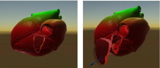
In 40 situational clinical cases, patients with tumors and metastases in the kidneys and heart were successfully operated using the developed software. A separate clinical case of a patient with cardiac myxoma surgery was performed in the cardiology center of Stavropol Region (figure 3).
Figure 3 – Simulation of operation on cardiac myxoma
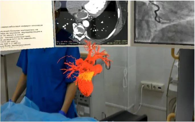
Two programs: DoctorCT-Slicer and HoloDoctor with HoloSurgeri module were used for 3D reconstruction of the heart obtained with CT (using bolus contrast and injector) [9]. A multispiral computed tomograph was used to construct a three-dimensional multilayer model of the heart. The data of a patient (a 57-year-old woman) who complained of chest pain, shortness of breath and high blood pressure were processed.
During the CT of the heart, it was revealed that the wall formation on the right wall of the left atrium is fixed on the leg, the nodal structure with calcifications, the contours are clear. The formation extends to the mouth of the right upper pulmonary vein, 27.8 x 17,3x13,9 mm in size.
DIAGNOSIS: a Neoplasm in the left atrium, myxoma of the left atrium.
Using DoctorCT-Slicer prepared and identified by the color of the neoplasm in the left atrium (tumor), while preserving the reconstructed 3D model of the heart for the implementation of the simulation of the surgical intervention in real time, based on specific morphological characteristics of the patient in simulation software HoloDoctor (figure 4).
Pharmacophore, 10(4) 2019, Pages 43-48
A B
Figure 4 – Preparation of 3D model in the program software DoctorCT-Slicer A – marker of the tumor secretion
B – section of the heart with the tumor
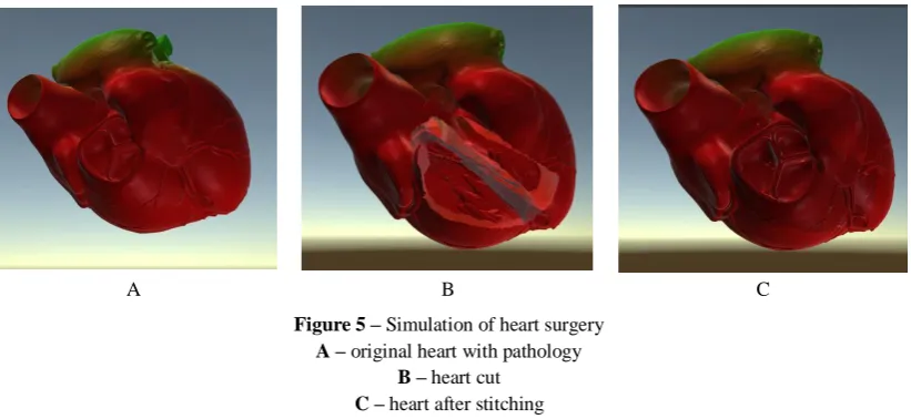
A B C
Figure 5 – Simulation of heart surgery A – original heart with pathology
B – heart cut C – heart after stitching
The high level of detail of the model provides a realistic image, allowing users to work with screen resolution up to 4K inclusive.
The cuts are made by simply pressing the mouse button, which is tied to the emulation of a scalpel. It makes simulation simple and effective.
The simulation is built as the first stage to create a simulator that will allow surgeons to simulate the operati on in the preparatory stage.
The simulation system allows to import volumetric data in the DICOM format of different modalities (CT, MRI, positron immersion tomography (PIT), x-ray angiography, 3D models of the organ and organ systems (multilayer models) obtained in the aftermath of reconstruction with CT, MRI and other methods.
The system (module) allowed the surgeon to operate on the heart with conditional prompts, i.e. when the surgeon held a scalpel in the wrong direction (near a large vessel), the system signaled that there is a large vessel (in this case, part of the coronary artery).
Conclusion
Pharmacophore, 10(4) 2019, Pages 43-48
List of abbreviations: CT - computed tomography; MRI - magnetic resonance imaging, STL - stereolithographic file, obj- object.
References
1. G. J. Hite, A. E. Mishvelov, E. A. Melchenko, А.А. Vlasov, O.I. Anfinogenova, C. V. Nuzhnaya, et al. (2019), “Holodoctor Planning Software Real-Time Surgical Intervention”, Pharmacophore, 10(3), 57-60.
2. Stalling, D.; Westerhoff, M.; Hege, H.-C.. C.D. Hansen and C.R. Johnson, ed. "Amira: A Highly Interactive System for Visual Data Analysis". The Visualization Handbook. Elsevier: 2005, p 749–767 doi:10.1016/b978-012387582-2/50040-x.
3. Medical Mistakes Kill 100,000+ Americans A Year. // Journal of the American Medical Association. Electronical source: http://www.yourmedicaldetective.com/drgrisanti/dangerous_medicine.htm .
4. Zhang Y., Ge HW., Li NC., Yu CF., Guo HF. Evaluation of three-dimensional printing for laparoscopic partial nephrectomy of renal tumors: a preliminary report. World J. Urol. 2016;34(4):533 – 537. DOI - https://doi.org/10.1007/s00345-015-1530-7.
5. Sokolova I. O. Creation and use of the program for studying the anatomy of the human cardiovascular system with the function of augmented reality / I. O. Sokolova, O. V. Ivanov, D. A. Belyakov, T. B. Belyakova // Scientific forum: Innovative science: collection of articles on materials VIII international conference. Moscow 2017, № 7 (8): 26-30
6. Areshidze DA, Mischenko DV, Makartseva LA, Kucher SA, Kozlova MA, Timchenko LD, Rzhepakovsky IV,
Nagdalian AA, Pushkin SV. Some Functional Measures of the Organism of Rats at Modeling of Ischemic Heart Disease in Two Different Ways. Entomology and Applied Science Letters. 2018 Jan 1;5(4):19-29.
7. Andrén O, Fall K, Franzén L, Andersson SO, Johansson JE, Rubin MA. How well does the Gleason score predict prostate cancer death? A 20yearfollowup of a population based cohort in Sweden. J Urol. 2006;175: 1337–1340. doi: 10.1016/S00225347(05)007342
8. Rohin K Iyer Loraine L Y Chiu, GordanaVunjak-Novakovic and MilicaRadisic. Biofabrication enables efficient interrogation and optimization of sequential culture of endothelial cells, ibroblasts and cardiomyocytes for formation of vascular cords in cardiac tissue engineering //Biofabrication – 2012. - №4. – С. 1-11.
9. Hight G Ya, Mishvelov A E, Nuzhnaya C V et al. New Image Modeling Features For Planning Surgical Interventions. Research Journal of Pharmaceutical, Biological and Chemical Sciences. 2019, 10 (1):140-143. 10. Sovilj, S., Magjarevic, R., Lovell, N., Dokos, S. Realistic 3D bidomain model of whole heart electrical activity and