R E S E A R C H A R T I C L E
Open Access
Optic nerve crush induces spatial and temporal
gene expression patterns in retina and optic
nerve of BALB/cJ mice
Tasneem P Sharma
1,2, Colleen M McDowell
1,2, Yang Liu
1,2, Alex H Wagner
3,4, David Thole
3, Benjamin P Faga
4,
Robert J Wordinger
1,2, Terry A Braun
3,4and Abbot F Clark
1,2*Abstract
Background:Central nervous system (CNS) trauma and neurodegenerative disorders trigger a cascade of cellular and molecular events resulting in neuronal apoptosis and regenerative failure. The pathogenic mechanisms and gene expression changes associated with these detrimental events can be effectively studied using a rodent optic nerve crush (ONC) model. The purpose of this study was to use a mouse ONC model to: (a) evaluate changes in retina and optic nerve (ON) gene expression, (b) identify neurodegenerative pathogenic pathways and (c) discover potential new therapeutic targets.
Results:Only 54% of total neurons survived in the ganglion cell layer (GCL) 28 days post crush. Using Bayesian Estimation of Temporal Regulation (BETR) gene expression analysis, we identified significantly altered expression of 1,723 and 2,110 genes in the retina and ON, respectively. Meta-analysis of altered gene expression (≥1.5,≤-1.5, p < 0.05) using Partek and DAVID demonstrated 28 up and 20 down-regulated retinal gene clusters and 57 up and 41 down-regulated optic nerve clusters. Regulated gene clusters included regenerative change, synaptic plasticity, axonogenesis, neuron projection, and neuron differentiation. Expression of selected genes (Vsnl1,Syt1,SynprandNrn1) from retinal and ON neuronal clusters were quantitatively and qualitatively examined for their relation to axonal neurodegeneration by immunohistochemistry and qRT-PCR.
Conclusion:A number of detrimental gene expression changes occur that contribute to trauma-induced
neurodegeneration after injury to ON axons.Nrn1(synaptic plasticity gene),SynprandSyt1 (synaptic vesicle fusion genes), andVsnl1(neuron differentiation associated gene) were a few of the potentially unique genes identified that were down-regulated spatially and temporally in our rodent ONC model. Bioinformatic meta-analysis identified significant tissue-specific and time-dependent gene clusters associated with regenerative changes, synaptic plasticity, axonogenesis, neuron projection, and neuron differentiation. These ONC induced neuronal loss and regenerative failure associated clusters can be extrapolated to changes occurring in other forms of CNS trauma or in clinical neurodegenerative pathological settings. In conclusion, this study identified potential therapeutic targets to address two key mechanisms of CNS trauma and neurodegeneration: neuronal loss and regenerative failure.
Keywords:Central nervous system, Optic nerve crush, Retinal ganglion cell, Apoptosis, Axotomy, Neurodegeneration, Regeneration, Microarray, Gene expression
* Correspondence:[email protected]
1North Texas Eye Research Institute, Ft. Worth, TX, USA
2Department of Cell Biology & Immunology, NTERI, UNTHSC, Ft. Worth, TX, USA Full list of author information is available at the end of the article
Background
Central nervous system (CNS) trauma and neurodegener-ative disorders trigger a cascade of cellular events resulting in extensive damage to neurons [1-5]. The non-permissive regenerative environment is due to expression of inhibi-tory cues [3,6-12], glial scarring [5,13], slow clearance of axonal debris [14], and CNS inflammation [15,16]. Regen-erative failure is a critical endpoint of these destructive triggers culminating in neuronal apoptosis [3,17,18] and inhibition of functional recovery.
The rodent optic nerve crush (ONC) model is an effect-ive model for CNS trauma and regeneration failure [19-25]. The easy accessibility of the optic nerve (ON), an extension of the CNS, and the reproducibility of the ONC model make it an effective tool to study CNS trauma. Changes in gene expression in rodent ONC models have been previously studied [22,26-30] and include gap associ-ated protein 43 (Gap43) [31-33], glial fibrillary acidic pro-tein (Gfap) [34-36] and neurofilament deregulation after crush injury [37]. Furthermore, progressive retinal gan-glion cell (RGC) degeneration has been associated with loss of trophic support [38,39], stimulation of inflamma-tory processes/immune regulation [40,41], and apoptotic effectors [39,42-45]. In addition, multiple injury models have been utilized to assess the fate of RGCs after ocular injuries that include ischemia/reperfusion, ON irradiation, ON transections, and traumatic ON injury in rodent and primate models [22,30,46-50].
Although previous studies with CNS trauma models have addressed gene expression changes related to neur-onal apoptosis [18,26,39,51], current gaps still exist for identifying long-term neuroprotective and regeneration inducing targets. Additionally, most expression studies for the ONC model have only been performed in the retina or the optic nerve head [3,22,29]. We adopted a distinct strategy from previously published literature by: (a) simultaneously focusing on both the retina and ON, (b) detailing an extended time-course after acute axonal trauma and (c) centering on neurodegeneration and regenerative failure. To pinpoint specific degenerative pathways and identify crucial genes involved with patho-logical axonal injuries, it is essential to create an extensive molecular gene profile underlying neuronal degeneration and regeneration failure mechanisms. Our study systemat-ically and temporally identified these degenerative mecha-nisms that ensue after such an insult. To prevent the progression of the disease, new drug therapies geared to-wards neuroprotection and effective axonal regeneration are required. The purpose of this study was to detect and quantify progressive temporal degenerative changes by: (a) analyzing gene clusters in the retina and ON using Affy-metrix microarrays in the neural, immune, and glial cells following ONC and (b) identifying temporal and spatial expression patterns of key gene targets within the retina
and ON after trauma. These data will allow the identifica-tion of a wide range of potential therapeutic targets associ-ated with neuronal loss and regenerative failure.
Results
This study highlights common as well as distinct gene expression responses of the retina and ON to ONC in-jury. To better understand the molecular mechanisms associated with neurodegenerative processes after injury, we first examined the survival of neurons in the ganglion cell layer (GCL) after acute axonal trauma by histo-logical examination of the retinas over an extended 28-day period, which is a well-established time line for RGC death [19]. Second, we identified significant cluster-based changes occurring sequentially in the retina and ON by meta-analysis of the array data. Third, we identified key clusters associated with neuron degeneration to isolate potential underlying damaging gene expression changes occurring within the retina and ON. Lastly, the expression of selective genes was confirmed quantitatively and quali-tatively to validate our array data and examine expression of potential therapeutic targets that are affected by CNS trauma.
Survival of neurons and specificity of gene expression changes following ONC
There was a progressive decrease of neurons in the retinal ganglion cell layer (RGCL) following ONC as assessed by Nissl stained retinal flat mounts (Additional file 1: Figure S1A). Approximately 50% of the cells in the RGCL are RGCs, while the remaining cells consist of displaced ama-crine cells, astrocytes, and microglia [19,52-55]. ONC dir-ectly damages the ON, eventually leading to the selective death of RGCs. The severity of injury to the RGCs after ONC can vary between studies and depends on: the spe-cies and strain of animal used, the quantity of axons af-fected by the crush, the distance from the globe at which the lesion is performed, the amount of force applied at the site of the lesion, the method used to evaluate damage, and the length of time post crush [37,46,56-58]. A signifi-cant sequential decline of RGCL neurons is seen as early as 14 days post crush (dpc) within our model (81.43% ± 16.9% survival, p < 0.01) with increased decline by 21 dpc (58.72% ± 5.70% survival, p < 0.001) and culminating in al-most complete loss of RGCs by 28 dpc (54.21% ± 8.27% survival, p < 0.001) (Additional file 1: Figure S1B).
significant temporal changes of genes in pooled samples using the Bayesian estimation of temporal regulation (BETR) analysis [59]. This evaluation allowed us to delin-eate the differences in percentage of gene changes occur-ring temporally after ONC within the retinal and ON datasets. BETR probabilities were determined for the total 18,786 genes identified within each dataset. BETR probabil-ities ranged from 0 to 1 with 0 being the least significantly changed genes temporally and 1 being the most. Genes were then classified into frequency bins based on the range of BETR probabilities.
In the retinal dataset, only 9.17% (1,723 genes out of 18,786 genes) had the highest BETR probabilities within frequency bin 10 (BETR probability - 0.9 to1.0) indicat-ing only a small specific percentage of total genes were altered temporally after ONC trauma (Additional file 2: Figure S2A, Additional file 3: Table S1A). Furthermore, within the ON dataset, only 11.23% (2,110 genes out of 18,786 genes) were in the highest BETR probability range (Additional file 2: Figure S2B, Additional file 3: Table S1B). The small subset of genes identified by BETR analysis correlates with regenerative failure and degeneration that occurs within the retina and ON.
Cluster specific gene classification following ONC
To extract meaningful biological information from the array data, we used the public data-mining tool Database for Annotation, Visualization and Integrated Discovery (DAVID) to cluster all differentially expressed genes into mechanistic biological categories. Temporal cluster classifi-cation is crucial for identifying the neuronal loss mecha-nisms that are sequentially regulated after trauma. Based on PARTEK fold change levels (≥1.5 and≤ −1.5 compared to the corresponding contralateral control eyes, q-value defined by the FDR analogue of the p < 0.05), we temporally categorized the clusters within three gene ontologies (GO); molecular function (MF), biological process (BP) and cellu-lar component (CC) according to the Mus musculus genome within the DAVID database.
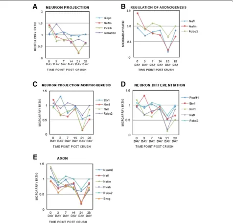
A total of 28 up-regulated clusters and 20 down-regulated clusters were significantly identified in the retinal dataset (p < 0.05) and 57 up-regulated clusters and 41 down-regulated clusters were identified within the ON dataset (Tables 1, 2, 3 and 4). To outline neurodegenerative mecha-nisms, key clusters were identified relating to neuronal loss and regeneration failure from both the retinal (Figure 1) and ON (Figure 2) clusters previously classified in Tables 1, 2, 3 and 4. Each of these key clusters contained a group of genes significantly (p < 0.05) correlating with that specific cluster. The temporal patterns of the microarray gene ratios were graphed according to their association with these clusters for the retina (Figure 1) and ON (Figure 2).
Retinal clusters associated with neuronal loss and regen-eration failure included the clusters neuron projection,
regulation of axonogenesis, neuron projection morpho-genesis, neuron differentiation and axon clusters (Figure 1). Of particular interest was the gene Neuritin 1 (Nrn1), which was identified within the neuron projection morphogenesis and neuron differentiation clusters (Figure 1C, D). NRN1 is a secreted GPI-linked protein that stimulates axonal and dendritic arbor growth [60]. Table 1 Temporal classification of up-regulated retinal gene cluster changes following ONC
Gene ontology Clusters Time point
P value
Molecular function Structural eye protein 3 dpc 1.90E-06
Eye development 3 dpc 4.50E-03
Extracellular matrix binding 3 dpc 5.30E-03
Calcium ion binding 7 dpc 3.30E-02
Structural eye lens protein 21 dpc 2.20E-11
Structural molecular activity 21 dpc 4.60E-06
Biological process Response to wounding 3 dpc 1.00E-04
Inflammatory response 3 dpc 3.80E-04
Defense response 3 dpc 5.90E-04
Positive regulation of immune system response
3 dpc 1.20E-02
Rho protein signal transduction 3 dpc 5.90E-03
Regulation of signal proliferation 3 dpc 2.70E-02
Defense response 7 dpc 5.40E-04
Inflammatory response 7 dpc 1.60E-02
Response to wounding 7 dpc 4.70E-02
Sensory perception 14 dpc 8.80E-03
Neurological system process 14 dpc 2.30E-02
G-protein coupled receptor signaling pathway
14 dpc 3.90E-02
Macromolecular complex assembly
28 dpc 1.30E-02
DNA packaging 28 dpc 3.10E-02
Positive regulation of protein kinase activity
28 dpc 4.50E-02
Cellular component
Extracellular region part 3 dpc 3.20E-03
Extracellular matrix 3 dpc 7.60E-03
Lysosome 3 dpc 3.40E-02
Extracellular region part 7 dpc 3.50E-02
Microsome 14 dpc 1.70E-02
Intermediate filament 14 dpc 3.70E-02
Ribosome 21 dpc 5.00E-03
Gene expression fold-change values were grouped individually from naïve eyes and ONC eyes out to 28 days post crush (dpc). Genes were highlighted based on fold values for up-regulated (≥1.5) retinal datasets. The selected genes were analyzed by gene ontology (GO) based cluster identification at each time point using DAVID. Significance was determined using the Benjamini multiple test correction, GO enrichment scoreχ2
Down-regulation of Nrn1 mRNA expression within the microarray was observed to be biphasic with an initial de-cline through 7 dpc, a slight increase at 14 dpc and a fur-ther decrease by 21 dpc (Figure 1C, D). These biphasic patterns may indicate a transient attempt at neuroprotec-tion/neuroregeneration early in the response to injury.
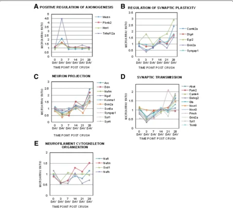
ON clusters associated with neuronal loss and regener-ation identified from the ON cluster tables (Tables 3 and 4) included positive regulation of axonogenesis, regulation of synaptic plasticity, neuron projection, synaptic transmission and neurofilament cytoskeleton organization (Figure 2).
Neuron projection and synaptic transmission clusters both identified key genes called synaptotagmins(Syt)that participate in axonal regeneration, including synaptic pro-jection and proper axonal targeting. Expression of Syt genes was elevated in the ON at 21 dpc (Figures 2C, D).
By analyzing the retina and ON simultaneously, we were able to observe the temporal response of gene ex-pression in the retina and ON individually as well as in comparison to each other. Neurofilament (NF) genes were identified in most of the retinal and optic nerve clusters. Decreased expression of neurofilament medium (Nefm) and light chain (Nefl) genes in the retina at 3 and 21 dpc (Figure 1) preceded neuronal loss after axonal damage (Additional file 1: Figure S1B). However, by 28 dpc Nefm and Nefl expression was elevated in the ON (Figures 2C and E). This pattern of NF expression is consistent with previous studies identifying NF dysregu-lation during neurodegeneration [61-74].
Validation of key target genes having differential expression by qRT-PCR
Analysis of pooled microarray samples does not account for the potential variations that exist between samples and may mask individual sample differences. To confirm individual samples follow the same trend of expression as the microarray data, we used qRT-PCR to determine the expression levels of target genes in each sample. For the retina, we verified two genes (Nrn1and Vsnl1) that have been previously identified as RGC markers [75,76]. Nrn1had similar expression patterns asVsnl1(Figure 3A and B, respectively), and expression of each gene was significantly correlated with the corresponding micro-array ratios (Nrn1 R2= 0.96,Vsnl1 R2= 0.73) (p < 0.05) (Additional file 4: Tables S2A and B). Both genes dis-played a biphasic level of expression with significantly de-creased expression from basal naïve levels at 3 and 21 dpc and modestly decreased expression at 14 dpc (p < 0.05, n = 5) (Figure 3).
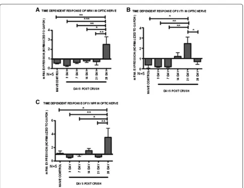
In the ON dataset, Nrn1, synaptotagmin 1 (Syt1) and synaptoporin (Synpr)expression levels were validated by qRT-PCR. We observed significantly increased expres-sion of Nrn1at 28 dpc versus all time-points (p < 0.05, n = 5) (Figure 4A). The qRT-PCR results significantly correlate with the microarray data (R2= 0.86, p < 0.05) (Additional file 4: Tables S2A and B). Syt1 expression was significantly up-regulated at 21 dpc (p < 0.05, n = 5), in contrast to all the other time points (Figure 4B). These results were similar to Syt1 microarray ratios, in which Syt1 expression was elevated only at the 28 dpc period. The shift in the time course of gene up-regulation is most likely due to various individual samples that were masked in the pooled microarray samples. Therefore, the linear regression correlation between both sets of data was less than 0.5 (R2= 0.07) (Additional file 4: Tables S2A and B). Increased expression of synaptoporin (Synpr) was observed at 28 dpc (Figure 4C) and correlated significantly with gene microarray ratios (R2= 0.71, p < 0.05, n = 5) (Additional file 4: Table S2B). Expression ofSynprhas been shown in neurons whileSyt1has been Table 2 Temporal classification of down-regulated retinal
gene cluster changes following ONC
Gene ontology Clusters Time point
P value
Molecular function
Structural eye lens protein 7 dpc 3.80E-15
Structural eye lens protein 28 dpc 2.60E-14
Pattern binding 28 dpc 3.80E-02
Biological process
Chromatin assembly 7 dpc 4.80E-04
Regulation of axonogenesis 21 dpc 7.40E-03
G-protein coupled receptor signaling pathway
21 dpc 2.00E-04
Neurological system process 21 dpc 7.10E-03
Intermediate filament bundle process
21 dpc 6.40E-05
Microtubule based process 21 dpc 5.10E-03
Axonogenesis 21 dpc 1.70E-02
Neuron projection morphogenesis 21 dpc 2.00E-02
Neuron differentiation 21 dpc 4.20E-02
Cell morphogenesis involved in differentiation
28 dpc 1.20E-02
Cellular component
Nucleosome 7 dpc 7.50E-05
Neuron projection 21 dpc 5.00E-05
Axon 21 dpc 6.50E-05
Neurofilament 21 dpc 1.60E-04
Intrinsic to membrane 21 dpc 1.30E-02
Neuron projection 28 dpc 9.90E-03
Chromosome 28 dpc 3.10E-02
Gene expression fold-change values were grouped individually from naïve eyes and ONC eyes out to 28 days post crush (dpc). Genes were highlighted based on fold values for down-regulated (≤ −1.5) retinal datasets. The selected genes were analyzed by gene ontology (GO) based cluster identification at each time point using DAVID. Significance was determined using the Benjamini multiple test correction, GO enrichment scoreχ2
shown in both neurons and is critical in fusion events of astrocytes [77-81]. In addition to synapse formation,Syt1 has also been shown to regulate the formation of axonal filopodia and branching [80]. The induction of bothSynpr and Syt1 expression may be related to synaptic vesicle fusion and release, and the roles of both genes in ONC need to be further explored.
Immunohistochemical analysis of validated gene targets
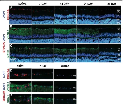
Whole retinas were utilized for microarray analysis, po-tentially masking the changes specific to the RGCs, as they comprise only about 0.5% of the whole retina [82]. To de-termine temporal protein expression patterns occurring specifically within the RGCs of the GCL, we performed retinal immunostaining. We first tested the expression of Brn3a (brain-specific homeobox/POU domain protein 3A), a well-known marker for RGCs [57,83,84]. As expected, we observed a progressive decrease in Brn3a expression after ONC within the GCL (Figure 5A and D). These results demonstrate a temporal decline in RGCs after axonal injury.
Retinal immunostaining for Vsnl1 and Nrn1 proteins confirmed apparent temporal changes in protein expres-sion after ONC. Within the naïve retinal sections, Table 3 Temporal classification of up-regulated ON gene
cluster changes following ONC
Gene ontology Clusters Time point
P value
Molecular function
Chemokine activity 3 dpc 6.50E-06
Growth factor binding 3 dpc 1.10E-03
Actin binding 3 dpc 9.10E-03
Serine type endopeptidase inhibitor activity
7 dpc 7.10E-04
Chemokine activity 14 dpc 6.70E-09
Cytokine activity 14 dpc 6.00E-06
Chemokine activity 21 dpc 1.60E-04
Cytokine binding 21 dpc 4.30E-04
Ion channel activity 28 dpc 1.20E-09
Calcium ion binding 28 dpc 7.70E-07
GABA receptor activity 28 dpc 4.30E-04
Neurotransmitter binding 28 dpc 4.00E-03
Calcium channel activity 28 dpc 9.10E-03
Protein kinase activator activity 28 dpc 8.80E-03
Biological process
Defense response 3 dpc 6.20E-05
Translation 3 dpc 1.20E-03
Cell cycle 3 dpc 3.10E-06
Leukocyte activation 3 dpc 2.10E-04
Actin cytoskeleton organization 3 dpc 6.80E-03
Regulation of adaptive immune response
3 dpc 3.60E-04
Positive regulation of programmed cell death
3 dpc 1.20E-03
Positive regulation of axonogenesis 3 dpc 2.10E-02
Sensory perception 7 dpc 2.90E-02
Immune response 14 dpc 3.40E-08
Chemotaxis 14 dpc 2.60E-07
Response to wounding 14 dpc 1.10E-06
Cell activation 14 dpc 5.70E-05
Defense response 21 dpc 1.60E-08
Response to wounding 21 dpc 4.00E-06
Chemotaxis 21 dpc 2.60E-06
Regulation of adaptive immune response
21 dpc 5.40E-06
Phagocytosis 21 dpc 7.10E-03
Neuropeptide signaling pathway 21 dpc 2.50E-02
Ion transport 28 dpc 2.70E-06
Transmission of nerve impulse 28 dpc 6.30E-08
Synaptic transmission 28 dpc 1.20E-06
Synaptic vesicle transport 28 dpc 4.60E-03
Regulation of synaptic plasticity 28 dpc 2.40E-03
Regulation of synaptic transmission 28 dpc 6.40E-03
Table 3 Temporal classification of up-regulated ON gene cluster changes following ONC(Continued)
Synaptogenesis 28 dpc 1.50E-02
Cell adhesion 28 dpc 3.20E-02
Cellular component
Chromosome 3 dpc 1.50E-09
Extracellular region part 3 dpc 8.80E-08
Collagen 3 dpc 6.00E-03
Focal adhesion 3 dpc 7.20E-03
Anchoring junction 3 dpc 2.50E-02
Extracellular region 7 dpc 9.80E-04
Extracellular region 14 dpc 3.30E-03
Cell surface 14 dpc 2.80E-04
Extracellular region 21 dpc 2.60E-04
Synapse part 28 dpc 1.90E-13
Postsynaptic membrane 28 dpc 1.90E-07
Neuron projection 28 dpc 3.30E-07
Dendrite 28 dpc 2.30E-05
Synaptosome 28 dpc 3.10E-04
Postsynaptic density 28 dpc 5.60E-03
Synaptic vesicle 28 dpc 2.60E-03
Gene expression fold-change values were grouped individually from naïve eyes and ONC eyes out to 28 days post crush (dpc). Genes were highlighted based on fold values for up-regulated (≥1.5) optic nerve datasets. The selected genes were analyzed by gene ontology (GO) based cluster identification at each time point using DAVID. Significance was determined using the Benjamini multiple test correction, GO enrichment scoreχ2
approximately 50% of the GCL cells were positive for Vsnl1/Brn3a and 47% were positive for Nrn1/Brn3a (Figure 5E and F). A biphasic protein expression pat-tern was observed for Vsnl1 with decreased expression in the nerve fiber layer (NFL) and inner plexiform layer (IPL) at 7 dpc, increased expression at 14 dpc com-pared to the naive retina, and a complete loss of ex-pression by 28 dpc (Figure 5B). Focusing on the GCL, the staining pattern also changed at 7 dpc and became more cytoplasmic, in contrast to the diffuse pattern ob-served in the naïve retinas (Figure 5E). These data ver-ify theVsnl1mRNA expression data (Figure 3A).
A similar biphasic expression pattern was observed for Nrn1 with peak expression at 14 dpc (Figure 5C) and in-creased nerve fiber layer staining pattern with the GCL at 7 dpc (Figure 5F). Compared to Vsnl1, Nrn1 immuno-staining was observed in the ganglion cells and NFL, but not as extensively within the IPL. In addition, previous studies of retinal Nrn1 in-situ hybridization exhibited predominant expression within the ganglion cell layer [85], which agrees with our IHC study.
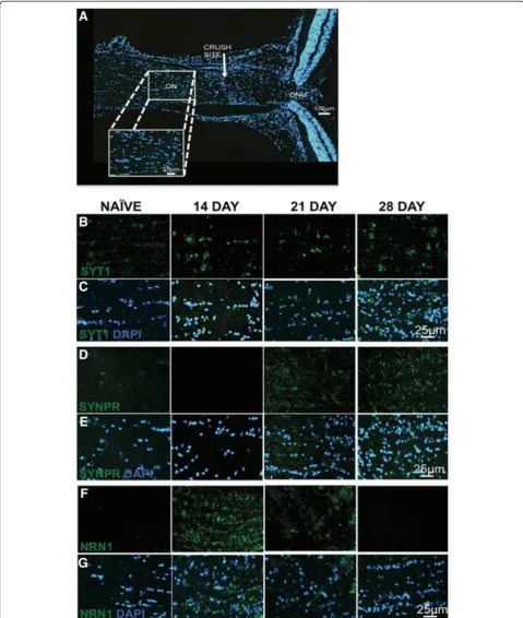
Temporal Syt1, Synpr and Nrn1 protein expression patterns were determined in longitudinal sections of the ON. Images were examined at each time point for each protein as represented by Figure 6A. At 7 dpc, all pro-teins were individually co-labeled with Nfl to show localization of the axons and the pattern of staining for each protein within the ON (Additional file 5: Figure S3 G-I). The expression pattern of Synpr and Nrn1 was not as intense as Syt1 (Additional file 5: Figures S3 G-I) but still colocalized with Nfl staining. In contrast, the stain-ing pattern of Syt1 colocalized with Nfl and also within the cells surrounding the ON axons (Additional file 5: Figure S3 G). Increased expression of Syt1 was evident at 14, 21 and 28 dpc (Figure 6B and C). Elevated levels of Syt1 were seen within the cytoplasmic region of ON cells through the time course post crush (Figure 6B). Synpr protein expression within the ON was evident at 21 and 28 dpc (Figure 6D and E). Similar to Syt1 expres-sion, Synpr cytoplasmic staining was observed within the Table 4 Temporal classification of down-regulated ON
gene cluster changes following ONC
Gene ontology Clusters Time point
P value
Molecular function
Calmodulin binding 3 dpc 6.50E-04
Voltage gated ion channel activity
3 dpc 6.70E-04
Ion binding 3 dpc 3.90E-04
Enzyme binding 3 dpc 5.80E-03
GABA receptor activity 3 dpc 6.70E-03
Ligand gated ion channel activity
3 dpc 1.80E-02
Calmodulin binding 7 dpc 1.30E-02
Calcium dependent phospholipid binding
7 dpc 1.40E-04
Nuclease activity 21 dpc 2.50E-02
Microtubule binding 21 dpc 2.50E-02
Motor activity 21 dpc 1.80E-02
Cytokine activity 28 dpc 2.90E-02
Biological process
Potassium ion transport 3 dpc 4.80E-05
Cation transport 3 dpc 5.30E-04
Neurotransmitter transport 3 dpc 3.80E-03
Synaptic transmission 3 dpc 1.40E-02
Calcium ion transport 3 dpc 1.10E-02
Neurofilament cytoskeleton organization
3 dpc 1.20E-03
Intermediate filament cytoskeleton organization
3 dpc 2.00E-03
Neurotransmitter transport 3 dpc 3.80E-03
MAPKKK cascade 3 dpc 2.40E-02
Microtubule based process 7 dpc 1.10E-02
Visual perception 14 dpc 4.40E-05
Cognition 14 dpc 6.50E-03
Neurological process 14 dpc 1.40E-02
Microtubule based process 21 dpc 1.30E-05
Lipid biosynthetic process 21 dpc 2.20E-02
Visual perception 28 dpc 3.20E-10
Sensory perception 28 dpc 2.90E-02
Eye development 28 dpc 4.80E-05
Immune response 28 dpc 4.70E-02
Cellular component
Synapse 3 dpc 4.30E-06
Cell junction 3 dpc 6.30E-04
Post synaptic membrane 3 dpc 1.10E-03
Presynaptic membrane 3 dpc 5.20E-05
Synapse part 7 dpc 2.90E-04
Cell junction 7 dpc 3.90E-03
Table 4 Temporal classification of down-regulated ON gene cluster changes following ONC(Continued)
Cell projection 7 dpc 3.10E-03
Clathrin coated vesicle 7 dpc 1.00E-02
Cytoskeleton 21 dpc 2.30E-02
Anchored to membrane 28 dpc 4.30E-02
Gene expression fold-change values were grouped individually from naïve eyes and ONC eyes out to 28 days post crush (dpc). Genes were highlighted based on fold values for down-regulated (≤ −1.5) optic nerve datasets. The selected genes were analyzed by gene ontology (GO) based cluster identifica-tion at each time point using DAVID. Significance was determined using the Benjamini multiple test correction, GO enrichment scoreχ2
ON cells (Figure 6D). The temporal protein expression pattern of Syt1 and Synpr followed the mRNA expression patterns (Figure 4B and C).
In contrast to Syt1 and Synpr, Nrn1 ON protein ex-pression levels (Figure 6F and G) did not match mRNA expression data (Figure 4A). Increased expression of Nrn1 was observed at 14 and 21 dpc (Figure 6F), while increased mRNA expression was observed at 28 dpc (Figure 4A). The offset in the time course for protein ex-pression compared to mRNA exex-pression can be expected
due to both mRNA half-life stability and rates of protein synthesis to degradation.
Discussion
Signaling pathways involved in RGC degeneration are quite complex, and identifying correct target molecules that can mitigate neuronal degeneration and failed re-generation are necessary to develop new neuroprotec-tion strategies. We utilized the ONC mouse model to understand the mechanisms involved in RGC death.
ONC directly damages the ON, leading to a progressive loss of RGCs. We identified temporal gene expression changes in the retina and ON after ONC. Key genes asso-ciated with neuronal loss and regenerative failure were identified in both retina and ON, and the expression changes were validated by qRT-PCR and immunostaining. Previously it has been shown that genetic background has an influence on susceptibility to neuronal damage in different inbred mouse lines after neurodegenerative stimuli [20]. C57BL/6 mice are more resistant to ONC, while BALB/c mice are more susceptible to this axonal in-jury. However, both strains display similar susceptibility to
spinal cord injury [20,86]. The differences observed be-tween strains could be partly due to variability in immune response, differences in neuronal stress pathways, and/or activation of alternate cell death pathways [20,87]. In addition, albino rodents are more susceptible to light-induced retinal damage, causing photoreceptor cell death and subsequent retinal degeneration [88,89]. However, ret-inal degeneration not induced by external factors has not been previously reported in BALB/cJ mice. Thus, our BALB/cJ ONC model is extremely useful for studying the potential mechanisms underlying neuronal cell death due to its susceptibility to crush injury.
Figure 2Temporal changes of specific optic nerve gene clusters related to neuronal loss and regeneration failure.Neuron specific and axonal regeneration related neuronal clusters were selected from the optic nerve GO tables and the microarray ratios of the genes within each of these clusters graphed temporally (0 to 28 days post crush (dpc)). Neuronal clusters identified included(A)positive regulation of axonogenesis,
Previous ONC studies have observed changes in gene expression within the retina [22,46,90-93] and glial based responses within the ON [29]. Analyzing the retina and ON simultaneously allowed the identification of individ-ual clusters related to neuronal loss and regenerative failure within each tissue separately as well as allowed us to observe the temporal response of gene expression oc-curring in both the ON and retina with progressive in-jury to these two tissues.
Neurofilament genes were identified in both the retina and ON datasets. Atypical accumulations of NFs are asso-ciated with several neurodegenerative disorders [61-74], and dysregulation of NFs and NF aggregation accompany axonal damage after CNS trauma. NFs have also been associated with CNS diseases and axonal degenerative processes [94]. We show temporal differences in neuro-filament expression between the retina and ON sug-gesting crucial gene changes occur after trauma in the retina and ON. There is progressive decline of retinal Nfl expression compared to the elevated expression within the ON out to 28 dpc. These results are consistent with a model in which axonal damage precedes retinal neuronal degeneration and accumulation of damage asso-ciated genes occurs within the ON before soma degener-ation. The changes in expression patterns identified in our ONC model correlate with previous studies identifying NF dysregulation during neurodegeneration [61-74].
The RGCL comprises multiple cell types including RGCs, amacrine cells, astrocytes, microglia, and vascular cells that interact with the RGC somas. After ONC, these cells also initiate degenerative pathways causing RGC apoptosis [95,96]. Thus, the deregulation of genes observed within the retina is not restricted to RGCs and also represent gene expression of the surrounding cells. Glial fi-brillary acidic protein (Gfap), a marker of astrogliosis, is
up-regulated after CNS trauma and is used as a universal index of retinal injury [34,96]. Gfap is initially up-regulated after ONC [35,36] and showed a similar ex-pression pattern in our retinal dataset. After injury of CNS axons, glial responses around the affected area are increased, and this may contribute to trauma-induced neurodegeneration [97]. By identifying key clusters as-sociated with degeneration of neurons and axonopathy, we were able to isolate potential target genes (Vsnl1, Nrn1, Syt1, Synpr).
Vsnl1 gene is a member of the neuronal subfamily of EF-hand calcium sensor proteins. These proteins play vital roles in cellular signal transduction and neuropro-tection/neurotoxicity and have been implicated in neu-rodegenerative diseases [98,99]. Vsnl1 is predominantly expressed in isolated immuno-panned rat RGCs [75] and has also been shown to specifically label the inner retina (amacrine and RGCs) and the inner plexiform layer of rat, chicken, and bovine retinas [100]. In our study, expression of the Vsnl1gene was down-regulated after ONC, which may prevent the survival of RGCs. Al-though the precise functional roles ofVsnl1 are still un-clear, Vsnl1 proteins may play key roles in membrane trafficking, neuronal signaling, and differentiation [99]. As ON axonal transport is inhibited after ONC, de-creased levels ofVsnl1may contribute to the deleterious effects on axonal transport mechanisms seen in ONC.
Functionally, Nrn1 acts as a ligand to the insulin recep-tor [101] and cleavage of the GPI anchor by phospholipase C allows the soluble secreted form to be cell independent [102]. The GPI membrane bound anchor of Nrn1 allows growth promotion as it can stimulate axonal plasticity, dendritic arborization, and synapse maturation in the CNS [60,102]. Conditional knockout of theNrn1gene de-lays development, maturation of axons and dendritic
arbors, synaptic maturation, and effective learning [103]. Neurotrophins such as nerve growth factor (NGF), brain-derived neurotrophic factor (BDNF) and neurotrophin-3 (NT-3) as well as neuronal activity can potentiate the ex-pression of Nrn1 [104,105]. NGF induces expression of Nrn1, which increases neurite outgrowth in a variety of experimental models [104,106,107]. Our studies suggest that after axonal insult, RGCs initially increase Nrn1 ex-pression for axonal regeneration to overcome obstructed transport mechanisms. These regenerative supportive mechanisms are lost 14 dpc because by then most of the RGCs have been damaged, and the survival of these neu-rons has progressively decreased. The correlation of ret-inal protein expression of Nrn1 at 14 dpc mimics the elevated expression of Nrn1 at the same time-point within the ON. Taken together, these data suggest that the dy-namic regulation ofNrn1 may be an effort for axonal re-generation after ONC.
The relative abundance of protein expression may not be proportional to the relative mRNA levels. This lack of correlation in mRNA and protein expression levels could be due to mRNA stability and/or rates of protein synthe-sis and/or degradation. The slight increase in retinal mRNA expression at 14 dpc (compared to 3 and 7 dpc) is maybe increasing the translation of the Nrn1 within the RGCs soma and Nfl, which is then transported downstream to the ON axons.
The optic nerve includes not only the axons of the RGCs but also astrocytes, microglia, and oligodendro-cytes that interact with RGC axons as well as each other [29]. Thus, the expression of genes observed within the ON may represent the beneficial or detrimental effects of neighboring cells surrounding the RGC axons. Differ-entially regulated genes within the ON expression microarray also identified other key genes associated with synaptic transmission (Syt1andSynpr) and synaptic
Figure 4mRNA expression patterns of selected optic nerve genes following ONC insult.Pooled microarray mRNA expression changes were validated in individual samples by qRT-PCR. Relative fold change in each sample was determined based on a 2 fold exponential using mRNA expression values normalized to Gapdh and the contralateral control eye. Fold values of each gene presented as mean ± SEM.(A)Neuritin 1 (Nrn1),(B)Synaptotagmin 1 (Syt1) and(C)Synaptoporin (Synpr). Statistical significance for each time-point determined by one-way ANOVA -Tukey
plasticity gene (Nrn1)that participate in axonal regener-ation, including synaptic projection, and proper axonal targeting.
A collection of signaling mechanisms link both axonal tips and dendritic terminals to neuronal soma and nucleus by motor-dependent transport machineries. Signaling com-plexes could be transported either in endosomes, or as non-endosomal complexes associated with importins and dynein [108]. Essential membrane components of synaptic vesicles and synaptic transmission associated proteins are translated in the soma and get transported to the growing distal ends of extending neurites after crush in-jury [109,110]. In addition, synaptic vesicles are localized
to small vesicles within the neuron, particularly in neur-onal axneur-onal processes [111]. Eventually, as axneur-onal trans-port is inhibited after ONC due to glial scarring [5,13], there is decreased transport of proteins involved in neuro-protection and synaptic plasticity. This causes deleterious effects, eventually leading to decreased synaptic plasticity and transmission at distal ends.
Syt proteins act as synaptic calcium sensors for vesicle fusion in conjunction with SNAREs that facilitate intra-cellular membrane fusion events [112-114]. Syts have a conserved mechanism of action and are crucial for neur-onal Ca2+-triggered vesicle fusion [115]. Previous studies have shown Syt1 to participate in axonal regeneration,
Figure 5Expression of Brn3a, Vsnl1 and Nrn1 in the retina following ONC.Progressive loss of RGCs observed with Brn3a and a biphasic pattern of expression observed for Vsnl1 and Nrn1.Time course of retina sections from naïve, 7, 14, 21 and 28 dpc (days post crush) are indicated on the top of the panel. Fluorescence micrographs of retinal tissue sections were immunolabeled with:(A)Brn3a (red),(B)Vsnl1 (green) and
including synaptic projection and proper axonal target-ing [80,116]. In our study, Syt1 was identified in ON neuron projection and synaptic transmission clusters. It appears that the ON attempts to initiate synaptic projec-tion following ONC trauma as shown by the biphasic mRNA and protein expression of Syt1.
Similarly, Synprsare essential membrane components of synaptic vesicles [79]. Synpr has restricted distribution within the CNS and is present in the telencephalic struc-tures, hippocampus, olfactory bulb, and retina [77,117-119]. Synpr plays potential roles in the modulation of synaptic transmission and specificity to neuronal circuitry [79]. In-creased protein expression of Synpr in the ON was ob-served at 21 and 28 dpc. The induction of both Synpr and Syt1 expression may be related to synaptic vesicle fusion and release. After trauma, these synaptic vesicles get trans-ported to the growing distal ends of extending neurites [109,110]. Eventually, as the RGCs are trying to overcome regenerative failure, they may increase expression of Syt proteins within their axons in attempt to induce synaptic plasticity and transmission at distal ends. Elevated expres-sion of Synpr and Nrn1 suggests they are mediating synap-tic differentiation as synapsynap-tic organizing proteins, but the deregulation of mRNA expression and eventually protein expression may be a futile attempt at ON regeneration in late pathogenesis.
We have explored temporal gene expression changes after ONC axonal injury that can be extrapolated to other CNS traumas. Although there are gene expression differences between the retina and brain, similar differ-ences also occur within discrete regions of the brain as each part of the brain has different motoric, sensory, and cognitive functions. For example, gene expression in the cerebellum differs the most from the other regions of the brain [120,121] and has also been reported in inbred strains of mouse brain [122]. In addition, inter-individual differences have also been reported within a species [121]. As is the case while studying any trauma or disease model, only a generic evaluation can be made in terms of rele-vance to other regions in the CNS. In conclusion, the ONC model has identified two key mechanisms of CNS trauma and neurodegeneration: neuronal loss and regen-erative failure. Dysregulation of Vsnl1, Syt1, Synpr and Nrn1gene expression may play an important role in neu-rodegeneration and potentially provide unique targets for intervention.
Conclusions
The current study delineates the gene expression pro-file associated with neurodegeneration and regenerative failure after ONC-induced CNS trauma. CNS trauma causes degeneration of neurons and axonopathy, which is evident in neurodegenerative diseases such as Parkinson’s, Alzheimer’s, and glaucoma [1-5]. The susceptibility of the
neurons to acute axonal injury allowed the identification of gene expression changes that occur before neuronal loss. Using the reproducible ONC model of CNS trauma, we were able to: (a) examine gene expression changes within the retina and ON, and (b) visualize protein expres-sion patterns of key selected genes associated with neuron loss and regenerative failure within the retina and ON after ONC. BETR analysis of microarray gene expression data was utilized to show that a select small subset of genes was affected at multiple time points following ONC. Bioinformatic meta-analysis identified gene clusters asso-ciated with regenerative changes, synaptic plasticity, axo-nogenesis, neuron projection, and neurodegeneration. A neurite synaptic plasticity gene (Nrn1), synaptic vesicle fu-sion genes (SynprandSyt1), and neuron differentiation as-sociated gene (Vsnl1) were a few of the key temporally regulated genes identified in our study. In conclusion, ana-lysis of these gene arrays and protein expression patterns allowed the detection, quantification and visualization of key differentially regulated genes after ONC. This study has identified potential pathogenic genes and possible new therapeutic targets to address two key mechanisms of CNS trauma: neuronal loss and regenerative failure.
Methods Animals
BALB/cJ mice aged 2–4 months were utilized for all the experiments and were obtained from the Jackson Laboratories (Bar Harbor, ME). The mice were housed and maintained in a 12-hour light/dark cycle and fed ad libitum. All procedures were performed in accordance with Association for Research in Vision and Ophthalmol-ogy Statement on the Use of Animals in Ophthalmic and Vision Research and the University of North Texas Health Science Center (UNTHSC) Institutional Animal Care and Use Committee regulations (IACUC protocol # 2011/ 2012-58-A04, approved October 8th 2012).
Optic nerve crush model
The ON of the left eye was crushed 0.5 mm posterior from the globe for 4 seconds using the Nickell’s tech-nique [19]. Briefly, mice were anesthetized by intraperi-toneal injection of ketamine (100 mg/kg) and xylazine (10 mg/kg) and an incision was made along the superior orbital margin. The ON (left) was exposed and crushed using a self-closing jeweler’s forceps to ensure reprodu-cibility and constant force. Extreme care was taken not to damage the ocular blood vessels. Indirect ophthal-moscopy was performed to ensure retinal circulation was not blocked. The contralateral eye was used as the uncrushed control.
Characterization of optic nerve crush model
To quantify cell loss from the retinal RGCL, retinas from fixed eyes were dissected, flat mounted and Nissl stained with cresyl violet stain as previously described [19,87,123]. Eyes were fixed in 4% paraformaldehyde in phosphate buffered saline (PBS; 0.1M phosphate and 100mM NaCl buffer (pH 7.4)) for an hour at room temperature. After fixation, the eyes were rinsed with PBS, and the posterior cup isolated and placed in 0.3% Triton-X 100 PBS for 16 hours at 22°C. The tissues were then placed in 3% H2O2 and NaH2PO4 overnight. The retinas were dissected, cut into four quadrants, and mounted RGC side up on positively charged glass slides (Fisher Scientific, Chicago, IL). The slides were then air dried and flattened with coverslips using 10 g weights. The dried retinas were stained with 1% cresyl violet acet-ate in 0.25% acetacet-ate for 30–45 seconds. After staining, the retinas were dehydrated in 90% and 100% ethanol and cleared with xylene to reduce background staining and mounted with a coverslip.
To determine the density of remaining RGCL neurons within each retina, two digital images from each rant (peripheral and mid-peripheral region - four quad-rants/retina) were captured at 400 X magnification. A total of 8 images per retina were counted using Adobe Photoshop software. Cell counts were analyzed by com-paring the experimental retinas against the contralateral control retinas (cell counts ± SD) at each time point. Quantification of percentage neuron survival following ONC from 3 to 28 dpc was performed. Data points (Additional file 1: Figure S1 B) represent mean ± SD of surviving neurons after crush normalized to contralat-eral control eyes. Statistics were determined using one-way ANOVA -Tukey post hoc test, ** p < 0.01, *** p < 0.001, n = 8-9 eyes/time-point.
RNA processing
Fresh retina and ON samples (from the globe to the chi-asm) were cleanly dissected without any contamination from surrounding tissue. In brief, after euthanization
each globe was harvested from the mouse eye socket at the globe and optic nerve head (ONH) juncture. The globe was transferred to a clean petri dish and opened along the limbus. The retina was harvested from the posterior cup and the ONH removed using a trephine. For the ON, the skull was opened and each left and right ON between the globe and chiasm was harvested separ-ately. All samples were collected in 1 ml of TRIzol (Invi-trogen, Grand Island, NY) and homogenized using 5 mm steel beads in the TissueLyser LT (Qiagen, Valencia, CA) for five minutes at 50 oscillations/second. For phase separation 50μl of BAN Phase Separation Reagent (Molecular Research Center, Cincinnati, Ohio) was added to the homogenized samples, and samples were centri-fuged at 14,000 rpm for 15 minutes. The upper aqueous phase was transferred to an RNeasy mini column (Qiagen, Valencia, CA) and processed according to manufacturer’s protocol. The total RNA was re-suspended in 20 μl of nuclease-free water and quantified using the Thermo Scientific NanoDrop 2000 (NanoDrop products, Wil-mington, DE). Integrity of the RNA was measured by calculating the RNA integrity number (RIN) using the Agilent Bioanalyzer (Agilent Technologies, Santa Clara, CA), and samples with RIN values greater than 7 were used for microarray analysis.
Affymetrix gene chip arrays
scanner with 7G upgrade, and data were collected using the using the GeneChip operating software (GCOS) v1.4.
Bioinformatic analysis of gene expression datasets
Microarray data were imported into the Partek Genomics Suite 6.6 software (Partek Inc., Louis, MO) and normal-ized based on the robust multi-array average (RMA). To further confirm the purity of each extracted tissue, we ex-amined the expression of retina specific genes in the ON tissue and ON genes in the retina tissue. There was greater expression of retina specific markers: Rho,Nr2e3, NrlandCrxin the retinal samples, while these genes were at the lower limits of detection in the ON samples. Con-versely, there was greater expression of myelin marker genesMag,Mobp,Mog,MbpandPlp1in the ON samples compared to the retina samples. In addition, we tested ex-pression levels ofRho andMbp by qPCR in both tissues (Additional file 6: Figure S4).
For the microarray analysis, the ONC samples were compared to the control samples and the microarray ra-tios and log2 fold values calculated at each time point. Up and down-regulated genes were identified for both datasets (retina and ON) with a selective filter of ≥1.5 and≤ −1.5 fold values. The fold values were based on theq-value defined by the FDR analogue of the p <0.05. The genes were further analyzed using the publicly avail-able bioinformatics software Database for Annotation, Visualization and Integrated Discovery (DAVID). Gene ontology (GO) based cluster analysis was performed to identify possible enrichment of genes (GO enrichment score calculated using aχ2test) using differentially regu-lated genes per time point. The Fishers Exact p value is calculated by DAVID to identify GO enrichment based clusters and any p < 0.05 were considered to be signifi-cant based on the Benjamini multiple test correction and were enriched in the annotation category [124,125]. Neuronal clusters were identified at specific time points and their genes graphed temporally under each GO category.
Identification of specific gene expression changes following ONC by Bayesian estimation of temporal regulation analysis
Analysis of time-course microarray data was performed using Bayesian Estimation of Temporal Regulation (BETR) analysis to account for any variations between individual samples within the pooled samples. The BETR expression probabilities were estimated using Probe Logarithmic In-tensity Error with GC-background correction, a routine built into the Affymetrix Power Tools toolkit. Expression estimates for 11 housekeeping genes across all time-points were used to create a linear model between the average expression level and variance of each gene and housekeep-ing genes. This model was used to simulate additional
readings for all estimated transcripts at each time point, which were subsequently used as additional inputs to the BETR [59] R package. From this algorithm output final BETR probabilities were determined for the total 18,786 genes identified within each of the retina and ON gene ex-pression datasets (Additional file 3: Table S1 A, B). BETR probabilities ranged from 0 to 1 with 0 being the least sig-nificantly changed genes temporally and 1 being the most. Genes were then classified into frequency bins based on the range of BETR probabilities. Each frequency bin iden-tified a range of 0.1 differences in BETR probabilities and bins ranged from the lowest 1 (BETR probability range of 0–0.1) to frequency bin 10 (BETR probability range of 0.9-1) (Additional file 3: Table S1A, B). We consid-ered low BETR probabilities of frequency bins <5 to re-flect no significant changes in gene expression, while high BETR probabilities (0.9-1.0) within frequency bin 10 to represent significant changes in gene expression over time.
Microarray confirmation through real-time qRT-PCR
qRT-PCR and microarray ratios, and the R2coefficient of determination calculated and p < 0.05 were considered statistically significant (Additional file 4: Table S2 B).
Immunohistochemistry
IHC was performed to validate protein expression of qRT-PCR confirmed genes and to localize target proteins in the retina and ON. Whole eyes were harvested and fixed in 4% paraformaldehyde for 2 hours at room temperature. After fixation, the tissue was placed in 20% sucrose over-night at 4°C and embedded in optical cutting temperature (OCT) the next day. Sections (10 μm) were cut using a cryostat (Leica Biosystems - Richmond, IL). Cross sections of retina were transferred to Superfrost glass slides (Fisher Scientific - Chicago, IL). Slides were incubated in PBS for 10 minutes and blocked with SuperBlock™Blocking Buffer (Fisher Scientific, Chicago, IL) at room temperature for one hour. Primary antibodies (Additional file 9: Table S5) were diluted in Superblock™. Each slide was probed with the respective primary antibody and incubated overnight at 4°C. Sections were then washed 3 times with PBS for 10 minutes each and incubated with Alexa Fluor secondary antibody (Additional file 9: Table S5) for 1 hour at room temperature. Slides were rinsed three times with PBS and mounted with ProLong® Gold anti-fade reagent with DAPI (Molecular Probes, Grand Island, NY). Sections were observed and captured using a Nikon Eclipse Ti-U Micro-scope (Nikon, Melville, NY) containing the Nuance Multi-spectral imaging system and analyzed using Adobe Photoshop CS5 software. Negative control images of ret-ina and ON sections with no primary antibody are pre-sented in Additional file 5: Figures S3 A-F.
Availability of supporting data
GEO Accession Number: Series GSE44708.
Additional files
Additional file 1: Figure S1.A, B: Optic nerve crush (ONC) significantly reduces neurons in the retinal ganglion cell layer (RGCL).
Additional file 2: Figure S2.A, B: Frequency distribution of genes altered following optic nerve crush.
Additional file 3: Table S1.A, B: BETR probabilities based retinal and ON genes distributed within frequency bins.
Additional file 4: Table S2.A, B: Microarray ratios and linear regression correlation values of selected target genes.
Additional file 5: Figure S3.A-I: Naïve control images and expression of Syt1, Synpr, Nrn1 and Nfl in the ON at 7 days post crush.
Additional file 6: Figure S4.A, B: Expression of tissue specific genes within normal retina and ON samples.
Additional file 7: Table S3.Primers for key genes validated from the retina and optic nerve datasets.
Additional file 8: Table S4.qRT-PCR cycles performed for confirming retina and optic nerve dataset gene expression levels.
Additional file 9: Table S5.Antibodies against key proteins validated by IHC.
Abbreviations
CNS:Central nervous system; SCI: Spinal cord injury; ONC: Optic nerve crush; RGC: Retinal ganglion cells; BETR: Bayesian Estimation of Temporal Regulation; DAVID: Database for Annotation, Visualization, and Integrated Discovery; MF: Molecular function; BP: Biological process; CC: Cellular component; NFL: Nerve fiber layer; GCL: Ganglion cell layer; DPC: Days post crush; OCT: Optical cutting temperature; PBS: Phosphate buffer saline solution; IHC: Immunohistochemistry; ANOVA: Analysis of variance; DAPI: 4′ ,6-diamidino-2-phenylindole; qRT-PCR: Quantitative real time polymerase chain reaction; NCBI: National center for biotechnology information; GO: Gene ontology; RIN: RNA integrity number.
Competing interests
The authors declare that they have no competing interests.
Authors’contributions
TPS performed the RNA extraction, tissue sample staining, bioinformatics Partek and DAVID analysis, qRT-PCR and correlation analysis of all optic nerve crush samples; including writing all sections of the manuscript. CM and YL both performed the optic nerve crush and subsequently CM did the Nissl stained retinal flat mount neuron survival analysis while YL prepared the immunohistochemistry sections. BF processed image data. DT performed differential expression analysis and AW analyzed the microarray data using the BETR analysis. AFC, RJW and TAB conceived the study, actively participated in the design and coordination of the study, reviewed all the data, and helped draft the manuscript. All authors read and approved the final manuscript.
Acknowledgements
This study was supported by a grant (W81XWH-10-2-0003) from the Department of Defense.
Author details
1North Texas Eye Research Institute, Ft. Worth, TX, USA.2Department of Cell Biology & Immunology, NTERI, UNTHSC, Ft. Worth, TX, USA.3Center for Bioinformatics and Computational Biology, University of Iowa, Iowa, IA, USA. 4
Department of Biomedical Engineering, University of Iowa, Iowa, IA, USA.
Received: 19 December 2013 Accepted: 18 April 2014 Published: 27 April 2014
References
1. Schwartz M:Optic nerve crush: protection and regeneration.Brain Res Bull
2004,62(6):467–471.
2. Ohlsson M, Mattsson P, Svensson M:A temporal study of axonal degeneration and glial scar formation following a standardized crush injury of the optic nerve in the adult rat.Restor Neurol Neurosci2004,
22(1):1–10.
3. Magharious M, D’Onofrio PM, Hollander A, Zhu P, Chen J, Koeberle PD:
Quantitative iTRAQ analysis of retinal ganglion cell degeneration after optic nerve crush.J Proteome Res2011,10(8):3344–3362.
4. Wohlfart G:Degeneration and regeneration in the nervous system. Recent advances.World Neurol1961,2:187–198.
5. Windle WF:Regeneration of axons in the vertebrate central nervous system.Physiol Rev1956,36(4):427–440.
6. Huber AB, Schwab ME:Nogo-A, a potent inhibitor of neurite outgrowth and regeneration.Biol Chem2000,381(5–6):407–419.
7. Huber AB, Weinmann O, Brosamle C, Oertle T, Schwab ME:Patterns of Nogo mRNA and protein expression in the developing and adult rat and after CNS lesions.J Neurosci2002,22(9):3553–3567.
8. Filbin MT:Myelin-associated inhibitors of axonal regeneration in the adult mammalian CNS.Nat Rev Neurosci2003,4(9):703–713.
9. Tang S, Qiu J, Nikulina E, Filbin MT:Soluble myelin-associated glycoprotein released from damaged white matter inhibits axonal regeneration.Mol Cell Neurosci2001,18(3):259–269.
10. Winzeler AM, Mandemakers WJ, Sun MZ, Stafford M, Phillips CT, Barres BA:
11. Kopp MA, Liebscher T, Niedeggen A, Laufer S, Brommer B, Jungehulsing GJ, Strittmatter SM, Dirnagl U, Schwab JM:Small-molecule-induced Rho-inhibition: NSAIDs after spinal cord injury.Cell Tissue Res2012,
349(1):119–132.
12. Sandvig A, Berry M, Barrett LB, Butt A, Logan A:Myelin-, reactive glia-, and scar-derived CNS axon growth inhibitors: expression, receptor signaling, and correlation with axon regeneration.Glia2004,46(3):225–251. 13. Silver J, Miller JH:Regeneration beyond the glial scar.Nat Rev Neurosci
2004,5(2):146–156.
14. Lawson LJ, Frost L, Risbridger J, Fearn S, Perry VH:Quantification of the mononuclear phagocyte response to Wallerian degeneration of the optic nerve.J Neurocytol1994,23(12):729–744.
15. Lazarov-Spiegler O, Rapalino O, Agranov G, Schwartz M:Restricted inflammatory reaction in the CNS: a key impediment to axonal regeneration?Mol Med Today1998,4(8):337–342.
16. Jaerve A, Muller HW:Chemokines in CNS injury and repair.Cell Tissue Res
2012,349(1):229–248.
17. Monnier PP, D’Onofrio PM, Magharious M, Hollander AC, Tassew N, Szydlowska K, Tymianski M, Koeberle PD:Involvement of caspase-6 and caspase-8 in neuronal apoptosis and the regenerative failure of injured retinal ganglion cells.J Neurosci2011,31(29):10494–10505.
18. Quigley HA, Nickells RW, Kerrigan LA, Pease ME, Thibault DJ, Zack DJ:
Retinal ganglion cell death in experimental glaucoma and after axotomy occurs by apoptosis.Invest Ophthalmol Vis Sci1995,36(5):774–786. 19. Li Y, Schlamp CL, Nickells RW:Experimental induction of retinal ganglion
cell death in adult mice.Invest Ophthalmol Vis Sci1999,40(5):1004–1008. 20. Li Y, Semaan SJ, Schlamp CL, Nickells RW:Dominant inheritance of retinal
ganglion cell resistance to optic nerve crush in mice.BMC Neurosci2007,
8:19.
21. Barron KD, Dentinger MP, Krohel G, Easton SK, Mankes R:Qualitative and quantitative ultrastructural observations on retinal ganglion cell layer of rat after intraorbital optic nerve crush.J Neurocytol1986,15(3):345–362. 22. Templeton JP, Nassr M, Vazquez-Chona F, Freeman-Anderson NE, Orr WE, Williams RW, Geisert EE:Differential response of C57BL/6J mouse and DBA/2J mouse to optic nerve crush.BMC Neurosci2009,10:90.
23. Misantone LJ, Gershenbaum M, Murray M:Viability of retinal ganglion cells after optic nerve crush in adult rats.J Neurocytol1984,13(3):449–465. 24. Bahr M:Live or let die - retinal ganglion cell death and survival during
development and in the lesioned adult CNS.Trends Neurosci2000,
23(10):483–490.
25. Klocker N, Zerfowski M, Gellrich NC, Bahr M:Morphological and functional analysis of an incomplete CNS fiber tract lesion: graded crush of the rat optic nerve.J Neurosci Methods2001,110(1–2):147–153.
26. Agudo M, Perez-Marin MC, Lonngren U, Sobrado P, Conesa A, Canovas I, Salinas-Navarro M, Miralles-Imperial J, Hallbook F, Vidal-Sanz M:Time course profiling of the retinal transcriptome after optic nerve transection and optic nerve crush.Mol Vis2008,14:1050–1063.
27. Tang Z, Arjunan P, Lee C, Li Y, Kumar A, Hou X, Wang B, Wardega P, Zhang F, Dong L, Zhang Y, Zhang SZ, Ding H, Fariss RN, Becker KG, Lennartsson J, Nagai N, Cao Y, Li X:Survival effect of PDGF-CC rescues neurons from apoptosis in both brain and retina by regulating GSK3beta phosphorylation.J Exp Med
2010,207(4):867–880.
28. Lukas TJ, Wang AL, Yuan M, Neufeld AH:Early cellular signaling responses to axonal injury.Cell Commun Signal: CCS2009,7:5.
29. Qu J, Jakobs TC:The time course of gene expression during reactive gliosis in the optic nerve.PLoS One2013,8(6):e67094.
30. Sharma A, Pollett MA, Plant GW, Harvey AR:Changes in mRNA expression of class 3 semaphorins and their receptors in the adult rat retino-collicular system after unilateral optic nerve injury.Invest Ophthalmol Vis Sci2012,53(13):8367–8377.
31. Blaugrund E, Lavie V, Cohen I, Solomon A, Schreyer DJ, Schwartz M:Axonal regeneration is associated with glial migration: comparison between the injured optic nerves of fish and rats.J Comp Neurol1993,330(1):105–112. 32. Doster SK, Lozano AM, Aguayo AJ, Willard MB:Expression of the
growth-associated protein GAP-43 in adult rat retinal ganglion cells following axon injury.Neuron1991,6(4):635–647.
33. Leon S, Yin Y, Nguyen J, Irwin N, Benowitz LI:Lens injury stimulates axon regeneration in the mature rat optic nerve.J Neurosci2000,20(12):4615–4626. 34. Ridet JL, Malhotra SK, Privat A, Gage FH:Reactive astrocytes: cellular
and molecular cues to biological function.Trends Neurosci1997,
20(12):570–577.
35. Dibas A, Oku H, Fukuhara M, Kurimoto T, Ikeda T, Patil RV, Sharif NA, Yorio T:
Changes in ocular aquaporin expression following optic nerve crush. Mol Vis2010,16:330–340.
36. Woldemussie E, Wijono M, Ruiz G:Muller cell response to laser-induced increase in intraocular pressure in rats.Glia2004,47(2):109–119. 37. Parrilla-Reverter G, Agudo M, Nadal-Nicolas F, Alarcon-Martinez L,
Jimenez-Lopez M, Salinas-Navarro M, Sobrado-Calvo P, Bernal-Garro JM, Villegas-Perez MP, Vidal-Sanz M:Time-course of the retinal nerve fibre layer degeneration after complete intra-orbital optic nerve transection or crush: a comparative study.Vis Res2009,49(23):2808–2825.
38. Koeberle PD, Bahr M:Growth and guidance cues for regenerating axons: where have they gone?J Neurobiol2004,59(1):162–180.
39. Kermer P, Klocker N, Bahr M:Neuronal death after brain injury. Models, mechanisms, and therapeutic strategies in vivo.Cell Tissue Res1999,
298(3):383–395.
40. Koeberle PD, Gauldie J, Ball AK:Effects of adenoviral-mediated gene transfer of interleukin-10, interleukin-4, and transforming growth factor-beta on the survival of axotomized retinal ganglion cells. Neuroscience2004,125(4):903–920.
41. Kipnis J, Yoles E, Schori H, Hauben E, Shaked I, Schwartz M:Neuronal survival after CNS insult is determined by a genetically encoded autoimmune response.J Neurosci2001,21(13):4564–4571.
42. Isenmann S, Wahl C, Krajewski S, Reed JC, Bahr M:Up-regulation of Bax protein in degenerating retinal ganglion cells precedes apoptotic cell death after optic nerve lesion in the rat.Eur J Neurosci1997,
9(8):1763–1772.
43. Kermer P, Ankerhold R, Klocker N, Krajewski S, Reed JC, Bahr M:Caspase-9: involvement in secondary death of axotomized rat retinal ganglion cells in vivo.Brain Res Mol Brain Res2000,85(1–2):144–150.
44. Kermer P, Klocker N, Labes M, Bahr M:Inhibition of CPP32-like proteases rescues axotomized retinal ganglion cells from secondary cell death in vivo.J Neurosci1998,18(12):4656–4662.
45. Kikuchi M, Tenneti L, Lipton SA:Role of p38 mitogen-activated protein kinase in axotomy-induced apoptosis of rat retinal ganglion cells. J Neurosci2000,20(13):5037–5044.
46. Galindo-Romero C, Aviles-Trigueros M, Jimenez-Lopez M, Valiente-Soriano FJ, Salinas-Navarro M, Nadal-Nicolas F, Villegas-Perez MP, Vidal-Sanz M, Agudo-Barriuso M:Axotomy-induced retinal ganglion cell death in adult mice: quantitative and topographic time course analyses.Exp Eye Res
2011,92(5):377–387.
47. Kim BJ, Braun TA, Wordinger RJ, Clark AF:Progressive morphological changes and impaired retinal function associated with temporal regulation of gene expression after retinal ischemia/reperfusion injury in mice.Mol Neurodegener2013,8:21.
48. Xia Y, Chen J, Xiong L, Liu J, Liu X, Ma L, Zhang Q, You C, Chen J, Liu X, Wang X, Ju Y:Retinal whole genome microarray analysis and early morphological changes in the optic nerves of monkeys after an intraorbital nerve irradiated injury.Mol Vis2011,17:2920–2933. 49. Jehle T, Dimitriu C, Auer S, Knoth R, Vidal-Sanz M, Gozes I, Lagreze WA:The
neuropeptide NAP provides neuroprotection against retinal ganglion cell damage after retinal ischemia and optic nerve crush.Albrecht Von Graefes Arch Klin Exp Ophthalmol2008,246(9):1255–1263.
50. Agudo M, Perez-Marin MC, Sobrado-Calvo P, Lonngren U, Salinas-Navarro M, Canovas I, Nadal-Nicolas FM, Miralles-Imperial J, Hallbook F, Vidal-Sanz M:
Immediate upregulation of proteins belonging to different branches of the apoptotic cascade in the retina after optic nerve transection and optic nerve crush.Invest Ophthalmol Vis Sci2009,50(1):424–431. 51. Goldenberg-Cohen N, Dratviman-Storobinsky O, El Dadon Bar S, Cheporko
Y, Hochhauser E:Protective effect of bax ablation against cell loss in the retinal ganglion layer induced by optic nerve crush in transgenic mice. J Neuroophthalmol2011,31(4):331–338.
52. Haverkamp S, Inta D, Monyer H, Wassle H:Expression analysis of green fluorescent protein in retinal neurons of four transgenic mouse lines. Neuroscience2009,160(1):126–139.
53. Raymond ID, Vila A, Huynh UC, Brecha NC:Cyan fluorescent protein expression in ganglion and amacrine cells in a thy1-CFP transgenic mouse retina.Mol Vis2008,14:1559–1574.
54. Masland RH:Neuronal diversity in the retina.Curr Opin Neurobiol2001,
11(4):431–436.