R E S E A R C H
Open Access
Tumor necrosis factor alpha has an early
protective effect on retinal ganglion cells after
optic nerve crush
Caitlin E Mac Nair
1,2, Kimberly A Fernandes
3, Cassandra L Schlamp
1, Richard T Libby
3,4,5and Robert W Nickells
1*Abstract
Background:Glaucoma is an optic neuropathy that is characterized by the loss of retinal ganglion cells (RGCs) initiated by damage to axons in the optic nerve. The degeneration and death of RGCs has been thought to occur in two waves. The first is axogenic, caused by direct insult to the axon. The second is somatic, and is thought to be caused by the production of inflammatory cytokines from the activated retinal innate immune cells. One of the cytokines consistently linked to glaucoma and RGC damage has been TNFα. Despite strong evidence implicating this protein in neurodegeneration, a direct injection of TNFαdoes not mimic the rapid loss of RGCs observed after acute optic nerve trauma or exposure to excitotoxins. This suggests that our understanding of TNFαsignaling is incomplete.
Methods:RGC death was induced by optic nerve crush in mice. The role of TNFαin this process was examined by quantitative PCR ofTnfαgene expression, and quantification of cell loss inTnfα−/−mice or in wild-type animals receiving an intraocular injection of exongenous TNFαeither before or after crush. Signaling pathways downstream of TNFαwere examined by immunolabeling for JUN protein accumulation or activation of EGFP expression in NFκB reporter mice.
Results:Optic nerve crush caused a modest increase inTnfαgene expression, with kinetics similar to the activation of both macroglia and microglia. A pre-injection of TNFαattenuated ganglion cell loss after crush, while ganglion cell loss was more severe inTnfα−/−mice. Conversely, over the long term, a single exposure to TNFαinduced extrinsic apoptosis in RGCs. Müller cells responded to exogenous TNFαby accumulating JUN and activating NFκB. Conclusion:Early after optic nerve crush, TNFαappears to have a protective role for RGCs, which may be mediated through Müller cells.
Keywords:Secondary degeneration, Neuroinflammation, TNFα, Retinal ganglion cell, Macroglia
Background
Optic neuropathies, of which glaucoma is the most com-mon, are characterized by axonal degeneration in the optic nerve and apoptotic death of retinal ganglion cell (RGC) somas, leading to irreversible vision loss [1-3]. While the exact mechanisms that initiate RGC injury have not been clearly established, animal models of ele-vated intraocular pressure, axotomy, and optic nerve crush mimic the apoptotic pathways observed during
glaucomatous neurodegeneration [4-9]. Although direct axonal injury ultimately leads to RGC somatic death, resident innate immune cells have long been suspected of playing a critical role during glaucoma [10-15]. Den-dritic cells have been shown to infiltrate the damaged retina after crush injury [16]; however, it is the retinal glial cells, specifically macroglia (astrocytes and Müller cells) and microglia that comprise the principal popula-tions of resident immune cells in the retina. Under nor-mal circumstances these cells maintain retinal health, but after an injury they undergo an activation response to behave as innate immune cells by presenting antigens and releasing cytokines and other small molecules into
* Correspondence:[email protected] 1
Department of Ophthalmology and Visual Sciences, University of Wisconsin, 571A MSC–1300 University Ave., Madison, WI 53706, USA
Full list of author information is available at the end of the article
the retinal tissue [17-19]. These signals initiate damage repair and remove critically injured neurons [15,20]; how-ever, the effect of prolonged glial activation on RGC sur-vival continues to be debated. Some research suggests that the innate immune response is critical for RGC protection after injury [21,22], while research in stroke and ischemia models demonstrate greater neuronal loss from activated glia [14,23,24]. More specifically in the latter paradigm, glial activation is thought to cause a second wave of RGC loss, termed secondary degeneration [7,11,25].
The model of secondary degeneration proposes that ganglion cell death during glaucoma occurs in two waves: first, that axonal injury culminates in the death of a subset of RGCs and the activation of retinal glia; and second, that the activated glia then produce cytotoxic molecules, such as inflammatory cytokines, that critically damage surviving RGCs [7,11,25-27]. It has been hypothesized that these cy-tokines are generated from either macroglia, principally Müller cells [28], or microglia [20], or both. Supporting evidence for this model comes from studies showing that minocycline, a broad spectrum anti-inflammatory drug, protects RGCs against optic nerve axotomy, experimental glaucoma, and optic nerve crush [7,29-31], implicating a damaging role for the immune response after injury. While many inflammatory cytokines have been linked to RGC de-generation [21,32,33], TNFα has been consistently associ-ated with glaucomatous neuropathy [10,11,23,34-37].
TNFαis a pro-inflammatory cytokine that is elevated in several neurological diseases including multiple sclerosis [33], Alzheimer’s disease [38] and ischemia [24]. It is gen-erated in the retinas of human glaucoma patients [35] as well as animal models of retinal injury [11,23,28,36,39-42]. Additionally, the receptors through which TNFα signals, TNFαreceptor 1 (TNFR1) and TNFαreceptor 2 (TNFR2), are also upregulated after retinal injury [35,43,44]. Isolat-ing TNFαfrom the complex degenerative signaling path-ways activated by RGC injury has yielded conflicting results about the role of this cytokine in RGC damage. TNFαis thought to contribute to RGC pathology follow-ing NMDA injection and optic nerve crush, which re-spectively cause RGC death within hours to days [5,7,45]; yet an intraocular injection of TNFαrequires 2 weeks to cause axonal injury and 8 weeks before RGC somatic loss is significant [10,36,46]. Although TNFαinjection does ul-timately result in RGC loss, the disconnect in the timing of RGC damage suggests that TNFαmay not simply flip a switch initiating degeneration, but may instead trigger a cascade of signaling networks that indirectly culminate in neuronal damage over time.
A possible explanation for this disconnect may be the opposing roles for TNFR1 and TNFR2 [43,47,48]. In human glaucoma, TNFR1 has been linked with the up-regulation of pro-apoptotic proteins including BAX and CASP1 [37], and TNFR1 deficiency protected neuronal
cell cultures from glutamate excitotoxicity [47], and in-creased RGC survival in a mouse model of optic nerve crush [11,37]. Conversely, TNFR2 deficiency increased neuronal susceptibility to glutamate [47], and caused greater RGC loss in a mouse model of ischemia/reperfu-sion [43]. Given that TNFαappears to play an important role during retinal injury, there is a clear need to better understand through which pathway(s) this cytokine is signaling. The present study investigates further the role of TNFα in the pathology of RGCs after optic nerve damage in mice. After optic nerve crush we detected a modest increase in Tnfα gene expression. Experimental evidence suggests that this inflammatory cytokine may have a protective role early in the RGC death process.
Materials and methods Animals
Adult C57BL/6J mice (Jackson Laboratory, Bar Harbor, ME, USA) were handled in accordance with the Associ-ation for Research in Vision and Ophthalmology statement on the use of animals in research. All experimental proto-cols and the ethical care of the mice were reviewed and approved by the Institutional Animal Care and Use Committee of the University of Wisconsin. Mice were housed in microisolator cages and kept on a 12-hour light/dark cycle and maintained on a 4% fat diet (8604 M/R; Harland Teklad, Madison, WI, USA). Bax-deficient mice were generated from breeding Bax+/− animals on a C57BL/6J background. Tnfα−/−mice were obtained from the Jackson Laboratory and as a gift from Dr Matyas Sandor at the Univeristy of Wisconsin. NFκB expression was monitored with cis-NFκBEGFP reporter mice [49] that were obtained from Dr Christian Jobin at the University of North Carolina. All genotypes were on the C57BL/6J background.
Optic nerve crush surgery
Intraocular injections
Mice were anesthetized with ketamine/xylazine and a drop of proparacaine was applied to numb the eye. A small hole was made through the conjunctiva and scleral tissue with a 30G needle, and then a 30G beveled Nanofil needle attached to a Nanofil syringe (World Precision In-struments, Inc., Sarasota, FL, USA) was inserted through the hole and a 2 μl volume of either 50 ng or 100 ng TNFα (Sigma, St Louis, MO, USA) was slowly delivered to the vitreous over 60 seconds. Care was taken not to damage the lens. After delivery, the needle was held in the eye for an additional 30 seconds before being retracted. A subcutaneous injection of buprenex was delivered to alle-viate pain and the mouse was allowed to recover.
RNA isolation and quantitative analysis of mRNA expression by quantitative PCR
Mice were euthanized with a lethal overdose of pento-barbital sodium prior to tissue harvest. Retinal tissue was collected and flash frozen on dry ice. At least three retinas were analyzed for each condition tested. Total RNA was isolated from the tissue using a solution of 50% phenol containing 1.67 M guanidine thiocyanate, 14.3 mM sodium acetate, 10.4 mM sodium citrate, 0.3%
β-mercaptoethanol, and 0.005% Sarkosyl. Retinal tissue was sonicated in 1 ml of the phenol solution with 10 pulses at 50% power using a Branson Sonifier SLPe En-ergy Cell Disruptor (All-Spec Industries, Willmington, NC, USA). The RNA was then extracted with chloroform and precipitated with isopropanol. The pellet was washed in 70% ethanol and dried before being resuspended in DEPC-treated water (Fisher Scientific, Waltham, MA, USA). The total RNA concentration was determined using a BioPhotometer (Eppendorf, Hamburg, Germany). A DNase treatment with DNase I (Promega, Madison, WI, USA) was then performed on 4 μg of RNA to eliminate contaminating genomic DNA. The DNase-treated RNA samples were extracted with phenol and chloroform, and precipitated with ethanol. The pellet was washed with 70% ethanol and dried before being resuspended in DEPC-treated water (Fisher Scientific). Finally the RNA was converted to cDNA with oligo(dT) 15 primers and Moloney murine leukemia virus reverse transcriptase (Promega).
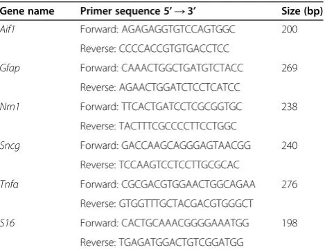
The cDNA samples were then diluted and 100 ng was analyzed by quantitative PCR (qPCR) for changes in gene expression of Aif1,Gfap, Nrn1,Sncg,Tnfαand S16 ribosomal protein mRNA. The cDNA was added to di-luted SYBR Green PCR master mix (Applied Biosystems, Grand Island, NY, USA) with 0.25μM of each primer in a 20 μl reaction volume. Each cDNA sample was run in triplicate on an ABI 7300 Real Time PCR system (Applied Biosystems), superimposed on a standard curve to deter-mine absolute transcript quantities, and normalized to
S16. Cycling conditions were 95°C (15 seconds) and 60°C (60 seconds) for 40 cycles with a dissociation step. Primer sequences are listed in Table 1.
Cell counts from retinal whole mounts
After euthanasia the superior portion of the eye was marked with a cautery, and then the whole eye was enucleated and fixed in 4% paraformaldehyde. After 50 minutes, the eye was rinsed in PBS and the anterior segment removed to create an eye cup. The retina was removed from the eye cup and placed with the ganglion cell layer (GCL) facing up onto a Superfrost Plus slide (Fisher Scientific); three additional relaxing cuts were made to allow the retina to lay flat. The whole mounts were stained with 300 ng/ml 4’ ,6-diamidino-2-phenylin-dole (DAPI; Fisher Scientific) and then thoroughly rinsed in PBS before being covered with Immu-mount (Fisher Scientific), coverslipped and stored at 4°C in the dark. Images were captured at 400× from all around the periphery of the retina, and nuclear counts were ob-tained from 24 distinct fields (120 μm2) for each retina and averaged together. Only rounded nuclei with at least one nucleolus, typical of both RGCs and amacrine cells in this layer, were included in the counts. Endothe-lial cells exhibiting elongated nuclei and no nucleolus, and densely staining astrocytes were excluded [52]. The GCL cell counts for each experimental retina were com-pared to the cell counts for the corresponding contralat-eral retina using the following formula to yield a percent change: [(cell count experimental)–(cell count control)]/ (cell count control) × 100. Retinal ganglion cells represent about 50% of the GCL population [53]. It should be noted that while Bax−/− mice have twice as many neurons as wild-type mice, the RGCs still represent about 50% of the GCL population [54].
Table 1 Quantitative PCR primer sequences
Gene name Primer sequence 5’→3’ Size (bp)
Aif1 Forward: AGAGAGGTGTCCAGTGGC 200
Reverse: CCCCACCGTGTGACCTCC
Gfap Forward: CAAACTGGCTGATGTCTACC 269
Reverse: AGAACTGGATCTCCTCATCC
Nrn1 Forward: TTCACTGATCCTCGCGGTGC 238
Reverse: TACTTTCGCCCCTTCCTGGC
Sncg Forward: GACCAAGCAGGGAGTAACGG 240
Reverse: TCCAAGTCCTCCTTGCGCAC
Tnfα Forward: CGCGACGTGGAACTGGCAGAA 276
Reverse: GTGGTTTGCTACGACGTGGGCT
S16 Forward: CACTGCAAACGGGGAAATGG 198
Immunofluorescent labeling
Whole eyes were fixed in 4% paraformaldehyde before the anterior segment was removed to create an eye cup. The eye cups were then rinsed in PBS, post-fixed over-night in 0.4% paraformaldehyde, and equilibrated in 30% sucrose in PBS. The eye cups were embedded in optimal cutting temperature compound (Fisher Scientific) in blocks and frozen on dry ice. Frozen sections were cut at 10 to 14μm. Slides were rinsed in PBS and then blocked in 0.2% Triton-X, 1% BSA, and 5% donkey serum in PBS for 1 hour at room temperature. Primary antibodies (see Table 2) were incubated overnight at 4°C in PBS containing 1% BSA. Slides were thoroughly rinsed in PBS and incubated in Texas Red-conjugated or FITC-conjugated secondary anti-bodies (Jackson ImmunoResearch, Inc., West Grove, PA, USA) in the dark for 2 hours at room temperature in PBS containing 1% BSA. Slides were thoroughly rinsed in PBS before being incubated with 300 ng/ml DAPI for 5 minutes at room temperature. Finally, the slides were rinsed in PBS and coverslipped with Immu-Mount and stored at 4°C in the dark.
Whole mounts labeled with BRN3A were stained as previously described by Nadal-Nicolas and colleagues [55], with minor modifications. Briefly, following fixation of the globe, the anterior segment was removed and the eye cups were incubated in PBS containing 0.5% Triton-X100 and 2% donkey serum (Jackson ImmunoResearch, Inc.) for 1.5 hours at room temperature. They were then transferred into the same buffer containing primary anti-body (see Table 2) overnight at 4°C. After incubation, the eye cups were thoroughly rinsed in PBS with 0.5% Triton-X100, and then fixed for an additional 10 minutes in 4% paraformaldehyde. Eye cups were rinsed in PBS and whole mounted onto Fisher Plus slides, and then incubated in 2% Triton-X100 and 2% donkey serum with 1:500 secondary antibody (Jackson ImmunoResearch) for 2 hours at room temperature. The whole mounts were rinsed in PBS and stained with 300 ng/ml DAPI for 5 minutes at room temperature. After a final wash with PBS, the slides were coverslipped with Immu-Mount and photographed.
Microscopy
All immunofluorescent photographs were acquired using a Zeiss Axioplan 2 Imaging microscope (Carl Zeiss Micro-imaging, Inc., Thornwood, NY, USA) with a digital black and white camera. Images were analyzed using the Zeiss Axiovision Image Analysis software v4.6 (Carl Zeiss Microimaging, Inc.).
Statistical analyses
Means from qPCR quantification are reported with the standard deviation of the mean, and cell counts are re-ported with standard error. Statistical significance between two means was determined using a two-sided Student’s
t-test.Pvalues were considered significant at a value equal to or less than 0.05.
Results
TNFαexpression is stimulated following optic nerve crush injury
An increase in Tnfα mRNA expression has been corre-lated with RGC and optic nerve injury; however, the time course of this expression to the best of our know-ledge has not been documented. Therefore, the changes inTnfαmRNA were analyzed by qPCR at 1, 3, 5, 7, and 14 days after optic nerve crush. Absolute mRNA levels in the retina were low across all time points in the injured retina. By 3 days after optic nerve injury, TNFαexpression was significantly elevated in the injured retina compared to the contralateral eye, and remained significantly higher at 5 and 7 days after injury (Figure 1A;P<0.05). Fourteen days after injury, the difference in TNFαmRNA levels was no longer significant (Figure 1A;P=0.94). This pattern of expression mirrored the increase in markers for glial acti-vation, specifically Aif1expression in microglia andGfap expression in macroglia (Figure 1B). Conversely, tran-scripts of two genes selectively expressed in RGCs were downregulated during this time frame (P <0.05 for all genes and time points, crush relative to contralateral naïve eyes, Figure 1C), consistent with previous observations indicative of RGC damage from crush injury [56-59]. Western blots and enzyme-linked immunosorbent assay
Table 2 Primary antibodies
Protein name Acronym Species Dilution Company Catalog #
Allograft inflammatory factor 1 AIF1 Polyclonal rabbit 1:1000 WAKOa 019-19741
BRN3A BRN3A Monoclonal mouse 1:50 Milliporeb MAB1585
Caspase 3 CASP3 Polyclonal rabbit 1:1000 R&Dc AF835
Glial fibrillary acidic protein GFAP Polyclonal rabbit 1:1000 DAKOd Z0334
JUN JUN Polyclonal rabbit 1:1000 Abcame Ab40766
Transcription factor SOX-9 SOX9 Polyclonal rabbit 1:1000 Millipore AB5535
Tumor necrosis factor alpha TNFα Polyclonal goat 1:100 R&D AF-410-NA
a
data did not reveal a quantifiable change in TNFα pro-tein (data not shown).
TNFαmediates retinal ganglion cell death through extrinsic apoptosis
An intraocular injection of TNFα induces RGC death and optic nerve degeneration [10,36,46], indicating that these cells are responsive to the toxic effects of this cyto-kine. Some discrepancies exist, however, in the mode of action by TNFα. First, toxicity to the RGC somas only manifests after several months from the initial exposure [36,46], making it unclear how TNFαparticipates in path-ologies like N-methyl-D-aspartate injection and optic nerve damage, which lead to RGC death more rapidly [5]. Second, RGC death induced by axonal damage is abso-lutely dependent on the intrinsic apoptotic pathway, since these cells are completely refractory to this insult when lacking the pro-apoptoticBaxgene [54,60,61]. Conversely, the predicted mode of action for TNFα is through the extrinsic pathway, independent of BAX activation [15]. To test which apoptotic pathway is essential for TNFα -induced RGC death, wild-type and Bax−/− mice were given an intraocular injection of TNFαand then assayed for cell loss at 6 and 8 weeks after injection.
Consistent with other studies, injected TNFα induced cell loss, but only after 8 weeks (Figure 2A). The total number of cells in wild-type mice significantly declined 8 weeks after treatment with 10 ng and 100 ng TNFα (8.97% and 19.41%, respectively; P <0.005, compared to PBS-injected eyes). Similarly, Bax-deficient RGCs were also susceptible to 10 ng and 100 ng TNFα, with the total number of RGC layer neurons declining by 8.11% and 15.03% (P<0.001, compared to PBS-injected eyes). PBS in-jections alone did not cause significant cell loss (wild types
P=0.36, knockoutsP=0.40, relative to contralateral eye). The effect of exogenous TNFα was not limited to a small percentage of RGCs, however. In similar experi-ments we stained retinal whole mounts for the RGC marker BRN3A, which can be depleted in damaged RGCs well in advance of cell death [56,62]. At 8 weeks after in-jection of 100 ng TNFα, there was undetectable BRN3A staining in retinas of both wild-type and Bax−/−mice ex-posed to this cytokine (Figure 2B-E). Thus, even though there is only a loss of 30 to 40% of the RGCs (after correc-tion of the percentage of RGCs that make up the total number of neurons in the ganglion cell layer [53]), ex-ogenous TNFαappears to cause some level of damage to the entire population of these cells.
TNFαdeficiency exacerbates retinal ganglion cell injury after optic nerve crush
To determine if TNFαcontributes to RGC pathology after optic nerve crush, we performed surgery onTnfα−/−mice and obtained cell counts after 7, 14, and 21 days
(Figure 3A). Although deletion of TNFR1 has previously been shown to confer significant resistance to acute optic nerve damage in mice [11], we observed greater cell loss in the Tnfα-deficient mice compared to the wild types. Although RGC loss was not significantly different 7 days after injury between the wild type and Tnfα−/− mice (8.84% compared to 7.15% total cell loss, respectively;
P=0.50), by 14 daysTnfα−/−mice had lost 19.46% of the total cells, while the wild-type mice only showed an 11.61% decline in their total RGC layer neurons (P<0.001). Cell loss continued to decline in both genotypes 21 days after crush, but there was again significantly greater loss in
Tnfα−/−
mice relative to the wild types (26.55% compared to 20.32%, respectively;P<0.001). In complimentary exper-iments, we also quantified the level of Caspase 3 (CASP3) activation after optic nerve crush inTnfα−/−mice. Previous studies have documented that CASP3 activity peaks be-tween 3 and 5 days in this experimental paradigm [50,63]. Consistent with the cell count data,Tnfα−/−mice exhibited significantly more CASP3 activity by 3 days after optic nerve crush compared to wild-type mice (P <0.05), al-though by 5 days the percent of CASP3+ cells was not statistically different (Figure 3B,C).
A single injection of TNFαprotects retinal ganglion cells when delivered prior to optic nerve crush
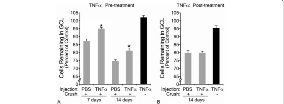
Dissecting out a single cytokine from a complex map of signaling pathways by genetic deletion is likely an over-simplification of any disease pathology. It is more plausible that TNFαis part of a larger orchestrated in-jury response, and without auxiliary inin-jury signals this cytokine may be limited in its effectiveness. We there-fore tested the effect of TNFα when delivered in con-junction with injury to RGCs by optic nerve crush. We tested two conditions: first, the cytokine was delivered prior to optic nerve crush to pre-activate the TNFα sig-naling network, which is expectedly enhanced following crush injury.
When an intraocular injection of 100 ng TNFα was delivered 5 days prior to crush injury, RGC loss was re-duced by almost 50% 1 week after crush, compared to PBS-injected crushed mice (Figure 4A, P <0.001). This protective effect persisted, relative to PBS-injected eyes, to 14 days post-crush, although cell density decreased in both cohorts of mice (P <0.001). A second condition was also tested in which 100 ng TNFα was delivered 7 days after optic nerve crush; however, there was no statistically significant difference in cell survival be-tween any of the groups subjected to crush (Figure 4B,
P >0.05). These results support our findings above that TNFα may protect RGCs from optic nerve injury and, importantly, will not exacerbate damage when applied after the initial insult to the optic nerve.
Figure 2TNFαmediates extrinsic apoptosis of retinal ganglion cells in a dose-dependent manner. (A)A single intraocular injection of TNFαwas delivered and the total number of cells in the ganglion cell layer (GCL) was obtained from 4’ ,6-diamidino-2-phenylindole-stained retinal whole mounts after 8 weeks. No cell loss was detected at 6 weeks (data not shown). Wild-type (black bars) andBax−/−mice (gray bars) exhibited dose-dependent loss of total cells, indicating cell death occurred via aBax-independent mechanism. The percent change was calculated by comparing the injected eye to the control eye. Statistics were calculated relative to the PBS-injected control group and are presented as mean ± SE (*P<0.005, **P<0.001); n≥3 for all genotypes and conditions.
(B-E)Retinal whole mounts were also stained for the retinal ganglion cell-specific transcription factor BRN3A. Approximately 40% of the neurons in the GCL stain for BRN3A in control retinas of both wild-type
TNFαis not critical for glial activation byAif1orGfap monitoring
The data shown in Figures 3 and 4 illustrate a positive role for TNFαafter RGC injury; however, the mechanisms of protection are still unclear. Others have reported that TNFαcan act in an autocrine manner to augment the glial response after an injury, particularly in the astrocytes and microglia [64-67], which may help protect neurons from subsequent damage [48,68]. Therefore, in the absence of TNFα, the retinal glia may inadequately respond to an injury and render neurons more susceptible to degener-ation. We next tested whether TNFαwas critical for glial
activation by comparing the microglial and macroglial ac-tivation responses after optic nerve crush in wild-type and
Tnfα−/−
mice.
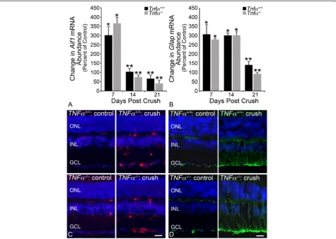
The absence of TNFαminimally affected glial activation, as a function of Aif1andGfapexpression; wild-type and
Tnfα−/−
mice showed almost identical activation trends. Microglial activation (Aif1) peaked in the injured retinas 7 days after crush (301% in wild-type mice, 366% in
Tnfα−/−
mice, P <0.001 relative to contralateral eye) and then declined at 14 and 21 days to 66.6% and 41.9%, respectively (Figure 5A, P <0.05). The increase in Aif1 expression was also detectable by immunofluorescence
Figure 3Mice deficient forTnfαhave greater retinal ganglion cell loss from crush than wild-type mice. (A)Wild-type andTnfα−/−mice were subjected to optic nerve crush and analyzed for total retinal ganglion cell layer neuronal cells remaining at 7, 14, and 21 days after injury. The decline in cell numbers was not statistically significant between the two genotypes at 7 days, but at 14 and 21 daysTnfα−/−mice had significantly more cell loss (*P<0.001 for both time points).(B,C)Retinal whole mounts from wild-type andTnfα−/−mice were analyzed for Caspase 3 (CASP3) activation 3 and 5 days after optic nerve crush. At 3 days,Tnfα−/−mice has a significantly greater number of CASP3+ cells (*P<0.05), but by 5 days the difference between the two genotypes was no longer significant. Results are presented at mean ± SE(A,C); n≥3 for all genotypes. Scale bar(B)=50μm. GCL, ganglion cell layer.
labeling in retinal sections, with positive cells manifesting in the inner and outer plexiform layers as well as the gan-glion cell layer (Figure 5C). Macroglial activation (Gfap) in wild-type and Tnfα−/− mice also trended similarly, with expression rising by 7 days to 305.6% and 277.0% in the wild types andTnfα-deficient mice, respectively (Figure 5B,
P<0.001 relative to contralateral eye).GfapmRNA levels remained steadily elevated at 14 days after crush (301.7% in wild types, 299.9% inTnfα−/−mice), before considerably declining at 21 days to 139.2% and 91.56% (P<0.001 rela-tive to contralateral eye). Glial fibrillary acid protein underwent a distinct morphological change with processes labeling through the retinal layers, consistent with Müller cell activation [20,30], and in the ganglion cell layer where astrocytes reside (Figure 5D). Overall, there was
no significant distinction between microglial or macro-glial activation in the wild-type andTnfα−/−mice.
TNFαactivates NFκB and causes JUN accumulation in Müller cells
Although TNFαdoes not appear to be required for glial activation as a function of Aif1 or Gfap expression, it may be a critical extracellular signal that initiates pro-tective networks within the activated glia after a retinal injury. Two known targets of TNFαsignaling are the tran-scription factors NFκB [40,47] and JUN [69], and these two proteins have been shown to work cooperatively to regu-late gene transcription [70-73]. The activation of NFκB has been correlated with resistance to ligand-mediated apop-tosis [74,75], and JUN has been linked with both protective
and apoptotic pathways in injured RGCs [51]. It is possible that TNFαis mediating protection through one or both of these transcription factors, so we next explored the effect of TNFαon NFκB activation and JUN accumulation.
Using cis-NFκBEGFP reporter mice [49], in which en-hanced green fluorescent protein (EGFP) is transcribed by activated NFκB, we first examined whether optic nerve in-jury activated NFκB in the retina. We expected to find that NFκB activation would correlate with the time course of TNFαexpression observed after crush; however, retinal sections did not reveal a detectable increase in NFκB ac-tivity following optic nerve crush (Figure 6B). We next tested the effect of an intraocular injection of TNFα into one eye of the reporter mice. Unlike crush, exogenous TNFαcaused a clear activation of NFκB as early as 1 day after treatment (Figure 6D), and remained prevalent by 3 days before returning to baseline levels at 5 days (data not shown). The activity of NFκB was independent of the injection procedure, as eyes injected with PBS did not show any evidence of EGFP expression (Figure 6C). NFκB activation by TNFαco-localized with SOX9, which select-ively labels Müller cells [76]. Interestingly, only a subset of Müller cells appeared to exhibit NFκB activation.
Optic nerve crush has previously been shown to cause nuclear accumulation of JUN in RGCs [50,51]. Similar to NFκB activation, however, an intraocular injection of TNFα caused a significant increase in JUN nuclear accu-mulation in the inner nuclear layer, and to a lesser extent in the GCL (Figure 7F). This accumulation occurred as early as 7 hours after TNFα exposure, and co-localized with SOX2, a marker for Müller glia and amacrine cells [77,78]. A morphologically distinct population of SOX2-positive cells in the GCL also showed an accumulation of JUN, which could be displaced amacrine cells or astro-cytes (Figure 7F, arrows). While TNFαcaused JUN accu-mulation in the majority of the Müller cell population, only a subset of the JUN-positive cells also exhibited NFκB activation.
Discussion
The role of TNFαin neurodegeneration has been exten-sively studied, yet caveats still remain in understanding the mechanism by which it contributes to disease. In the con-text of RGC injury and death, TNFαhas been considered by some researchers as detrimental and has been modeled as a secondary inducer of RGC loss [7,11,13,25-27,79]. In accordance with the literature supporting this theory, we found thatTnfαmRNA is elevated in our model of optic nerve crush, and that the cytokine does lead to a delayed loss of RGCs through an extrinsic apoptotic mechanism. However, a single intraocular injection of TNFαdoes not cause rapid RGC loss as is seen with other ocular injury models to which TNFα has been linked. Additional re-search supports that TNFαis beneficial and protective to
neurons [43,47,48,80]. Consistent with this we found that genetic deletion of theTnfαgene rendered mice more sus-ceptible to optic nerve injury, and that pre-treatment with exogenous TNFα promoted RGC survival after crush. It appears contradictory that TNFαis both detrimental and protective to RGCs; however, our data present a poten-tially critical timing component that has not previously been studied in regards to TNFα signaling in the retina. More specifically, it appears that early TNFα exposure prior to an injury may be protective, while chronic TNFα
expression may eventually culminate in neuronal damage and loss.
This phenomenon of neuronal protection from pre-conditioning has been previously observed in stroke patients, in which those with a history of transient ische-mia attacks fared better following a cerebral infarction than those without a similar history [81]. This protection has been mimicked in cell culture models of neuronal insult and animal models of brain ischemia with TNFα exposure prior to a damaging stimulus [47,82]. Early treatment with TNFαhas been positively correlated with survival pathways mediated by a number of proteins, in-cluding phosphoinositide-3-kinase [47], the transcription factor NFκB, and the histone acetyltransferase CREB bind-ing protein [82]. The upregulation of CREB bindbind-ing pro-tein was observed only in neurons, even when co-cultured with astrocytes [82], suggesting that TNFαwas acting dir-ectly on the neurons and not being mediated through the glia. However, in the retina, astrocytes are only one sub-population of glial cells - Müller cells and microglia also contribute to retinal health and injury repair, complicat-ing the mechanism by which TNFα may be promoting RGC survival.
TNFαsignals through TNFR1 and TNFR2, and follow-ing retinal ischemia and a mouse model of glaucoma, both receptors are upregulated in cells of the inner nu-clear layer and GCL [43,44]. TNFR1 is a transmembrane protein with an intracellular death domain that, upon activation, can interact with adaptor proteins and initiate apoptosis through CASP8 [37,83,84], and several studies
have shown that Tnfr1−/− mice exhibit significantly less RGC loss than wild-type mice after injury [11,43]. In comparison, TNFR2 does not contain a death domain and has been linked with sustained NFκB activation [43,47]. Unlike the RGC protection seen after injury with
Tnfr1deficiency,Tnfr2−/−mice fair worse than wild-type mice following ischemia [43] and glutamate excitotoxi-city [47]. Interestingly, the Tnfα−/− mice in our study more closely reflect the enhanced RGC pathology seen inTnfr2−/− mice after injury. This might suggest a pref-erence for TNFαto bind TNFR2, possibly explaining the protection afforded when TNFR1 is genetically ablated, restricting TNFαto signal through the TNFR2 protective networks. Alternatively, Tnfr2 deficiency may enhance apoptotic signals through TNFR1, rendering central ner-vous system tissue more susceptible to injury. In both scenarios, it seems critical to understand the proteins downstream of each receptor that are being affected after injury, and in particular which retinal cell types are responding to this cytokine.
It is important to note that while we have shown an early protective potential of TNFα, the long-term conse-quence of TNFα exposure still appears to be detrimen-tal. This dual function of TNFα may reflect different responses of individual cell types to this cytokine, which is consistent with a recent publication by Dvoriantchikova and Ivanov [85]. Their research found that, in response to TNFα, RGC cultures exhibited sustained JNK activation and death, while astrocytes upregulated NFκB and pro-moted survival [85]. Therefore, the localization of TNFα
expression and the cells responding to this cytokine will influence whether TNFα has a beneficial or detrimental effect on RGCs. TNFαis expressed by a number of innate immune responders, and has been co-localized to optic nerve head, nerve fiber layer, GCL, and the inner nuclear layer of human glaucoma patients [34,35,43,86], and is up-regulated by macroglia and microglia in the optic nerve and optic nerve head [11,34]. Additional studies have shown that dendritic cells also infiltrate the retina follow-ing a similar optic nerve crush paradigm described here [16,87], and it is conceivable that they may be the source of TNFα. The proximity of TNFα production to specific cell types in the retina may generate the differential pro-tective versus detrimental effects. The propro-tective effects may occur through an indirect mechanism, by TNFα -induced changes in retinal glia. Based on the rapid induction of NFκB activity and JUN accumulation after exposure to exogenous TNFα, we attribute this rapid re-sponse to Müller cells (see below). Conversely, the detri-mental effects may result from a direct interaction with the RGCs. Kitaoka and colleagues noted that intravitreal injection of TNFα in rabbits resulted in relatively early-onset axonal damage followed by soma death many weeks after TNFαexposure [40].
Unlike the delayed effect of TNFα on RGC somas, we have shown that Müller cells respond rapidly to TNFα within 1 day of exposure by accumulating JUN and upreg-ulating NFκB, two known targets downstream of TNFα [85]. It is important to note that JUN is activated by phos-phorylation; however, the p-JUN antibody is less reliable than that for JUN due to cross-reactivity [50]. Therefore, the data presented in this manuscript are documented as nuclear accumulation of JUN rather than activation. Add-itionally, JUN is known to autoregulate its own expression following its activation [88], and JUN levels have been used as a surrogate of JUN activity. In response to an in-traocular delivery of TNFα, both JUN and NFκB exhibit nuclear activity, and it is interesting that the primary glial cells responding to TNFα are the Müller cells. However, while JUN accumulation was present in all of the Müller cells, only a subset exhibited NFκB activity. This might suggest that JUN is upstream of NFκB, and that all of the Müller cells have not yet been able to activate the latter gene, although the literature suggests a more complex interplay between these transcription factors [71,73]. It is also unclear if either of these pathways are involved in the protective effect of TNFα. However, given the strong asso-ciation of NFκB with survival pathways [47,73], it is pos-sible that the cells expressing this transcription factor might be mediating the protective effect seen in our stud-ies involving intravitreal injection of exogenous TNFα. A paradox with this interpretation, however, is that neither JUN nor NFκB were activated after optic nerve crush. While this may have been a function of reduced or more
localized levels of TNFα production (such as by infil-trating dendritic cells), it remains unclear how the endogenous TNFα signaling response provides a pro-tective environment for RGCs. Further studies involving cell-specific ablation of one or both of these transcrip-tion factors are needed to decipher whether they play a role in the endogenous TNFαprotective effect.
The signaling pathways activated in the injured retina are complex, but we have identified a critical timing component in TNFαsignaling: specifically, that early ex-posure to this cytokine protects RGCs from subsequent optic nerve damage. A considerable amount of literature has identified a damaging role for TNFα, yet this research has indirectly focused on the effect of TNFαsignaling late after injury. Future studies should consider the advantage of early immune activation in the retina, specifically with an emphasis on Müller cell activation. By bolstering pro-tective pathways early, rather than eliminating cytokine signaling entirely, RGC loss may be minimized following a severe insult and improve the prognosis for patients with optic neuropathies, such as glaucoma.
Conclusion
Our studies underscore that while long-term exposure to TNFαis toxic to RGCs, this cytokine appears to initi-ate protective pathways that improve RGC survival im-mediately following optic nerve injury. The mechanism of protection may be occurring through TNFαactivation of Müller cells.
Abbreviations
CASP:Caspase; DAPI: 4’,6-diamidino-2-phenylindole; EGFP: enhanced green fluorescent protein; GCL: ganglion cell layer; PBS: phosphate-buffered saline; PCR: polymerase chain reaction; qPCR: quantitative polymerase chain reaction; RGC: retinal ganglion cell; TNFα: tumor necrosis factor alpha; TNFR1: tumor necrosis factor alpha receptor 1; TNFR2: tumor necrosis factor alpha receptor 2.
Competing interests
The authors declare that they have no competing interests.
Authors’contributions
RWN and RTL conceived the study and participated in its design and coordination. CEM conducted the optic nerve crush and intraocular injections on wild-type,Tnfα−/−mice, andcis-NFκBEGFPreporter mice, and subsequent immunofluorescence and quantitative PCR. KAF conducted the JUN studies and performed optic nerve crush onTnfα−/−mice. CLS participated in study design and data analysis. CEM and RWN drafted the manuscript. All authors read and approved the final manuscript.
Acknowledgements
The authors would like to thank Dr Matyas Sandor for theTnfα−/−mice, and Dr Christian Jobin for thecis-NFκBEGFPreporter mice. This work was supported by the National Eye Institute R01 EY018606 (RTL), R01 EY012223 (RWN), R01 EY018869 (RWN), NEI P30 EY016665 (Ophthalmology and Visual Sciences), NRSA T32 GM081061 (CEM), a Glaucoma Research Foundation Shaffer grant (RWN), and Research to Prevent Blindness Inc. unrestricted funding to the Department of Ophthalmology and Visual Sciences.
Author details 1
Molecular Pathology Graduate Program, University of Wisconsin, 3170-10 K/L MFCB–1685 Highland Ave., Madison, WI 53705, USA.3Flaum Eye Institute, University of Rochester Medical Center, 601 Elmwood Ave., Box 659, Rochester, NY 14642, USA.4Department of Biomedical Genetics, University of Rochester Medical Center, 601 Elmwood Ave., Box 633, Rochester, NY 14642, USA.5The Center for Visual Sciences, University of Rochester Medical Center, 274 Meliora Hall, RC Box 270270, Rochester, NY 14627, USA.
Received: 10 June 2014 Accepted: 3 November 2014
References
1. Whitmore AV, Libby RT, John SW:Glaucoma: thinking in new ways - a role for autonomous axonal self-destruction and other compartmentalised processes?Prog Retin Eye Res2005,24:639–662.
2. Nickells RW:From ocular hypertension to ganglion cell death: a theoretical sequence of events leading to glaucoma.Can J Ophthalmol 2007,42:278–287.
3. Nickells RW:Ganglion cell death in glaucoma: from mice to men. Vet Ophthalmol2007,10(Suppl 1):88–94.
4. Quigley HA, Nickells RW, Kerrigan LA, Pease ME, Thibault DJ, Zack DJ:
Retinal ganglion cell death in experimental glaucoma and after axotomy occurs by apoptosis.Invest Ophthalmol Vis Sci1995,36:774–786. 5. Li Y, Schlamp CL, Nickells RW:Experimental induction of retinal ganglion
cell death in adult mice.Invest Ophthalmol Vis Sci1999,40:1004–1008. 6. Nickells RW:Apoptosis of retinal ganglion cells in glaucoma: an update
of the molecular pathways involved in cell death.Surv Ophthalmol1999,
43(Suppl 1):S151–S161.
7. Yoles E, Muller S, Schwartz M:NMDA-receptor antagonist protects neurons from secondary degeneration after partial optic nerve crush. J Neurotrauma1997,14:665–675.
8. Levkovitch-Verbin H, Harris-Cerruti C, Groner Y, Wheeler LA, Schwartz M, Yoles E:RGC death in mice after optic nerve crush injury: oxidative stress and neuroprotection.Invest Ophthalmol Vis Sci2000,41:4169–4174. 9. Li Y, Semaan SJ, Schlamp CL, Nickells RW:Dominant inheritance of retinal
ganglion cell resistance to optic nerve crush in mice.BMC Neurosci2007,
8:19.
10. Madigan MC, Sadun AA, Rao NS, Dugel PU, Tenhula WN, Gill PS:Tumor necrosis factor-alpha (TNF-alpha)-induced optic neuropathy in rabbits. Neurol Res1996,18:176–184.
11. Tezel G, Yang X, Yang J, Wax MB:Role of tumor necrosis factor receptor-1 in the death of retinal ganglion cells following optic nerve crush injury in mice.Brain Res2004,996:202–212.
12. Zhang S, Wang H, Lu Q, Qing G, Wang N, Wang Y, Li S, Yang D, Yan F:
Detection of early neuron degeneration and accompanying glial responses in the visual pathway in a rat model of acute intraocular hypertension.Brain Res2009,1303:131–143.
13. Fitzgerald M, Bartlett CA, Harvey AR, Dunlop SA:Early events of secondary degeneration after partial optic nerve transection: an
immunohistochemical study.J Neurotrauma2010,27:439–452. 14. Sivakumar V, Foulds WS, Luu CD, Ling EA, Kaur C:Retinal ganglion cell
death is induced by microglia derived pro-inflammatory cytokines in the hypoxic neonatal retina.J Pathol2011,224:245–260.
15. Almasieh M, Wilson AM, Morquette B, Cueva Vargas JL, Di Polo A:The molecular basis of retinal ganglion cell death in glaucoma.Prog Retin Eye Res2012,31:152–181.
16. Lehmann U, Heuss ND, McPherson SW, Roehrich H, Gregerson DS:
Dendritic cells are early responders to retinal injury.Neurobiol Dis2010,
40:177–184.
17. Gerhardinger C, Costa MB, Coulombe MC, Toth I, Hoehn T, Grosu P:
Expression of acute-phase response proteins in retinal Muller cells in diabetes.Invest Ophthalmol Vis Sci2005,46:349–357.
18. Templeton JP, Freeman NE, Nickerson JM, Jablonski MM, Rex TS, Williams RW, Geisert EE:Innate immune network in the retina activated by optic nerve crush.Invest Ophthalmol Vis Sci2013,54:2599–2606.
19. Garcia M, Vecino E:Role of Muller glia in neuroprotection and regeneration in the retina.Histol Histopathol2003,18:1205–1218. 20. Bosco A, Steele MR, Vetter ML:Early microglia activation in a mouse
model of chronic glaucoma.J Comp Neurol2011,519:599–620. 21. Wax MB, Tezel G:Immunoregulation of retinal ganglion cell fate in
glaucoma.Exp Eye Res2009,88:825–830.
22. Baltmr A, Duggan J, Nizari S, Salt TE, Cordeiro MF:Neuroprotection in glaucoma - is there a future role?Exp Eye Res2010,91:554–566. 23. Tezel G, Wax MB:Increased production of tumor necrosis factor-alpha by
glial cells exposed to simulated ischemia or elevated hydrostatic pressure induces apoptosis in cocultured retinal ganglion cells.J Neurosci 2000,20:8693–8700.
24. Dvoriantchikova G, Barakat D, Brambilla R, Agudelo C, Hernandez E, Bethea JR, Shestopalov VI, Ivanov D:Inactivation of astroglial NF-kappa B promotes survival of retinal neurons following ischemic injury.Eur J Neurosci2009,
30:175–185.
25. Levkovitch-Verbin H, Quigley HA, Kerrigan-Baumrind LA, D’Anna SA, Kerrigan D, Pease ME:Optic nerve transection in monkeys may result in secondary degeneration of retinal ganglion cells.Invest Ophthalmol Vis Sci2001,
42:975–982.
26. Levkovitch-Verbin H, Dardik R, Vander S, Melamed S:Mechanism of retinal ganglion cells death in secondary degeneration of the optic nerve. Exp Eye Res2010,91:127–134.
27. Yoles E, Schwartz M:Potential neuroprotective therapy for glaucomatous optic neuropathy.Surv Ophthalmol1998,42:367–372.
28. Lebrun-Julien F, Duplan L, Pernet V, Osswald I, Sapieha P, Bourgeois P, Dickson K, Bowie D, Barker PA, Di Polo A:Excitotoxic death of retinal neurons in vivo occurs via a non-cell-autonomous mechanism.J Neurosci 2009,29:5536–5545.
29. Baptiste DC, Powell KJ, Jollimore CA, Hamilton C, LeVatte TL, Archibald ML, Chauhan BC, Robertson GS, Kelly ME:Effects of minocycline and tetracycline on retinal ganglion cell survival after axotomy.Neuroscience 2005,134:575–582.
30. Bosco A, Inman DM, Steele MR, Wu G, Soto I, Marsh-Armstrong N, Hubbard WC, Calkins DJ, Horner PJ, Vetter ML:Reduced retina microglial activation and improved optic nerve integrity with minocycline treatment in the DBA/2 J mouse model of glaucoma.Invest Ophthalmol Vis Sci2008,49:1437–1446.
31. Levkovitch-Verbin H, Kalev-Landoy M, Habot-Wilner Z, Melamed S:Minocycline delays death of retinal ganglion cells in experimental glaucoma and after optic nerve transection.Arch Ophthalmol2006,124:520–526.
32. Kuchtey J, Rezaei KA, Jaru-Ampornpan P, Sternberg P Jr, Kuchtey RW:Multiplex cytokine analysis reveals elevated concentration of interleukin-8 in glaucomatous aqueous humor.Invest Ophthalmol Vis Sci2010,51:6441–6447. 33. Kerschensteiner M, Meinl E, Hohlfeld R:Neuro-immune crosstalk in CNS
diseases.Results Probl Cell Differ2010,51:197–216.
34. Yuan L, Neufeld AH:Tumor necrosis factor-alpha: a potentially neurodestructive cytokine produced by glia in the human glaucomatous optic nerve head.Glia2000,32:42–50.
35. Tezel G, Li LY, Patil RV, Wax MB:TNF-alpha and TNF-alpha receptor-1 in the retina of normal and glaucomatous eyes.Invest Ophthalmol Vis Sci 2001,42:1787–1794.
36. Nakazawa T, Nakazawa C, Matsubara A, Noda K, Hisatomi T, She H, Michaud N, Hafezi-Moghadam A, Miller JW, Benowitz LI:Tumor necrosis factor-alpha mediates oligodendrocyte death and delayed retinal ganglion cell loss in a mouse model of glaucoma.J Neurosci2006,26:12633–12641.
37. Yang X, Luo C, Cai J, Powell DW, Yu D, Kuehn MH, Tezel G:
Neurodegenerative and inflammatory pathway components linked to TNF-alpha/TNFR1 signaling in the glaucomatous human retina.Invest Ophthalmol Vis Sci2011,52:8442–8454.
38. Tezel G:TNF-alpha signaling in glaucomatous neurodegeneration.Prog Brain Res2008,173:409–421.
39. de Kozak Y, Naud MC, Bellot J, Faure JP, Hicks D:Differential tumor necrosis factor expression by resident retinal cells from experimental uveitis-susceptible and -resistant rat strains.J Neuroimmunol1994,55:1–9. 40. Kitaoka Y, Kitaoka Y, Kwong JM, Ross-Cisneros FN, Wang J, Tsai RK, Sadun
AA, Lam TT:TNF-alpha-induced optic nerve degeneration and nuclear factor-kappaB p65.Invest Ophthalmol Vis Sci2006,47:1448–1457. 41. Roh M, Zhang Y, Murakami Y, Thanos A, Lee SC, Vavvas DG, Benowitz LI,
Miller JW:Etanercept, a widely used inhibitor of tumor necrosis factor-alpha (TNF-alpha): prevents retinal ganglion cell loss in a rat model of glaucoma.PLoS One2012,7:e40065.
42. Husain S, Liou GI, Crosson CE:Opioid receptor activation: suppression of ischemia/reperfusion-induced production of TNF-alpha in the retina. Invest Ophthalmol Vis Sci2011,52:2577–2583.
43. Fontaine V, Mohand-Said S, Hanoteau N, Fuchs C, Pfizenmaier K, Eisel U:
(TNF) in retinal ischemia: opposite roles of TNF receptor 1 and TNF receptor 2.J Neurosci2002,22:RC216.
44. Howell GR, Walton DO, King BL, Libby RT, John SW:Datgan, a reusable software system for facile interrogation and visualization of complex transcription profiling data.BMC Genomics2011,12:429.
45. Reichstein D, Ren L, Filippopoulos T, Mittag T, Danias J:Apoptotic retinal ganglion cell death in the DBA/2 mouse model of glaucoma.Exp Eye Res 2007,84:13–21.
46. Madigan MC, Rao NS, Tenhula WN, Sadun AA:Preliminary morphometric study of tumor necrosis factor-alpha (TNF alpha)-induced rabbit optic neuropathy.Neurol Res1996,18:233–236.
47. Marchetti L, Klein M, Schlett K, Pfizenmaier K, Eisel UL:Tumor necrosis factor (TNF)-mediated neuroprotection against glutamate-induced excitotoxicity is enhanced by N-methyl-D-aspartate receptor activation: essential role of a TNF receptor 2-mediated phosphatidylinositol 3-kinase-dependent NF-kappa B pathway.J Biol Chem2004,279:32869–32881.
48. Veroni C, Gabriele L, Canini I, Castiello L, Coccia E, Remoli ME, Columba-Cabezas S, Arico E, Aloisi F, Agresti C:Activation of TNF receptor 2 in microglia promotes induction of anti-inflammatory pathways.Mol Cell Neurosci2010,45:234–244.
49. Magness ST, Jijon H, Van Houten FN, Sharpless NE, Brenner DA, Jobin C:In vivo pattern of lipopolysaccharide and anti-CD3-induced NF-kappa B activation using a novel gene-targeted enhanced GFP reporter gene mouse.J Immunol2004,173:1561–1570.
50. Fernandes KA, Harder JM, Fornarola LB, Freeman RS, Clark AF, Pang IH, John SW, Libby RT:JNK2 and JNK3 are major regulators of axonal injury-induced retinal ganglion cell death.Neurobiol Dis2012,46:393–401. 51. Fernandes KA, Harder JM, Kim J, Libby RT:JUN regulates early
transcriptional responses to axonal injury in retinal ganglion cells.Exp Eye Res2013,112C:106–117.
52. Janssen KT, Mac Nair CE, Dietz JA, Schlamp CL, Nickells RW:Nuclear atrophy of retinal ganglion cells precedes the bax-dependent stage of apoptosis.Invest Ophthalmol Vis Sci2013,54:1805–1815.
53. Schlamp CL, Montgomery AD, Mac Nair CE, Schuart C, Willmer DJ, Nickells RW:
Evaluation of the percentage of ganglion cells in the ganglion cell layer of the rodent retina.Mol Vis2013,19:1387–1396.
54. Li Y, Schlamp CL, Poulsen KP, Nickells RW:Bax-dependent and independent pathways of retinal ganglion cell death induced by different damaging stimuli.Exp Eye Res2000,71:209–213.
55. Nadal-Nicolas FM, Jimenez-Lopez M, Salinas-Navarro M, Sobrado-Calvo P, Alburquerque-Bejar JJ, Vidal-Sanz M, Agudo-Barriuso M:Whole number, distribution and co-expression of brn3 transcription factors in retinal ganglion cells of adult albino and pigmented rats.PLoS One2012,7:e49830. 56. Pelzel HR, Schlamp CL, Nickells RW:Histone H4 deacetylation plays a
critical role in early gene silencing during neuronal apoptosis. BMC Neurosci2010,11:62.
57. Buckingham BP, Inman DM, Lambert W, Oglesby E, Calkins DJ, Steele MR, Vetter ML, Marsh-Armstrong N, Horner PJ:Progressive ganglion cell degeneration precedes neuronal loss in a mouse model of glaucoma. J Neurosci2008,28:2735–2744.
58. Soto I, Oglesby E, Buckingham BP, Son JL, Roberson ED, Steele MR, Inman DM, Vetter ML, Horner PJ, Marsh-Armstrong N:Retinal ganglion cells downregulate gene expression and lose their axons within the optic nerve head in a mouse glaucoma model.J Neurosci2008,28:548–561.
59. Schlamp CL, Johnson EC, Li Y, Morrison JC, Nickells RW:Changes in Thy1 gene expression associated with damaged retinal ganglion cells.Mol Vis 2001,7:192–201.
60. Libby RT, Li Y, Savinova OV, Barter J, Smith RS, Nickells RW, John SW:
Susceptibility to neurodegeneration in a glaucoma is modified by Bax gene dosage.PLoS Genet2005,1:17–26.
61. Nickells RW, Semaan SJ, Schlamp CL:Involvement of the Bcl2 gene family in the signaling and control of retinal ganglion cell death.Prog Brain Res 2008,173:423–435.
62. Weishaupt JH, Klocker N, Bahr M:Axotomy-induced early down-regulation of POU-IV class transcription factors Brn-3a and Brn-3b in retinal ganglion cells.J Mol Neurosci2005,26:17–25.
63. Harder JM, Libby RT:BBC3 (PUMA) regulates developmental apoptosis but not axonal injury induced death in the retina.Mol Neurodegener2011,6:50. 64. Selmaj KW, Farooq M, Norton WT, Raine CS, Brosnan CF:Proliferation of
astrocytes in vitro in response to cytokines: a primary role for tumor necrosis factor.J Immunol1990,144:129–135.
65. Merrill JE:Effects of interleukin-1 and tumor necrosis factor-alpha on astrocytes, microglia, oligodendrocytes, and glial precursors in vitro. Dev Neurosci1991,13:130–137.
66. Ganter S, Northoff H, Mannel D, Gebicke-Harter PJ:Growth control of cultured microglia.J Neurosci Res1992,33:218–230.
67. Kuno R, Wang J, Kawanokuchi J, Takeuchi H, Mizuno T, Suzumura A:
Autocrine activation of microglia by tumor necrosis factor-alpha. J Neuroimmunol2005,162:89–96.
68. Dhandapani KM, Hadman M, De Sevilla L, Wade MF, Mahesh VB, Brann DW:
Astrocyte protection of neurons: role of transforming growth factor-beta signaling via a c-Jun-AP-1 protective pathway.J Biol Chem2003,
278:43329–43339.
69. Liu J, Minemoto Y, Lin A:c-Jun N-terminal protein kinase 1 (JNK1), but not JNK2, is essential for tumor necrosis factor alpha-induced c-Jun kinase activation and apoptosis.Mol Cell Biol2004,24:10844–10856. 70. Stein B, Baldwin AS Jr, Ballard DW, Greene WC, Angel P, Herrlich P:
Cross-coupling of the NF-kappa B p65 and Fos/Jun transcription factors produces potentiated biological function.EMBO J1993,12:3879–3891. 71. Fujioka S, Niu J, Schmidt C, Sclabas GM, Peng B, Uwagawa T, Li Z, Evans DB,
Abbruzzese JL, Chiao PJ:NF-kappaB and AP-1 connection: mechanism of NF-kappaB-dependent regulation of AP-1 activity.Mol Cell Biol2004,
24:7806–7819.
72. Dann SG, Allison WT, Veldhoen K, Johnson T, Hawryshyn CW:Chromatin immunoprecipitation assay on the rainbow trout opsin proximal promoters illustrates binding of NF-kappaB and c-jun to the SWS1 promoter in the retina.Exp Eye Res2004,78:1015–1024.
73. Maggirwar SB, Ramirez S, Tong N, Gelbard HA, Dewhurst S:Functional interplay between nuclear factor-kappaB and c-Jun integrated by coactivator p300 determines the survival of nerve growth factor-dependent PC12 cells.J Neurochem2000,74:527–539.
74. Mizrahi K, Kagan S, Stein J, Yaniv I, Zipori D, Askenasy N:Resistance of hematopoietic progenitors to Fas-mediated apoptosis is actively sustained by NFkappaB with a characteristic transcriptional signature. Stem Cells Dev2013,6:676–686.
75. Varfolomeev EE, Ashkenazi A:Tumor necrosis factor: an apoptosis JuNKie? Cell2004,116:491–497.
76. Shen W, Fruttiger M, Zhu L, Chung SH, Barnett NL, Kirk JK, Lee S, Coorey NJ, Killingsworth M, Sherman LS, Gillies MC:Conditional Muller cell ablation causes independent neuronal and vascular pathologies in a novel transgenic model.J Neurosci2012,32:15715–15727.
77. Le RD, Rayner K, Rex M, Wigmore PM, Scotting PJ:The transcription factor cSox2 and neuropeptide Y define a novel subgroup of amacrine cells in the retina.J Anat2002,200:51–56.
78. Lin YP, Ouchi Y, Satoh S, Watanabe S:Sox2 plays a role in the induction of amacrine and Muller glial cells in mouse retinal progenitor cells.Invest Ophthalmol Vis Sci2009,50:68–74.
79. Schwartz M, Belkin M, Yoles E, Solomon A:Potential treatment modalities for glaucomatous neuropathy: neuroprotection and neuroregeneration. J Glaucoma1996,5:427–432.
80. Bartsch JW, Wildeboer D, Koller G, Naus S, Rittger A, Moss ML, Minai Y, Jockusch H:Tumor necrosis factor-alpha (TNF-alpha) regulates shedding of TNF-alpha receptor 1 by the metalloprotease-disintegrin ADAM8: evidence for a protease-regulated feedback loop in neuroprotection. J Neurosci2010,30:12210–12218.
81. Moncayo J, de Freitas GR, Bogousslavsky J, Altieri M, van Melle G:Do transient ischemic attacks have a neuroprotective effect?Neurology2000,
54:2089–2094.
82. Saha RN, Ghosh A, Palencia CA, Fung YK, Dudek SM, Pahan K:TNF-alpha preconditioning protects neurons via neuron-specific up-regulation of CREB-binding protein.J Immunol2009,183:2068–2078.
83. Naude PJ, den Boer JA, Luiten PG, Eisel UL:Tumor necrosis factor receptor cross-talk.FEBS J2011,278:888–898.
84. Bender LM, Morgan MJ, Thomas LR, Liu ZG, Thorburn A:The adaptor protein TRADD activates distinct mechanisms of apoptosis from the nucleus and the cytoplasm.Cell Death Differ2005,12:473–481. 85. Dvoriantchikova G, Ivanov D:Tumor necrosis factor-alpha mediates
activation of NF-kappaB and JNK signaling cascades in retinal ganglion cells and astrocytes in opposite ways.Eur J Neurosci2014,8:3171–3178. 86. Yan X, Tezel G, Wax MB, Edward DP:Matrix metalloproteinases and tumor
87. Heuss ND, Pierson MJ, Montaniel KR, McPherson SW, Lehmann U, Hussong SA, Ferrington DA, Low WC, Gregerson DS:Retinal dendritic cell recruitment, but not function, was inhibited in MyD88 and TRIF deficient mice. J Neuroinflammation2014,11:143.
88. Angel P, Hattori K, Smeal T, Karin M:The jun proto-oncogene is positively autoregulated by its product, Jun/AP-1.Cell1988,55:875–885.
doi:10.1186/s12974-014-0194-3
Cite this article as:Mac Nairet al.:Tumor necrosis factor alpha has an early protective effect on retinal ganglion cells after optic nerve crush. Journal of Neuroinflammation201411:194.
Submit your next manuscript to BioMed Central and take full advantage of:
• Convenient online submission
• Thorough peer review
• No space constraints or color figure charges
• Immediate publication on acceptance
• Inclusion in PubMed, CAS, Scopus and Google Scholar
• Research which is freely available for redistribution